The Role of TRP Channels and PMCA in Brain Disorders: Intracellular Calcium and pH Homeostasis - Frontiers
←
→
Page content transcription
If your browser does not render page correctly, please read the page content below
MINI REVIEW
                                                                                                                                          published: 28 January 2021
                                                                                                                                      doi: 10.3389/fcell.2021.584388

                                            The Role of TRP Channels and PMCA
                                            in Brain Disorders: Intracellular
                                            Calcium and pH Homeostasis
                                            Sung-Min Hwang 1† , Ji Yeon Lee 2† , Chul-Kyu Park 1* and Yong Ho Kim 1*
                                            1
                                             Gachon Pain Center, Department of Physiology, Gachon University College of Medicine, Incheon, South Korea, 2 Gil Medical
                                            Center, Department of Anesthesiology and Pain Medicine, Gachon University, Incheon, South Korea

                                            Brain disorders include neurodegenerative diseases (NDs) with different conditions
                                            that primarily affect the neurons and glia in the brain. However, the risk factors and
                            Edited by:      pathophysiological mechanisms of NDs have not been fully elucidated. Homeostasis
                    Sandra Derouiche,
    National Institute for Physiological
                                            of intracellular Ca2+ concentration and intracellular pH (pHi ) is crucial for cell function.
               Sciences (NIPS), Japan       The regulatory processes of these ionic mechanisms may be absent or excessive in
                      Reviewed by:          pathological conditions, leading to a loss of cell death in distinct regions of ND patients.
                           Jun Zhou,
                                            Herein, we review the potential involvement of transient receptor potential (TRP) channels
     German Cancer Research Center
                   (DKFZ), Germany          in NDs, where disrupted Ca2+ homeostasis leads to cell death. The capability of TRP
                       Lee J. Martin,       channels to restore or excite the cell through Ca2+ regulation depending on the level
           Johns Hopkins University,
                       United States
                                            of plasma membrane Ca2+ ATPase (PMCA) activity is discussed in detail. As PMCA
               Luciene Bruno Vieira,        simultaneously affects intracellular Ca2+ regulation as well as pHi , TRP channels and
          Federal University of Minas
                                            PMCA thus play vital roles in modulating ionic homeostasis in various cell types or specific
                        Gerais, Brazil
                                            regions of the brain where the TRP channels and PMCA are expressed. For this reason,
                   *Correspondence:
                         Yong Ho Kim        the dysfunction of TRP channels and/or PMCA under pathological conditions disrupts
                 euro16@gachon.ac.kr        neuronal homeostasis due to abnormal Ca2+ and pH levels in the brain, resulting in
                        Chul-Kyu Park
                pck0708@gachon.ac.kr
                                            various NDs. This review addresses the function of TRP channels and PMCA in controlling
                                            intracellular Ca2+ and pH, which may provide novel targets for treating NDs.
     † These   authors have contributed
                    equally to this work    Keywords: TRP channels, brain pathology, neurodegenerative diseases, calcium, pH, homeostasis, neuron

                     Specialty section:
          This article was submitted to     INTRODUCTION
                               Signaling,
                 a section of the journal   Calcium (Ca2+ ) is a second messenger involved in numerous signal transduction pathways,
   Frontiers in Cell and Developmental      including cell proliferation, cell growth, neuronal excitability, metabolism, apoptosis, and
                                 Biology
                                            differentiation (Berridge et al., 2000; Gleichmann and Mattson, 2011; Maklad et al., 2019).
             Received: 17 July 2020         Intracellular Ca2+ has a complex role in brain signaling and regulates brain physiology to
         Accepted: 06 January 2021          maintain neuronal integrity (Marambaud et al., 2009; Bezprozvanny, 2010; Kawamoto et al.,
         Published: 28 January 2021
                                            2012). Ca2+ influx across the plasma membrane is important for fundamental brain functions
                               Citation:    which are mainly mediated by glutamate receptor channels, voltage-gated Ca2+ channels,
   Hwang S-M, Lee JY, Park C-K and          sodium-calcium exchanger, and transient receptor potential (TRP) channels (Bezprozvanny,
       Kim YH (2021) The Role of TRP
                                            2010; Cross et al., 2010; Gees et al., 2010; Cuomo et al., 2015; Kumar et al., 2016). Thus,
         Channels and PMCA in Brain
   Disorders: Intracellular Calcium and
                                            Ca2+ signaling affects a variety of neuronal functions in diverse physiological roles, and
                      pH Homeostasis.       Ca2+ must be tightly regulated to avoid uncontrolled responses that can lead to pathological
       Front. Cell Dev. Biol. 9:584388.     conditions (Kumar et al., 2016). However, sustained increase in Ca2+ influx induces endoplasmic
      doi: 10.3389/fcell.2021.584388        reticulum stress, mitochondrial dysfunction, and various proteases, resulting in neuronal cell death

Frontiers in Cell and Developmental Biology | www.frontiersin.org                 1                                         January 2021 | Volume 9 | Article 584388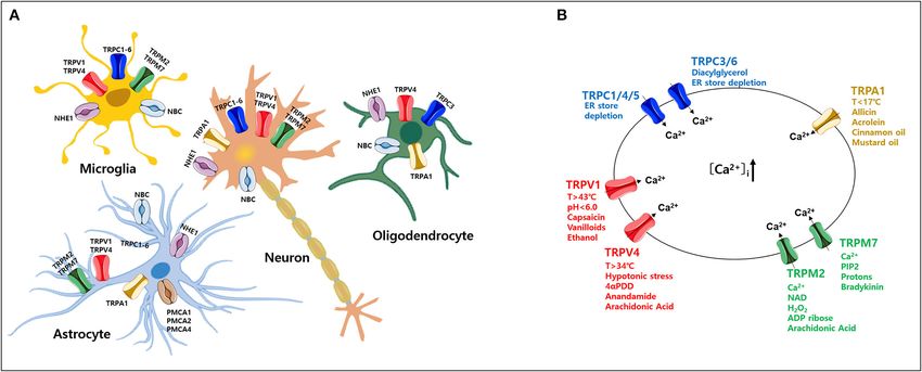
Hwang et al.                                                                                                    TRP in Neurodegenerative Diseases

(Bezprozvanny, 2010; Kawamoto et al., 2012). Indeed, impaired             Conversely, Ca2+ extrusion is dependent on Ca2+ pumps and
cell function caused by reactive nitrogen (oxygen) species and            Na+ /Ca2+ exchangers (Strehler and Thayer, 2018). Among these,
abnormal pH homeostasis also underpins the pathophysiology                plasma membrane Ca2+ ATPases (PMCAs) actively extrude
of neurodegenerative diseases (NDs) (Piacentini et al., 2008;             Ca2+ ions out of cells (Boczek et al., 2019). Thus, these pumps
Bezprozvanny, 2010; Gleichmann and Mattson, 2011; Zundorf                 are important gatekeepers for maintaining intracellular Ca2+
and Reiser, 2011; Harguindey et al., 2017, 2019; Popugaeva et al.,        homeostasis in cells (Stafford et al., 2017; Boczek et al., 2019).
2017). In particular, the maintenance of Ca2+ and pH levels               However, PMCA dysfunction causes altered Ca2+ homeostasis
is involved in a variety of NDs, including Alzheimer’s disease            and leads to a persistent increase in cytosolic Ca2+ , which can
(AD), Parkinson’s disease (PD), Huntington’s disease (HD),                be neurotoxic and can accelerate the development of NDs and
amyotrophic lateral sclerosis (ALS), and age-related disorders            cognitive impairments as the person ages (Strehler and Thayer,
(Harguindey et al., 2007; Kumar et al., 2009; Smaili et al.,              2018; Boczek et al., 2019). In particular, it is possible that the
2009; Ruffin et al., 2014; Hong et al., 2020; Thapak et al.,              regulation of Ca2+ concentration might be more sensitive in
2020). Extensive literature indicates that an excessive increase          which the cells are expressed both TRP and PMCA in the
in cytosolic Ca2+ and H+ constitutes both direct and indirect             particular brain region (Figure 1). Thereby, abnormal expression
ND-induced processes (Marambaud et al., 2009; Smaili et al.,              of either TRP or PMCA subtype may be more likely to cause ND
2009; Bezprozvanny, 2010; Ruffin et al., 2014; Zhao et al., 2016;         than other parts of the brain (Figure 2) (Minke, 2006; Stafford
Harguindey et al., 2017).                                                 et al., 2017). In addition, PMCA activity is associated with
    TRP channels constitute a large family of membrane Ca2+               intracellular acidification (Hwang et al., 2011) which is associated
channels involved in a wide range of processes including                  with neurological conditions observed among AD patients and
thermoregulation, osmosis, pH, stretch, and chemical signaling            other ND patients (Kato et al., 1998; Hamakawa et al., 2004;
(Kaneko and Szallasi, 2014). Functionally, activation of TRP              Mandal et al., 2012; Ruffin et al., 2014; Tyrtyshnaia et al., 2016).
channels influences Ca2+ signaling by allowing Ca2+ to enter                 It is crucial to investigate whether increased Ca2+ and (or)
the cell (cell depolarization), which may activate voltage-gated          acidification are risk factors that affects ND-induced processes
Ca2+ channels (Nilius and Owsianik, 2011; Vennekens et al.,               (Chesler, 2003; Hwang et al., 2011; Ruffin et al., 2014; Cuomo
2012). TRP channels in neuronal cells regulate voltage-gated              et al., 2015; Stafford et al., 2017; Boczek et al., 2019). Here,
Ca2+ , K+ , and Na+ channels, whereas TRP channel regulation              we review the involvement of TRP channels and PMCA in the
in glial cells results in reduced Ca2+ entry via ORAI by                  pathophysiology of NDs.
membrane depolarization, or increased Ca2+ influx through
the hyperpolarization of the membrane (Gees et al., 2010). In
the central nervous system, TRP channels are widely expressed
throughout the brain and play an essential role in regulating Ca2+        BRAIN DISORDERS
homeostasis associated with various cellular functions, including
synaptic plasticity, synaptogenesis, and synaptic transmission            Neurodegenerative Diseases
in a specific region of the brain (Venkatachalam and Montell,             NDs such as AD, PD, HD, and ALS are age-related conditions
2007; Kaneko and Szallasi, 2014; Jardin et al., 2017; Chi et al.,         characterized by uncontrolled neuronal death in the brain (Hong
2018; Hong et al., 2020). In addition, TRP subtype channels               et al., 2020; Slanzi et al., 2020; Thapak et al., 2020). To date,
are expressed simultaneously or separately in neurons and                 several studies have reported that NDs are associated with protein
glia, fulfilling critical roles in cell homeostasis, development,         aggregation, oxidative stress, inflammation, and abnormal Ca2+
neurogenesis, and synaptic plasticity (Vennekens et al., 2012).           homeostasis (Sprenkle et al., 2017). The impairment of Ca2+
Several members of the TRP subtype are highly expressed in                homeostasis is known to result in increased susceptibility to NDs
neurons and glia (Moran et al., 2004; Butenko et al., 2012; Ho            (Kumar et al., 2009; Smaili et al., 2009; Bezprozvanny, 2010;
et al., 2014; Ronco et al., 2014; Verkhratsky et al., 2014; Liu           Gleichmann and Mattson, 2011; Kawamoto et al., 2012; Bagur
et al., 2017; Rakers et al., 2017) (Table 1). Thus, diverse TRP           and Hajnoczky, 2017). In particular, this impairment is associated
channels expressed in the brain are involved in the progression           with changes in Ca2+ buffering capacity, deregulation of Ca2+
of NDs such as Parkinson’s and Alzheimer’s. In particular,                channel activity, and alteration in other calcium regulatory
increased intracellular Ca2+ via TRP channels contributes to              proteins that occur in some types of neurons and glial cells in
various pathophysiological events (Venkatachalam and Montell,             certain brain regions (Zundorf and Reiser, 2011; Nikoletopoulou
2007; Kaneko and Szallasi, 2014; Moran, 2018; Hong et al., 2020)          and Tavernarakis, 2012). There is also increased Ca2+ influx
as well as brain disorders such as AD, PD, stroke, epilepsy, and          mediated by abnormal TRP channel activation (Sawamura et al.,
migraine (Table 1)(Morelli et al., 2013; Kaneko and Szallasi, 2014;       2017). Similarly, Ca2+ extrusion through PMCA has been shown
Kumar et al., 2016; Moran, 2018; Hong et al., 2020; Liu et al.,           to decrease in aged neurons (Jiang et al., 2012). For this reason,
2020).                                                                    these NDs are associated with Ca2+ channels in neurons and
    The normal regulation of intracellular Ca2+ levels involves           glial cells (astrocytes, microglia, and oligodendrocytes), which
mechanisms that control the specific uptake and extrusion                 are important for neuronal survival, myelin formation, neuronal
mechanisms across the cell membrane (Kawamoto et al., 2012;               support, and regulation of local neuron activity (neurons-glial
Strehler and Thayer, 2018). Ca2+ influx is mediated by several            signaling) (Zhang and Liao, 2015; Cornillot et al., 2019; Enders
voltage- and ligand-gated channels as well as transporters.               et al., 2020).

Frontiers in Cell and Developmental Biology | www.frontiersin.org     2                                    January 2021 | Volume 9 | Article 584388
Hwang et al. TRP in Neurodegenerative Diseases
TABLE 1 | A summary of the transient receptor potential (TRP) subtypes found in distribution of central nervous system (CNS) cell types.
TRP channels Expression in brain Expression in glia Disorders References
TRPC TRPC1 - Cerebellum, Astrocyte, microglia, NDs, ADs, Riccio et al., 2002; Bollimuntha
subfamily hippocampus, PD, HD, et al., 2005, 2006; Selvaraj et al.,
forebrain 2009, 2012; Hong et al., 2015
- Dopaminergic
neuron (Human/mouse)
TRPC3 - Cerebellum, Astrocyte, NDs, ADs, Rosker et al., 2004; Wu et al.,
hippocampus, PDs 2004; Yamamoto et al., 2007;
forebrain Mizoguchi et al., 2014
- Dopaminergic
neuron (Human)
TRPC4 Cerebellum, Astrocyte, Epilepsy Wang et al., 2007; Wu et al.,
hippocampus, 2008; Von Spiczak et al., 2010;
forebrain Tai et al., 2011
TRPC5 - Cerebellum, forebrain Astrocyte, NDs, PDs, Shin et al., 2010; Tai et al., 2011;
- Hippocampus (mouse) Epilepsy Kaczmarek et al., 2012
TRPC6 Cerebellum, Astrocyte, microglia NDs, ADs Lessard et al., 2005; Wang et al.,
hippocampus, 2015; Liu et al., 2017; Lu et al.,
forebrain, striatum 2017
TRPM TRPM2 - Hippocampus, Astrocyte, microglia NDs, ADs, Fonfria et al., 2005; Kaneko
subfamily forebrain PDs et al., 2006; Hermosura et al.,
- Cerebellum (human), 2008; Ostapchenko et al., 2015
cortex (rat)
TRPM7 - Cerebellum, Astrocyte, microglia NDs, ADs, Aarts and Tymianski, 2005;
forebrain, PDs, Epilepsy Hermosura et al., 2005; Chen X.
- Hippocampus et al., 2010; Coombes et al.,
(human) 2011; Oakes et al., 2019
- cortex (mouse)
TRPV TRPV1 - Basal ganglia, Astrocyte, microglia NDs, AD, HD, Lastres-Becker et al., 2003; Kim
subfamily hindbrain Cerebellum epilepsy et al., 2005; Gibson et al., 2008;
- Hippocampus (rat/mouse), Li et al., 2008; Lee et al., 2011;
Balleza-Tapia et al., 2018
TRPV4 Cerebellum, Astrocyte, microglia NDs, AD, Auer-Grumbach et al., 2010;
hippocampus, Chen D. H. et al., 2010;
Landoure et al., 2010; Klein
et al., 2011; Wang et al., 2019
TRPA TRPA1 Cerebellum, Astrocyte, oligodendrocyte AD Shigetomi et al., 2011; Lee et al.,
subfamily hippocampus, 2016; Saghy et al., 2016;
Bolcskei et al., 2018
PMCA, plasma membrane Ca2+ ATPase; AD, Alzheimer’s disease; PD, Parkinson’s disease; ND, neurodegenerative disease.
Pathophysiological Role of TRP Channels TRP vanilloid (TRPV), TRP ankyrin (TRPA), TRP melastatin
TRP channels are non-selective, Ca2+ -permeable channels that (TRPM), TRP polycystin (TRPP), and TRP mucolipin (TRPML)
regulate diverse cellular functions in neurons (Nilius, 2007; (Nilius, 2007; Selvaraj et al., 2010; Nishida et al., 2015; Sawamura
Venkatachalam and Montell, 2007; Sawamura et al., 2017). Based et al., 2017). Most TRP channels are non-selective channels with
on functional characterization of TRP channels by a wide range consistent Ca2+ permeability (Samanta et al., 2018) and each
of stimuli (Zheng, 2013), aberrant activity of TRP channels TRP subtype responds to various temperatures, ligands, as well
likely initiates and/or propagates ND processes, especially cell as specific agonists and activators (Figure 1B) (Luo et al., 2020).
death, via increased intracellular Ca2+ in various brain regions TRP channels are tetramers formed by monomers that share a
(Moran, 2018; Hong et al., 2020; Huang et al., 2020). Here, common structure comprising six transmembrane domains and
we focus on the function of TRP channels associated with containing cation-selective pores (Hellmich and Gaudet, 2014).
Ca2+ signaling in neurons and glial cells (Figure 1A) (Nilius, Numerous studies have reported that these TRP channels are
2007; Bollimuntha et al., 2011; Zheng, 2013; Zhang and Liao, related to neuronal cell death that is associated with abnormal
2015; Jardin et al., 2017; Sawamura et al., 2017; Hasan and Ca2+ homeostasis (Gees et al., 2010; Sawamura et al., 2017).
Zhang, 2018; Samanta et al., 2018; Cornillot et al., 2019; Enders
et al., 2020; Wang et al., 2020). Based on sequence homology, TRPC (Classic or Canonical)
the TRP family currently comprises 28 mammalian channels TRPC was the first TRP group identified in mammals (Selvaraj
and is subdivided into six subfamilies: TRP canonical (TRPC), et al., 2010). The TRPC subfamily contains members: TRPC1-7
Frontiers in Cell and Developmental Biology | www.frontiersin.org 3 January 2021 | Volume 9 | Article 584388Hwang et al. TRP in Neurodegenerative Diseases
FIGURE 1 | Expression of various transient receptor potential (TRP) subtypes and calcium (Ca2+ ) influx by their agonists in the mammalian central nervous system
(CNS). (A) Expression profile of various TRP channels, NHE1, and NBC, in mammalian CNS cell types. (B) Ca2+ influx through activation of TRP subtypes by various
agonists or activators in the mammalian CNS. TRP, transient receptor potential; PMCA, plasma membrane Ca2+ ATPase; NBC, Na+ /HCO− 3 cotransporters; NHE,
Na+ /H+ exchangers.
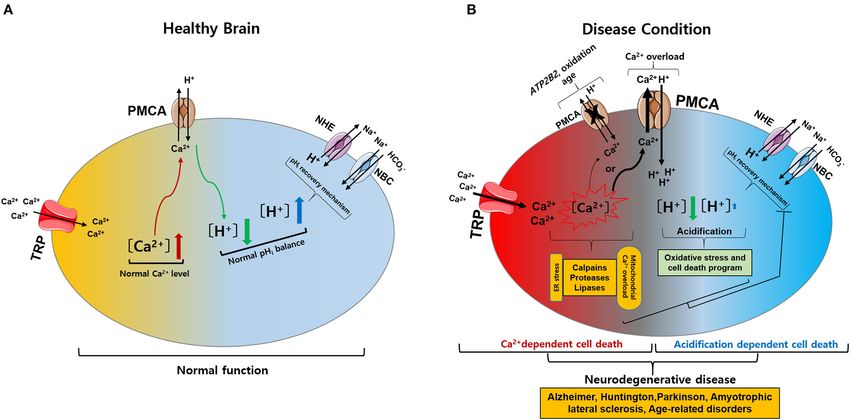
FIGURE 2 | Intracellular calcium (Ca2+ ) and pH (pHi ) signaling by activation of TRP and PMCA in healthy and diseased condition of the brain. (A) Normal physiological
function of intracellular Ca2+ and pHi homeostasis. The activation of TRP channels leads to Ca2+ influx into the cytosol. Increased Ca2+ levels are regulated by PMCA.
The activation of PMCA can cause acidification. Acidification conditions are mediated by pHi recovery functions regulated by NBC and NHE. (B) Neurodegenerative
diseases caused by pathophysiological functions of intracellular Ca2+ and pHi homeostasis. (1) The activation of TRP channels leads to excess Ca2+ influx and
overload Ca2+ is maintained due to ATP2B2, oxidation, and age-related downregulation of PMCA: Ca2+ -dependent cell death. (2) PMCA overexpression due to
cytoplasmic Ca2+ overload cause persistent acidification from inhibition of the pHi recovery mechanism by oxidative stress or cell death program: acidification
dependent cell death. Ultimately, abnormal intracellular Ca2+ and pHi levels impair neuronal function, resulting in neurodegenerative diseases. TRP, transient receptor
potential; PMCA, plasma membrane Ca2+ ATPase; NBC, Na+ /HCO− 3 cotransporters; NHE, Na /H exchangers.
+ +
Frontiers in Cell and Developmental Biology | www.frontiersin.org 4 January 2021 | Volume 9 | Article 584388Hwang et al. TRP in Neurodegenerative Diseases
(Wang et al., 2020). With the exception of TRPC2, all TRPC disorders, including AD, PD, and bipolar disorder (Akyuva and
channels are widely expressed in the brain from the embryonic Naziroglu, 2020). In addition, an increase in intracellular Ca2+
period to adulthood (Douglas et al., 2003). TRPC channels and Aβ induced by TRPM2 activity induces neuronal cell death
can form functional channels by heteromeric interactions, in the rat striatum (Belrose and Jackson, 2018). Mg2+ is the
functioning as non-selective Ca2+ entry channels with distinct second most abundant cation and essential cofactor in various
activation modes (Villereal, 2006). Thus, TRPC channels play an enzymatic reactions (Ryazanova et al., 2010). TRPM2 is expressed
important role in regulating basic neuronal processes. TRPC1 by both microglia and astrocytes, which regulate gliosis and
is highly expressed and involved in the early development and immune cell function (Wang et al., 2016; Huang et al., 2017).
proliferation of neurons (Yamamoto et al., 2005; Hentschke et al., TRPM7 is permeable to Mg2+ and maintains Mg2+ homeostasis
2006) as well as synaptic transmission (Broker-Lai et al., 2017; (Ryazanova et al., 2010). In mouse cortical neurons, inhibition of
Wang et al., 2020). TRPC1 and TRPC4 have been reported TRPM7 expression protects against neuronal cell damage (Asrar
to regulate neuronal cell death in response to seizures in and Aarts, 2013; Huang et al., 2020). TRPM7 is also found in
the hippocampus and septum (Broker-Lai et al., 2017). The astrocytes and microglia to control migration, proliferation, and
TRPC1/4/5 channel has been expressed in the somatosensory invasion (Siddiqui et al., 2014; Zeng et al., 2015).
cortex, hippocampus, and motor cortex of adult rats (Riccio
et al., 2002; Moran et al., 2004; Fowler et al., 2007). In TRPV (Vanilloid)
particular, the dense expression of TRPC3 regulates hippocampal TRPV channels form homo- or heterotetrameric complexes and
neuronal excitability and memory function (Neuner et al., are non-selective cation channels (Startek et al., 2019). The TRPV
2015). The abnormal increase in sustained cytosolic Ca2+ by subfamily consists of six members (TRPV1-6) that are located
TRPC5 activation causes neuronal damage through the calpain- mostly on the plasma membrane (Zhai et al., 2020). Recent
caspase-dependent pathway and the CaM kinase as seen in HD studies on pathological TRPV1 expression in the brain have
(Hong et al., 2015). Spinocerebellar ataxia type 14 (SCA14) is been performed (Mickle et al., 2015). TRPV1 activation induces
an autosomal dominant ND caused by a mutation in protein caspase-3 dependent programmed cell death through Ca2+ -
kinase Cγ (Wong et al., 2018). This mutation of SCA14 has mediated signaling, resulting in cell death of cortical neurons
been demonstrated to cause phosphorylation failure in TRPC3 (Ho et al., 2012; Song et al., 2013) and also triggers cell death
channels, resulting in persistent Ca2+ entry that may contribute through L-type Ca2+ channels and Ca2+ influx in rat cortical
to neurodegeneration (Adachi et al., 2008). On the other hand, neurons (Shirakawa et al., 2008). The activation of cannabinoid 1
TRPC3 or TRPC6 promotes neurotrophin action on brain- (CB1) receptors stimulates TRPV1 activity, leading to increased
derived neurotrophic factor (BDNF) by improving neuronal intracellular Ca2+ and cell death of mesencephalic dopaminergic
survival through Ca2+ influx (Huang et al., 2011). All TRPC neurons (Kim et al., 2005, 2008). TRPV1 activation induces
channels are expressed in astrocytes and TRPC1 and TRPC3 play apoptotic cell death in rat cortical neurons, leading to chronic
a critical role in astrocyte store-operated Ca2+ entry, which is epilepsy distinguished by abnormal brain activity (Fu et al.,
induced by endoplasmic reticulum depletion (Verkhratsky et al., 2009). TRPV1 activation in microglia plays a positive role
2014). TRPC1 and TRPC6 are also expressed in rat microglia in promoting microglial phagocytosis in damaged cells while
(Zhang and Liao, 2015). Thus, some TRPC channels exhibit disrupting mitochondria and increasing ROS production (Kim
different functions in normal physiological or pathological et al., 2006; Hassan et al., 2014). TRPV1 has been shown to affect
events, depending on Ca2+ signaling in the brain (Huang et al., the migration of astrocytes (Ho et al., 2014). Abnormal function
2011; Li et al., 2012; Neuner et al., 2015). of TRPV4 leads to neuronal dysfunction and axonal degeneration
due to increased Ca2+ via Ca2+ /calmodulin-dependent protein
TRPM (Melastatin) kinase II (CaMKII) (Woolums et al., 2020). TRPV4 plays a role
Of all TRP channels, the TRPM subfamily has the largest in regulating the osmotic pressure in the brain and is highly
and most diverse expression levels and has been strongly expressed throughout glial cells associated with ND (Liedtke and
implicated in NDs (Samanta et al., 2018). The TRPM channel Friedman, 2003; Rakers et al., 2017). Thus, these channels play an
consists of eight members (TRPM1-8) and shares common important role in Ca2+ homeostasis and are therapeutic targets
structural characteristics with other TRP channels (Huang for various disorders.
et al., 2020). However, they have a variety of C-terminal
sections with active enzyme domains and a unique N-terminal TRPA (Ankyrin)
without ankyrin repeats involved in channel assembly and TRPA1 was first identified as an ankyrin-like transmembrane
trafficking (Huang et al., 2020). A distinctive feature of TRPM protein and the solitary member of the mammalian TRPA
channels is the regulation of Ca2+ and magnesium (Mg2+ ) subfamily (Yang and Li, 2016). TRPA1 is a non-selective cation
homeostasis, and TRPM (2–7) are mainly expressed in the CNS. channel formed by homo- or heterotetramer subunits with
In addition, TRPM2 is activated by a wide range of factors a cytosolic N-terminal domain (16 ankyrin repeat sequence)
including NAD+ -related metabolites, adenosine diphosphate- and C-terminal Ca2+ -binding domains (Nilius et al., 2011;
ribose, oxidative stress, and depletion of glutathione (GSH) (Sita Fernandes et al., 2012). The TRPA1 channel responds to a
et al., 2018). Increased levels of reactive oxygen species (ROS) variety of ligands, such as temperature, osmotic changes, and
due to GSH depletion causes TRPM2-dependent Ca2+ influx to endogenous compounds (Nishida et al., 2015). To date, the
induce neuronal cell death, suggesting that several neurological reported role of TRPA1 in neurons is the mediation of pain,
Frontiers in Cell and Developmental Biology | www.frontiersin.org 5 January 2021 | Volume 9 | Article 584388Hwang et al. TRP in Neurodegenerative Diseases
cold, inflammation, and itch sensation (Fernandes et al., 2012). homeostasis (Bezprozvanny, 2010) are likely to underpin Ca2+ -
Recent reports indicate that TRPA1 hyperactivation causes Aβ dependent neuronal death in NDs (Sawamura et al., 2017; Hong
oligomer-mediated rapid Ca2+ signaling (Bosson et al., 2017; et al., 2020).
Hong et al., 2020). Additionally, ablation of TRPA1 in APP/PS1
transgenic mice attenuated the progression of AD, improved
learning and memory conditions, and reduced Aβ plaques and PATHOPHYSIOLOGICAL ROLE OF
cytokines (Lee et al., 2016). Similarly, TRPA1 channels promote PLASMA MEMBRANE CALCIUM ATPases
Ca2+ hyperactivity of astrocytes and then contribute to synaptic
dysfunction due to the oligomeric forms of Aβ peptide (Lee et al., Of the various proteins involved in Ca2+ signaling, PMCA is the
2016; Bosson et al., 2017; Logashina et al., 2019; Alavi et al., most sensitive Ca2+ detector that regulates Ca2+ homeostasis
2020). In addition, TRPA1 mediates Ca2+ signaling in astrocytes, (Boczek et al., 2019). PMCA exists in four known isoforms
resulting in dysregulation of synaptic activity in AD (Bosson (Boczek et al., 2019). In both mice and humans, PMCAs 1–
et al., 2017). 4 exhibit anatomically distinct expression patterns, such that
isoforms 1 and 4 are ubiquitously expressed in all tissue types,
whereas PMCA2 and PMCA3 are tissue-specific and exclusive
Other Channels in neurons of the brain (Kip et al., 2006). In addition, PMCA1,
TRPML and TRPP have limited similarity to other TRP family 2, and 4 were detected in rat cortical astrocytes (Fresu et al.,
members (Samanta et al., 2018; Huang et al., 2020). TRPML 1999) (Table 2). The general structure of PMCA consists of 10
channels (TRPML1-3) are Ca2+ permeable cation channels transmembrane domains (TM) with the N- and C-terminal ends
that each contain six transmembrane segments with helices on the cytosolic side (Stafford et al., 2017). The physiological
(S1–S6) and a pore site comprised of S5, S6, and two pore functions of PMCA include the regulation and maintenance
helices (PH1 and PH2) (Schmiege et al., 2018; Tedeschi et al., of optimal Ca2+ homeostasis (Bagur and Hajnoczky, 2017).
2019). TRPML channels are mostly located in intracellular PMCA is an ATP-driven Ca2+ pump that maintains low resting
compartments instead of the plasma membrane (Clement et al., intracellular Ca2+ concentration ([Ca2+ ]i) to prevent cytotoxic
2020). TRPP channels share high protein sequence similarity Ca2+ overload-mediated cell death through activation of ion
with TRPML channels and are located in the primary cilia channels such as TRP (Zundorf and Reiser, 2011). In addition,
consisting of TRPP1 (also known as PKD1) and TRPP2 (PKD2) PMCA is involved in Ca2+ -induced intracellular acidification
(Samanta et al., 2018). To date, evidence indicates that various by countertransport of H+ ions (Vale-Gonzalez et al., 2006;
TRP channels are expressed in the CNS and play important Majdi et al., 2016). Thus, PMCA plays a vital role in controlling
roles in the development of several NDs (Sawamura et al., 2017; cell survival and cell death (Stafford et al., 2017). PMCA
Samanta et al., 2018). In particular, TRP channels and Ca2+ expression changes significantly during brain development
TABLE 2 | A summary of the transient receptor potential (TRP) subtypes found in distribution of central nervous system (CNS) cell types.
PMCA subfamily Expression in brain Expression Disorders References
in glia
PMCA1 - Ubiquitous in brain Rat cortical AD, PD Stauffer et al., 1995;
(human and rat). astrocytes Fresu et al., 1999; Brini
- Cerebellum, cerebral et al., 2013
cortex, brain
stem (Human)
PMCA2 - Cerebellar purkinje Rat cortical AD, PD, cerebellar Stauffer et al., 1995;
neurons (human/mouse) astrocytes ataxias, sensory Fresu et al., 1999;
- cerebellum, cerebral neuron diseases Kurnellas et al., 2007;
cortex, brain Empson et al., 2010;
stem (Human) Hajieva et al., 2018;
Strehler and Thayer,
2018
PMCA3 - Cerebellum, cerebral Limited Cerebellar ataxias, Stauffer et al., 1995;
cortex (Human) sensory neuron Zanni et al., 2012;
- Cerebellum and diseases Strehler and Thayer,
hippocampus (Rat) 2018
PMCA4 - Ubiquitous in Rat cortical AD, PD Stauffer et al., 1995;
brain (human/rat) astrocytes Fresu et al., 1999; Brini
- Cerebellum, cerebral et al., 2013; Zaidi et al.,
cortex, brain 2018
stem (Human)
PMCA, plasma membrane Ca2+ ATPase; AD, Alzheimer’s disease; PD, Parkinson’s disease.
Frontiers in Cell and Developmental Biology | www.frontiersin.org 6 January 2021 | Volume 9 | Article 584388Hwang et al. TRP in Neurodegenerative Diseases
(Boczek et al., 2019). One of the characteristics of brain aging activation of PMCA in the rat trigeminal ganglion (Hwang
is a Ca2+ homeostasis disorder, which can result in detrimental et al., 2011). Under normal conditions, acidification conditions
consequences on neuronal function (Boczek et al., 2019). are promptly returned to and maintained at normal pH levels
Overall, PMCAs have been attributed a housekeeping role in through a physiological pHi recovery mechanism involving
maintaining intracellular Ca2+ levels through precise regulation the regulation of Na+ /H+ exchangers (NHE) and Na+ -HCO− 3
of Ca2+ homeostasis (Strehler et al., 2007). However, the cotransporter (NBCs) in the brain (Chesler, 2003; Sinning and
altered composition of PMCA is associated with a less efficient Hubner, 2013; Ruffin et al., 2014; Bose et al., 2015). NHE1
Ca2+ extrusion system, increasing the risk of neurodegenerative is abundantly expressed in all neuronal cells and astrocytes,
processes (Strehler and Thayer, 2018). ATP2B2 is a deafness- regulating cell volume homeostasis and pHi (Song et al., 2020).
associated gene that encodes PMCA2 (Smits et al., 2019). A NBC1 is also widely expressed in astrocytes throughout the
recent study reported a link between PMCA2 and autism brain (Annunziato et al., 2013) (Figure 1A). However, functional
spectrum disorder (ASD) (Yang et al., 2013). ASD is a group of inhibition of pHi recovery mechanism in pathological conditions
neurodevelopmental disorders that results in deficits in social leads to excessive intracellular acidification (Majdi et al., 2016).
interaction (Chaste and Leboyer, 2012; Fatemi et al., 2012). Therefore, although the exact underlying mechanism that causes
Intracellular Ca2+ levels are crucial for regulating neuronal intracellular acidification in brain neurons is unknown. However,
survival, differentiation, and migration (Bezprozvanny, 2010). it appears that persistent intracellular acidification condition
Perturbations in these processes underlie the pathogenesis of promotes irreversible neuronal damage and induces amyloid
autism spectrum disorders (Gilbert and Man, 2017). ATP2B3 aggregation in the brains of patients with AD (Xiong et al., 2008;
mutations are associated with X-linked cerebellar ataxia and Ruffin et al., 2014).
Ca2+ extrusion disorders in patients with cerebellar ataxia
and developmental delay (Zanni et al., 2012; Mazzitelli and CONCLUSION
Adamo, 2014; Cali et al., 2015). Several neurotoxic agents,
such as oxidation and age, downregulate PMCA function and Intracellular Ca2+ and pH regulation play vital roles in both
increase susceptibility to NDs (Zaidi, 2010). In particular, the physiological and pathological conditions. Abnormal changes
internalization of PMCA2 initiated by protease function in rat in Ca2+ or pH typically cause cell death. TRP channels are
hippocampal pyramidal cells after glutamate exposure or kainate- involved in Ca2+ influx, which affects neuronal and glial
induced seizures, in which loss of PMCA function occurs, may functions under normal physiological conditions. However,
contribute to Ca2+ dysregulation and lead to neuronal cell death altered expression of TRP channels can lead to excess Ca2+ influx,
(Pottorf et al., 2006; Stafford et al., 2017). A decrease in PMCA and intracellular Ca2+ overload is maintained due to ATP2B2,
activity and increased Ca2+ may cause cell death depending on oxidation, and aging-related downregulation of PMCA, leading
the degree of cytosolic accumulation of tau and Aβ in AD (Boczek to Ca2+ -dependent cell death. Alternatively, overexpression of
et al., 2019). In addition, PMCA expression is decreased in the PMCA due to cytoplasmic Ca2+ overload causes continuous
cortex of postmortem brains of patients with AD (Berrocal et al., acidification from inhibition of the pHi recovery mechanisms
2019; Boczek et al., 2019). by oxidative stress or programmed cell death, resulting in
acidification-dependent cell death (Figure 2) (Harguindey et al.,
pH REGULATION BY PMCA IN 2017, 2019). To date, TRP channels have been investigated for
NEURODEGENERATIVE DISEASES their role in NDs. However, targeting TRP channels and PMCA,
including Ca2+ and pH regulation, as a treatment for NDs
As mentioned above, PMCAs have a Ca2+ extrusion function requires a deeper understanding of their function in both health
on the membrane and another important function, namely H+ and disease. This review describes potential therapeutic targets
uptake (Stafford et al., 2017). Since PMCA is responsible for for NDs by discussing TRP channels and PMCA responsible for
control of Ca2+ extrusion and H+ uptake rates, it provides the disruption of intracellular Ca2+ and pH homeostasis that
an important link between Ca2+ signaling and intracellular underpin ND development.
pH (pHi ) in neurons (Hwang et al., 2011). Mechanisms that
maintain strict pH homeostasis in the brain control neuronal AUTHOR CONTRIBUTIONS
excitability, synaptic transmission, neurotransmitter uptake,
nociception, and inflammation (Chesler, 2003; Dhaka et al., C-KP and YK conceived and supervised the project. S-MH, JL,
2009; Casey et al., 2010; Hwang et al., 2011). Changes in C-KP, and YK wrote the paper. All authors contributed to the
pH caused via pH-sensitive or pH-regulated ion channels article and approved the submitted version.
are detrimental to brain function and can cause multiple
degenerative diseases (Ruffin et al., 2014). Neuronal excitability is FUNDING
particularly sensitive to changes in intracellular and extracellular
pH mediated by various ion channels (Parker and Boron, This work was supported by grants from the National
2013). The activation of TRPV1 has been reported to induce Research Foundation of Korea (NRF-2017M3C7A1025602
a rise in Ca2+ and cause intracellular acidification via the and NRF-2019R1C1C1010822).
Frontiers in Cell and Developmental Biology | www.frontiersin.org 7 January 2021 | Volume 9 | Article 584388Hwang et al. TRP in Neurodegenerative Diseases
REFERENCES Brini, M., Cali, T., Ottolini, D., and Carafoli, E. (2013). The plasma
membrane calcium pump in health and disease. FEBS J. 280, 5385–5397.
Aarts, M. M., and Tymianski, M. (2005). TRPMs and neuronal cell death. Pflugers doi: 10.1111/febs.12193
Arch. 451, 243–249. doi: 10.1007/s00424-005-1439-x Broker-Lai, J., Kollewe, A., Schindeldecker, B., Pohle, J., Nguyen Chi, V., Mathar,
Adachi, N., Kobayashi, T., Takahashi, H., Kawasaki, T., Shirai, Y., Ueyama, T., I., et al. (2017). Heteromeric channels formed by TRPC1, TRPC4 and TRPC5
et al. (2008). Enzymological analysis of mutant protein kinase Cgamma causing define hippocampal synaptic transmission and working memory. EMBO J. 36,
spinocerebellar ataxia type 14 and dysfunction in Ca2+ homeostasis. J. Biol. 2770–2789. doi: 10.15252/embj.201696369
Chem. 283, 19854–19863. doi: 10.1074/jbc.M801492200 Butenko, O., Dzamba, D., Benesova, J., Honsa, P., Benfenati, V., Rusnakova, V.,
Akyuva, Y., and Naziroglu, M. (2020). Resveratrol attenuates hypoxia- et al. (2012). The increased activity of TRPV4 channel in the astrocytes of the
induced neuronal cell death, inflammation and mitochondrial adult rat hippocampus after cerebral hypoxia/ischemia. PLoS ONE 7:e39959.
oxidative stress by modulation of TRPM2 channel. Sci. Rep. 10:6449. doi: 10.1371/journal.pone.0039959
doi: 10.1038/s41598-020-63577-5 Cali, T., Lopreiato, R., Shimony, J., Vineyard, M., Frizzarin, M., Zanni, G.,
Alavi, M. S., Shamsizadeh, A., Karimi, G., and Roohbakhsh, A. (2020). Transient et al. (2015). A novel mutation in isoform 3 of the plasma membrane Ca2+
receptor potential ankyrin 1 (TRPA1)-mediated toxicity: friend or foe? Toxicol. pump impairs cellular Ca2+ homeostasis in a patient with cerebellar ataxia
Mech. Methods 30, 1–18. doi: 10.1080/15376516.2019.1652872 and laminin subunit 1alpha mutations. J. Biol. Chem. 290, 16132–16141.
Annunziato, L., Boscia, F., and Pignataro, G. (2013). Ionic transporter activity in doi: 10.1074/jbc.M115.656496
astrocytes, microglia, and oligodendrocytes during brain ischemia. J. Cereb. Casey, J. R., Grinstein, S., and Orlowski, J. (2010). Sensors and regulators of
Blood Flow Metab. 33, 969–982. doi: 10.1038/jcbfm.2013.44 intracellular pH. Nat. Rev. Mol. Cell Biol. 11, 50–61. doi: 10.1038/nrm2820
Asrar, S., and Aarts, M. (2013). TRPM7, the cytoskeleton and neuronal death. Chaste, P., and Leboyer, M. (2012). Autism risk factors: genes, environment,
Channels (Austin) 7, 6–16. doi: 10.4161/chan.22824 and gene-environment interactions. Dialogues Clin. Neurosci. 14, 281–292.
Auer-Grumbach, M., Olschewski, A., Papic, L., Kremer, H., Mcentagart, M. E., doi: 10.31887/DCNS.2012.14.3/pchaste
Uhrig, S., et al. (2010). Alterations in the ankyrin domain of TRPV4 cause Chen, D. H., Sul, Y., Weiss, M., Hillel, A., Lipe, H., Wolff, J., et al. (2010). CMT2C
congenital distal SMA, scapuloperoneal SMA and HMSN2C. Nat. Genet. 42, with vocal cord paresis associated with short stature and mutations in the
160–164. doi: 10.1038/ng.508 TRPV4 gene. Neurology 75, 1968–1975. doi: 10.1212/WNL.0b013e3181ffe4bb
Bagur, R., and Hajnoczky, G. (2017). Intracellular Ca(2+) Sensing: its Chen, X., Numata, T., Li, M., Mori, Y., Orser, B. A., Jackson, M. F., et al.
role in calcium homeostasis and signaling. Mol. Cell 66, 780–788. (2010). The modulation of TRPM7 currents by nafamostat mesilate depends
doi: 10.1016/j.molcel.2017.05.028 directly upon extracellular concentrations of divalent cations. Mol. Brain 3:38.
Balleza-Tapia, H., Crux, S., Andrade-Talavera, Y., Dolz-Gaiton, P., Papadia, D., doi: 10.1186/1756-6606-3-38
Chen, G., et al. (2018). TrpV1 receptor activation rescues neuronal function Chesler, M. (2003). Regulation and modulation of pH in the brain. Physiol. Rev. 83,
and network gamma oscillations from Abeta-induced impairment in mouse 1183–1221. doi: 10.1152/physrev.00010.2003
hippocampus in vitro. Elife 7:37703. doi: 10.7554/eLife.37703.025 Chi, H., Chang, H. Y., and Sang, T. K. (2018). Neuronal cell death
Belrose, J. C., and Jackson, M. F. (2018). TRPM2: a candidate therapeutic mechanisms in major neurodegenerative diseases. Int. J. Mol. Sci. 19:3082.
target for treating neurological diseases. Acta Pharmacol. Sin. 39, 722–732. doi: 10.3390/ijms19103082
doi: 10.1038/aps.2018.31 Clement, D., Goodridge, J. P., Grimm, C., Patel, S., and Malmberg,
Berridge, M. J., Lipp, P., and Bootman, M. D. (2000). The versatility and K. J. (2020). TRP channels as interior designers: remodeling the
universality of calcium signalling. Nat. Rev. Mol. Cell Biol. 1, 11–21. endolysosomal compartment in natural killer cells. Front. Immunol. 11:753.
doi: 10.1038/35036035 doi: 10.3389/fimmu.2020.00753
Berrocal, M., Caballero-Bermejo, M., Gutierrez-Merino, C., and Mata, A. M. Coombes, E., Jiang, J., Chu, X. P., Inoue, K., Seeds, J., Branigan, D., et al.
(2019). Methylene blue blocks and reverses the inhibitory effect of tau on (2011). Pathophysiologically relevant levels of hydrogen peroxide induce
PMCA function. Int. J. Mol. Sci. 20:3521. doi: 10.3390/ijms20143521 glutamate-independent neurodegeneration that involves activation of transient
Bezprozvanny, I. B. (2010). Calcium signaling and neurodegeneration. Acta receptor potential melastatin 7 channels. Antioxid. Redox Signal 14, 1815–1827.
Naturae 2, 72–82. doi: 10.32607/20758251-2010-2-1-72-80 doi: 10.1089/ars.2010.3549
Boczek, T., Radzik, T., Ferenc, B., and Zylinska, L. (2019). The puzzling role of Cornillot, M., Giacco, V., and Hamilton, N. B. (2019). The role of TRP
neuron-specific PMCA isoforms in the aging process. Int. J. Mol. Sci. 20:6338. channels in white matter function and ischaemia. Neurosci. Lett. 690, 202–209.
doi: 10.3390/ijms20246338 doi: 10.1016/j.neulet.2018.10.042
Bolcskei, K., Kriszta, G., Saghy, E., Payrits, M., Sipos, E., Vranesics, A., et al. Cross, J. L., Meloni, B. P., Bakker, A. J., Lee, S., and Knuckey, N. W. (2010). Modes
(2018). Behavioural alterations and morphological changes are attenuated of neuronal calcium entry and homeostasis following cerebral ischemia. Stroke
by the lack of TRPA1 receptors in the cuprizone-induced demyelination Res. Treat. 2010:316862. doi: 10.4061/2010/316862
model in mice. J. Neuroimmunol. 320, 1–10. doi: 10.1016/j.jneuroim.2018. Cuomo, O., Vinciguerra, A., Cerullo, P., Anzilotti, S., Brancaccio, P., Bilo, L.,
03.020 et al. (2015). Ionic homeostasis in brain conditioning. Front. Neurosci. 9:277.
Bollimuntha, S., Ebadi, M., and Singh, B. B. (2006). TRPC1 protects human SH- doi: 10.3389/fnins.2015.00277
SY5Y cells against salsolinol-induced cytotoxicity by inhibiting apoptosis. Brain Dhaka, A., Uzzell, V., Dubin, A. E., Mathur, J., Petrus, M., Bandell, M., et al. (2009).
Res. 1099, 141–149. doi: 10.1016/j.brainres.2006.04.104 TRPV1 is activated by both acidic and basic pH. J. Neurosci. 29, 153–158.
Bollimuntha, S., Selvaraj, S., and Singh, B. B. (2011). Emerging roles of canonical doi: 10.1523/JNEUROSCI.4901-08.2009
TRP channels in neuronal function. Adv. Exp. Med. Biol. 704, 573–593. Douglas, R. M., Xue, J., Chen, J. Y., Haddad, C. G., Alper, S. L., and Haddad,
doi: 10.1007/978-94-007-0265-3_31 G. G. (2003). Chronic intermittent hypoxia decreases the expression of Na/H
Bollimuntha, S., Singh, B. B., Shavali, S., Sharma, S. K., and Ebadi, M. exchangers and HCO3-dependent transporters in mouse CNS. J. Appl. Physiol.
(2005). TRPC1-mediated inhibition of 1-methyl-4-phenylpyridinium ion 95, 292–299. doi: 10.1152/japplphysiol.01089.2002
neurotoxicity in human SH-SY5Y neuroblastoma cells. J. Biol. Chem. 280, Empson, R. M., Akemann, W., and Knopfel, T. (2010). The role of the calcium
2132–2140. doi: 10.1074/jbc.M407384200 transporter protein plasma membrane calcium ATPase PMCA2 in cerebellar
Bose, T., Cieslar-Pobuda, A., and Wiechec, E. (2015). Role of ion channels in Purkinje neuron function. Funct. Neurol. 25, 153–158.
regulating Ca(2)(+) homeostasis during the interplay between immune and Enders, M., Heider, T., Ludwig, A., and Kuerten, S. (2020). Strategies for
cancer cells. Cell Death Dis. 6:e1648. neuroprotection in multiple sclerosis and the role of calcium. Int. J. Mol. Sci.
Bosson, A., Paumier, A., Boisseau, S., Jacquier-Sarlin, M., Buisson, A., 21:1663. doi: 10.3390/ijms21051663
and Albrieux, M. (2017). TRPA1 channels promote astrocytic Ca(2+) Fatemi, S. H., Aldinger, K. A., Ashwood, P., Bauman, M. L., Blaha, C.
hyperactivity and synaptic dysfunction mediated by oligomeric forms of D., Blatt, G. J., et al. (2012). Consensus paper: pathological role of
amyloid-beta peptide. Mol. Neurodegener. 12:53. doi: 10.1186/s13024-017- the cerebellum in autism. Cerebellum 11, 777–807. doi: 10.1007/s12311-01
0194-8 2-0355-9
Frontiers in Cell and Developmental Biology | www.frontiersin.org 8 January 2021 | Volume 9 | Article 584388Hwang et al. TRP in Neurodegenerative Diseases Fernandes, E. S., Fernandes, M. A., and Keeble, J. E. (2012). The functions of neurodegenerative disorders. Proc. Natl. Acad. Sci. U. S. A. 102, 11510–11515. TRPA1 and TRPV1: moving away from sensory nerves. Br. J. Pharmacol. 166, doi: 10.1073/pnas.0505149102 510–521. doi: 10.1111/j.1476-5381.2012.01851.x Ho, K. W., Lambert, W. S., and Calkins, D. J. (2014). Activation of the TRPV1 Fonfria, E., Marshall, I. C., Boyfield, I., Skaper, S. D., Hughes, J. P., Owen, D. cation channel contributes to stress-induced astrocyte migration. Glia 62, E., et al. (2005). Amyloid beta-peptide(1-42) and hydrogen peroxide-induced 1435–1451. doi: 10.1002/glia.22691 toxicity are mediated by TRPM2 in rat primary striatal cultures. J. Neurochem. Ho, K. W., Ward, N. J., and Calkins, D. J. (2012). TRPV1: a stress response protein 95, 715–723. doi: 10.1111/j.1471-4159.2005.03396.x in the central nervous system. Am. J. Neurodegener. Dis. 1, 1–14. Fowler, M. A., Sidiropoulou, K., Ozkan, E. D., Phillips, C. W., and Cooper, D. C. Hong, C., Jeong, B., Park, H. J., Chung, J. Y., Lee, J. E., Kim, J., et al. (2020). TRP (2007). Corticolimbic expression of TRPC4 and TRPC5 channels in the rodent channels as emerging therapeutic targets for neurodegenerative diseases. Front. brain. PLoS ONE 2:e573. doi: 10.1371/journal.pone.0000573 Physiol. 11:238. doi: 10.3389/fphys.2020.00238 Fresu, L., Dehpour, A., Genazzani, A. A., Carafoli, E., and Guerini, D. (1999). Hong, C., Seo, H., Kwak, M., Jeon, J., Jang, J., Jeong, E. M., et al. (2015). Increased Plasma membrane calcium ATPase isoforms in astrocytes. Glia 28, 150–155. TRPC5 glutathionylation contributes to striatal neuron loss in Huntington’s doi: 10.1002/(SICI)1098-1136(199911)28:23.0.CO;2-7 disease. Brain 138, 3030–3047. doi: 10.1093/brain/awv188 Fu, M., Xie, Z., and Zuo, H. (2009). TRPV1: a potential Huang, J., Du, W., Yao, H., and Wang, Y. (2011). “TRPC channels in neuronal target for antiepileptogenesis. Med. Hypotheses 73, 100–102. survival,” in TRP Channels, ed M. X. Zhu (Boca Raton, FL: CRC Press/Taylor & doi: 10.1016/j.mehy.2009.01.005 Francis), 1–23. Gees, M., Colsoul, B., and Nilius, B. (2010). The role of transient receptor potential Huang, S., Turlova, E., Li, F., Bao, M. H., Szeto, V., Wong, R., et al. cation channels in Ca2+ signaling. Cold Spring Harb. Perspect. Biol. 2:a003962. (2017). Transient receptor potential melastatin 2 channels (TRPM2) mediate doi: 10.1101/cshperspect.a003962 neonatal hypoxic-ischemic brain injury in mice. Exp. Neurol. 296, 32–40. Gibson, H. E., Edwards, J. G., Page, R. S., Van Hook, M. J., and Kauer, J. A. (2008). doi: 10.1016/j.expneurol.2017.06.023 TRPV1 channels mediate long-term depression at synapses on hippocampal Huang, Y., Fliegert, R., Guse, A. H., Lu, W., and Du, J. (2020). A structural interneurons. Neuron 57, 746–759. doi: 10.1016/j.neuron.2007.12.027 overview of the ion channels of the TRPM family. Cell Calcium 85:102111. Gilbert, J., and Man, H. Y. (2017). Fundamental elements in autism: from doi: 10.1016/j.ceca.2019.102111 neurogenesis and neurite growth to synaptic plasticity. Front. Cell. Neurosci. Hwang, S. M., Koo, N. Y., Jin, M., Davies, A. J., Chun, G. S., Choi, S. Y., et al. 11:359. doi: 10.3389/fncel.2017.00359 (2011). Intracellular acidification is associated with changes in free cytosolic Gleichmann, M., and Mattson, M. P. (2011). Neuronal calcium calcium and inhibition of action potentials in rat trigeminal ganglion. J. Biol. homeostasis and dysregulation. Antioxid. Redox Signal 14, 1261–1273. Chem. 286, 1719–1729. doi: 10.1074/jbc.M109.090951 doi: 10.1089/ars.2010.3386 Jardin, I., Lopez, J. J., Diez, R., Sanchez-Collado, J., Cantonero, C., Hajieva, P., Baeken, M. W., and Moosmann, B. (2018). The role of Plasma Albarran, L., et al. (2017). TRPs in pain sensation. Front. Physiol. 8:392. Membrane Calcium ATPases (PMCAs) in neurodegenerative disorders. doi: 10.3389/fphys.2017.00392 Neurosci. Lett. 663, 29–38. doi: 10.1016/j.neulet.2017.09.033 Jiang, L., Bechtel, M. D., Galeva, N. A., Williams, T. D., Michaelis, E. K., and Hamakawa, H., Murashita, J., Yamada, N., Inubushi, T., Kato, N., and Kato, Michaelis, M. L. (2012). Decreases in plasma membrane Ca(2)(+)-ATPase in T. (2004). Reduced intracellular pH in the basal ganglia and whole brain brain synaptic membrane rafts from aged rats. J. Neurochem. 123, 689–699. measured by 31P-MRS in bipolar disorder. Psychiatry Clin. Neurosci. 58, 82–88. doi: 10.1111/j.1471-4159.2012.07918.x doi: 10.1111/j.1440-1819.2004.01197.x Kaczmarek, J. S., Riccio, A., and Clapham, D. E. (2012). Calpain cleaves Harguindey, S., Polo Orozco, J., Alfarouk, K. O., and Devesa, J. (2019). Hydrogen and activates the TRPC5 channel to participate in semaphorin 3A-induced ion dynamics of cancer and a new molecular, biochemical and metabolic neuronal growth cone collapse. Proc. Natl. Acad. Sci. U. S. A. 109, 7888–7892. approach to the etiopathogenesis and treatment of brain malignancies. Int. J. doi: 10.1073/pnas.1205869109 Mol. Sci. 20:4278. doi: 10.3390/ijms20174278 Kaneko, S., Kawakami, S., Hara, Y., Wakamori, M., Itoh, E., Minami, T., et al. Harguindey, S., Reshkin, S. J., Orive, G., Arranz, J. L., and Anitua, E. (2006). A critical role of TRPM2 in neuronal cell death by hydrogen peroxide. (2007). Growth and trophic factors, pH and the Na+/H+ exchanger J. Pharmacol. Sci. 101, 66–76. doi: 10.1254/jphs.FP0060128 in Alzheimer’s disease, other neurodegenerative diseases and cancer: new Kaneko, Y., and Szallasi, A. (2014). Transient receptor potential (TRP) channels: a therapeutic possibilities and potential dangers. Curr. Alzheimer Res. 4, 53–65. clinical perspective. Br. J. Pharmacol. 171, 2474–2507. doi: 10.1111/bph.12414 doi: 10.2174/156720507779939841 Kato, T., Murashita, J., Kamiya, A., Shioiri, T., Kato, N., and Inubushi, T. Harguindey, S., Stanciu, D., Devesa, J., Alfarouk, K., Cardone, R. A., Polo Orozco, (1998). Decreased brain intracellular pH measured by 31P-MRS in bipolar J. D., et al. (2017). Cellular acidification as a new approach to cancer treatment disorder: a confirmation in drug-free patients and correlation with white and to the understanding and therapeutics of neurodegenerative diseases. matter hyperintensity. Eur. Arch. Psychiatry Clin. Neurosci. 248, 301–306. Semin. Cancer Biol. 43, 157–179. doi: 10.1016/j.semcancer.2017.02.003 doi: 10.1007/s004060050054 Hasan, R., and Zhang, X. (2018). Ca(2+) regulation of TRP ion channels. Int. J. Kawamoto, E. M., Vivar, C., and Camandola, S. (2012). Physiology and Mol. Sci. 19:1256. doi: 10.3390/ijms19041256 pathology of calcium signaling in the brain. Front. Pharmacol. 3:61. Hassan, S., Eldeeb, K., Millns, P. J., Bennett, A. J., Alexander, S. P., and Kendall, doi: 10.3389/fphar.2012.00061 D. A. (2014). Cannabidiol enhances microglial phagocytosis via transient Kim, S. R., Bok, E., Chung, Y. C., Chung, E. S., and Jin, B. K. (2008). Interactions receptor potential (TRP) channel activation. Br. J. Pharmacol. 171, 2426–2439. between CB(1) receptors and TRPV1 channels mediated by 12-HPETE are doi: 10.1111/bph.12615 cytotoxic to mesencephalic dopaminergic neurons. Br. J. Pharmacol. 155, Hellmich, U. A., and Gaudet, R. (2014). Structural biology of TRP channels. Handb. 253–264. doi: 10.1038/bjp.2008.246 Exp. Pharmacol. 223, 963–990. doi: 10.1007/978-3-319-05161-1_10 Kim, S. R., Kim, S. U., Oh, U., and Jin, B. K. (2006). Transient receptor potential Hentschke, M., Wiemann, M., Hentschke, S., Kurth, I., Hermans-Borgmeyer, I., vanilloid subtype 1 mediates microglial cell death in vivo and in vitro via Ca2+- Seidenbecher, T., et al. (2006). Mice with a targeted disruption of the Cl- mediated mitochondrial damage and cytochrome c release. J. Immunol. 177, /HCO3- exchanger AE3 display a reduced seizure threshold. Mol. Cell. Biol. 26, 4322–4329. doi: 10.4049/jimmunol.177.7.4322 182–191. doi: 10.1128/MCB.26.1.182-191.2006 Kim, S. R., Lee, D. Y., Chung, E. S., Oh, U. T., Kim, S. U., and Jin, B. K. Hermosura, M. C., Cui, A. M., Go, R. C., Davenport, B., Shetler, C. M., Heizer, (2005). Transient receptor potential vanilloid subtype 1 mediates cell death J. W., et al. (2008). Altered functional properties of a TRPM2 variant in of mesencephalic dopaminergic neurons in vivo and in vitro. J. Neurosci. 25, Guamanian ALS and PD. Proc. Natl. Acad. Sci. U. S. A. 105, 18029–18034. 662–671. doi: 10.1523/JNEUROSCI.4166-04.2005 doi: 10.1073/pnas.0808218105 Kip, S. N., Gray, N. W., Burette, A., Canbay, A., Weinberg, R. J., and Strehler, E. Hermosura, M. C., Nayakanti, H., Dorovkov, M. V., Calderon, F. R., Ryazanov, E. (2006). Changes in the expression of plasma membrane calcium extrusion A. G., Haymer, D. S., et al. (2005). A TRPM7 variant shows altered sensitivity systems during the maturation of hippocampal neurons. Hippocampus 16, to magnesium that may contribute to the pathogenesis of two Guamanian 20–34. doi: 10.1002/hipo.20129 Frontiers in Cell and Developmental Biology | www.frontiersin.org 9 January 2021 | Volume 9 | Article 584388
Hwang et al.                                                                                                                                    TRP in Neurodegenerative Diseases

Klein, C. J., Shi, Y., Fecto, F., Donaghy, M., Nicholson, G., Mcentagart,                         Marambaud, P., Dreses-Werringloer, U., and Vingtdeux, V. (2009).
    M. E., et al. (2011). TRPV4 mutations and cytotoxic hypercalcemia                                Calcium signaling in neurodegeneration. Mol. Neurodegener. 4:20.
    in axonal Charcot-Marie-Tooth neuropathies. Neurology 76, 887–894.                               doi: 10.1186/1750-1326-4-20
    doi: 10.1212/WNL.0b013e31820f2de3                                                             Mazzitelli, L. R., and Adamo, H. P. (2014). Hyperactivation of the human plasma
Kumar, A., Bodhinathan, K., and Foster, T. C. (2009). Susceptibility to                              membrane Ca2+ pump PMCA h4xb by mutation of Glu99 to Lys. J. Biol. Chem.
    calcium dysregulation during brain aging. Front. Aging Neurosci. 1:2.                            289, 10761–10768. doi: 10.1074/jbc.M113.535583
    doi: 10.3389/neuro.24.002.2009                                                                Mickle, A. D., Shepherd, A. J., and Mohapatra, D. P. (2015). Sensory TRP channels:
Kumar, P., Kumar, D., Jha, S. K., Jha, N. K., and Ambasta, R. K. (2016). Ion                         the key transducers of nociception and pain. Prog. Mol. Biol. Transl. Sci. 131,
    channels in neurological disorders. Adv. Protein Chem. Struct. Biol. 103,                        73–118. doi: 10.1016/bs.pmbts.2015.01.002
    97–136. doi: 10.1016/bs.apcsb.2015.10.006                                                     Minke, B. (2006). TRP channels and Ca2+ signaling. Cell Calcium 40, 261–275.
Kurnellas, M. P., Lee, A. K., Szczepanowski, K., and Elkabes, S. (2007). Role                        doi: 10.1016/j.ceca.2006.05.002
    of plasma membrane calcium ATPase isoform 2 in neuronal function in                           Mizoguchi, Y., Kato, T. A., Seki, Y., Ohgidani, M., Sagata, N., Horikawa, H.,
    the cerebellum and spinal cord. Ann. N. Y. Acad. Sci. 1099, 287–291.                             et al. (2014). Brain-derived neurotrophic factor (BDNF) induces sustained
    doi: 10.1196/annals.1387.025                                                                     intracellular Ca2+ elevation through the up-regulation of surface transient
Landoure, G., Zdebik, A. A., Martinez, T. L., Burnett, B. G., Stanescu, H. C., Inada,                receptor potential 3 (TRPC3) channels in rodent microglia. J. Biol. Chem. 289,
    H., et al. (2010). Mutations in TRPV4 cause Charcot-Marie-Tooth disease type                     18549–18555. doi: 10.1074/jbc.M114.555334
    2C. Nat. Genet. 42, 170–174. doi: 10.1038/ng.512                                              Moran,       M.     M.     (2018).   TRP      channels      as    potential    drug
Lastres-Becker, I., De Miguel, R., De Petrocellis, L., Makriyannis, A., Di                           targets.      Annu.      Rev.     Pharmacol.       Toxicol.     58,     309–330.
    Marzo, V., and Fernandez-Ruiz, J. (2003). Compounds acting at the                                doi: 10.1146/annurev-pharmtox-010617-052832
    endocannabinoid and/or endovanilloid systems reduce hyperkinesia in                           Moran, M. M., Xu, H., and Clapham, D. E. (2004). TRP ion channels in the nervous
    a rat model of Huntington’s disease. J. Neurochem. 84, 1097–1109.                                system. Curr. Opin. Neurobiol. 14, 362–369. doi: 10.1016/j.conb.2004.05.003
    doi: 10.1046/j.1471-4159.2003.01595.x                                                         Morelli, M. B., Amantini, C., Liberati, S., Santoni, M., and Nabissi, M. (2013).
Lee, K. I., Lee, H. T., Lin, H. C., Tsay, H. J., Tsai, F. C., Shyue, S. K., et al. (2016).           TRP channels: new potential therapeutic approaches in CNS neuropathies. CNS
    Role of transient receptor potential ankyrin 1 channels in Alzheimer’s disease.                  Neurol. Disord. Drug Targets 12, 274–293. doi: 10.2174/18715273113129990056
    J. Neuroinflammation 13:92. doi: 10.1186/s12974-016-0557-z                                    Neuner, S. M., Wilmott, L. A., Hope, K. A., Hoffmann, B., Chong, J. A.,
Lee, T. H., Lee, J. G., Yon, J. M., Oh, K. W., Baek, I. J., Nahm, S. S., et al. (2011).              Abramowitz, J., et al. (2015). TRPC3 channels critically regulate hippocampal
    Capsaicin prevents kainic acid-induced epileptogenesis in mice. Neurochem.                       excitability and contextual fear memory. Behav. Brain Res. 281, 69–77.
    Int. 58, 634–640. doi: 10.1016/j.neuint.2011.01.027                                              doi: 10.1016/j.bbr.2014.12.018
Lessard, C. B., Lussier, M. P., Cayouette, S., Bourque, G., and Boulay, G. (2005).                Nikoletopoulou, V., and Tavernarakis, N. (2012). Calcium homeostasis in aging
    The overexpression of presenilin2 and Alzheimer’s-disease-linked presenilin2                     neurons. Front. Genet. 3:200. doi: 10.3389/fgene.2012.00200
    variants influences TRPC6-enhanced Ca2+ entry into HEK293 cells. Cell.                        Nilius, B. (2007). TRP channels in disease. Biochim. Biophys. Acta 1772, 805–812.
    Signal 17, 437–445. doi: 10.1016/j.cellsig.2004.09.005                                           doi: 10.1016/j.bbadis.2007.02.002
Li, H. B., Mao, R. R., Zhang, J. C., Yang, Y., Cao, J., and Xu, L. (2008). Antistress             Nilius, B., and Owsianik, G. (2011). The transient receptor potential family of ion
    effect of TRPV1 channel on synaptic plasticity and spatial memory. Biol.                         channels. Genome Biol. 12:218. doi: 10.1186/gb-2011-12-3-218
    Psychiatry 64, 286–292. doi: 10.1016/j.biopsych.2008.02.020                                   Nilius, B., Prenen, J., and Owsianik, G. (2011). Irritating channels: the case of
Li, W., Calfa, G., Larimore, J., and Pozzo-Miller, L. (2012). Activity-dependent                     TRPA1. J. Physiol. 589, 1543–1549. doi: 10.1113/jphysiol.2010.200717
    BDNF release and TRPC signaling is impaired in hippocampal neurons                            Nishida, M., Kuwahara, K., Kozai, D., Sakaguchi, R., and Mori, Y. (2015).
    of Mecp2 mutant mice. Proc. Natl. Acad. Sci. U. S. A. 109, 17087–17092.                          “TRP channels: their function and potentiality as drug targets,” in Innovative
    doi: 10.1073/pnas.1205271109                                                                     Medicine: Basic Research and Development, eds K. Nakao, N. Minato, and S.
Liedtke, W., and Friedman, J. M. (2003). Abnormal osmotic regulation                                 Uemoto (Tokyo: Springer), 195–218. doi: 10.1007/978-4-431-55651-0_17
    in trpv4-/- mice. Proc. Natl. Acad. Sci. U. S. A. 100, 13698–13703.                           Oakes, M., Law, W. J., and Komuniecki, R. (2019). Cannabinoids stimulate
    doi: 10.1073/pnas.1735416100                                                                     the trp channel-dependent release of both serotonin and dopamine
Liu, N., Wu, J., Chen, Y., and Zhao, J. (2020). Channels that cooperate with TRPV4                   to modulate behavior in C. elegans. J. Neurosci. 39, 4142–4152.
    in the brain. J. Mol. Neurosci. 70, 1812–1820. doi: 10.1007/s12031-020-01574-z                   doi: 10.1523/JNEUROSCI.2371-18.2019
Liu, N., Zhuang, Y., Zhou, Z., Zhao, J., Chen, Q., and Zheng, J. (2017). NF-kappaB                Ostapchenko, V. G., Chen, M., Guzman, M. S., Xie, Y. F., Lavine, N.,
    dependent up-regulation of TRPC6 by Abeta in BV-2 microglia cells increases                      Fan, J., et al. (2015). The Transient Receptor Potential Melastatin
    COX-2 expression and contributes to hippocampus neuron damage. Neurosci.                         2 (TRPM2) channel contributes to beta-amyloid oligomer-related
    Lett. 651, 1–8. doi: 10.1016/j.neulet.2017.04.056                                                neurotoxicity and memory impairment. J. Neurosci. 35, 15157–15169.
Logashina, Y. A., Korolkova, Y. V., Kozlov, S. A., and Andreev, Y. A.                                doi: 10.1523/JNEUROSCI.4081-14.2015
    (2019). TRPA1 channel as a regulator of neurogenic inflammation and                           Parker, M. D., and Boron, W. F. (2013). The divergence, actions, roles, and
    pain: structure, function, role in pathophysiology, and therapeutic potential                    relatives of sodium-coupled bicarbonate transporters. Physiol. Rev. 93, 803–959.
    of ligands. Biochemistry Mosc. 84, 101–118. doi: 10.1134/S00062979190                            doi: 10.1152/physrev.00023.2012
    20020                                                                                         Piacentini, R., Gangitano, C., Ceccariglia, S., Del Fa, A., Azzena, G. B.,
Lu, R., He, Q., and Wang, J. (2017). TRPC channels and Alzheimer’s disease. Adv.                     Michetti, F., et al. (2008). Dysregulation of intracellular calcium homeostasis
    Exp. Med. Biol. 976, 73–83. doi: 10.1007/978-94-024-1088-4_7                                     is responsible for neuronal death in an experimental model of selective
Luo, L., Song, S., Ezenwukwa, C. C., Jalali, S., Sun, B., and Sun, D. (2020).                        hippocampal degeneration induced by trimethyltin. J. Neurochem. 105,
    Ion channels and transporters in microglial function in physiology and brain                     2109–2121. doi: 10.1111/j.1471-4159.2008.05297.x
    diseases. Neurochem. Int. 142:104925. doi: 10.1016/j.neuint.2020.104925                       Popugaeva, E., Pchitskaya, E., and Bezprozvanny, I. (2017). Dysregulation
Majdi, A., Mahmoudi, J., Sadigh-Eteghad, S., Golzari, S. E., Sabermarouf, B.,                        of neuronal calcium homeostasis in Alzheimer’s disease - a therapeutic
    and Reyhani-Rad, S. (2016). Permissive role of cytosolic pH acidification in                     opportunity? Biochem. Biophys. Res. Commun. 483, 998–1004.
    neurodegeneration: a closer look at its causes and consequences. J. Neurosci.                    doi: 10.1016/j.bbrc.2016.09.053
    Res. 94, 879–887. doi: 10.1002/jnr.23757                                                      Pottorf, W. J. II, Johanns, T. M., Derrington, S. M., Strehler, E. E., Enyedi, A.,
Maklad, A., Sharma, A., and Azimi, I. (2019). Calcium signaling in brain cancers:                    and Thayer, S. A. (2006). Glutamate-induced protease-mediated loss of plasma
    roles and therapeutic targeting. Cancers 11:145. doi: 10.3390/cancers11020145                    membrane Ca2+ pump activity in rat hippocampal neurons. J. Neurochem. 98,
Mandal, P. K., Akolkar, H., and Tripathi, M. (2012). Mapping of hippocampal                          1646–1656. doi: 10.1111/j.1471-4159.2006.04063.x
    pH and neurochemicals from in vivo multi-voxel 31P study in healthy normal                    Rakers, C., Schmid, M., and Petzold, G. C. (2017). TRPV4 channels contribute
    young male/female, mild cognitive impairment, and Alzheimer’s disease. J.                        to calcium transients in astrocytes and neurons during peri-infarct
    Alzheimers Dis. 31, S75–86. doi: 10.3233/JAD-2012-120166                                         depolarizations in a stroke model. Glia 65, 1550–1561. doi: 10.1002/glia.23183

Frontiers in Cell and Developmental Biology | www.frontiersin.org                            10                                            January 2021 | Volume 9 | Article 584388
You can also read


