Potential Strategies to Improve the Effectiveness of Drug Therapy by Changing Factors Related to Tumor Microenvironment
←
→
Page content transcription
If your browser does not render page correctly, please read the page content below
REVIEW
                                                                                                                                           published: 10 August 2021
                                                                                                                                      doi: 10.3389/fcell.2021.705280

                                            Potential Strategies to Improve the
                                            Effectiveness of Drug Therapy by
                                            Changing Factors Related to Tumor
                                            Microenvironment
                                            Dehong Cao 1† , Xiaokaiti Naiyila 1,2† , Jinze Li 1,2† , Yin Huang 1,2† , Zeyu Chen 1,2 , Bo Chen 1,2 ,
                                            Jin Li 1,2 , Jianbing Guo 1 , Qiang Dong 1 , Jianzhong Ai 1 , Lu Yang 1 , Liangren Liu 1* and
                                            Qiang Wei 1*
                                            1
                                             Department of Urology/Institute of Urology, West China Hospital, Sichuan University, Chengdu, China, 2 West China School
                                            of Medicine, Sichuan University, Chengdu, China

                         Edited by:
                                            A tumor microenvironment (TME) is composed of various cell types and extracellular
                         Kevin J. Ni,       components. It contains tumor cells and is nourished by a network of blood vessels. The
         St George Hospital, Australia      TME not only plays a significant role in the occurrence, development, and metastasis of
                      Reviewed by:          tumors but also has a far-reaching impact on the effect of therapeutics. Continuous
                            Tianyi Liu,
              University of California,     interaction between tumor cells and the environment, which is mediated by their
        San Francisco, United States        environment, may lead to drug resistance. In this review, we focus on the key cellular
               Bandana Chakravarti,
Sanjay Gandhi Post Graduate Institute
                                            components of the TME and the potential strategies to improve the effectiveness of drug
  of Medical Sciences (SGPGI), India        therapy by changing their related factors.
                     *Correspondence:
                                            Keywords: tumor microenvironment, cancer-associated fibroblasts, tumor-associated macrophages, drug
                            Liangren Liu    therapy, targeted therapy
                liuliangren@scu.edu.cn
                              Qiang Wei
                weiqiang339@126.com
     † These
                                            INTRODUCTION
               authors have contributed
                    equally to this work    Tumor microenvironment (TME) refers to the cellular environment in which tumor cells
                                            and cancer stem cells (CSCs) exist. It can directly promote angiogenesis, invasion, metastasis,
                    Specialty section:
                                            and chronic inflammation, and help maintain the stemness of the tumor (Denton et al.,
         This article was submitted to
    Molecular and Cellular Oncology,
                                            2018). Different TMEs have not only adverse effects on the occurrence of tumors but also
                a section of the journal    favorable consequences for patients. The composition of TME includes local stromal cells (such
  Frontiers in Cell and Developmental       as resident fibroblasts and macrophages), remotely recruited cells (such as endothelial cells),
                                Biology     immune cells (including myeloid cells and lymphoid cells), bone marrow-derived inflammatory
            Received: 05 May 2021           cells, extracellular matrix (ECM), blood vessels, and signal molecules (Del Prete et al., 2017).
            Accepted: 13 July 2021          Among them, tumor-associated myeloid cells (TAMCs) also include five different myeloid cell
          Published: 10 August 2021         groups: tumor-associated macrophages (TAMs), monocytes expressing angiopoietin-2 receptor
                                Citation:   Tie2 (Tie2 expressing monocytes or TEM), myeloid suppressor cells (MDSCs), and tumor-
        Cao D, Naiyila X, Li J, Huang Y,    associated dendritic cells (Kim and Bae, 2016). Together, they surround tumor cells while
Chen Z, Chen B, Li J, Guo J, Dong Q,        being nourished by a network of blood vessels. The TME plays a key role in the occurrence,
  Ai J, Yang L, Liu L and Wei Q (2021)      development, and metastasis of tumors. It also has a far-reaching impact on the effect of
        Potential Strategies to Improve
                                            therapeutics, and recent studies have shown that targeted the TME is clinically feasible (Table 1).
 the Effectiveness of Drug Therapy by
  Changing Factors Related to Tumor
                                            Non-malignant cells in the TME usually stimulate uncontrolled proliferation of cells and play a
                     Microenvironment.      tumor-promoting function in the overall processes of carcinogenesis. In contrast, malignant cells
        Front. Cell Dev. Biol. 9:705280.    can metastasize to healthy tissues in other parts of the body through the lymph or circulatory
       doi: 10.3389/fcell.2021.705280       system (Tu et al., 2014). As TME plays a decisive role in the progress of tumor treatment,

Frontiers in Cell and Developmental Biology | www.frontiersin.org                 1                                          August 2021 | Volume 9 | Article 705280
Cao et al. Tumor Microenvironment and Drug Therapy
it is essential to further understand the components associated CSCs is expected to become an effective means of tumor-
with TME in order to provide more precise treatment for targeted therapy.
different types of cancer.
CANCER-RELATED FIBROBLASTS
CANCER STEM CELLS AND TUMOR
MICROENVIRONMENT Cancer-associated fibroblasts are the most common type of host
cells in the TME. It is now generally accepted that CAFs are
Bonnet and Dick (1997) first confirmed the existence of CSCs in a heterogeneous population with distinct functions which can
patients with acute myeloid leukemia and subsequently detected serve as positive and negative regulators of tumor progression
CSCs in other primary tumor tissues and cell lines (Kinugasa (Kalluri, 2016). Under the influence of the microenvironment,
et al., 2014; Lau et al., 2017). CSCs refer to the subpopulations of CAFs obtain an activated phenotype that is different from that
tumor cells present in tumor masses, which are characterized by of normal fibroblasts. It can promote tumor progression and
tumorigenicity and self-renewal properties (Magee et al., 2012). regulate the composition of ECM by secreting soluble factors
There is increasing evidence that CSCs play a key role in tumor and interacting with other types of cells (Piccard et al., 2012).
recurrence, metastasis, and therapeutic resistance (Najafi et al., In patients with prostate cancer, CAF in the TME can promote
2019a). TME induces the interaction between cancer cells and a cell proliferation and sphere formation through paracrine signals,
variety of tissue cells. The functional characteristics of CSCs are thus promoting the growth of tumor stem cells. Studies have
affected by differentiated cancer cells and activated extracellular confirmed that the presence of a large amount of CAF in
signals mediated by fibroblasts, macrophages, epithelial cells, the tumor stroma is associated with poor prognosis in lung,
endothelial cells, and blood cells, which provide the necessary breast, and pancreatic cancer (Räsänen and Vaheri, 2010). CAF
growth elements for tumor cells and play an important role in can promote tumor progression by maintaining the continuous
promoting and maintaining the stemness of CSCs (Rafii et al., proliferation and growth of tumor cells at the metastatic site
2002; Byrne et al., 2005; Kopp et al., 2006; Huang et al., 2010). (Li and Wang, 2011).
Recent studies have shown that in addition to changes in proto-
oncogenes, the occurrence and metastasis of tumors are closely Source and Function of CAF
related to their microenvironment. Most activated CAFs originate from resident fibroblasts, which
In the TME, cancer-associated fibroblasts (CAFs) can promote can recruit and activate many growth factors and cytokines,
and maintain the stem cell-like properties of liver cancer such as transforming growth factor β, fibroblast growth factor-
cells through the IL-6/STAT3/Notch signaling pathway (Xiong 2, and platelet-derived growth factor (PDGF). It has been
et al., 2018). In contrast, TAMs activate STAT3 and the found that these growth factors and cytokines are abundant
hedgehog signaling pathway by secreting milk fat globule in TME (Räsänen and Vaheri, 2010). CAFs can also be
surface growth factor 8 and IL-6, thereby affecting the self- derived from bone marrow mesenchymal stem cells (Figure 1),
renewal and chemotherapy resistance of CSCs (Jinushi et al., transforming from resident epithelium or endothelial cells in the
2011). Fan et al. (2014) also found that TAMs in liver cancer tumor stroma via EMT or endothelial–mesenchymal transition
promote CSC phenotypes through the induction of epithelial– (EndMT), respectively (Kidd et al., 2012). The functions of
mesenchymal transition (EMT) by transforming growth factor activated CAFs include the synthesis and secretion of ECM
β1 (TGF-β1). Moreover, IL-6 and NO secreted by MDSCs and the release of proteolytic enzymes, such as heparanase and
can activate STAT3 and NOTCH signaling pathways, stimulate matrix metalloproteinases (MMPs), leading to ECM remodeling
the expression of microRNA101 in CSCs, and promote the (Kessenbrock et al., 2010; Wu and Dai, 2017).
expression of C-terminal binding protein-2 (CtBP2). The CtBP2 Cancer-associated fibroblasts can interact with tumor cells
protein acts as a transcriptional auxiliary inhibitor factor that through direct contact and can also secrete a variety of cytokines
can directly target the core genes of stem cells Nanog and through paracrine methods to promote the occurrence and
Sox2, and ultimately lead to the enhancement of the stemness development of cancer (Kalluri, 2016; Salimifard et al., 2020).
of CSCs (Cui et al., 2013; Peng et al., 2016). Remarkably, Orimo et al. (2005) have shown that CXCL12 (stromal cell-
these microenvironmental factors can also maintain the dryness derived factor-1, SDF-1) secreted by CAFs directly stimulates
of CSCs through Wntβ-catenin, FGFR, and MEK signaling tumor growth by acting through the cognate receptor, CXCR4,
pathways (Borah et al., 2015; Krishnamurthy and Kurzrock, which is expressed by carcinoma cells. In addition, CAF-secreted
2018; Jin, 2020). CSCs can also regulate the expression and/or vascular cell adhesion molecule-1 (VCAM-1) also promotes
secretion of cytokines such as NFAT, NF-κB, and STAT signaling the proliferation, migration, and invasion of tumor cells by
pathways through SOX2 and other genes, thereby regulating activating the AKT and MAPK signals of lung cancer cells
TME and recruiting TAMs to create an environment for the (Zhou et al., 2020). Recently, Seino et al. (2018) found that
further development of tumors (Mou et al., 2015; Zeng et al., CAFs can provide a Wnt-producing niche to support the in vivo
2018). This undoubtedly supports the close connection between growth of the Wnt-deficient pancreatic ductal adenocarcinoma
CSCs and TME. Considering that CSCs play a key role in (PDAC) organoid mode. CAFs are also an important source
the process of tumor occurrence, development, and recurrence, of growth factors and cytokines [including hepatocyte growth
the microenvironment regulation strategy for the growth of factor (HGF), vascular endothelial growth factor (VEGF), PDGF,
Frontiers in Cell and Developmental Biology | www.frontiersin.org 2 August 2021 | Volume 9 | Article 705280Cao et al. Tumor Microenvironment and Drug Therapy
TABLE 1 | Most recent clinical trials of TME targeted therapies.
Target Inhibitors/antibodies Clinical trial phase Reference
Treg cells
PD-1/PD-L1 Nivolumab (PD-1 inhibitor) FDA-approved
Pembrolizumab (PD-1 inhibitor)
Durvalumab (PD- L1 inhibitor)
Atezolizumab (PD- L1 inhibitor)
Avelumab (PD- L1 inhibitor) Cemiplimab
(PD-1 inhibitor)
CTLA4 Ipilimumab (anti-CTLA4 monoclonal FDA-approved
antibody)
LAG-3 Relatlimab (anti-LAG-3 mAb) Phase I/II clinical trial Phase II NCT01968109 NCT02614833
Eftilagimod alpha (LAG-3Ig fusion clinical trial
protein)
OX40 MEDI6383 (OX40 agonist) Phase I clinical trial NCT02221960
IDO Navoximod (IDO inhibitor) Linrodostat Phase I clinical trial Phase III clinical NCT02048709 NCT03661320
mesylate (IDO inhibitor) trial
CAFs
MMPs Rebimastat (MMP inhibitor) Phase II clinical trial NCT00040755
CXCR2 Reparixin (CXCR1/2 inhibitor ) Phase II clinical trial NCT01861054
BMS-813160 (CXCR2 antagonist ) Phase I/II clinical trial NCT03496662
AMD3100 (CXCR4 Inhibitor) Phase I/II clinical trial Lecavalier-Barsoum et al., 2018
CXCL12/CXCR4 LY2510924 (CXCR4 antagonist) Phase II clinical trial Phase I clinical NCT01439568 NCT01837095
trial Phase II clinical trial NCT02826486
Balixafortide (CXCR4 antagonist)
Motixafortide (CXCR4 antagonist)
TGF-β GC1008 (anti-TGF-β monoclonal Phase II clinical trial NCT01401062
antibody)
TAMs
CSF-1R PLX3397 (CSF-1R inhibitor) Phase I/II clinical trial NCT01596751
CSF-1R AMG820 (anti-CSF-1R monoclonal Phase I/II clinical trial NCT02713529
antibody)
Deplete macrophages Zoledronate, clodronate, ibandronate Phase III clinical trial NCT00127205 NCT00009945
TLR7 852A (TLR7 agonist) Imiquimod (TLR7 Phase II clinical trial NCT00319748 NCT00899574
agonist) NCT00821964
CCR2 PF-4136309 (CCR2 inhibitor) Phase I clinical trial NCT01413022
MDSCs
PDE-5 Tadalafil (PDE-5 inhibitors) Phase II clinical trial NCT00752115
iNOS and arginase NCX4016 (Nitric oxide-releasing aspirin Phase I clinical trial NCT00331786
derivative)
MDSC differentiation All-trans retinoic acid Inducing Phase II clinical trial NCT00617409
Hypoxia
Hypoxia TH-302 (hypoxia-activated prodrug) Phase III clinical trial Phase I/II NCT01746979 NCT00394628
AQ4N (hypoxia-activated prodrug) clinical trial
ECM
Hyaluronan PEGPH20 (recombinant hyaluronidase) Phase II clinical trial Phase III clinical NCT01839487 NCT02715804
trial
Tumor vasculatures
VEGFRs, PDGFRs, KIT Sorafenib (tyrosine kinase inhibitor) FDA-approved
Sunitinib (tyrosine kinase inhibitor)
DLL4 OMP21M18 (anti-DLL4 monoclonal Phase I clinical trial NCT01189968
antibody)
Notch1 OMP52M51 (anti-Notch1 monoclonal Phase I clinical trial NCT01778439
antibody)
γ -Secretase MK0752 (γ -secretase inhibitor) Phase I clinical trial NCT00106145
PD-1, programmed cell death-1; PD-L1, programmed death-ligand 1; CTLA4, cytotoxic T lymphocyte-associated antigen-4; LAG-3, lymphocyte activation gene-3; IDO,
indoleamine 2,3-dioxygenase; CAFs, cancer-associated fibroblasts; MMPs, matrix metalloproteinases; SDF-1, stromal-derived factor 1; CXCR, chemokine (C-X-C motif)
receptor; TGF-β, transforming growth factor beta; CSF-1R, stimulating factor-1 receptor; TLR7, Toll-like receptor 7; MDSC, myeloid-derived suppressor cell; PDE-5,
phosphodiesterase-5-inhibitor; ECM, extracellular matrix; VEGFR, vascular endothelial growth factor receptor; PDGFR, platelet-derived growth factor receptor; DLL4,
Delta-like 4.
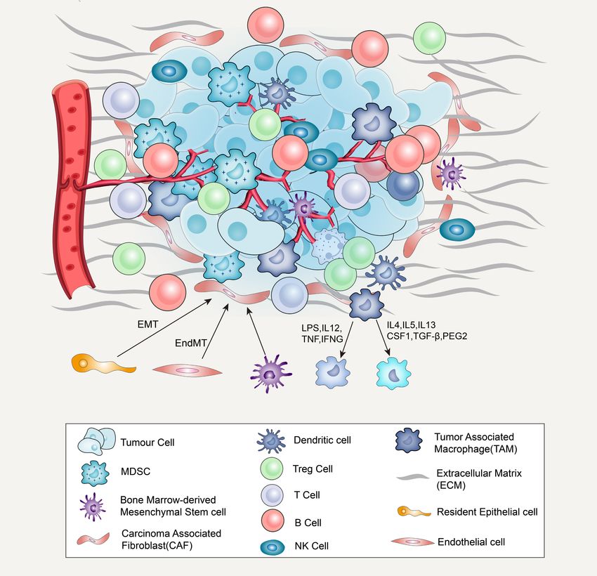
Frontiers in Cell and Developmental Biology | www.frontiersin.org 3 August 2021 | Volume 9 | Article 705280Cao et al. Tumor Microenvironment and Drug Therapy FIGURE 1 | Major cellular constituents and matrix component of the TME, including cancer cells, immune cells (T-cells, B-cells, NK cells, dendritic cells, MDSCs, TAMs), CAFs and ECM. CAF derived from bone marrow mesenchymal stem cells and transform through epithelial–mesenchymal transition (EMT) or endothelial mesenchymal transition (EndMT) from resident epithelium or endothelial cells (A). When macrophages are exposed to LPS, MAMPs, IL12, TNF, IFNG, or another TLR agonists, they will transition to M1-like. When exposed to IL4, IL5, IL10, IL13, CSF1, TGFβ1, and PGE2, it will transition to M2-like state (B). etc.], which can stimulate the growth of tumor cells in vitro recruiting endothelial progenitor cells (EPCs) into carcinomas, and lead to therapeutic drug resistance (Straussman et al., 2012; an effect mediated in part by CXCL12 (Orimo et al., 2005). Erez et al., 2013; Paraiso and Smalley, 2013). CXCL12 can activate the PI3K/AKT signaling pathway in tumor Angiogenesis in tumor tissues can provide oxygen and cells, upregulate the expression of VEGF in tumor tissues, nutrients for tumor cell metabolism and promote tumor growth and promote angiogenesis (Wen et al., 2019). VEGF activates and metastasis. Many studies have shown that CAFs can release the main signaling pathway in tumor angiogenesis by binding a variety of stimulating factors that promote angiogenesis and to its cognate receptor, VEGFR (Claesson-Welsh and Welsh, play an important role in the recruitment and proliferation of 2013). Mirkeshavarz et al. (2017) found that CAFs can secrete tumor vascular endothelial cells and the formation of vascular interleukin-6 (IL-6) and VEGF to induce angiogenesis in oral sprouts (Benyahia et al., 2017). CAFs promote angiogenesis by cancer, and that IL-6 can induce the secretion of VEGF in Frontiers in Cell and Developmental Biology | www.frontiersin.org 4 August 2021 | Volume 9 | Article 705280
Cao et al.                                                                                                  Tumor Microenvironment and Drug Therapy

CAF cell lines. CAF can also release active growth factors                  significant side effects due to lack of specificity, such as cachexia
from the ECM by expressing MMPs, which indirectly promotes                  and anemia (Roberts et al., 2013; Tran et al., 2013). Because of
angiogenesis (Najafi et al., 2019b) and serves as one of the sources        the lack of specific markers for CAF, this method is not feasible
of MMP9 (Boire et al., 2005) and MMP13 (Vosseler et al., 2009).             at present, so the need to know more about the mechanism by
Both these substances have been shown to release VEGF from the              which CAF works remains important for the development of
ECM to increase angiogenesis in tumors (Lederle et al., 2010).              more targeted treatments.
   Cancer-associated fibroblasts interact with tumor cells                     In a parallel study, pharmacological stimulation of the VDR
through inflammatory signals, thereby affecting tumor cell                  was successfully performed in activated pancreatic stellate cells
migration and invasion. The CAF-mediated CXCL12/CXCR4                       (PSCs). VDR is the main genomic inhibitor that is activated by
axis plays a key role in tumor cell proliferation, invasion, and            PSCs. In addition, treatment with the VDR ligand calcipotriol
migration. The CXCL12/CXCR4 axis can activate the MEK/ERK,                  induced matrix remodeling, which can inhibit tumor-related
PI3K/AKT, and Wnt/β-catenin pathways to promote EMT,                        inflammation and fibrosis, and also improves the transport of
thereby promoting tumor invasion and metastasis (Guo et al.,                gemcitabine to the tumor area, thus reversing chemotherapy
2016; Zhou et al., 2019; Mortezaee, 2020). It also activates the            resistance in the pancreatic ductal adenocarcinoma model
PI3K, MAPK, and ERK1/2 signaling pathways, promotes the                     (Sherman et al., 2014). Due to the complex interaction between
secretion of MMPs, reduces the adhesion of tumor cells, and                 CAF and other cells in the tumor environment, targeting some
increases their invasion and metastasis ability (Wu and Dai,                CAF subsets may cause multiple responses in the TME, which
2017). In addition, a recent study found that CAF-secreted                  may have multiple effects depending on the individual. To
CXCL-1 can stimulate the migration and invasion of oral                     eradicate cancer, the synergistic combination of CAF-targeted
cancer cells, that there is an interdependent relationship                  therapy and other effective treatments (such as immunotherapy)
between CAFs and cancer cells in the oral squamous carcinoma                should also be considered.
microenvironment, and that CXCL-1 can upregulate MMP-1                         Furthermore, the CXCL12/CXCR4 axis activates multiple
in CAF expression and activity (Wei et al., 2019). In addition,             signaling pathways to promote tumor cell proliferation, invasion,
CAFs can change the structure and physical properties of the                distant metastasis, and inhibit apoptosis. Therefore, the screening
ECM, thereby affecting tumor cell migration and invasion                    of antagonists targeting the CXCL12/CXCR4 signaling pathway
(Egeblad et al., 2010).                                                     is a promising target for tumor therapy. Lecavalier-Barsoum
                                                                            et al. (2018) found that the CXCR4 inhibitor AMD3100 can
                                                                            inhibit the CXCL12/CXCR4 axis in the treatment of patients
Drug Resistance and Targeted Therapy                                        with advanced disseminated high-grade serous ovarian cancer,
of CAF                                                                      and the combination of AMD3100 and low-dose paclitaxel can
The fight against drug resistance remains a major challenge in              inhibit the growth of ovarian cancer cells. In osteosarcoma,
tumor treatment. CAFs mediate a variety of tumor resistance                 AMD3100 blocks the invasion and metastasis of osteosarcoma
to chemotherapeutic drugs. CAFs can act on tumor cells by                   to the lung by inhibiting the JNK and AKT pathways (Liao
secreting cytokines, activating downstream signaling pathways in            et al., 2015). Another CXCR4 antagonist, AMD3465, can inhibit
tumor cells, and promoting tumor resistance (Chen and Song,                 the proliferation, colony formation, invasion, and migration of
2019). Studies have shown that CAFs can enhance EMT and                     bladder cancer cells through the CXCL12/CXCR4/β-catenin axis
cisplatin resistance in non-small cell lung cancer induced by               (Zhang et al., 2018).
transforming growth factor β by releasing high levels of IL-                   Micro RNA and siRNA can silence gene expression through
6, while cisplatin, in turn, promotes cancer cells to produce               post-transcriptional regulatory mechanisms, which may be
transforming growth factor β, resulting in CAF activation                   another viable way to inhibit CXCR4 expression. In breast
(Figure 1). CAFs can also promote chemotherapy resistance                   cancer cells, siRNA targeting CXCR4 inhibited the migration
in tumor cells by secreting exosomes. Gemcitabine (GEM) is                  of breast cancer cells in vitro (Burger et al., 2011). miR-126
currently a chemotherapy drug that is commonly used in the                  can also inactivate the RhoA signaling pathway in colon cancer
treatment of pancreatic cancer. Fang et al. (2019) found that               by reducing the expression of CXCR4 and inducing a tumor
exosomal miR-106b derived from CAFs plays an important                      suppressor effect (Yuan et al., 2016). These studies show that
role in GEM resistance in pancreatic cancer. Recently, Zhang                miRNA or siRNA targeting CXCR4 is of great significance in
et al. (2020) showed that exosomal miR-522 secreted by CAFs                 tumor treatment research. CTCE-9908 is composed of dimers of
prevents the death of cancer cells by targeting ALOX15 and                  CXCL12, which is a competitive inhibitor of CXCL12 targeting
blocking the accumulation of lipid-ROS. In addition, a new                  CXCR4 and can inhibit the secretion of CXCL12 (Guo et al.,
mechanism for obtaining gastric cancer drug resistance through              2016). Huang et al. (2009) reported that CTCE-9908 can target
the intercellular signaling pathways of USP7, hnRNPA1, exo-                 the CXCL12/CXCR4 axis and inhibit primary tumor growth and
miR-522, and ALOX15 has been observed.                                      metastasis of breast cancer. Hassan et al. (2011) also found that
    Direct ablation of CAF can promote the regression of                    CTCE-9908 combined with the anti-angiogenic agent DC101 also
immunogenic tumors (Feig et al., 2013), which has been explored             reduced the volume of the primary tumor and distant metastasis
in several recent studies, where these cells are cleared by injection       compared with DC101 alone. Moreover, an in vitro experiment
of diphtheria toxin or targeting FAP-specific chimeric antigen              proved that CTCE-9908 can inhibit the growth, invasion, and
receptor T cells; direct ablation of CAF, however, can lead to              metastasis of prostate cancer (Wong et al., 2014). This evidence

Frontiers in Cell and Developmental Biology | www.frontiersin.org       5                                      August 2021 | Volume 9 | Article 705280
Cao et al. Tumor Microenvironment and Drug Therapy
supports CTCE-9908 as an efficacious novel agent to prevent and (Sun et al., 2018). The exosomes of tumor cells can stimulate
treat the spread of metastatic cancer. At present, cancer treatment TAMs to secrete cytokines and enhance tumor invasion and
methods targeting CAFs and the CXCL12/CXCR4 axis are being metastasis (Trivedi et al., 2016).
explored and developed rapidly.
Drug Resistance and Targeted Therapy
of TAM
TUMOR-ASSOCIATED MACROPHAGE Tumor-associated macrophages can promote tumor repair
response by coordinating tissue damage and limit the anti-
Tumor-associated macrophages account for a large proportion tumor activity of conventional chemotherapy and radiotherapy
of most malignant tumors. They promote tumor progression at by providing a protective niche for CSCs (Mantovani et al.,
different levels by promoting genetic instability, cultivating CSCs, 2017). There is increasing evidence that macrophages play a
supporting metastasis, and taming protective adaptive immunity central role in both normal and diseased tissue remodeling,
(Mantovani et al., 2017). TAMs can be divided into M1-like including angiogenesis, basement membrane rupture, leukocyte
and M2-like types. When macrophages are exposed to cytokines infiltration, and immunosuppression. Therefore, TAMs have
such as bacterial lipopolysaccharide (LPS), microbe-associated become a promising target for the development of new anticancer
molecular patterns (MAMPs), IL12, TNF, interferon-γ (IFNG), treatments. These methods are mainly focused on the depletion
or other Toll-like receptor (TLR) agonists, they will be in a of M2-like TAMs and/or promotion of their transformation to
pro-inflammatory and anti-tumor state, hence M1-like. When M1-like phenotype (Cassetta and Pollard, 2018; Pradel et al.,
exposed to IL4, IL5, IL10, IL13, CSF1, TFGB1, and prostaglandin 2018). However, the effectiveness of this method may be limited
E2 (PGE2), it transitions from a pro-inflammatory state to an by a variety of factors, such as alternative immunosuppressive
anti-inflammatory and pro-tumor state, that is, to an M2-like cells that can compensate for TAMs, the existence of innate
state (Murray et al., 2014). TAMs have a high degree of functional and acquired drug resistance mechanisms, and the emergence of
plasticity and can quickly adapt to changing microenvironment strong immunosuppression after cessation of treatment (Quail
(Gubin et al., 2018). The necrotic and anoxic regions of the and Joyce, 2017). PLX-3397 is a small-molecule inhibitor of
TME contain M2-like TAMs, with low fluidity, limited antigen the CSF-1 pathway. It is not only an effective tyrosine kinase
presentation ability, and secrete a large number of tumor support inhibitor of CSF-1R, but also targeted at cKit and FLT3.
factors (Wenes et al., 2016). The metabolic spectrum of TAMs is Blocking CSF-1/CSF-1R can reduce TAMS and reprogramming
in a dynamic model, which can change with the nutritional needs TAMS in the TME and enhance the activation of T cells in
of malignant tumor cells and changes in TME. It also has a far- the TME by enhancing antigen presentation. The downstream
reaching impact on the survival of TAMs, cancer progression, and effect blocked by CSF-1/CSF-1R hinders the growth of the
tumor-targeted immune response. tumor (Zhu et al., 2014). In a mouse model of preclinical
The most abundant inflammatory or immune cell type is lung adenocarcinoma, PLX-3397 has been shown to change
near the CAF-populated areas in the tumor stroma, indicating the distribution of TAMs in the TME and reduce tumor
a close interaction between TAMs and CAF. In prostate cancer, load (Cuccarese et al., 2017). In the syngeneic mouse model
CAF-mediated CXCL12/CXCR4 axis induces the differentiation of BRAFV600E mutant melanoma, PLX-3397 combined with
of monocytes and possibly M1 cells into pro-tumor M2 cells. adoptive cell metastasis immunotherapy showed a decrease in
Conversely, TAMs with the M2 phenotype activate CAFs, thereby TAMs (Mok et al., 2014). In similar melanoma mouse models,
promoting tumor malignancy (Augsten et al., 2014; Comito et al., PLX-3397 combined with BRAF inhibitor PLX4032 significantly
2014). In vitro co-culture experiments showed that CAF-like reduced M2 phenotypic macrophage recruitment, resulting in
BM-MSCs enhanced the invasiveness of TAM-like macrophages. significant tumor growth inhibition (Ngiow et al., 2016). In
These macrophages strongly stimulate the proliferation and addition, recent studies have shown that M2-like TAMs, which
invasion of CAFs, thereby synergistically promoting the seem to be regulators of lysosomal pH, express high levels
development of neuroblastoma (Hashimoto et al., 2016). of vacuolar ATP enzymes and are expected to become a new
Tumor-associated macrophages release TNF-α to increase drug target (Kuchuk et al., 2018; Liu et al., 2019). Targeting
MMPs secreted by tumor cells and tumor stromal cells, destroy TAMs has proven to be a promising strategy, and with the
basement membrane tissue, and promote tumor metastasis deepening of preclinical development of TAM-targeted drugs
(Shuman Moss et al., 2012). TAMs also stimulate vascular and the new progress in the study of TAM mechanism, TAM-
endothelium to secrete VEGF by synthesizing and secreting the targeted therapy will become an important supplement to
Wnt7b protein to regulate angiogenesis (Yeo et al., 2014). TNF- anticancer drugs.
α binds to tumor necrosis factor receptor 1 (TNFR-1), activates
the VEGFC/VEGFR3 pathway, and promotes lymphangiogenesis
(Ran and Montgomery, 2012). In addition, transforming (TGF-β) MYELOGENOUS SUPPRESSOR CELLS
secreted by TAMs can induce EMT of colorectal cancer cells,
thereby promoting the invasion and metastasis of colorectal Myelogenous suppressor cells (MDSCs) are a heterogeneous
cancer cells (Yang et al., 2019). Notably, exosomes are one of the population composed of bone marrow progenitor cells and
components in TME, which carry a variety of active substances immature bone marrow cells (IMCs) (Gabrilovich et al., 2012).
and are the mediator of information transmission between cells Under normal physiological conditions, IMCs produced in the
Frontiers in Cell and Developmental Biology | www.frontiersin.org 6 August 2021 | Volume 9 | Article 705280Cao et al. Tumor Microenvironment and Drug Therapy
bone marrow can rapidly differentiate into mature granulocytes, lymphoma in combination with immune checkpoint inhibitors.
macrophages, or dendritic cells. In tumors and other pathological Systemic administration of AZD9150 significantly decreased
conditions, IMCs cannot normally differentiate into mature bone granulocyte MDSCs in peripheral blood mononuclear cells
marrow cells under the action of cytokines, thus forming MDSCs (PBMCs) (Reilley et al., 2018). Current targeting strategies mainly
with immunosuppressive functions, including T cell suppression include induction of differentiation into mature cells, inhibiting
and innate immune regulation (Kumar et al., 2016). In the TME, its expansion and recruitment, and blocking its immune
immunosuppressive cytokines such as IL-10 and TGF-β secreted characteristics. Studies have shown that some neutralizing
by MDSCs are important factors that inhibit the anti-tumor antibodies or inhibitors targeting chemokine systems (CXCR4,
immune response and promote tumor progression (Yaseen et al., CXCR2, and CCL2) and tumor-derived factors (CSF1, GM-CSF,
2020; Salminen, 2021). Studies have shown that TGF-β can and IL-6) can inhibit the expansion or recruitment of MDSC
inhibit the cytotoxic activity of cytotoxic T and NK cells by (Bayne et al., 2012; Sumida et al., 2012; Highfill et al., 2014).
reducing the production of interferon-γ (IFN-γ). On the other For example, the chemokine receptor CCR5 plays a key role
hand, TGF-β can also inhibit the proliferation of anti-tumor in the chemotaxis of MDSCs to TME (Weber et al., 2018).
immune active cells and inhibit anti-tumor immunity from the However, not all MDSCs express CCR5. In melanoma mice,
root (Salminen et al., 2018). Bone marrow mesenchymal stem MDSCs expressing CCR5 have stronger immunosuppressive
cells play a role in inducing proliferation in the TME due to ability than MDSCs that do not express CCR5. Blocking CCR5
the interaction between cytokines and chemokines in the tumor can inhibit the recruitment and immunosuppressive activity of
inflammatory environment. Conversely, MDSCs can stimulate MDSCs and improve the survival rate of melanoma patients
angiogenesis by producing matrix metalloproteinase 9, pro-factor (Blattner et al., 2018).
2, and VEGF, which further induces the migration of cancer cells It has been found that some drugs, such as phosphodiesterase-
to endothelial cells and promotes the metastasis of cancer cells 5 inhibitors (sildenafil, cyclooxygenase-2 inhibitors
(Lee et al., 2018; Yang et al., 2020). (acetylsalicylic acid and celecoxib), vardenafil and tadalafil and
Myelogenous suppressor cells produce high levels of bardoxolone methyl, can directly block the immunosuppressive
inhibitory molecules, such as Arg1, reactive oxygen species activity of MDSCs and restore T cell response (Serafini et al.,
(ROS), inducible nitric oxide synthase (iNOS), and prostaglandin 2006; Nagaraj et al., 2010; Fujita et al., 2011; Obermajer et al.,
E2 (PGE2), to directly inhibit the anti-tumor immune response 2011). Recent studies have found that MDSC-specific peptide-Fc
induced by effector T cells (Kusmartsev et al., 2004; Gabrilovich fusion protein therapy can completely deplete MDSCs in the
and Nagaraj, 2009; Condamine et al., 2015; He et al., 2018). blood, spleen, and tumor without affecting other immune cells,
MDSCs can also inhibit the immune response by inducing and inhibit tumor growth process (Qin et al., 2014), which
regulatory T cells (Tregs), promoting the development of provides a new idea for inhibiting tumor growth in vivo. In
macrophages into M2 phenotypes, and differentiating into patients and animal models, the failure of anti-angiogenic
TAMs (Huang et al., 2006; Weber et al., 2018). Deng et al. therapy based on inhibition of the VEGF pathway is often
(2017) found that MDSC-exosomes can directly accelerate concomitant with an increase in the number of MDSCs or
the proliferation and metastasis of tumor cells by delivering TAMs infiltrating tumor tissues (Lu-Emerson et al., 2013;
miR-126a, which indicates that MDSCs have a new regulatory Gabrusiewicz et al., 2014). Along this line of thinking, anti-VEGF
mechanism on tumor cells. MDSC-induced immunosuppression therapy is thought to upregulate alternative angiogenic factors
promotes tumor progression by promoting EMT, accelerating (prokinin-1 and proagonin-2) produced by myeloid cells, which
immune escape, and enhancing the formation of metastatic may accidentally produce anti-angiogenic effects and limit
lesions (Veglia et al., 2018). Additionally, MDSCs enhance the tumor recurrence.
stemness of tumor cells, promote angiogenesis by secreting IL6 Recent studies have shown that the accumulation of MDSCs
and NO, and promote tumor growth, invasion, and metastasis in tumors limits the effect of anti-programmed death 1 (PD1)
directly or indirectly by inhibiting T cells or natural killer cells in the treatment of rhabdomyosarcoma checkpoint blockage.
(Condamine et al., 2015, 2016). Inhibition of MDSC metastasis with an anti-CXCR2 antibody can
enhance the efficacy of anti-PD1 (Highfill et al., 2014). In a tumor
model of tolerant mice, the removal of MDSCs with gemcitabine
Drug Resistance and Targeted Therapy combined with immunotherapy can effectively break the self-
of MDSC tolerance and induce strong anti-tumor immunity (Ko et al.,
The key roles played by MDSCs in the TME show that 2007). Several chemotherapeutic drugs, such as anthracyclines,
it is necessary to target them effectively by blocking or platinum derivatives, and doxorubicin, can induce immunogenic
deleting them. Although they play a key role in tumor cell death, thus activating an effective anti-tumor adaptive
progression, there are no FDA-approved drugs or treatments response (Kroemer et al., 2013). The chemical process for
that directly target MDSCs. At present, clinical trials are enhancing the anticancer effect of these drugs includes increasing
underway to target the activities of iNOS Arg1 and STAT3, the antigen presentation ability of dendritic cells and the
metabolism through CD36, transport through CXCR2, and other subsequent CD8+ T cell response (Bracci et al., 2014). Although
mechanisms for different types of cancer (Fleming et al., 2018). the current targeted therapy targeting only MDSCs does not
The antisense oligonucleotide STAT3 inhibitor AZD9150 has strengthen clinical outcomes, it may play an important role in
been used in phase 1b clinical trials of diffuse large B-cell anticancer immunotherapy in the future.
Frontiers in Cell and Developmental Biology | www.frontiersin.org 7 August 2021 | Volume 9 | Article 705280Cao et al. Tumor Microenvironment and Drug Therapy
CONCLUSION AUTHOR CONTRIBUTIONS
Most of the treatments are focused on a certain aspect of DC, XN, JL, YH, ZC, BC, JL, and JG: wrote the review article
the TME. Although some of these therapeutic responses prepared and assembled the figure and table. QD, JA, LL,
have produced positive results, a more effective way is to and QW: critically organized and revised the manuscript by
promote inflammatory innate immune cells, such as CD8+ incorporating significant reports. All authors contributed to the
T cells, and to alter many aspects of TME through a article and approved the submitted version.
strong inflammatory response. Breakthrough drug resistance
remains a major clinical challenge. The response of tumor
cells to treatment depends not only on the regulation FUNDING
of the TME but also on the aberration of its genome.
Targeted therapy cannot focus on the complete depletion This work was funded by the National Natural Science
of all inherent cells in the TME, as this may cause Foundation of China (Grant No. 82000721), Post-Doctor
severe complications in the patient. The solution must Research Project, West China Hospital, Sichuan University
be a complex combination, with focus on developing (Grant No. 2019HXBH089), Health commission of Sichuan
multidrug management that targets both tumor cells and province (Grant No. 20PJ036), and Programs from the
TME to overcome resistance and improve prognosis as Department of Science and Technology of Sichuan Province
much as possible. (Grant No. 2020YJ0054).
REFERENCES Claesson-Welsh, L., and Welsh, M. (2013). VEGFA and tumour angiogenesis.
J. Intern. Med. 273, 114–127. doi: 10.1111/joim.12019
Augsten, M., Sjöberg, E., Frings, O., Vorrink, S. U., Frijhoff, J., and Olsson, E. Comito, G., Giannoni, E., Segura, C. P., Barcellos-de-Souza, P., Raspollini, M. R.,
(2014). Borg Å, Östman A: cancer-associated fibroblasts expressing CXCL14 Baroni, G., et al. (2014). Cancer-associated fibroblasts and M2-polarized
rely upon NOS1-derived nitric oxide signaling for their tumor-supporting macrophages synergize during prostate carcinoma progression. Oncogene 33,
properties. Cancer Res. 74, 2999–3010. doi: 10.1158/0008-5472.can-13-2740 2423–2431. doi: 10.1038/onc.2013.191
Bayne, L. J., Beatty, G. L., Jhala, N., Clark, C. E., Rhim, A. D., Stanger, Condamine, T., Dominguez, G. A., Youn, J. I., Kossenkov, A. V., Mony, S., Alicea-
B. Z., et al. (2012). Tumor-derived granulocyte-macrophage colony-stimulating Torres, K., et al. (2016). Lectin-type oxidized LDL receptor-1 distinguishes
factor regulates myeloid inflammation and T cell immunity in pancreatic population of human polymorphonuclear myeloid-derived suppressor cells in
cancer. Cancer Cell 21, 822–835. doi: 10.1016/j.ccr.2012.04.025 cancer patients. Sci. Immunol. 1:aaf8943. doi: 10.1126/sciimmunol.aaf8943
Benyahia, Z., Dussault, N., Cayol, M., Sigaud, R., Berenguer-Daizé, C., Delfino, Condamine, T., Ramachandran, I., Youn, J. I., and Gabrilovich, D. I. (2015).
C., et al. (2017). Stromal fibroblasts present in breast carcinomas promote Regulation of tumor metastasis by myeloid-derived suppressor cells. Annu. Rev.
tumor growth and angiogenesis through adrenomedullin secretion. Oncotarget Med. 66, 97–110. doi: 10.1146/annurev-med-051013-052304
8, 15744–15762. doi: 10.18632/oncotarget.14999 Cuccarese, M. F., Dubach, J. M., Pfirschke, C., Engblom, C., Garris, C., Miller,
Blattner, C., Fleming, V., Weber, R., Himmelhan, B., Altevogt, P., Gebhardt, C., M. A., et al. (2017). Heterogeneity of macrophage infiltration and therapeutic
et al. (2018). CCR5(+) Myeloid-Derived Suppressor Cells Are Enriched and response in lung carcinoma revealed by 3D organ imaging. Nat. Commun.
Activated in Melanoma Lesions. Cancer Res. 78, 157–167. doi: 10.1158/0008- 8:14293.
5472.can-17-0348 Cui, T. X., Kryczek, I., Zhao, L., Zhao, E., Kuick, R., Roh, M. H., et al. (2013).
Boire, A., Covic, L., Agarwal, A., Jacques, S., Sherifi, S., and Kuliopulos, A. Myeloid-derived suppressor cells enhance stemness of cancer cells by inducing
(2005). PAR1 is a matrix metalloprotease-1 receptor that promotes invasion and microRNA101 and suppressing the corepressor CtBP2. Immunity 39, 611–621.
tumorigenesis of breast cancer cells. Cell 120, 303–313. doi: 10.1016/j.cell.2004. doi: 10.1016/j.immuni.2013.08.025
12.018 Del Prete, A., Schioppa, T., Tiberio, L., Stabile, H., and Sozzani, S. (2017). Leukocyte
Bonnet, D., and Dick, J. E. (1997). Human acute myeloid leukemia is organized trafficking in tumor microenvironment. Curr. Opin. Pharmacol. 35, 40–47.
as a hierarchy that originates from a primitive hematopoietic cell. Nat. Med. 3, doi: 10.1016/j.coph.2017.05.004
730–737. doi: 10.1038/nm0797-730 Deng, Z., Rong, Y., Teng, Y., Zhuang, X., Samykutty, A., Mu, J., et al.
Borah, A., Raveendran, S., Rochani, A., Maekawa, T., and Kumar, D. S. (2015). (2017). Exosomes miR-126a released from MDSC induced by DOX treatment
Targeting self-renewal pathways in cancer stem cells: clinical implications for promotes lung metastasis. Oncogene 36, 639–651. doi: 10.1038/onc.2016.229
cancer therapy. Oncogenesis 4:e177. doi: 10.1038/oncsis.2015.35 Denton, A. E., Roberts, E. W., and Fearon, D. T. (2018). Stromal Cells in the Tumor
Bracci, L., Schiavoni, G., Sistigu, A., and Belardelli, F. (2014). Immune-based Microenvironment. Adv. Exp. Med. Biol. 1060, 99–114.
mechanisms of cytotoxic chemotherapy: implications for the design of novel Egeblad, M., Rasch, M. G., and Weaver, V. M. (2010). Dynamic interplay between
and rationale-based combined treatments against cancer. Cell Death Differ. 21, the collagen scaffold and tumor evolution. Curr. Opin. Cell Biol. 22, 697–706.
15–25. doi: 10.1038/cdd.2013.67 doi: 10.1016/j.ceb.2010.08.015
Burger, J. A., Stewart, D. J., Wald, O., and Peled, A. (2011). Potential of CXCR4 Erez, N., Glanz, S., Raz, Y., Avivi, C., and Barshack, I. (2013). Cancer associated
antagonists for the treatment of metastatic lung cancer. Expert Rev. Anticancer fibroblasts express pro-inflammatory factors in human breast and ovarian
Ther. 11, 621–630. doi: 10.1586/era.11.11 tumors. Biochem. Biophys. Res. Commun. 437, 397–402. doi: 10.1016/j.bbrc.
Byrne, A. M., Bouchier-Hayes, D. J., and Harmey, J. H. (2005). Angiogenic and cell 2013.06.089
survival functions of vascular endothelial growth factor (VEGF). J. Cell. Mol. Fan, Q. M., Jing, Y. Y., Yu, G. F., Kou, X. R., Ye, F., Gao, L., et al. (2014).
Med. 9, 777–794. doi: 10.1111/j.1582-4934.2005.tb00379.x Tumor-associated macrophages promote cancer stem cell-like properties via
Cassetta, L., and Pollard, J. W. (2018). Targeting macrophages: therapeutic transforming growth factor-beta1-induced epithelial-mesenchymal transition
approaches in cancer. Nat. Rev. Drug Discov. 17, 887–904. doi: 10.1038/nrd. in hepatocellular carcinoma. Cancer Lett. 352, 160–168. doi: 10.1016/j.canlet.
2018.169 2014.05.008
Chen, X., and Song, E. (2019). Turning foes to friends: targeting cancer-associated Fang, Y., Zhou, W., Rong, Y., Kuang, T., Xu, X., Wu, W., et al. (2019).
fibroblasts. Nat. Rev. Drug Discov. 18, 99–115. doi: 10.1038/s41573-018-0004-1 Exosomal miRNA-106b from cancer-associated fibroblast promotes
Frontiers in Cell and Developmental Biology | www.frontiersin.org 8 August 2021 | Volume 9 | Article 705280Cao et al. Tumor Microenvironment and Drug Therapy
gemcitabine resistance in pancreatic cancer. Exp. Cell Res. 383:111543. Kalluri, R. (2016). The biology and function of fibroblasts in cancer. Nat. Rev.
doi: 10.1016/j.yexcr.2019.111543 Cancer 16, 582–598. doi: 10.1038/nrc.2016.73
Feig, C., Jones, J. O., Kraman, M., Wells, R. J., Deonarine, A., Chan, D. S., Kessenbrock, K., Plaks, V., and Werb, Z. (2010). Matrix metalloproteinases:
et al. (2013). Targeting CXCL12 from FAP-expressing carcinoma-associated regulators of the tumor microenvironment. Cell 141, 52–67. doi: 10.1016/j.cell.
fibroblasts synergizes with anti-PD-L1 immunotherapy in pancreatic cancer. 2010.03.015
Proc. Natl. Acad. Sci. U. S. A. 110, 20212–20217. doi: 10.1073/pnas.1320318110 Kidd, S., Spaeth, E., Watson, K., Burks, J., Lu, H., Klopp, A., et al. (2012). Origins of
Fleming, V., Hu, X., Weber, R., Nagibin, V., Groth, C., Altevogt, P., et al. (2018). the tumor microenvironment: quantitative assessment of adipose-derived and
Targeting Myeloid-Derived Suppressor Cells to Bypass Tumor-Induced bone marrow-derived stroma. PLoS One 7:e30563. doi: 10.1371/journal.pone.
Immunosuppression. Front. Immunol. 9:398. doi: 10.3389/fimmu.2018. 0030563
00398 Kim, J., and Bae, J. S. (2016). Tumor-Associated Macrophages and Neutrophils in
Fujita, M., Kohanbash, G., Fellows-Mayle, W., Hamilton, R. L., Komohara, Y., Tumor Microenvironment. Mediators Inflamm. 2016:6058147.
Decker, S. A., et al. (2011). COX-2 blockade suppresses gliomagenesis by Kinugasa, Y., Matsui, T., and Takakura, N. (2014). CD44 expressed on cancer-
inhibiting myeloid-derived suppressor cells. Cancer Res. 71, 2664–2674. doi: associated fibroblasts is a functional molecule supporting the stemness and drug
10.1158/0008-5472.can-10-3055 resistance of malignant cancer cells in the tumor microenvironment. Stem Cells
Gabrilovich, D. I., and Nagaraj, S. (2009). Myeloid-derived suppressor cells as 32, 145–156. doi: 10.1002/stem.1556
regulators of the immune system. Nat. Rev. Immunol. 9, 162–174. doi: 10.1038/ Ko, H. J., Kim, Y. J., Kim, Y. S., Chang, W. S., Ko, S. Y., Chang, S. Y., et al. (2007).
nri2506 A combination of chemoimmunotherapies can efficiently break self-tolerance
Gabrilovich, D. I., Ostrand-Rosenberg, S., and Bronte, V. (2012). Coordinated and induce antitumor immunity in a tolerogenic murine tumor model. Cancer
regulation of myeloid cells by tumours. Nat. Rev. Immunol. 12, 253–268. doi: Res. 67, 7477–7486. doi: 10.1158/0008-5472.can-06-4639
10.1038/nri3175 Kopp, H. G., Ramos, C. A., and Rafii, S. (2006). Contribution of endothelial
Gabrusiewicz, K., Liu, D., Cortes-Santiago, N., Hossain, M. B., Conrad, progenitors and proangiogenic hematopoietic cells to vascularization of tumor
C. A., Aldape, K. D., et al. (2014). Anti-vascular endothelial growth factor and ischemic tissue. Curr. Opin. Hematol. 13, 175–181. doi: 10.1097/01.moh.
therapy-induced glioma invasion is associated with accumulation of Tie2- 0000219664.26528.da
expressing monocytes. Oncotarget 5, 2208–2220. doi: 10.18632/oncotarget. Krishnamurthy, N., and Kurzrock, R. (2018). Targeting the Wnt/beta-catenin
1893 pathway in cancer: update on effectors and inhibitors. Cancer Treat. Rev. 62,
Gubin, M. M., Esaulova, E., Ward, J. P., Malkova, O. N., Runci, D., Wong, P., 50–60. doi: 10.1016/j.ctrv.2017.11.002
et al. (2018). High-Dimensional Analysis Delineates Myeloid and Lymphoid Kroemer, G., Galluzzi, L., Kepp, O., and Zitvogel, L. (2013). Immunogenic cell
Compartment Remodeling during Successful Immune-Checkpoint Cancer death in cancer therapy. Annu. Rev. Immunol. 31, 51–72.
Therapy. Cell 175, 1014–1030.e19. Kuchuk, O., Tuccitto, A., Citterio, D., Huber, V., Camisaschi, C., Milione, M.,
Guo, F., Wang, Y., Liu, J., Mok, S. C., Xue, F., and Zhang, W. (2016). et al. (2018). pH regulators to target the tumor immune microenvironment in
CXCL12/CXCR4: a symbiotic bridge linking cancer cells and their stromal human hepatocellular carcinoma. Oncoimmunology 7:e1445452. doi: 10.1080/
neighbors in oncogenic communication networks. Oncogene 35, 816–826. doi: 2162402x.2018.1445452
10.1038/onc.2015.139 Kumar, V., Patel, S., Tcyganov, E., and Gabrilovich, D. I. (2016). The Nature of
Hashimoto, O., Yoshida, M., Koma, Y., Yanai, T., Hasegawa, D., Kosaka, Y., et al. Myeloid-Derived Suppressor Cells in the Tumor Microenvironment. Trends
(2016). Collaboration of cancer-associated fibroblasts and tumour-associated Immunol. 37, 208–220.
macrophages for neuroblastoma development. J. Pathol. 240, 211–223. doi: Kusmartsev, S., Nefedova, Y., Yoder, D., and Gabrilovich, D. I. (2004). Antigen-
10.1002/path.4769 specific inhibition of CD8+ T cell response by immature myeloid cells in cancer
Hassan, S., Buchanan, M., Jahan, K., Aguilar-Mahecha, A., Gaboury, L., Muller, is mediated by reactive oxygen species. J. Immunol. 172, 989–999. doi: 10.4049/
W. J., et al. (2011). CXCR4 peptide antagonist inhibits primary breast tumor jimmunol.172.2.989
growth, metastasis and enhances the efficacy of anti-VEGF treatment or Lau, E. Y., Ho, N. P., and Lee, T. K. (2017). Cancer Stem Cells and Their
docetaxel in a transgenic mouse model. Int. J. Cancer 129, 225–232. doi: Microenvironment: biology and Therapeutic Implications. Stem Cells Int.
10.1002/ijc.25665 2017:3714190.
He, Y. M., Li, X., Perego, M., Nefedova, Y., Kossenkov, A. V., Jensen, E. A., et al. Lecavalier-Barsoum, M., Chaudary, N., Han, K., Koritzinsky, M., Hill, R., and
(2018). Transitory presence of myeloid-derived suppressor cells in neonates is Milosevic, M. (2018). Targeting the CXCL12/CXCR4 pathway and myeloid
critical for control of inflammation. Nat. Med. 24, 224–231. doi: 10.1038/nm. cells to improve radiation treatment of locally advanced cervical cancer. Int.
4467 J. Cancer 143, 1017–1028. doi: 10.1002/ijc.31297
Highfill, S. L., Cui, Y., Giles, A. J., Smith, J. P., Zhang, H., Morse, E., et al. (2014). Lederle, W., Hartenstein, B., Meides, A., Kunzelmann, H., Werb, Z., Angel, P., et al.
Disruption of CXCR2-mediated MDSC tumor trafficking enhances anti-PD1 (2010). MMP13 as a stromal mediator in controlling persistent angiogenesis in
efficacy. Sci. Transl. Med. 6:237ra267. skin carcinoma. Carcinogenesis 31, 1175–1184. doi: 10.1093/carcin/bgp248
Huang, B., Pan, P. Y., Li, Q., Sato, A. I., Levy, D. E., Bromberg, J., et al. (2006). Lee, S. E., Lim, J. Y., Kim, T. W., Jeon, Y. W., Yoon, J. H., Cho, B. S., et al.
Gr-1+CD115+ immature myeloid suppressor cells mediate the development (2018). Matrix Metalloproteinase-9 in Monocytic Myeloid-Derived Suppressor
of tumor-induced T regulatory cells and T-cell anergy in tumor-bearing host. Cells Correlate with Early Infections and Clinical Outcomes in Allogeneic
Cancer Res. 66, 1123–1131. doi: 10.1158/0008-5472.can-05-1299 Hematopoietic Stem Cell Transplantation. Biol. Blood Marrow Transplant. 24,
Huang, E. H., Singh, B., Cristofanilli, M., Gelovani, J., Wei, C., Vincent, L., et al. 32–42. doi: 10.1016/j.bbmt.2017.08.017
(2009). A CXCR4 antagonist CTCE-9908 inhibits primary tumor growth and Li, B., and Wang, J. H. (2011). Fibroblasts and myofibroblasts in wound healing:
metastasis of breast cancer. J. Surg. Res. 155, 231–236. doi: 10.1016/j.jss.2008. force generation and measurement. J. Tissue Viability 20, 108–120. doi: 10.1016/
06.044 j.jtv.2009.11.004
Huang, M., Li, Y., Zhang, H., and Nan, F. (2010). Breast cancer stromal Liao, Y. X., Fu, Z. Z., Zhou, C. H., Shan, L. C., Wang, Z. Y., Yin, F., et al. (2015).
fibroblasts promote the generation of CD44+CD24- cells through SDF- AMD3100 reduces CXCR4-mediated survival and metastasis of osteosarcoma
1/CXCR4 interaction. J. Exp. Clin. Cancer Res. 29:80. by inhibiting JNK and Akt, but not p38 or Erk1/2, pathways in in vitro and
Jin, W. (2020). Role of JAK/STAT3 Signaling in the Regulation of Metastasis, the mouse experiments. Oncol. Rep. 34, 33–42. doi: 10.3892/or.2015.3992
Transition of Cancer Stem Cells, and Chemoresistance of Cancer by Epithelial- Liu, N., Luo, J., Kuang, D., Xu, S., Duan, Y., Xia, Y., et al. (2019). Lactate
Mesenchymal Transition. Cells 9:217. doi: 10.3390/cells9010217 inhibits ATP6V0d2 expression in tumor-associated macrophages to promote
Jinushi, M., Chiba, S., Yoshiyama, H., Masutomi, K., Kinoshita, I., Dosaka-Akita, HIF-2α-mediated tumor progression. J. Clin. Invest. 129, 631–646. doi: 10.
H., et al. (2011). Tumor-associated macrophages regulate tumorigenicity and 1172/jci123027
anticancer drug responses of cancer stem/initiating cells. Proc. Natl. Acad. Sci. Lu-Emerson, C., Snuderl, M., Kirkpatrick, N. D., Goveia, J., Davidson, C., Huang,
U. S. A. 108, 12425–12430. doi: 10.1073/pnas.1106645108 Y., et al. (2013). Increase in tumor-associated macrophages after antiangiogenic
Frontiers in Cell and Developmental Biology | www.frontiersin.org 9 August 2021 | Volume 9 | Article 705280Cao et al. Tumor Microenvironment and Drug Therapy therapy is associated with poor survival among patients with recurrent Rafii, S., Lyden, D., Benezra, R., Hattori, K., and Heissig, B. (2002). Vascular and glioblastoma. Neuro Oncol. 15, 1079–1087. doi: 10.1093/neuonc/not082 haematopoietic stem cells: novel targets for anti-angiogenesis therapy? Nat. Rev. Magee, J. A., Piskounova, E., and Morrison, S. J. (2012). Cancer stem cells: impact, Cancer 2, 826–835. doi: 10.1038/nrc925 heterogeneity, and uncertainty. Cancer Cell 21, 283–296. doi: 10.1016/j.ccr. Ran, S., and Montgomery, K. E. (2012). Macrophage-mediated lymphangiogenesis: 2012.03.003 the emerging role of macrophages as lymphatic endothelial progenitors. Mantovani, A., Marchesi, F., Malesci, A., Laghi, L., and Allavena, P. (2017). Cancers 4, 618–657. doi: 10.3390/cancers4030618 Tumour-associated macrophages as treatment targets in oncology. Nat. Rev. Räsänen, K., and Vaheri, A. (2010). Activation of fibroblasts in cancer stroma. Exp. Clin. Oncol. 14, 399–416. doi: 10.1038/nrclinonc.2016.217 Cell Res. 316, 2713–2722. doi: 10.1016/j.yexcr.2010.04.032 Mirkeshavarz, M., Ganjibakhsh, M., Aminishakib, P., Farzaneh, P., Mahdavi, N., Reilley, M. J., McCoon, P., Cook, C., Lyne, P., Kurzrock, R., Kim, Y., et al. Vakhshiteh, F., et al. (2017). Interleukin-6 secreted by oral cancer- associated (2018). STAT3 antisense oligonucleotide AZD9150 in a subset of patients with fibroblast accelerated VEGF expression in tumor and stroma cells. Cell. Mol. heavily pretreated lymphoma: results of a phase 1b trial. J. Immunother. Cancer Biol. 63, 131–136. doi: 10.14715/cmb/2017.63.10.21 6:119. Mok, S., Koya, R. C., Tsui, C., Xu, J., Robert, L., Wu, L., et al. (2014). Inhibition Roberts, E. W., Deonarine, A., Jones, J. O., Denton, A. E., Feig, C., Lyons, S. K., of CSF-1 receptor improves the antitumor efficacy of adoptive cell transfer et al. (2013). Depletion of stromal cells expressing fibroblast activation protein- immunotherapy. Cancer Res. 74, 153–161. doi: 10.1158/0008-5472.can-13- α from skeletal muscle and bone marrow results in cachexia and anemia. J. Exp. 1816 Med. 210, 1137–1151. doi: 10.1084/jem.20122344 Mortezaee, K. (2020). CXCL12/CXCR4 axis in the microenvironment of solid Salimifard, S., Masjedi, A., Hojjat-Farsangi, M., Ghalamfarsa, G., Irandoust, M., tumors: a critical mediator of metastasis. Life Sci. 249:117534. doi: 10.1016/j. Azizi, G., et al. (2020). Cancer associated fibroblasts as novel promising lfs.2020.117534 therapeutic targets in breast cancer. Pathol. Res. Pract. 216:152915. doi: 10. Mou, W., Xu, Y., Ye, Y., Chen, S., Li, X., Gong, K., et al. (2015). Expression of Sox2 1016/j.prp.2020.152915 in breast cancer cells promotes the recruitment of M2 macrophages to tumor Salminen, A. (2021). Increased immunosuppression impairs tissue homeostasis microenvironment. Cancer Lett. 358, 115–123. doi: 10.1016/j.canlet.2014. with aging and age-related diseases. J. Mol. Med. 99, 1–20. doi: 10.1007/s00109- 11.004 020-01988-7 Murray, P. J., Allen, J. E., Biswas, S. K., Fisher, E. A., Gilroy, D. W., Goerdt, Salminen, A., Kauppinen, A., and Kaarniranta, K. (2018). Myeloid-derived S., et al. (2014). Macrophage activation and polarization: nomenclature and suppressor cells (MDSC): an important partner in cellular/tissue senescence. experimental guidelines. Immunity 41, 14–20. doi: 10.1016/j.immuni.2014. Biogerontology 19, 325–339. doi: 10.1007/s10522-018-9762-8 06.008 Seino, T., Kawasaki, S., Shimokawa, M., Tamagawa, H., Toshimitsu, K., Fujii, Nagaraj, S., Youn, J. I., Weber, H., Iclozan, C., Lu, L., Cotter, M. J., et al. M., et al. (2018). Human Pancreatic Tumor Organoids Reveal Loss of Stem (2010). Anti-inflammatory triterpenoid blocks immune suppressive function Cell Niche Factor Dependence during Disease Progression. Cell Stem Cell 22, of MDSCs and improves immune response in cancer. Clin. Cancer Res. 16, 454–467.e6. 1812–1823. doi: 10.1158/1078-0432.ccr-09-3272 Serafini, P., Meckel, K., Kelso, M., Noonan, K., Califano, J., Koch, W., et al. (2006). Najafi, M., Farhood, B., and Mortezaee, K. (2019a). Cancer stem cells (CSCs) in Phosphodiesterase-5 inhibition augments endogenous antitumor immunity by cancer progression and therapy. J. Cell. Physiol 234, 8381–8395. doi: 10.1002/ reducing myeloid-derived suppressor cell function. J. Exp. Med. 203, 2691– jcp.27740 2702. doi: 10.1084/jem.20061104 Najafi, M., Farhood, B., and Mortezaee, K. (2019b). Extracellular matrix (ECM) Sherman, M. H., Yu, R. T., Engle, D. D., Ding, N., Atkins, A. R., Tiriac, H., stiffness and degradation as cancer drivers. J. Cell. Biochem. 120, 2782–2790. et al. (2014). Vitamin D receptor-mediated stromal reprogramming suppresses doi: 10.1002/jcb.27681 pancreatitis and enhances pancreatic cancer therapy. Cell 159, 80–93. doi: Ngiow, S. F., Meeth, K. M., Stannard, K., Barkauskas, D. S., Bollag, G., 10.1016/j.cell.2014.08.007 Bosenberg, M., et al. (2016). Co-inhibition of colony stimulating factor-1 Shuman Moss, L. A., Jensen-Taubman, S., and Stetler-Stevenson, W. G. (2012). receptor and BRAF oncogene in mouse models of BRAF(V600E) melanoma. Matrix metalloproteinases: changing roles in tumor progression and metastasis. Oncoimmunology 5:e1089381. doi: 10.1080/2162402x.2015.1089381 Am. J. Pathol. 181, 1895–1899. Obermajer, N., Muthuswamy, R., Odunsi, K., Edwards, R. P., and Kalinski, P. Straussman, R., Morikawa, T., Shee, K., Barzily-Rokni, M., Qian, Z. R., Du, J., et al. (2011). PGE(2)-induced CXCL12 production and CXCR4 expression controls (2012). Tumour micro-environment elicits innate resistance to RAF inhibitors the accumulation of human MDSCs in ovarian cancer environment. Cancer Res. through HGF secretion. Nature 487, 500–504. doi: 10.1038/nature11183 71, 7463–7470. doi: 10.1158/0008-5472.can-11-2449 Sumida, K., Wakita, D., Narita, Y., Masuko, K., Terada, S., Watanabe, K., et al. Orimo, A., Gupta, P. B., Sgroi, D. C., Arenzana-Seisdedos, F., Delaunay, T., (2012). Anti-IL-6 receptor mAb eliminates myeloid-derived suppressor cells Naeem, R., et al. (2005). Stromal fibroblasts present in invasive human breast and inhibits tumor growth by enhancing T-cell responses. Eur. J. Immunol. 42, carcinomas promote tumor growth and angiogenesis through elevated SDF- 2060–2072. doi: 10.1002/eji.201142335 1/CXCL12 secretion. Cell 121, 335–348. doi: 10.1016/j.cell.2005.02.034 Sun, W., Luo, J. D., Jiang, H., and Duan, D. D. (2018). Tumor exosomes: a double- Paraiso, K. H., and Smalley, K. S. (2013). Fibroblast-mediated drug resistance in edged sword in cancer therapy. Acta Pharmacol. Sin. 39, 534–541. doi: 10.1038/ cancer. Biochem. Pharmacol. 85, 1033–1041. doi: 10.1016/j.bcp.2013.01.018 aps.2018.17 Peng, D., Tanikawa, T., Li, W., Zhao, L., Vatan, L., Szeliga, W., et al. (2016). Tran, E., Chinnasamy, D., Yu, Z., Morgan, R. A., Lee, C. C., Restifo, N. P., et al. Myeloid-Derived Suppressor Cells Endow Stem-like Qualities to Breast Cancer (2013). Immune targeting of fibroblast activation protein triggers recognition Cells through IL6/STAT3 and NO/NOTCH Cross-talk Signaling. Cancer Res. of multipotent bone marrow stromal cells and cachexia. J. Exp. Med. 210, 76, 3156–3165. doi: 10.1158/0008-5472.can-15-2528 1125–1135. doi: 10.1084/jem.20130110 Piccard, H., Muschel, R. J., and Opdenakker, G. (2012). On the dual roles and Trivedi, M., Talekar, M., Shah, P., Ouyang, Q., and Amiji, M. (2016). Modification polarized phenotypes of neutrophils in tumor development and progression. of tumor cell exosome content by transfection with wt-p53 and microRNA-125b Crit. Rev. Oncol. Hematol. 82, 296–309. doi: 10.1016/j.critrevonc.2011.06.004 expressing plasmid DNA and its effect on macrophage polarization. Oncogenesis Pradel, L. P., Franke, A., and Ries, C. H. (2018). Effects of IL-10 and T(h) 2 5:e250. doi: 10.1038/oncsis.2016.52 cytokines on human Mϕ phenotype and response to CSF1R inhibitor. J. Leukoc. Tu, E., Chia, P. Z., and Chen, W. (2014). TGFβ in T cell biology and tumor Biol. 103, 545–558. doi: 10.1002/jlb.5ma0717-282r immunity: angel or devil? Cytokine Growth Factor Rev. 25, 423–435. doi: Qin, H., Lerman, B., Sakamaki, I., Wei, G., Cha, S. C., Rao, S. S., et al. 10.1016/j.cytogfr.2014.07.014 (2014). Generation of a new therapeutic peptide that depletes myeloid-derived Veglia, F., Perego, M., and Gabrilovich, D. (2018). Myeloid-derived suppressor cells suppressor cells in tumor-bearing mice. Nat. Med. 20, 676–681. doi: 10.1038/ coming of age. Nat. Immunol. 19, 108–119. doi: 10.1038/s41590-017-0022-x nm.3560 Vosseler, S., Lederle, W., Airola, K., Obermueller, E., Fusenig, N. E., and Mueller, Quail, D. F., and Joyce, J. A. (2017). Molecular Pathways: deciphering Mechanisms M. M. (2009). Distinct progression-associated expression of tumor and stromal of Resistance to Macrophage-Targeted Therapies. Clin. Cancer Res. 23, 876–884. MMPs in HaCaT skin SCCs correlates with onset of invasion. Int. J. Cancer 125, doi: 10.1158/1078-0432.ccr-16-0133 2296–2306. doi: 10.1002/ijc.24589 Frontiers in Cell and Developmental Biology | www.frontiersin.org 10 August 2021 | Volume 9 | Article 705280
You can also read