A novel method for the in vivo isolation of circulating tumor cells from peripheral blood of cancer patients using a functionalized and structured ...
←
→
Page content transcription
If your browser does not render page correctly, please read the page content below
A novel method for the in vivo isolation of circulating
tumor cells from peripheral blood of cancer patients
 using a functionalized and structured medical wire

         Received February 17, 2012; Accepted April 3, 2012
                    DOI: 10.3892/ijo.2012.1557
INTERNATIONAL JOURNAL OF ONCOLOGY
A novel method for the in vivo isolation of circulating tumor cells
from peripheral blood of cancer patients using a
functionalized and structured medical wire
NADIA SAUCEDO-ZENI1, STEFFI MEWES1, ROBERT NIESTROJ1, LUKASZ GASIOROWSKI2, DAVID MURAWA3,
PIOTR NOWACZYK3, TATIANA TOMASI1, EKKEHARD WEBER1, GRZEGORZ DWORACKI2,
NILS G. MORGENTHALER1, HEIKE JANSEN4, CORINNA PROPPING4, KAROLINA STERZYNSKA5,
WOJCIECH DYSZKIEWICZ2, MACIEJ ZABEL5, MARION KIECHLE4,
UTE REUNING4, MANFRED SCHMITT4 and KLAUS LÜCKE1
1
GILUPI GmbH, Potsdam, Germany; 2Department of Thoracic Surgery, WCPiT, Poznan University of Medical Science;
3
First Department of Surgical Oncology and General Surgery, Wielkopolska Cancer Center, 61-866 Poznan, Poland;
4
Department of Obstetrics and Gynecology, Klinikum rechts der Isar, Technische Universitaet Muenchen,
Munich, Germany; 5Poznan University of Medical Science, Fredry 10, 61-701 Poznan, Poland
Received February 17, 2012; Accepted April 3, 2012
DOI: 10.3892/ijo.2012.1557
Abstract. The isolation of circulating tumor cells (CTCs) from 24 patients, with a median of 5.5 (0-50) CTCs in breast cancer
the blood of patients afflicted with solid malignant tumors (n=12) and 16 (2-515) CTCs in NSCLC (n=12). CTCs could be
becomes increasingly important as it may serve as a ‘liquid isolated across all tumor stages, including early stage cancer,
biopsy’ with the potential of monitoring the course of the cancer in which distant metastases were not yet diagnosed, while no
disease and its response to cancer therapy, with subsequent CTCs could be detected in healthy volunteers. In this observa-
molecular characterization. For this purpose, we functionalized tory study, no adverse effects were noted. Evidently, the FSMW
a structured medical Seldinger guidewire (FSMW), normally has the potential to become an important device to enrich CTCs
used to obtain safe access to blood vessels and other organ in vivo for monitoring the course of the cancer disease and the
cavities, with a chimeric monoclonal antibody directed to the efficacy of anticancer treatment.
cell surface expressed epithelial cell surface adhesion molecule
(EpCAM). This medical device was optimized in vitro and its Introduction
biocompatibility was tested according to the regulations for
medical devices and found to be safe with no noteworthy side Personalized cancer treatment is at present one of the most chal-
effects. Suitability, specificity and sensitivity of the FSMW to lenging goals in cancer research in order to improve health and
catch and enrich CTCs in vivo from circulating peripheral blood quality of life of cancer patients (1,2). Since many tumor cells
were tested in 24 breast cancer or non-small cell lung cancer are distinct on the molecular level (3), and modern cancer drugs
(NSCLC) patients and in 29 healthy volunteers. For this, the target selected molecular pathways, the definitive goal is to
FSMW was inserted through a standard venous cannula into identify cancer patients at risk and those who may benefit from
the cubital veins of healthy volunteers or cancer patients for a certain cancer therapy (1,2). For this purpose, the isolation
the duration of 30 min. After removal, CTCs were identified of circulating tumor cells (CTC) from the peripheral blood of
by immunocytochemical staining of EpCAM and/or cyto- patients afflicted with cancer becomes increasingly important
keratins and staining of their nuclei and counted. The FSMW and thus is in the focus of cancer research and the pharmaceu-
successfully enriched EpCAM-positive CTCs from 22 of the tical industry (4). Enrichment and enumeration of CTC offer the
potential as a prognostic cancer biomarker and may fulfill the
criteria for a surrogate biomarker to evaluate the response of
patients to cancer therapy (5,6). Molecular characterization of
Correspondence to: Dr Klaus Lücke, GILUPI GmbH, Am CTC is a rapidly developing research field aiming at revealing
Mühlenberg 11, D-14476 Potsdam, Germany novel drug targets and to investigate mechanisms of tumor
E-mail: klaus.luecke@gilupi.com metastasis (7).
Over the past decade, several in vitro methodological
Key words: circulating tumor cells, breast cancer, non-small cell approaches to isolate and detect rare CTC in the peripheral
lung cancer, epithelial cell adhesion molecule, medical wire blood of cancer patients have been reported, including flow
cytofluorometry (8), image-based immunological approaches
(9), fluidic microchip technology (10), and PCR methods
(11-13). At present, an antibody-coated magnetic particle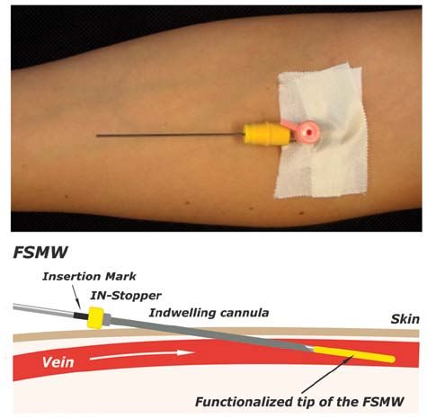
2                               SAUCEDO-ZENI et al: In vivo ISOLATION OF CIRCULATING TUMOR CELLS

isolation system targeting the epithelial cell surface EpCAM            SK-BR-3 breast cancer cells (CLS, Eppelheim, Germany),
is used in most studies designed for ex vivo quantification of          which were grown in cell culture flasks until cell monolayers
CTC in the blood of patients with advanced breast, colon, or            reached a confluency of ~80%. Adherent SK-BR-3 cells were
prostate cancer (14-16). Patients with metastasized cancer              detached with trypsin/EDTA (ethylene diamine tetraacetate)
diseases exhibit detectable numbers of CTC in their blood (17),         (Biochrom AG, Berlin, Germany), centrifuged, and suspended
however, since all ex vivo detection systems are limited by the         in phosphate-buffered saline (PBS). In addition, EDTA-anti-
blood volume that can be obtained from the patients or handled          coagulated blood samples obtained from healthy donors were
by the detection system (18), these technologies are of relatively      spiked with those cells and tested for binding to the FSMW.
low sensitivity.                                                        Furthermore, blood samples from breast or lung cancer patients
    To overcome the limitations of small blood sample volumes           were also assessed in the in vitro flow system to test for CTC
of the ex vivo CTC isolation techniques, new approaches are             binding to the FSMW.
needed for screening large blood volumes in vivo using for
example the principles of photoacoustic flow cytofluorometry            Cytotoxicity tests. To examine potential cytotoxic effects of the
(19). We developed an alternative medical device: a structured          FSMW, we performed direct FSMW cell contact and material
and functionalized medical wire (FSMW) based on a Seldinger             elution tests in vitro, based on the requirements for a class IIa
guidewire (20) that offers the opportunity of capturing CTC             medical device, as outlined in the ISO Guideline 10993-5
from the circulating blood of cancer patients. Like in other            (www.iso.org). For the elution test, eluates of one or three
ex vivo CTC-detection technologies, identification of CTC               FSMW devices (three to identify any variation in the produc-
captured by the FSMW is performed by phenotyping CTC with               tion process), and of reference materials known to be toxic
antibodies directed to cytokeratins and/or epithelial cell markers.     (copper wire, Goodfellow GmbH, Bad Nauheim, Germany) or
Occasionally trapped hematologic cells are identified by anti-          non-toxic (Teflon wire, PTFE, Goodfellow GmbH) for normal
bodies directed to respective hematologic cell surface markers.         human dermal fibroblasts (NHDF, C-12302, PromoCell GmbH,
Here, we describe a novel in vivo CTC-catching medical device,          Heidelberg, Germany) were generated (0.095 m2 FSMW surface
the FSMW, its biocompatibility and first results of its applica-        area eluted with 5 ml of RPMI-medium, FG 1235, Biochrom
tion for in vivo enrichment of CTC from the peripheral blood            AG, at 37˚C for 24 h), and their influence on cell morphology
of patients presenting with breast cancer or non-small cell lung        and viability of NHDF examined. Evaluation of potential
cancer (NSCLC).                                                         effects of these eluates on cell morphology was done qualita-
                                                                        tively by microscopic analysis for changes in cell morphology,
Materials and methods                                                   cell adhesive capacity, and cell disintegration. Viability of cells
                                                                        was tested by use of the colorimetric TTC assay (triphenyltetra-
Functionalized structured medical wire. The FSMW (Fig. 1) is            zolium chloride test).
based on a stainless steel medical wire (Seldinger guidewire) of            In brief, for the microscopical inspection, NHDF were
0.5 mm in diameter and 160 mm in length (EPflex, Dettingen,             exposed to FSMW eluates for 48 h at 37˚C. Eluates of reference
Germany). The first 20 mm are plated with a 2 µm thick gold layer       materials known to be toxic (copper wire, Goodfellow GmbH)
deposited on the device by galvanization (OTEK, Brieselang,             or non-toxic (Teflon wire, PTFE, Goodfellow GmbH) for
Germany). Subsequently, a hydrogel layer composed of a linear,          NHDF were tested in parallel. After 48 h of incubation, cells
synthetic polycarboxylate is attached to the gold layer (Xantec         were stained with the dye CFSE [5-(and-6)-carboxyfluorescein
Bioanalytics, Düsseldorf, Germany). The carboxyl groups                 diacetate succinimidyl ester; Invitrogen, Carlsbad, USA] and
present in the hydrogel are then activated with EDC (1-ethyl-           inspected under the fluorescence microscope. Impairment of cell
3-[3-dimethylaminopropyl] carbodiimide hydrochloride) and               morphology was scored according to a common reaction index
NHS (N-hydroxysuccinimide) (Sigma-Aldrich GmbH, Seelze,                 (none versus slight, moderate or severe).
Germany) allowing for functionalization via covalent coupling               To test for viability of NHDF, a colorimetric assay (TTC
of a chimeric antibody directed to the epithelial cell adhesion         assay) was performed, which allows for the quantitative
molecule CD326 (EpCAM; HEA 125, kindly provided by                      assessment of cell viability in the presence of material-derived
Dr G. Moldenhauer, German Cancer Research Center, DKFZ,                 eluates. After 48 h of incubation of NHDF with the eluates, a
Heidelberg, Germany) present on the surface of most CTC.                yellow tetrazolium compound (EZ4U, Cell Proliferation and
Functionalization of the FSMW is carried out under clean room           Cytotoxicity Assay, Biomedica Medizinprodukte GmbH &
conditions. The FSMW is a sterile medical device and intended           Co. KG, Vienna, Austria) was added (3 h, 37˚C) which was
for single in vivo use only. For the in vivo application, the sterile   converted due to metabolic mitochondrial cellular activity to a
FSMW is inserted into a standard 20G (pink color code) intra-           brick-red formazan. Change in color was recorded at 450 nm
venous cannula (Fig. 2).                                                by use of a microtiter plate reader (SPECTROstar-Omega,
                                                                        BMG Labtech, Ortenberg, Germany). For direct contact tests,
In vitro experiments. Prior to FSMW application in patients,            the FSMW device was placed on a layer of adherent NHDF,
a hemodynamic flow system was applied (Fig. 3). Within this             followed by a 24 h incubation of this set-up at 37˚C. Reference
system, blood was routed via flexible tubes through a flow              wires made of Teflon or copper were tested in parallel.
chamber into which up to two FSMW are inserted. The flow
rate, velocity, and flow direction can be regulated by a peristaltic    Acute systemic toxicity. To assess potential hazardous effects
pump. A flow rate of 20 ml/min was applied to reflect in vivo flow      of the FSMW in humans, possibly occurring, acute systemic
conditions within cubital veins (21). In this in vitro flow system      toxicity of the FSMW was tested. In a parallel clinical study,
the FSMW was tested for binding of cultured EpCAM-positive              aiming at the in vivo enrichment of trophoblasts from pregnant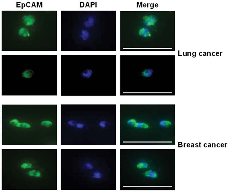
INTERNATIONAL JOURNAL OF ONCOLOGY 3
women, potential acute systemic toxicity of the eluates had 30 min, followed by the addition of secondary FITC-conjugated
already been monitored. According to these tests, no adverse anti-IgM (Santa Cruz Biotechnology Inc.), 1:200 (2 µg/ml), for
effects were caused by the FSMW (22). For evaluation of the 30 min at RT. For detection of adherent platelets, a fluorescence-
acute systemic toxicity of the FSMW, four test groups of five labeled antibody directed to CD41 was applied [anti-CD41a-PE,
mice each [Charles River, Sulzfeld, Germany; NMRI (Han) 1:100 (2 µg/ml) in PBS; Santa Cruz Biotechnology Inc.].
mice, female non-pregnant, nulliparous, 19-24 g body weight,
4-5 weeks old) were injected intravenously or intraperitoneally Study population. Patients were recruited at the Wielkopolska
with a single dose of four different extracts of the FSMW. The Cancer Center, Department of Surgical Oncology and General
amount of eluates administered were adjusted to body weight Surgery, and at the Poznan University of Medical Sciences, both
at a volume of 50 ml/kg for 0.9% (w/v) NaCl, 5% (v/v) ethanol, in Poznan, Poland. The healthy donor population was recruited
and cotton seed oil, and at 10 g/kg for polyethylene glycol-400. at the Department of Obstetrics and Gynecology, Klinikum
Four control groups of five mice each were treated in the same rechts der Isar, Technische Universitaet Muenchen, Munich,
manner with the corresponding extraction vehicle not previ- Germany. The studies were approved by the Institutional Ethics
ously exposed to the FSMW. Committees and written informed consents of the patients and
This dosing regime supplied an about 100 times higher dose volunteers were obtained.
of the FSMW than the expected dose in humans. The animals For the in vitro functionality test of the FSMW, we included
were followed up immediately after injection, and at 4, 24, 48 17 patients afflicted with breast cancer and 7 patients with
and 72 h intervals for body weight and toxic effects. Cage side NSCLC. For the in vivo functionality test of the FSMW, we
observations included spontaneous activity, lethargy, recumbent included 12 breast cancer patients and 12 NSCLC patients
position, convulsions, tremors, apnoea, asphyxia, vocalization, (Tables I and II). Patients of both cancer types had different
diarrhea, obvious changes in the skin and fur, eyes and mucous tumor stages and had not undergone surgery or received
membranes (salivation, discharge). At the end of the observation chemotherapy at the time of enrolment. Principal inclusion
period the animals were sacrificed. All animals were subjected to criteria for breast cancer and NSCLC patients were >18 years
gross necropsy. Any gross pathological changes were recorded. of age, histopathologically confirmed diagnosis of breast
The studies were approved by the Ethics Committee of the cancer or potentially resectable NSCLC with eligibility for
Medical University of Poznan, Poland. radical surgery. Exclusion criteria: history of psychiatric
disease, participation in other clinical trials, history of allergy,
Hemocompatibility test. Objectives of the hemocompatibility anaphylactic reactions, prior immunological diseases (anti-
test were assessment of the patient's risk to develop thrombosis, phospholipid antibody syndrome, Goodpasture's syndrome,
coagulation, and hemolysis after in vivo exposure to the FSMW. lupus erythematosus, polychondritis, rheumatoid arthritis,
The test method described in the ISO Guideline 10993-4:2002 sarcoidosis, scleroderma, Sjogren's syndrome, ANCA positive
(E) and according to Xu and coworkers (23) was employed. states), immunodeficiencies, prior infections with hepatitis
To assess any influence of the FSMW on the blood coagula- viruses, the cytomegalovirus (CMV), or infectious diseases
tion cascade, the plasma recalcification time was determined. such as tuberculosis, syphilis, or toxoplasmosis. Principal
The FSMW was incubated for 10 min at 37˚C in citrate anti- exclusion criteria for the 29 healthy volunteers (premenopausal
coagulated platelet-poor plasma, which was then treated with 24, postmenopausal 5) were >18 years of age, pregnancy or
CaCl2 to induce blood coagulation. Clotting time of plasma, breastfeeding, any kind of oncological or allergic disease
which had not been exposed to the FSMW, served as a control. including asthma and thromboembolic complications. Median
Fibrin formation was determined during a rigid up and down age of the volunteers was 27 (range 22-67).
movement of the FSMW in a plasma sample or in a plasma For in vitro studies of the FSMW in an in vitro flow system,
sample which was not exposed to the FSMW. Time intervals peripheral venous blood from patients afflicted with breast
until fibrin deposits were formed on the FSMW were recorded. cancer or NSCLC was harvested into EDTA-tubes (Sarstedt
For examination of any potential hemolytic risk exerted by the AG & Co., Nuembrecht, Germany). Blood samples were kept
FSMW, erythrocytes were isolated from EDTA anti-coagulated at RT and processed in the flow system at RT within 72 h after
blood of healthy donors and eluates of the FSMW added. After drawing of the blood.
incubation at room temperature (RT), the erythrocyte-eluate-
suspension was centrifuged and the hemolysis rate (% increase In vivo application of the FSMW. The FSMW was designed to
in cell-free hemoglobin concentration) determined photo- fit into a standard 20G intravenous cannula, which is placed into
metrically. Products that have no hemolytic potential should the cubital vein of a cancer patient or a healthy donor (Fig. 2).
exhibit a hemolytic rate of less than 5%. An IN-Stopper (Sarstedt AG & Co.) allows secure fixation to the
To assess non-specific blood cell adhesion to the FSMW intravenous cannula. The FSMW is slowly pushed forward into
device, the antibody-functionalized part of the FSMW was put the cannula until the EpCAM-antibody-functionalized FSMW
into an in vitro hemodynamic flow system (Fig. 3) which simulates surface of 2 cm in length is exposed to the blood flow within
the in vivo blood flow situation, including FSMW contact period, the lumen of the vein. The correct insertion is indicated by a
body temperature, and flow rate. After removal, the FSMW mark on the distal part of the FSMW, which is not inserted into
was inspected microscopically for blood cells deposited on the the cannula. The FSMW remains in the cubital vein for 30 min.
device. Then, in order to detect any fibrin deposits on the FSMW, In the present study, the insertion of the FSMW was done
the device was stained with antibodies directed to fibrin(ogen) before the respective patient underwent surgery of the primary
[mouse anti-human fibrin antibody, IgM, 1:100 (2 µg/ml) in tumor. During the procedure of FSMW application, the patient
PBS; Santa Cruz Biotechnology Inc., Heidelberg, Germany] for remained in a flat or supine position. The total volume of blood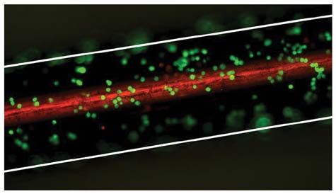
4                               SAUCEDO-ZENI et al: In vivo ISOLATION OF CIRCULATING TUMOR CELLS

Table I. Clinical characteristics of breast cancer patients assessed   Table II. Clinical characteristics of NSCLC patients assessed
for CTC enrichment in vivo and histomorphological features of          for CTC enrichment in vivo and histomorphological features
the primary tumor.                                                     of the primary tumor.

Classification
   No. of                                   Patients     Patients      Classification
                                                                          No. of  Patients                                   Patients
  patients                                  with 1-4     with ≥5         patients with 1-4                                   with ≥5
		                                          CTC (%)      CTC (%)       		         CTC (%)                                    CTC (%)

All patients                      12        3 (25)        9 (75)       All patients                      12       2 (16.7)    8 (66.7)
ER-/PR-status                                                          Histological classification
  Positive for either             10        3 (30)        7 (70)         Squamous cell lung               8       1 (12.5)    5 (62.5)
  Negative for both                2        0             2 (100)        carcinoma
                                                                         Adenocarcinoma                   3       1 (33.3)    2 (66.7)
HER2-status
                                                                         Large cell lung carcinoma        1       0           1 (100)
 Positive                           3       0             3 (100)
 Negative                           9       3 (33.3)      6 (66.7)     Histological grade
                                                                         G2                               9       2 (22.2)    6 (66.7)
Histological classification
                                                                         G3                               2       0           1 (50)
  Invasive-ductal                   7       1 (14.3)      6 (85.7)
                                                                         Unknown                          1       0           1 (100)
  Invasive-lobular                  3       1 (33.3)      2 (66.7)
  Invasive-ductal/                  1       1 (100)       0            Tumor stage
  invasive lobular                                                       T2N0M0                           4       1 (25)      2 (50)
  Invasive-ductal/                  1       0             1 (100)        T2N1M0                           3       0           3 (100)
  bifocal cancer                                                         T2N2M0                           2       1 (50)      1 (50)
                                                                         T3N0M0                           1       0           1 (100)
Adjuvant therapy
                                                                         T3N1M0                           2       0           1 (50)
  Chemotherapy                      3       1 (33.3)      2 (66.7)
  Endocrine therapy                 1       0             1 (100)
  No adjuvant therapy               8       2 (25)        6 (75)
Tumor stage                                                            stained with the nuclear dye 4,6-diamidino-2-phenylindole
  T1N1M0                            2       1 (50)        1 (50)       (DAPI; 1 µg/ml PBS; Life Technologies GmbH). Intensity of the
  T1N3M0                            1       0             1 (100)      immunocytochemical staining of CTC was evaluated using an
  T2N1M0                            3       1 (33.3)      2 (66.7)     Axio Imager.A1m microscope (Zeiss, Jena, Germany) equipped
  T2N3M0                            1       0             1 (100)      with an AxioCam digital camera system and the AxioVision 4.6
  T4N0M0                            1       0             1 (100)      software (Zeiss). EpCAM- or cytokeratin-positive cells included
  T4N2M0                            1       0             1 (100)      in the count had to disclose additional features such as a large
  T4N+N/A                           1       0             1 (100)      cell body (diameter 10-50 µm), an irregular cell shape, a large
  T1N1M1                            1       1 (100)       0            irregularly shaped nucleus, and a high nuclear to cytoplasmic
                                                                       ratio (17,25,26). Cells were counted on each FSMW by an oper-
  T3N+N/A                           1       0             1 (100)
                                                                       ator blinded to the clinical background of the patients. Results
                                                                       are given as number of CTC immobilized on the surface of the
                                                                       EpCAM-antibody-functionalized FSMW. In some cases, before
                                                                       inspection, cells were fixed with 4% (w/v) buffered paraformal-
coming into contact with the FSMW during the 30 min applica-           dehyde.
tion period is estimated at 1.5-3 liters (24).
                                                                       Results
Inspection of the FSMW for bound CTC. After removal of the
FSMW from the cubital vein, the FSMW was briefly and gently            Isolation and subsequent molecular characterization of CTC
washed in PBS, followed by incubation in PBS containing                from the blood of cancer patients becomes increasingly impor-
2% (w/v) bovine serum albumin (BSA, Carl Roth GmbH,                    tant as it may serve as a ‘liquid biopsy’ with the potential of
Karlsruhe, Germany, purity grade ≥98%), for 30 min at RT.              monitoring the course of the cancer disease and response to
Characterization of CTC captured by the FSMW was done by               cancer therapy. For this purpose, but different than currently
immunocytochemical staining for EpCAM or cytokeratins 4, 5,            employed ex vivo CTC enrichment protocols, we applied a
8, 9, and 18. Cells attached to the FSMW were incubated with           structured medical Seldinger guidewire (FSMW), functional-
an FITC-conjugated mouse monoclonal antibody directed to               ized with a chimeric monoclonal antibody directed to EpCAM,
EpCAM [1:100 in PBS (10 µg/ml); Acris Antibodies GmbH,                 to be used in vivo to catch and enrich CTC from the peripheral
Herford, Germany] and a phycoerythrin (PE)-conjugated rabbit           blood pool (Fig. 1). The FSMW was first optimized in vitro
antibody raised against CD45 [1:25 in PBS (2 µg/ml); Life              for its CTC catching ability and then tested for biocompat-
Technologies GmbH, Darmstadt, Germany]. Cells were counter-            ibility according to the ISO guidelines for medical devices.
INTERNATIONAL JOURNAL OF ONCOLOGY                                                                       5

Figure 1. Schematic drawing of the functionalized tip of the FSMW. Antibodies to the epithelial cell surface antigen EpCAM are attached to a polycarboxylate
hydrogel (1-5 µm) which is coated on a gold-plated (200 nm) Seldinger guidewire. Then the hydrogel is functionalized with antibodies to the EpCAM. This FSMW
interacts with target cells expressing EpCAM antigen on their surface, e.g., CTC of breast and lung cancer patients.

                                                                                  Figure 3. Schematic drawing of an in vitro flow system for repeated interaction
                                                                                  of CTC with the FSMW. This in vitro flow system allows the simulation of
Figure 2. Insertion of the FSMW into the cubital vein through a conventional      in vivo venous blood flow conditions. A bidirectional flow is maintained by a
cannula. Above: arm bend showing the place where the FSMW is inserted into        peristaltic pump to enable repeated interaction of blood with the FSMW. This
the cubital vein. Below: the FSMW is slowly pushed forward into the can-          flow system is used to test interaction of CTC present in anti-coagulated blood
nula until the anti-EpCAM-antibody-functionalized FSMW surface of 2 cm in         or anti-coagulated blood spiked with cultured tumor cells with up to two FSMW
length is exposed to the blood flow within the lumen of the vein. An IN-Stopper   simultaneously. The blood is not passing through the peristaltic pump to avoid
allows its secure fixation to the intravenous cannula. The correct length of      mechanical impairment of cells. The flow system contains 16 ml of blood. Flow
insertion is indicated by a mark on the distal part of the FSMW, which is not     conditions are 20 ml/min, 44 cycles per 30 min, at RT.
inserted into the cannula. The FSMW remains in the cubital vein for 30 min.

                                                                                  20 ml of anti-coagulated blood of healthy volunteers or blood
Subsequently, suitability of the FSMW to catch and enrich CTC                     obtained from breast cancer or NSCLC patients. The intention
in vivo from circulating peripheral blood (Fig. 2) was tested in                  of these experiments was to test: a) whether EpCAM-antibody
breast cancer and NSCLC patients in the framework of clinical                     mediated tumor cell immobilization on the FSMW occurs
trials, in comparison to healthy volunteers.                                      under conditions similar to venous blood flow and b) whether
                                                                                  non-malignant blood cells are interfering with the capture
In vitro evaluation of the CTC capture capability of the                          capability of the FSMW. Representative microscopic images
FSMW. Functionality of the FSMW in regard to its antibody-                        of FSMW-immobilized fluorescence-labeled SK-BR-3 cells
mediated CTC enrichment capability was tested in an in vitro                      from the in vitro flow system experiments are shown in Fig. 4.
dynamic flow system (Fig. 3) by capturing SK-BR-3 breast                          The functionality of the FSMW in the in vitro flow system was
cancer cells simulating human CTC which had been added to                         also tested with blood samples from breast cancer and NSCLC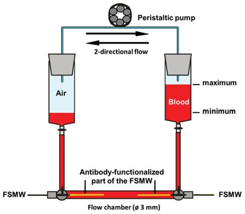
6                                    SAUCEDO-ZENI et al: In vivo ISOLATION OF CIRCULATING TUMOR CELLS

Figure 4. Representative image of cultured SK-BR-3 breast cancer cells cap-
tured by the FSMW. Cultured SK-BR-3 breast cancer cells were captured by
the FSMW, fixed with buffered 4% (w/v) paraformaldehyde and then stained
with FITC-labeled antibodies raised against EpCAM. The white lines denote         Figure 6. Test for potential adverse effects of eluates obtained from different
the borders of the FSMW. Green fluorescence indicates SK-BR-3 cells. Note:        wire materials by TTC assay according to ISO Guideline 10993-5. The ability
view of the FSMW from the top focal plane, thus only cells attached here are      of vital cells to convert a tetrazolium salt into a red formazan derivative was
in focus, cells in lower planes are not. The upper part of the FSMW appears in    employed to determine the influence of different wire eluates on the viability
red due to fluorescence illumination of the gold layer.                           of cultured NHDF. Reference eluates prepared from known non-cytotoxic
                                                                                  (Teflon) and cytotoxic (copper) wire materials and from eluates prepared from
                                                                                  different FSMW were added to cultured NHDF and formazan production
                                                                                  recorded photometrically at 450 nm after 3 h of incubation at RT. Obtained
                                                                                  optical density values are expressed as relative cell viability in percent. Eluates
                                                                                  were prepared from indicated numbers of test materials. Formazan production
                                                                                  by NHDF in the absence of any test wire eluates was set to 100%.

                                                                                  potential effects on the viability of cultured NHDF. Microscopic
                                                                                  inspection of NHDF after treatment with FSMW or Teflon
                                                                                  wire eluates did not disclose any changes in cell morphology.
                                                                                  In contrast, NHDF subjected to copper wire extract, detached
                                                                                  from the cell culture dish substratum and assumed a spindle-
                                                                                  shaped cell morphology (Fig. 5). Cellular mitochondrial activity
                                                                                  (as a surrogate marker for cell viability) assessed by the TTC
                                                                                  test remained unchanged when NHDF were exposed to the
                                                                                  FSMW eluate, compared to that observed in NHDF treated with
                                                                                  copper wire eluate, resulting in a more than 80% reduction of
                                                                                  cell viability (Fig. 6). Direct contact of NHDF with the FSMW
                                                                                  gave similar results. Even after a 24-h incubation period of the
Figure 5. Test for potential adverse effects of eluates obtained from different
                                                                                  FSMW on cultured NHDF, no perturbation of cell membrane
wire materials on cell integrity by assessment of cell morphology; according
to ISO guideline 10993-5. Cultured CFSE-stained NHDF were incubated               integrity was observed. In contrast, the copper wire as a positive
with different wire eluates for 24 h at 37˚C to demonstrate any influence of      control had a negative influence on NHDF since they detached
the eluates on cell morphology. (A), Reference Teflon wire eluate with known      from the substrate and showed a reduced proliferation rate.
non-cytotoxic effect on NHDF: no apparent changes in cell morphology. (B),
                                                                                      Tests for in vivo biocompatibility of the FSMW did not
Reference copper wire eluate with known cytotoxic effect on NHDF: severe
changes in cell morphology. (C), FSMW eluate prepared from a single FSMW:         reveal any signs of acute toxicity when FSMW eluates (n=4)
no apparent change in cell morphology. (D), FSMW eluate prepared from three       were injected intravenously or intraperitoneally into NMRI
FSMW: no apparent change in cell morphology. Staining of NHDF with CFSE           mice (n=5; for details see Materials and methods). No FSMW-
was performed to improve microscopic inspection.
                                                                                  related mortalities were recorded. All mice survived the test
                                                                                  period of 72 h independent of whether a negative control or a
                                                                                  FSMW eluate was applied and the mice showed normal food
patients. Under these conditions, the FSMW captured CTC in 7                      intake and unchanged body weight.
out of 17 (41%) anti-coagulated blood samples of breast cancer                        Hemocompatibility tests did not indicate any hemolytic
patients (range 1-44, median 5). Also, all anti-coagulated blood                  effects of FSMW eluates. The re-calcification time of platelet-
samples of NSCLC patients (n=7) turned out to be positive for                     poor plasma of citrate anti-coagulated blood from healthy
CTC (range 1-8, median 7).                                                        donors in the presence of the FSMW (n=5) was comparable to
                                                                                  the recalcification time of citrate anti-coagulated blood which
Biocompatibility of the FSMW device. To demonstrate the                           had not been exposed to the FSMW (n=5).
biocompatibility and safety of the FSMW for cancer patients
or healthy volunteers during its in vivo application, tests were                  In vivo application of the FSMW in healthy volunteers and in
performed according to ISO guidelines recommended for                             breast cancer and NSCLC patients. The FSMW was inserted
class IIa medical devices. Eluates prepared from the FSMW                         into the cubital veins of 29 healthy volunteers, 12 patients
as described in Materials and methods were tested for their                       afflicted with breast cancer and 12 patients presenting with
INTERNATIONAL JOURNAL OF ONCOLOGY 7
Table III. CTC enriched by in vivo use of the FSMW: summary
for lung and breast cancer patients.
Breast cancer (%) NSCLC (%)
No. of patients 12 12
Positive 10 (83.3) 12 (100)
CTC on FSMW
Range 0-50 2-515
Median 5.5 16
Mean ± SD 9.7±13.7 55.4±145
CTC counts
0 2 (16.7) 0
Figure 7. Number of CTC captured by the FSMW in vivo. Twelve NSCLC and8 SAUCEDO-ZENI et al: In vivo ISOLATION OF CIRCULATING TUMOR CELLS
of FSMW-captured CTC from breast cancer patients stained for to identify CTC. Nonetheless, in the past, also nucleic acid-based
EpCAM expression are depicted in Fig. 8. detection approaches were frequently used, with the disadvan-
In a series of clinical trials employing the CellSearch device tage that CTC enumeration or assessment of cell morphology is
(Veridex LLC, Warren, NJ) as an immunomagnetic isolation not possible by this technology (28,30,33-37).
technique for CTC in metastatic breast cancer patients (14,27), Different from these methods, we have developed a novel,
a disease-specific cut-off was used to define patient groups with proprietary in vivo CTC detecting technology which makes
a favorable (INTERNATIONAL JOURNAL OF ONCOLOGY 9
properties or of relative larger size than the majority of blood 3. Kan Z, Jaiswal BS, Stinson J, Janakiraman V, Bhatt D, Stern HM,
Yue P, Haverty PM, Bourgon R, Zheng J, Moorhead M,
cells (28,33,38,53,54), yet, not allowing an immediate visual Chaudhuri S, Tomsho LP, Peters BA, Pujara K, Cordes S, Davis DP,
microscopic control of the enriched CTC. Some technologies Carlton VE, Yuan W, Li L, Wang W, Eigenbrot C, Kaminker JS,
are using microfluidic filters or magnetic beads, also coated Eberhard DA, Waring P, Schuster SC, Modrusan Z, Zhang Z,
Stokoe D, De Sauvage FJ, Faham M and Seshagiri S: Diverse
with antibodies directed to EpCAM, in some cases combined somatic mutation patterns and pathway alterations in human
with magnetic rods, microposts, or herringbones to catch the cancers. Nature 466: 869-873, 2010.
antibody-labeled CTC (28,38). Yet, all these EpCAM-antibody- 4. Maheswaran S, Sequist LV, Nagrath S, Ulkus L, Brannigan B,
Collura CV, Inserra E, Diederichs S, Lafrate AJ, Bell DW,
based technologies assume that tumor cells of epithelial origin do Digumarthy S, Muzikansky A, Irimia D, Settleman J, Tompkins RG,
express the EpCAM antigen. This is not always so, epithelial cells Lynch TJ, Toner M and Haber DA: Detection of mutations in EGFR
may lack the EpCAM antigen. In this case, additional antibodies in circulating lung-cancer cells. N Engl J Med 359: 366-377, 2008.
5. Dotan E, Cohen SJ, Alpaugh KR and Meropol NJ: Circulating
to other epithelial surface antigens should be considered for cell tumor cells: evolving evidence and future challenges. Oncologist
trapping, such as CD49f, HER2, MUC1/2, or carcinoembryonic 14: 1070-1082, 2009.
antigen (CEA) (34,55). The novel FSMW is also making use 6. Krebs MG, Hou JM, Ward TH, Blackhall FH and Dive C:
Circulating tumour cells: their utility in cancer management and
of trapping epithelial-derived tumor cells with a high-affinity predicting outcomes. Ther Adv Med Oncol 2: 351-365, 2010.
antibody directed to EpCAM but other cell surface-directed 7. Hou JM, Krebs M, Ward T, Sloane R, Priest L, Hughes A, Clack G,
antibodies can be easily covalently attached to the hydrogel of Ranson M, Blackhall F and Dive C: Circulating tumor cells as a
window on metastasis biology in lung cancer. Am J Pathol 178:
the FSMW, alone or in combination with EpCAM-directed anti- 989-996, 2011.
bodies. That way, not only tumor cells of epithelial origin, but 8. Simpson SJ, Vachula M, Kennedy MJ, Kaizer H, Coon JS, Ghalie R,
also CTC derived from malignant melanomas, sarcomas, and Williams S and van Epps D: Detection of tumor cells in the bone
marrow, peripheral blood, and apheresis products of breast cancer
other types of cancer can be trapped. Even further, antibodies patients using flow cytometry. Exp Hematol 23: 1062-1068, 1995.
can be attached to the FSMW which are targeting circulating 9. Ring AE, Zabaglo L, Ormerod MG, Smith IE and Dowsett M:
non-tumor cells such as endothelial cells, rare forms of leukemia Detection of circulating epithelial cells in the blood of patients
with breast cancer: comparison of three techniques. Br J Cancer
cells, or trophoblast cells of pregnant women. Indeed, in another 92: 906-912, 2005.
clinical cell trapping approach employing a FSMW covalently 10. Nagrath S, Sequist LV, Maheswaran S, Bell DW, Irimia D,
modified with antibodies directed to HLA-G (a trophoblast Ulkus L, Smith MR, Kwak EL, Digumarthy S, Muzikansky A,
Ryan P, Balis UJ, Tompkins RG, Haber DA and Toner M: Isolation
surface antigen), circulating trophoblasts were caught from the of rare circulating tumour cells in cancer patients by microchip
peripheral blood of pregnant women and subjected to testing of technology. Nature 450: 1235-1239, 2007.
genetic fetal abnormalities. 11. Bossolasco P, Ricci C, Farina G, Soligo D, Pedretti D, Scanni A
and Deliliers GL: Detection of micrometastatic cells in breast
Recent results by Farace et al point to important discrep cancer by RT-pCR for the mammaglobin gene. Cancer Detect Prev
ancies between the numbers of CTC enumerated by different 26: 60-63, 2002.
enrichment technologies, also depending on the type of tumor 12. Iakovlev VV, Goswami RS, Vecchiarelli J, Arneson NC and
Done SJ: Quantitative detection of circulating epithelial cells by
(56). Specifically, Flores et al showed for breast and lung Q-RT-PCR. Breast Cancer Res Treat 107: 145-154, 2008.
cancer patients that even for one type of CTC enrichment 13. Xenidis N, Perraki M, Kafousi M, Apostolaki S, Bolonaki I,
technology, simply by using two different cell enrichment Stathopoulou A, Kalbakis K, Androulakis N, Kouroussis C, Pallis T,
Christophylakis C, Argyraki K, Lianidou ES, Stathopoulos S,
kits (CellSearch Epithelial Kit versus CellSearch Profile Kit) Georgoulias V and Mavroudis D: Predictive and prognostic value
on the Veridex CellSearch™ machine, up to 20-fold differ- of peripheral blood cytokeratin-19 mRNA-positive cells detected
ences in CTC yield were obtained by using the CellSearch by real-time polymerase chain reaction in node-negative breast
cancer patients. J Clin Oncol 24: 3756-3762, 2006.
Profile Kit (57). Thus, keeping these results in mind, further 14. Cristofanilli M, Budd GT, Ellis MJ, Stopeck A, Matera J, Miller MC,
preclinical studies are needed to compare performance Reuben JM, Doyle GV, Allard WJ, Terstappen LW and Hayes DF:
and yield of the novel in vivo FSMW CTC enrichment Circulating tumor cells, disease progression, and survival in meta-
static breast cancer. N Engl J Med 351: 781-791, 2004.
technology with other, established ex vivo CTC enrichment 15. Cohen SJ, Punt CJ, Iannotti N, Saidman BH, Sabbath KD,
technologies. Gabrail NY, Picus J, Morse M, Mitchell E, Miller MC, Doyle GV,
Tissing H, Terstappen LW and Meropol NJ: Relationship of circu-
lating tumor cells to tumor response, progression-free survival,
Acknowledgements and overall survival in patients with metastatic colorectal cancer.
J Clin Oncol 26: 3213-3221, 2008.
This work was supported by the Federal Ministry of Education 16. Riethdorf S, Fritsche H, Müller V, Rau T, Schindlbeck C, Rack B,
Janni W, Coith C, Beck K, Jänicke F, Jackson S, Gornet T,
and Research (BMBF), grant number 01EZ0863. The skilled Cristofanilli M and Pantel K: Detection of circulating tumor cells
technical assistance of Sandra Hippauf and Rosalinde Bräuer in peripheral blood of patients with metastatic breast cancer: a
is highly acknowledged. We also thank Professor Karl-Ludwig validation study of the CellSearch system. Clin Cancer Res 13:
920-928, 2007.
Laugwitz, Technische Universitaet Muenchen, Munich, 17. Allard WJ, Matera J, Miller MC, Repollet M, Connelly MC,
Germany, for advise on and assistance with the use of the auto- Rao C, Tibbe AG, Uhr JW and Terstappen LW: Tumor cells
mated fluorescence microscope. circulate in the peripheral blood of all major carcinomas but not
in healthy subjects or patients with nonmalignant diseases. Clin
Cancer Res 10: 6897-6904, 2004.
References 18. Tibbe AG, Miller MC and Terstappen LW: Statistical consider-
ations for enumeration of circulating tumor cells. Cytometry A
1. Doroshow JH and Parchment RE: Oncologic phase 0 trials incor- 71: 154-162, 2007.
porating clinical pharmacodynamics: from concept to patient. 19. Nedosekin DA, Sarimollaoglu M, Ye JH, Galanzha EI and
Clin Cancer Res 14: 3658-3663, 2008. Zharov VP: In vivo ultra-fast photoacoustic flow cytometry of
2. Kuhlmann J and Wensing G: The applications of biomarkers circulating human melanoma cells using near-infrared high-pulse
in early clinical drug development to improve decision-making rate lasers. Cytometry A 79: 825-833, 2011.
processes. Curr Clin Pharmacol 1: 185-191, 2006. 20. Barber CJ: Central venous catheter placement for intravenous
digital subtraction angiography: an assessment of technical
problems and success rate. Br J Radiol 62: 599-602, 1989.10 SAUCEDO-ZENI et al: In vivo ISOLATION OF CIRCULATING TUMOR CELLS
21. Stanton AW, Holroyd B, Northfield JW, Levick JR and Mortimer PS: 43. Nichols AC, Lowes LE, Szeto CC, Basmaji J, Dhaliwal S,
Forearm blood flow measured by venous occlusion plethysmo Chapeskie C, Todorovic B, Read N, Venkatesan V, Hammond A,
graphy in healthy subjects and in women with postmastectomy Palma DA, Winquist E, Ernst S, Fung K, Franklin JH, Yoo J,
oedema. Vascular Med 3: 3-8, 1998. Koropatnick J, Mymryk JS, Barrett JW and Allan AL: Detection
22. Florek E, Bręborowicz GH, Lücke K, Madejczyk M, Chuchracki M, of circulating tumor cells in advanced head and neck cancer using
Dworacki G, Zabel M and Giersig M: The acute systemic toxicity the Cellsearch system. Head Neck doi: 10.1002/hed.21941 [Epub
study for normal catheter and cell-select catheter (CSC). Arch ahead of print] 2011.
Perinatal Med 14: 20-31, 2008. 44. Lecharpentier A, Vielh P, Perez-Moreno P, Planchard D, Soria JC
23. Xu FJ, Li YL, Kang ET and Neoh KG: Heparin-coupled and Farace F: Detection of circulating tumour cells with a hybrid
poly(poly(ethylene-glycol) monomethacrylate)-Si(111)) hybrids and (epithelial/mesenchymal) phenotype in patients with metastatic
their blood compatible surfaces. Biomacromolecules 6: 1759-1768, non-small cell lung cancer. Br J Cancer 105: 1338-1341, 2011.
2005. 45. Cohen SJ, Punt CJ, Iannotti N, Saidman BH, Sabbath KD,
24. Fortune JB and Feustel P: Effect of patient position on size and Gabrail NY, Picus J, Morse MA, Mitchell E, Miller MC, Doyle GV,
location of the subclavian vein for percutaneous puncture. Arch Tissing H, Terstappen LW and Meropol NJ: Prognostic significance
Surg 138: 996-1000, 2003. of circulating tumor cells in patients with metastatic colorectal
25. Larson CJ, Moreno JG, Pienta KJ, Gross S, Repollet M, O'Hara SM, cancer. Ann Oncol 20: 1223-1229, 2009.
Russell T and Terstappen LW: Apoptosis of circulating tumor cells 46. De Bono JS, Scher HI, Montgomery RB, Parker C, Miller MC,
in prostate cancer patients. Cytometry A 62: 46-53, 2004. Tissing H, Doyle GV, Terstappen LW, Pienta KJ and Raghavan D:
26. Coumans FA, Doggen CJ, Attard G, De Bono JS and Terstappen LW: Circulating tumor cells predict survival benefit from treatment in
All circulating EpCAM+CK+CD45- objects predict overall survival metastatic castration-resistant prostate cancer. Clin Cancer Res
in castration-resistant prostate cancer. Ann Oncol 21: 1851-1857, 14: 6302-6309, 2008.
2010. 47. Poveda A, Kaye SB, McCormack R, Wang S, Parekh T, Ricci D,
27. Cristofanilli M, Hayes DF, Budd GT, Ellis MJ, Stopeck A, Lebedinsky CA, Tercero JC, Zintl P and Monk BJ: Circulating
Reuben JM, Doyle GV, Matera J, Allard WJ, Miller MC, tumor cells predict progression free survival and overall survival
Fritsche HA, Hortobagyi GN and Terstappen LW: Circulating in patients with relapsed/recurrent advanced ovarian cancer.
tumor cells: a novel prognostic factor for newly diagnosed meta- Gynecol Oncol 122: 567-572, 2011.
static breast cancer. J Clin Oncol 23: 1420-1430, 2005. 48. Bear HD: Measuring circulating tumor cells as a surrogate end
28. Zhe X, Cher ML and Bonfil RD: Circulating tumor cells: finding point for adjuvant therapy of breast cancer: what do they mean
the needle in the haystack. Am J Cancer Res 1: 740-751, 2011. and what should we do about them? J Clin Oncol 26: 1195-1197,
29. Steeg PS and Theodorescu D: Metastasis: a therapeutic target for 2008.
cancer. Nat Clin Pract Oncol 5: 206-219, 2008. 49. Pachmann K, Camara O, Kavallaris A, Krauspe S, Malarski N,
30. Graves H and Czerniecki BJ: Circulating tumor cells in breast Gajda M, Kroll T, Jörke C, Hammer U, Altendorf-Hofmann A,
cancer patients: an evolving role in patient prognosis and disease Rabenstein C, Pachmann U, Runnebaum I and Höffken K:
progression. Patholog Res Int 2011: 621090, 2011. Monitoring the response of circulating epithelial tumor cells to
31. Molloy TJ, Devriese LA, Helgason HH, Bosma AJ, Hauptmann M, adjuvant chemotherapy in breast cancer allows detection of patients
Voest EE, Schellens JH and van't Veer LJ: A multimarker QPCR- at risk of early relapse. J Clin Oncol 26: 1208-1215, 2008.
based platform for the detection of circulating tumour cells in 50. Bidard FC, Mathiot C, Delaloge S, Brain E, Giachetti S,
patients with early-stage breast cancer. Br J Cancer 104: 1913-1919, De Cremoux P, Marty M and Pierga JY: Single circulating tumor
2011. cell detection and overall survival in nonmetastatic breast cancer.
32. Li Q, Qi H, Zhou HX, Deng CY, Zhu H, Li JF, Wang XL and Li FR: Ann Oncol 21: 729-733, 2010.
Detection of micrometastases in peripheral blood of non-small 51. Hofman VJ, Ilie MI, Bonnetaud C, Selva E, Long E, Molina T,
cell lung cancer with a refined immunomagnetic nanoparticle Vignaud JM, Fléjou JF, Lantuejoul S, Piaton E, Butori C,
enrichment assay. Int J Nanomed 6: 2175-2181, 2011. Mourad N, Poudenx M, Bahadoran P, Sibon S, Guevara N,
33. O'Flaherty JD, Gray S, Richard D, Fennell D, O'Leary JJ, Santini J, Vénissac N, Mouroux J, Vielh P and Hofman PM:
Blackhall FH and O'Byrne KJ: Circulating tumour cells, their Cytopathologic detection of circulating tumor cells using the
role in metastasis, and their clinical utility in lung cancer. Lung isolation by size of epithelial tumor cell method: promises and
Cancer 76: 19-25, 2012. pitfalls. Am J Clin Pathol 135: 146-156, 2011.
34. Sun YF, Yang XR, Zhou J, Qiu SJ, Fan J and Xu Y: Circulating 52. Rolle A, Günzel R, Pachmann U, Willen B, Höffken K and
tumor cells: advances in detection methods, biological issues, and Pachmann K: Increase in number of circulating disseminated
clinical relevance. J Cancer Res Clin Oncol 137: 1151-1173, 2011. epithelial cells after surgery for non-small cell lung cancer
35. Alunni-Fabbroni M and Sandri MT: Circulating tumour cells in monitored by MAINTRAC(R) is a predictor for relapse: a
clinical practice: methods of detection and possible characteriza- preliminary report. World J Surg Oncol 3: 18, 2005.
tion. Methods 50: 289-297, 2010. 53. Fehm T, Solomayer EF, Meng S, Tucker T, Lane N, Wang J and
36. Ross JS and Slodkowska EA: Circulating and disseminated tumor Gebauer G: Methods for isolating circulating epithelial cells and
cells in the management of breast cancer. Am J Clin Pathol 132: criteria for their classification as carcinoma cells. Cytotherapy 7:
237-245, 2009. 171-185, 2005.
37. Allan AL and Keeney M: Circulating tumor cell analysis: technical 54. Gascoyne PR, Noshari J, Anderson TJ and Becker FF: Isolation of
and statistical considerations for application to the clinic. J Oncol rare cells from cell mixtures by dielectrophoresis. Electrophoresis
2010: 426218, 2010. 30: 1388-1398, 2009.
38. Van de Stolpe A, Pantel K, Sleijfer S, Terstappen LW and 55. Mostert B, Kraan J, Sieuwerts AM, van der Spoel P, Bolt-
Den Toonder JM: Circulating tumor cell isolation and diagnostics: De Vries J, Prager-van der Smissen WJ, Smid M, Timmermans AM,
toward routine clinical use. Cancer Res 71: 5955-5960, 2011. Martens JW, Gratama JW, Foekens JA and Sleijfer S: CD49f-based
39. Sleijfer S, Gratama JW, Sieuwerts AM, Kraan J, Martens JW selection of circulating tumor cells (CTC) improves detection
and Foekens JA: Circulating tumour cell detection on its way to across breast cancer subtypes. Cancer Lett 319: 49-55, 2012.
routine diagnostic implementation? Eur J Cancer 43: 2645-2650, 56. Farace F, Massard C, Vimond N, Drusch F, Jacques N, Billiot F,
2007. Laplanche A, Chauchereau A, Lacroix L, Planchard D,
40. Paterlini-Brechot P and Benali NL: Circulating tumor cells (CTC) Le Moulec S, André F, Fizazi K, Soria JC and Vielh P: A direct
detection: clinical impact and future directions. Cancer Lett 253: comparison of CellSearch and ISET for circulating tumour-cell
180-204, 2007. detection in patients with metastatic carcinomas. Br J Cancer
41. Swaby RF and Cristofanilli M: Circulating tumor cells in breast 105: 847-853, 2011.
cancer: a tool whose time has come of age. BMC Med 9: 43, 57. Flores LM, Kindelberger DW, Ligon AH, Capelletti M,
2011. Fiorentino M, Loda M, Cibas ES, Jänne PA and Krop IE: Improving
42. Blümke K, Bilkenroth U, Schmidt U, Melchior A, Füssel S, the yield of circulating tumour cells facilitates molecular charac-
Bartel F, Heynemann H, Fornara P, Taubert H, Wirth MP and terisation and recognition of discordant HER2 amplification in
Meye A: Detection of circulating tumor cells from renal carcinoma breast cancer. Br J Cancer 102: 1495-1502, 2010.
patients: experiences of a two-center study. Oncol Rep 14: 895-899,
2005.You can also read