NOTCH1 signaling promotes protein stability of HER3 through the AKT pathway in squamous cell carcinoma of head and neck
←
→
Page content transcription
If your browser does not render page correctly, please read the page content below
Oncogenesis                                                                                                                                          www.nature.com/oncsis

ARTICLE                  OPEN

NOTCH1 signaling promotes protein stability of HER3 through
the AKT pathway in squamous cell carcinoma of head and neck
                     1,2,3 ✉                                                                        2,4 ✉
Yi-Ping Wang                , I-Ju Liu2,5, Kai-Chi Chen          2,5
                                                                       and Han-Chung Wu

© The Author(s) 2021

    Epidermal growth factor receptor (EGFR) remains the sole druggable molecular target other than the PD1/PD-L1 pathway with
    meaningful clinical benefit in squamous cell carcinoma of head and neck (SCCHN). Human epidermal growth factor receptor 3
    (HER3) confers the resistance to EGFR-targeted treatment in SCCHN. Thus, it is essential to determine the distribution and regulatory
    mechanisms of HER3 in SCCHN. We explored the prevalence of HER3 expression and its distribution within SCCHN by
    immunohistochemical staining and clinicopathological correlations were analyzed. The regulatory mechanism of HER3 expression
    was then dissected in vitro, using RT-PCR, Western blotting, and immunoprecipitation in a set of SCCHN cell lines. Subsequent
    in vivo validation in the murine model was also performed. We found that concomitant high expression of HER3 and its ligand
    NRG1 in SCCHN is associated with the increased presence of regional lymphatic metastasis and the majority of HER3 is located on
    the differentiated tumor cells. Further investigation revealed that HER3 is under positive control of NOTCH1 through transcriptional
    activation and inhibition of protein degradation through the polyubiquitination machinery via AKT pathway and USP8
    deubiquitinating enzyme. In addition, loss of function of NOTCH1 suppresses HER3 expression through increased phosphorylation
    of serine 473 of AKT in SCCHN cells, and promotes the aggressiveness of the tumor cells. These data indicated that the level of HER3
    is regulated by NOTCH1 in SCCHN both transcriptionally and post-translationally, and NOTCH1 is in a higher hierarchy in the
    regulatory system of the AKT pathway. Since NOTCH1 is inactivated in approximately 10% of SCCHN cases and this aberration
    strongly impacts the AKT pathway and diminishes HER3, exclusion of patients with NOTCH1-inactivated SCCHN may be beneficial
    for future clinical trials of HER3-targeting antibodies.

    Oncogenesis (2021)10:59 ; https://doi.org/10.1038/s41389-021-00348-5

INTRODUCTION                                                                                benefit, other than immune-related pathways [2, 9–12]. Cetux-
Squamous cell carcinoma of the head and neck (SCCHN) poses a                                imab, a chimeric immunoglobulin G1 (IgG1) antibody against
grave threat to public health in South-Central Asia, Melanesia, and                         EGFR, is approved for use in conjunction with radiotherapy in
Central and Eastern Europe, with 830,000 new cases and 430,000                              locoregionally advanced SCCHN or as a single regimen in
SCCHN-related deaths reported worldwide annually [1]. This type                             platinum-refractory, recurrent/metastatic SCCHN [13, 14]. In recent
of cancer comprises two major subtypes, namely, the conven-                                 studies, mounting evidence suggests that compensatory
tional type SCCHNs and the HPV-associated oropharyngeal SCC                                 HER3 signaling and subsequent phosphatidylinositol-3-kinase
[2]. The former usually arises from potentially malignant disorders                         (PI3K)/AKT activation plays a major role in resistance to EGFR-
within the mucosa lining the oral cavity, oropharynx, hypophar-                             targeted treatments in SCCHN [15, 16]. Thus, several HER3-
ynx, and larynx [3], and this entity shares a similar biological                            targeting biological drugs are under development and have
behavior and hence a similar staging system [4]. Regional lymph                             entered clinical trials. HER3 exhibits a weak kinase activity
node involvement is a common feature of patients with                                       compared to other HER family members [17], however, the
advanced-stage SCCHN (stages III to IVB), which comprise about                              cytoplasmic domain of HER3 possesses six docking sites for the
two-thirds of total SCCHN cases [5]. Despite recent advances in the                         p85 regulatory subunit of PI3K. Upon binding of its ligand,
fields of surgery and oncology, the failure rate of locoregional                             neuregulin 1 (NRG1), HER3 serves as an efficient phosphotyrosine
control is estimated to be up to 60%, and the risk of distant                               scaffold to other HER family members and causes potent
metastasis is up to 30% [6, 7]. In addition, the overall survival of                        activation of downstream AKT signaling. Nevertheless, the
SCCHN patients has only increased by 5% over the last two                                   regulatory mechanisms underlying HER3 expression and turnover
decades [8].                                                                                have not been fully elucidated.
   Intensive research on the oncophysiology of SCCHN has been                                  In this study, we evaluated the levels of HER3 and NRG1 in
conducted, however, epidermal growth factor receptor (EGFR)                                 SCCHN and analyzed the associations with clinical outcomes.
remains the sole druggable molecular target with meaningful                                 Concomitant high expression of these two proteins in SCCHN was

1
 Faculty of Dentistry, School of Dentistry, National Taiwan University, Taipei, Taiwan. 2Institute of Cellular and Organismic Biology, Academia Sinica, Taipei, Taiwan. 3Department
of Dentistry, National Taiwan University Hospital, Taipei, Taiwan. 4Graduate Institute of Oral Biology, School of Dentistry, National Taiwan University, Taipei, Taiwan. 5These
authors contributed equally: I-Ju Liu, Kai-Chi Chen. ✉email: ypwang0530@ntu.edu.tw; hcw0928@gate.sinica.edu.tw

Received: 1 April 2021 Revised: 26 July 2021 Accepted: 29 July 2021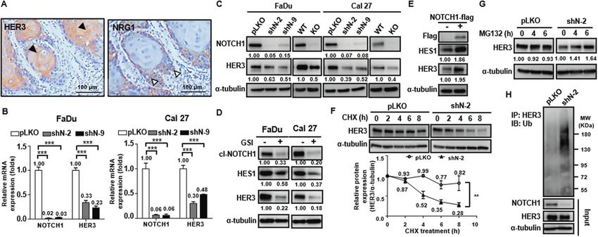
Y.-P. Wang et al.
                  2
                      associated with increased regional lymphatic metastasis. In                    analysis and negativity of mycoplasma contamination was routinely
                      addition, the majority of HER3 was found to be located on                      checked through PCR every week.
                      differentiated tumor cells in the center of infiltrative tumor nests.
                      The differentiation of keratinocytes in the stratified squamous                 Immunoprecipitation of HER3 and polyubiquitin assays
                      epithelium is regulated by several master molecules, including                 Cells were transfected with a pEF1α-HER3 plasmid for 24 h (hs), and cells
                      p63 and NOTCH1. As the differentiation progresses, the keratino-               were treated with MG132 (25 μM) for 5 h. Subsequently, cells were
                      cytes gradually lose expression of p63, and NOTCH1 becomes a                   mixed with lysis buffer (50 mM Tris-HCl, pH 7.4, 150 mM NaCl, 1% NP-40)
                      major determinant of cell fate [18, 19]. We demonstrated that                  supplemented with protease inhibitors (Roche, Switzerland). Equal
                      HER3 is under the control of NOTCH1, which stimulates HER3                     amounts of total proteins (500 μg) were incubated with the anti-HER3
                                                                                                     antibody at a dilution of 1:250 at 4 °C for 8 h. Then, proteins were
                      transcriptional activation and inhibits its protein degradation.
                                                                                                     incubated with 10 μl Dynabeads TM Protein G (Invitrogen, USA) at 4 °C for
                      Interestingly, loss of function of NOTCH1 suppressed the                       2 h to pull down the antibody-bound proteins. The mixtures were
                      expression of HER3 but boosted the phosphorylation of AKT                      washed and boiled in SDS sample buffer (50 mM Tris-HCl, pH 6.8, 2%
                      serine 473 (S473) in SCCHN cells, suggesting that NOTCH1 affects               SDS, 0.1% bromophenol blue, 10% glycerol, 12.5% β-mercaptoethanol).
                      the PI3K/AKT pathway more profoundly and independently of its                  The pulled-down proteins were analyzed for ubiquitinylation by Western
                      effects on HER3. Based on these findings, it may be beneficial to                blotting.
                      exclude patients with dysfunctional NOTCH1 from clinical trials on
                      HER3-targeting therapeutics, and expression of NRG1 may serve as               Phospho-RTK array assay
                      a valuable biomarker for enrollment in such trials.                            The profile of phosphorylated-receptor tyrosine kinases (RTKs) was
                                                                                                     analyzed using a Human Phospho-RTK Array Kit (R&D Systems, USA),
                                                                                                     according to the manufacturer’s instructions. The intensity score of each
                      MATERIALS AND METHODS                                                          array spot was measured with a UVP BioSpectrum image system.
                      Clinical specimens and clinicopathological staging                             Phosphorylated RTK protein levels were compared between shNOTCH1
                      The protocols and procedures in this study conformed to all relevant           SCCHN cells and the corresponding control cells (pLKO groups).
                      ethical and institutional guidelines and were conducted after the
                      acquisition of informed consent from all subjects and approval by the          Subcutaneous xenograft model
1234567890();,:

                      Institutional Review Board of National Taiwan University Hospital. Since       Female non-obese diabetic/severe combined immunodeficiency (NOD/
                      the first choice of treatment in laryngeal and pharyngeal SCC has been          SCID) mice were obtained from the Jackson Laboratory (Bar Harbor, Maine,
                      shifted to definitive chemoradiotherapy to achieve organ preservation [8],      USA) and were bred in the core facility of the ICOB at Academia Sinica,
                      the quantity of the specimens from those two regions has been dwindled         Taiwan. As per the regulation of Institutional Animal Care and Use
                      and the oral cavity is the only anatomical location that offers ample          Committee of Academia Sinica, Taiwan (IACUC No. 20-05-1468), mice of
                      surgical specimen of head and neck squamous cell carcinoma. We                 4–6 weeks old were injected subcutaneously with 2 × 10⁶ NOTCH1-wide
                      retrieved one hundred FFPE tissue blocks of oral squamous cell carcinoma       type (WT) and NOTCH1-knockout (KO) Cal 27 cells in each side of the
                      (OSCC) patients from the archive at the Department of Oral Pathology in        dorsolateral flank (n = 8). No randomization of mice was performed in this
                      the years 2006–2011 (NTUH IRB, No. 201412066RINA and No.                       experiment. Mice that did not grow tumors will be excluded from the
                      201901040RIND) to ensure adequate statistical power. The preset enrolled       experiment, however, all inoculated mice in this experiment developed
                      criteria were primary oral squamous cell carcinoma without previous            tumors as expected. The tumor volumes were calculated using the
                      treatment (such as inductive chemotherapy and radiation therapy) and the       following formula: tumor volume = length × (width)2 × 0.52. The data are
                      age of the patient was required to be older than 20 years. The exclusion       presented as the mean ± standard error of the mean.
                      criteria was set as recurrent cancers, positive history of previous chemo/
                      radiotherapy, and tissue size smaller than 0.7 × 0.5 cm. The diagnosis was
                      confirmed after a review of hematoxylin and eosin-stained (H&E) tissue          Statistics
                      sections by two qualified oral pathologists. The TNM staging at the time of     The association between clinicopathological parameters and expression
                      diagnosis for every case was re-evaluated according to the 8th edition of      status of HER3 and NRG1 in clinical specimens was analyzed by Fisher’s
                      staging criteria from the American Joint Committee on Cancer. Complete         exact test and Pearson’s chi-squared test. All in vitro comparisons were
                      records of follow-up were available in 87 out of 100 cases enrolled. The       performed with three technical replicates and three independent biological
                      recurrence of the tumor was defined as an SCC found less than 2 cm away         replicates (n = 3). Analyses of in vitro experiments were performed using
                      from and within 3 years of the previous primary tumor [20]. Areas that         two-tailed ANOVA. P-values less than 0.05 were considered statistically
                      were 1 cm away from the peritumoral epithelial dysplasia and devoid of an      significant for all tests. All statistical analyses were performed using SPSS
                      underlying inflammatory cell infiltrate were considered as normal mucosa         (SPSS Inc., USA).
                      (n = 11). The clinical characteristics of the enrolled cohort were summar-
                      ized in Supplementary Table S1.
                                                                                                     RESULTS
                      Immunohistochemical (IHC) staining of HER3 and NRG1                            Concomitant high expression of HER3 and NRG1 is associated
                      Tissue sections from enrolled cases were cut to the 4 μm thickness and         with nodal metastasis in SCCHN and HER3 is regulated by
                      stained by immunohistochemistry. Routine immunohistochemical staining          NOTCH1
                      was performed with monoclonal primary antibodies against HER3 and              To investigate the clinical significance of the HER3 pathway in
                      NRG1 (details in Supplementary Table S2), using a protocol described           SCCHN, we first evaluated the expression status of HER3 and its
                      previously [21]. Tissue sections incubated with normal mouse IgG (NMIgG)       ligand NRG1 in 100 cases of OSCC, which is the 4th leading cancer
                      instead of primary antibody served as negative controls. Perceptible and       type in the male population in Taiwan, through immunohisto-
                      convincing granular membrane staining, either partially or completely          chemical staining. Positive membranous stains of HER3 and NRG1
                      encircling the cells, was regarded as positive. All histopathological images
                                                                                                     were discerned in 95 cases. The mean and standard deviation of
                      were captured with an Olympus BX53 microscope and DP2-BSW image
                      acquisition software.
                                                                                                     the percentage of positive tumor cells was 72.93 ± 31.68% for
                                                                                                     HER3 and 81.65 ± 21.84% for NRG1, respectively. On the other
                                                                                                     hand, the percentage of the keratinoyctes positive for HER3 was
                      Cell lines and culture conditions                                              significantly lower in all eleven samples of the normal epithelium
                      FaDu (HPV-negative oropharyngeal squamous cell carcinoma) and Cal 27           (51.51 ± 5.09%, p = 0.0012) and no signal of NRG1 was seen in any
                      (OSCC) cell lines were purchased from the Bioresource Collection and
                      Research Center (BCRC, Hsinchu, Taiwan). Both cell lines were maintained       normal keratinocytes (Supplementary Fig. S1A). Analysis of the
                      in medium (MEM for FaDu cells and DMEM for Cal 27 cells) supplemented          TCGA dataset of SCCHN on UALCAN platform also revealed that
                      with 10% fetal bovine serum (FBS; Gibco, USA) and 100 μg/ml Penicillin/        the mRNA transcript number of NRG1 was significantly higher in
                      Streptomycin (P/S; Gibco, USA) in a humidified incubator at 37 °C with 5%       primary SCCHN than the normal counterparts (Supplementary Fig.
                      CO2. The authenticity of cell lines was confirmed by short tandem repeat        S1B). We found no significant correlation between these two

                                                                                                                                       Oncogenesis (2021)10:59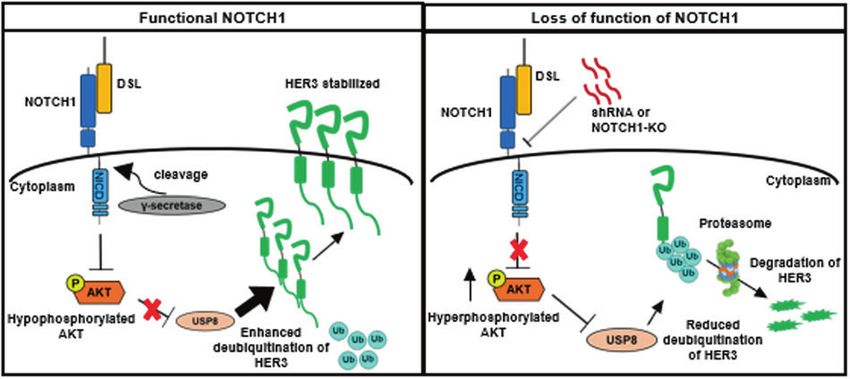
Y.-P. Wang et al.
                                                                                                                                                3
                                                                        the knockdown of NOTCH1 through various clones of shRNA in
 Table 1. Clinicopathological correlation of concomitant high
                                                                        both FaDu and Cal 27 cell lines (Fig. 1B). Efficient suppression of
 expression of HER3 and NRG1 and OSCC outcome.
                                                                        HER3 protein was also evident after knockdown and knockout of
                            Concomitant high              Pearson       NOTCH1 with different constructs of shRNA and CRISPR-Cas9
                            expression of HER3            p-value       system in SCCHN cells (Fig. 1C). Furthermore, we found that the
                            and NRG1                                    expression of HER3 was dose-dependently reduced by treatment
                                                                        of NRG1 in increasing concentrations in NOTCH1-knockout FaDu
Parameter                   (−)             (+)
                                                                        cells and Cal 27 cells (Supplementary Fig. S5), and this reduction
T                                                                       was also present in the NOTCH1-expressing counterparts when
              T1 + T2       48 (72.73%)     18 (52.94%)   0.079         exposed to NRG1 in high concentration (40 ng/ml). Since the
              T3 + T4       18 (27.27%)     16 (47.06%)                 activation of the canonical NOTCH1 pathway requires protein
                                                                        cleavage by γ-secretase to release the NOTCH intracellular domain
N
                                                                        (NICD) to trigger the downstream transcription, we further assayed
              N0            50 (75.76%)     17 (50.00%)   0.018         the impact of functional disability of NOTCH1 upon the regulation
              N1 +          16 (24.24%)     17 (50.00%)                 of HER3. After adding an inhibitor of γ-secretase, the protein levels
              N2 + N3                                                   of HES1 (a known downstream component of NOTCH1 pathway,
Stage                                                                   as a control) and HER3 were significantly reduced in SCCHN cells
              I + II        49 (74.24%)     17 (50.00%)   0.028         (Fig. 1D). Conversely, overexpression of NOTCH1 protein efficiently
                                                                        increased the expression of HES1 and HER3 (Fig. 1E). Taken
              III + IV      17 (25.76%)     17 (50.00%)                 together, it suggests that NOTCH1 positively regulates HER3 on
Recurrencea                                                             both mRNA and protein levels in SCCHN.
              Negative      46 (82.14%)     15 (48.39%)   0.002
              Positive      10 (17.86%)     16 (51.61%)                 Inhibition of NOTCH1 compromises HER3 protein stability
                                                                        The expression level of protein is determined by various
ENE  b
                                                                        mechanisms, including transcriptional activation and proteosomal
              Negative      12 (75.00%)     14 (82.35%)   1.000         degradation. Next, we examined the difference of protein stability
              Positive        4 (25.00%)     3 (17.65%)                 of HER3 with and without the suppressed function of NOTCH1 in
 a
  Complete records of follow-up were available in 87 out of 100 cases   SCCHN cells. The protein level of HER3 was prematurely decreased
 enrolled.                                                              to 52% of the original level at 4 h after inhibition of translational
 b
  ENE, extranonal extension.                                            activity through cycloheximide in NOTCH1-knockdown SCCHN
                                                                        cells, and it further dropped to 28% at post-treatment 8 h. In
                                                                        contrast, the protein level of HER3 remained as 99% and 82% of the
markers and parameters in the TNM system when evaluated in              original level in the control counterpart at the corresponding time
univariate analysis (data not shown). However, simultaneous high        points, respectively (Fig. 1F). These findings suggested NOTCH1
expression of HER3 and NRG1 within the tumor (percentages of            affects not only the transcriptional regulation but also the protein
positive neoplastic cells were more than the mean value of              stability of HER3 through manipulating the proteosomal degrada-
corresponding markers) was significantly associated with the             tion. To test our hypothesis, we added the proteosomal inhibitor
presence of metastasis and high cancer staging, and the presence        MG132 into the culture media of NOTCH1-knockdown SCCHN cells
of local recurrence (Table 1). Concomitant high expression of           and the control counterpart. The downregulation of HER3 protein
these two markers failed to demonstrate a significant correlation        in NOTCH1-knockdown cells could be reversed through the
of the presence of extracapsular spread at the time of diagnosis        addition of the proteasome inhibitor MG132 (Fig. 1G). Since the
(Table 1), and no significant difference of 5-year overall survival      polyubiquitin tag serves as the beacon for the 26 S proteasome and
was discerned between the concomitant NRG1 and HER3 high-               is a pivotal marker in protein degradation, we next investigated if
expressing subset and the rest of the enrolled cases (Supplemen-        NOTCH1 influences the polyubiquitination of the HER3 protein.
tary Fig. S2). In addition, we observed that the majority of the        After overexpression of HER3 in NOTCH1-knockdown SCCHN cells
HER3-positive cells were the differentiated cells in the center of      and the control counterpart, immunoprecipitation of HER3 was
the infiltrating tumor nests, and the NRG1-positive cells were           performed and the presence of ubiquitin on the protein was
primarily the basaloid cells at the periphery of the cell nests         probed with Western blotting. Enhanced polyubiquitination was
(Fig. 1A). This spatial discrepancy was discerned in 66% of the         discerned in NOTCH1-knockdown cells compared to the control
enrolled cases. This phenomenon drives us to explore if the             cells (Fig. 1H). In summary, these data indicated NOTCH1 not only
differentiation axis of the squamous epithelium takes a part in         participates in the transcription of HER3 but also enhances the
the regulation of HER3. Given the fact that NOTCH1 is a master          protein stability of HER3 through inhibition of polyubiquitination of
regulator in the terminal differentiation of the squamous               the HER3 protein.
epithelium and HER3 was mostly observed in the more
differentiated tumor cells, we evaluated the contribution of            Loss of NOTCH1 function leads to activation of AKT signaling
NOTCH1 to the expression of HER3. Immunohistochemical                   In order to discover the underlying mechanism of NOTCH1-
staining of NOTCH1 was performed in our cohort of OSCC. It              inhibited polyubiquitination of HER3, we explored the change of
revealed that the expression of NOTCH1 protein in the cases             signaling landscape after the knockdown of NOTCH1 in SCCHN
without nodal involvement was approximately two-fold higher             cells. The analysis of phosphorylated-receptor tyrosine kinase
than those cases with nodal diseases, and downregulation of             array revealed that the only alteration of the major signaling
NOTCH1 was closely associated with nodal involvement (p =               pathway in both cell lines with a meaningful fold-change after
0.0865, Supplementary Fig. S3B). Colocalization of NOTCH1 and           knockdown of NOTCH1 was the augmentation of phosphorylation
HER3 on the centrally-located differentiated neoplastic cells within    of serine 473 of AKT (6.71 folds in FaDu cells and 5.18 folds in Cal
the infiltrative tumor nests was also discerned in 51% of cases of       27 cells, Supplementary Fig. S6). This finding was unexpected
our enrolled cohort (Supplementary Fig. S4). However, this finding       since HER3 is known as an activator of AKT and downregulation of
did not confer any statistically significant difference in regard to     HER3 through inhibition of NOTCH1 was supposed to result in
clinicopathological features (data not shown). In vitro studies         suppression of the AKT pathway. This finding suggested that
showed that the level of HER3 mRNA was drastically reduced after        NOTCH1 is in a higher hierarchy in the AKT axis than HER3, since

Oncogenesis (2021)10:59
Y.-P. Wang et al.
4
Fig. 1 HER3 is preferentially expressed in differentiated tumor cells and inhibition of NOTCH1 suppresses HER3 mRNA and destabilizes
HER3 protein in SCCHN. A The majority of HER3-positive cells were differentiated cells in the center of the infiltrating tumor nests (black
arrowhead, membranous stain of HER3), while the signal for NRG1 was primarily found in basaloid cells at the periphery of the tumor nests
(white arrowhead, cytoplasmic stain of NRG1). B The mRNA level of HER3 was drastically reduced in FaDu and Cal 27 cells after
NOTCH1 silencing with different lentivirus-expressed shRNAs, when compared to control shRNA (pLKO, as the baseline of normalization).
Column: the average relative expression, compared to the pLKO group ± standard error (n = 3). ***p < 0.001 by two-way ANOVA. C The
protein level of HER3 was also greatly reduced in FaDu and Cal 27 cells after NOTCH1 silencing with different shRNAs or knockout with a
CRISPR-Cas9 system. D The expression of HER3 was significantly decreased after NOTCH1 signaling was inhibited with the treatment of a
γ-secretase inhibitor (GSI) for 24 h in SCCHN cells (FaDu, 50 μM; Cal 27, 20 μM). cl-NOTCH1, cleaved NOTCH1; HES1, a known downstream
component of NOTCH1 pathway that serves as an indicator of NOTCH1 signaling. E Conversely, overexpression of NOTCH1 protein efficiently
increased the expression of HES1 and HER3. F The protein stability assay revealed a steep decrease of HER3 protein and a lower residual
protein quantity in NOTCH1-knockdown FaDu cells compared to controls (28% vs. 82% at 8 h) after inhibition of translational activity by
cycloheximide (30 μg/μl). Dot: the average relative expression normalized to the original level ± standard error (n = 3). **p < 0.01 by two-way
ANOVA. G The downregulation of HER3 protein in NOTCH1-knockdown cells could be reversed by the addition of the proteasome inhibitor
MG-132 (25 μM). H Immunoprecipitation assay showed enhanced polyubiquitination of HER3 protein in NOTCH1-knockdown cells compared
to control cells (pLKO).
the effect of NOTCH1 upon the AKT pathway overrides that of the original level in the DMSO-treated control group (Fig. 3A).
HER3. In order to verify if this is a bystander effect caused by Immunoprecipitation of HER3 also showed that wortmannin
shRNA, we examined the phosphorylation of AKT in SCCHN after lowered the polyubiquitination of HER3 in both control SCCHN
manipulation of the function of NOTCH1 with various methods. cells and the corresponding NOTCH1-knockdown counterpart,
Increased phosphorylation of AKT was present when the NOTCH1 and the polyubiquitination of HER3 was more prominent in
was knockdown with different constructs of shRNA (Fig. 2A) and NOTCH1-knockdown cells than that discerned in the control cells
when the function of NOTCH1 was inhibited by a γ-secretase (Fig. 3B). Similar results were also present when the function of
inhibitor (Fig. 2B), while overexpression of NOTCH1 led to NOTCH1 was compromised by γ-secretase inhibitor in SCCHN cells
decreased phosphorylation of AKT (Fig. 2C). Furthermore, the (Fig. 3C). These findings confirmed that activation of the PI3K/AKT
addition of γ-secretase inhibitor to NOTCH1-knockdown and pathway enhances polyubiquitination of HER3, and downregula-
NOTCH1-knockout SCCHN cells did not boost the phosphorylation tion of NOTCH1 further boosts the degradative signal of HER3.
of AKT, indicating the AKT-stimulating effect was only associated Polyubiquitination is a multistep process and is controlled by a set
with NOTCH1 but not other substrates of γ-secretase (Fig. 2D and of enzymes, typically composed of activating enzymes, conjugat-
Fig. 2E, respectively). To seek out the possible underlying ing enzymes, and ligase. We examined the mRNA of two E3 ligases
mechanism of AKT activation after the loss of function of NOTCH1, (NEDD4 and NRDP1) in SCCHN cells after knockdown of NOTCH1
we examined the impact on the phosphorylation landscape of and no significant difference was noted compared to that in the
EGFR after knockdown of NOTCH1 in FaDu and Cal 27 cells. control counterpart (data not shown). Then, we examined
Although augmentation of phosphorylation of AKT was consis- components of the polyubiquitination process that are regulated
tently observed, the phosphorylation signatures of EGFR were by the AKT axis. Ubiquitin-specific peptidase 8 (USP8) is a
generally decreased after knockdown of NOTCH1 in FaDu cells, deubiquitinating enzyme under negative regulation of the PI3K/
while phosphorylation of EGFR at all tested loci were upregulated AKT pathway. When the function of USP8 was enhanced by
by the shN-9 clone in Cal 27 cells. (Supplementary Fig. S7). A more wortmannin, the protein level of HER3 in NOTCH1-knockdown
detailed multi-omic elucidation of these two cell lines and SCCHN cells increased (Fig. 3D, two lanes on the left). After the
translation into clinical specimens is needed in a future study to introduction of siRNA against USP8, HER3 protein were reduced
unravel the underlying cause of this discrepancy. due to suppressed deubiquitination, and a supplement of
wortmannin partially rescued the expression level of HER3
Activation of AKT contributes to destabilization of HER3 (Fig. 3D, two lanes on the right). Immunoprecipitation and
through negative regulation of USP8 subsequent immunoblotting also confirmed that the ubiquitina-
Next, we investigated whether activation of AKT is a pivotal tion of HER3 decreased in both control and NOTCH1-knockdown
component in NOTCH1-inhibited polyubiquitination of HER3. The SCCHN cells after treatment with wortmannin, which resulted in
protein level of re-expressed HER3 kept increasing in the 6 h under enhancement of USP8, and this suppression can be partially
treatment of wortmannin (10 μM) in NOTCH1-knockdown SCCHN reversed by inhibition of USP8 through siRNA (Fig. 3E). However,
cells, in contrast, the protein of HER3 dwindled to less than 50% of loss of NOTCH1 exerts a more profound impact on the biological
Oncogenesis (2021)10:59Y.-P. Wang et al.
5
Fig. 2 NOTCH1 negatively regulates AKT in SCCHN cells. A Increased phosphorylation of AKT was observed in FaDu and Cal 27 cells after
NOTCH1-knockdown with different shRNA constructs. B Phosphorylation of AKT was also enhanced by treatment with GSI in FaDu and Cal 27
cells. C Overexpression of NOTCH1 led to decreased phosphorylation of AKT in FaDu cells. D The addition of GSI to NOTCH1-knockdown FaDu
and Cal 27 cells did not further boost the phosphorylation of AKT. E Phosphorylation of AKT remained the same in NOTCH1-knockout FaDu
and Cal 27 cells after treatment with GSI.
behavior of SCCHN cells than that caused by decreased HER3. clinical samples [26]. Moreover, high NRG1 expression was
Knockout and knockdown of NOTCH1 in FaDu and Cal 27 cells reported to be associated with activated HER3 in HNSCC and
enhanced the cellular viability and the capabilities of colony was proposed as a selective biomarker [27]. However, no detailed
formation, migration, and invasion, albeit the effects of shRNA information about the spatial distribution of the receptor and
clone shN-9 were less prominent in migration assay of Cal 27 cells ligand were given in previous studies. In our study, we found that
and invasion assays in FaDu cells (Fig. 4A, D and Supplementary HER3- and NRG1-positive cells were prevalent in SCCHN from
Fig. S8). Murine in vivo subcutaneous xenograft model also Taiwanese patients, but a significant correlation with undesirable
demonstrated the expression of HER3 was positively correlated clinical features was only found for tumors with concomitant high
with NOTCH1 in the xenografted Cal 27 cells (Fig. 4E), and volumes levels of HER3 and NRG1. Moreover, the majority of HER3 was
of the tumors from NOTCH1-knockout Cal 27 cells were observed in differentiated squamous cells at the center of
significantly larger than those from the NOTCH1-wild type infiltrative tumor nests, while the NRG1 protein was mainly
counterpart (Fig. 4F, G). Phosphorylation of AKT was also detected in the basaloid cells at the periphery of tumor islands.
enhanced in NOTCH1-knockout tumors than that in the These observations prompted us to explore whether HER3
NOTCH1-wild type lesions (Supplementary Fig. S9). In summary, expression is under the regulation of differentiation pathways in
we demonstrated that NOTCH1 positively regulates HER3 through the squamous epithelium.
transcriptional control and increased deubiquitination and sub- NOTCH1 is a master regulator of terminal differentiation in the
sequent stabilization of the protein. Loss of NOTCH1 would stratified squamous epithelium, and its expression is mainly
activate the AKT pathway and suppress the deubiquitination of present on the differentiated keratinocytes of the spinous layer
HER3, in turn the degradation of HER3 will be enhanced (Fig. 5). [28, 29]. Once engaged by various ligands of the Delta-like and
Jagged protein families, two proteolytic cleavages (first by
ADAM10 and then by γ-secretase) take place on NOTCH1,
DISCUSSION releasing the Notch intracellular domain (NICD). The NICD
HER3 is a member of the HER family, and its primary ligand is subsequently enters the nucleus and stimulates transcription of
NRG1. Although HER3 has weak kinase activity compared to other target genes in cooperation with the DNA-binding protein, CBF1-
HER coreceptors, it can interact with EGFR to form kinase-active Suppressor of Hairless-LAG1 (CSL; also known as RBPJ), and the co-
hetero-oligomers upon binding of the cognate ligands to the activator, Mastermind-like transcriptional co-activator 1 (MAML1)
extracellular receptor domains [22]. HER3 serves as an efficient [30–32]. Our study revealed that downregulation of NOTCH1 in
phosphotyrosine scaffold upon transphosphorylation by other SCCHN cells caused decreased expression of HER3, and functional
HER family members and potently activates downstream signal- suppression of γ-secretase lead to similar results. In contrast,
ing. Moreover, the cytoplasmic domain of HER3 possesses high constitutive overexpression of NOTCH1 increases the expression
specificity for the p85 regulatory subunit of PI3K (six docking sites of HER3. These results were in agreement with the finding that
per HER3 molecule) [23] and HER3 plays a role as pivotal linker most of the positive immunostaining signal for HER3 was detected
between EGFR and PI3K/AKT signaling pathways. While HER3 in differentiated tumor cells of clinical samples. In addition, our
amplification and/or overexpression is known to occur, only data demonstrated that exposure to NRG1 leads to down-
sporadic somatic HER3 mutations have been reported [12, 24, 25]. regulation of HER3 in NOTCH1-low expressing cells. This finding
Protein-altering mutations of HER3 were found only in 1% (1 out supports the observation that the peripheral undifferentiated
of 74 cases) of human head and neck cancers [12]. On the other basaloid cells in tumor nests exhibit a low level of HER3 and
hand, previous studies have uncovered coexpression of HER3 and NOTCH1 and a high level of NRG1. Further experiments showed
NRG1, and subsequent autocrine signaling, in HNSCC cell lines and that loss of NOTCH1 function results in destabilization of HER3
Oncogenesis (2021)10:59Y.-P. Wang et al.
6
Fig. 3 NOTCH1 downregulation promotes degradation of HER3 protein via suppression of deubiquitinating enzyme USP8 through
activated AKT signaling. A The protein level of HER3 steadily increased over 6 h following treatment with wortmannin (10 μM) in HER3-
recapitulated, NOTCH1-knockdown SCCHN FaDu cells. In contrast, the HER3 protein level was less than 50% of the original level in the DMSO-
treated control group. Dot: average relative expression normalized to the original levels ± standard error (n = 3). **p < 0.01 by two-way ANOVA.
B Immunoprecipitation experiments also showed wortmannin lowered HER3 polyubiquitination in both control FaDu cells (two lanes on the
left) and NOTCH1-knockdown cells (two lanes on the right). Polyubiquitination of HER3 was more prominent in NOTCH1-knockdown cells
than in control cells. C Similar results were found when the NOTCH1 function was compromised by GSI. D When USP8 was stimulated by
wortmannin, the protein level of HER3 in NOTCH1-knockdown FaDu cells increased (two lanes on the left), while HER3 protein was reduced
after the introduction of siRNA against USP8 (two lanes on the right). In addition, treatment with wortmannin partially rescued the expression
of HER3. E Ubiquitination of HER3 was decreased in both control and NOTCH1-knockdown SCCHN cells after treatment with wortmannin,
which resulted in enhancement of USP8. This suppression could be partially reversed by the siRNA knockdown of USP8.
protein through increased phosphorylation of AKT at serine 437 conferred by NOTCH1 in SCCHN also exist [42–44]. This raises the
and augmented HER3 polyubiquitination. Polyubiquitination is a possibility that the result of NOTCH1 activation is contextual in
highly-conserved eukaryotic system for proteosomal degradation SCCHN and is influenced by the crosstalk between other signaling
of proteins [33]. After interaction with a ubiquitin-activating pathways [45]. Our data showed that loss of NOTCH1 function
enzyme (E1), ubiquitin is transferred to a ubiquitin-conjugating activates the AKT pathway and leads to suppression of HER3 in
enzyme (E2) and forms polyubiquitin chains. The polyubiquitin SCCHN cells. This novel finding is intriguing since HER3 is
chain will be attached to a target protein through a ubiquitin- considered to be a potent activator of PI3K, and it seems to be a
protein ligase (E3). The poly-ubiquitinated target protein is then paradox that loss of function of a tumor suppressor gene causes
usually destined to be recognized and degraded by the 26 S downregulation of an oncogenic receptor tyrosine kinase at first
proteasome. Interestingly, our data revealed that the expression of glance. Nevertheless, the relative abundance of EGFR and HER3
two major E3 ligases, NEDD4 and NRDP1, was not affected by the and the presence of EGF or NRG triggers different oligomeric
knockdown of NOTCH1. Yet ubiquitin modifications are reversible, combinations and the downstream pathways [46], and hetero-
as the conjugates can be removed from substrates by the action dimerization between EGFR and HER3 occurs only when HER3 is
of deubiquitinating enzymes (DUBs) [34]. The largest DUB family is engaged by NRG1 but not when EGFR is bound by EGF. In short,
the ubiquitin-specific proteases; the members of this family are the function of HER3 is determined by the abundance of NRG1,
essential regulators of a wide range of different cellular functions. and the combined evaluation of this pair of receptors and ligands
For instance, USP7 controls p53 stability by deubiquitination of is more appropriate in the clinical setting. In addition, activation of
p53 and Mdm2 [35], and USP1 is known to regulate the DNA AKT can trigger a negative feedback regulation upon HER3, as
replication processivity factor, PCNA, by cleaving the ubiquitinated demonstrated in breast cancers [15]. The exact mechanism of
protein [36]. Among USPs, USP8 is a negatively regulated NOTCH1-AKT coupling is currently unclear. NOTCH1-regulated MYC
substrate of the AKT pathway, as PTEN loss and AKT activation has been shown to be a transcriptional activator of PTEN [47], and
are associated with suppression of USP8 levels [37]. Previous downregulation of PTEN leads to increased AKT signaling. In
studies showed that USP8 participates in the endosomal sorting of addition, multiple factors influence the outcome of activated
transmembrane proteins [38, 39] and modulates their function NOTCH1 signaling [48]. For instance, the signaling strength of
and stability through deubiquitination. Furthermore, USP8 was NOTCH1 activation depends on the type of engaged ligand and
found to promote degradation of EGFR [38, 40] and hepatocyte the relative levels between the ligands and NOTCH1 per se [49].
growth factor receptor [38]. In the current study, we demonstrated Glycosylation of EGF repeats in the extracellular domain of
that the polyubiquitination and subsequent reduction of HER3 is NOTCH1 by Fringe protein in Golgi bodies also modulates the
enhanced by USP8 attenuation via activation of the AKT pathway signaling pattern of NOTCH1 [50]. These features render immuno-
in SCCHN cells with loss of NOTCH1 function. histochemical quantification alone against the protein backbone of
Although NOTCH1 is considered to be oncogenic in several NOTCH1 as a suboptimal indicator for the activity of the
types of malignancies, studies utilizing whole-exome sequencing downstream pathway. Other than the canonical pathway, NOTCH1
detected inactivating mutations of NOTCH1 in 12–15% of SCCHN can also exert biological function through a non-canonical, ligand-
cases [9, 12]. In addition, increased cutaneous tumorigenesis is independent route involving β-catenin [51]. A comprehensive
evident in animal models with disabled NOTCH1 [41]. These multi-omic analysis in SCCHN may shed light on finding the
findings suggest that NOTCH1 confers tumor-suppressive functions missing link between NOTCH1 and AKT, and an animal model with
in the stratified squamous epithelium, where SCCHN arises. an inducible knockout of NOTCH1 is needed to serve as a more
However, conflicting reports claiming oncogenic mechanisms authentic platform for the exploration of the role of NOTCH1 in
Oncogenesis (2021)10:59Y.-P. Wang et al.
7
Fig. 4 Loss of function of NOTCH1 down-regulates HER3 expression and augments tumor aggressiveness in SCCHN. Functional assays
were performed and a comparison between the NOTCH1-wild type (WT) and NOTCH1-knockout (KO) SCCHN cells. Knockout of
NOTCH1 significantly promotes the cellular viability (A), the capability of colony formation (B), migration (C), and invasion (D) in both FaDu
and Cal 27 cells. Data are presented as the mean ± SD. *p < 0.05; **p < 0.01 and ***p < 0.001 (E) Immunohistochemical staining revealed that
the expression of HER3 was positively correlated with NOTCH1 in the xenografted Cal 27 cells in the murine subcutaneous xenograft model.
The tumor volumes (F, G) of the NOTCH1-knockout tumors are significantly increased compared to those of the NOTCH1-wild type tumors.
Scale bar, 50 μm. Data are presented as the mean ± SEM. ***p < 0.001.
anti-HER3 antibody, may be a promising strategy, owing to the
high prevalence of HER3 expression in SCCHN. Thus, HER3 may
serve as a molecular beacon for drug delivery that overcomes
compensatory signaling crosstalk in malignant cells. This strategy is
currently under investigation [55], and the preclinical results show
good antitumor activity against HER3-expressing tumors with
tolerable safety profiles. In conclusion, our data suggest that HER3
is under the control of NOTCH1, which stimulates HER3 transcrip-
tional activation and inhibits its protein degradation. Interestingly,
loss of function of NOTCH1 suppressed the expression of HER3 but
boosted the phosphorylation of AKT serine 473 (S473) in SCCHN
Fig. 5 Graphical abstract. Schematic illustration shows the working cells, suggesting that NOTCH1 affects the PI3K/AKT pathway more
model of NOTCH1 regulation of HER3 protein. Functional profoundly and independently of its effects on HER3.
NOTCH1 signaling suppresses the phosphorylation of AKT serine
473, which inactivates the deubiquitinating enzyme USP8. Activated
USP8 then deceases the polyubiquitination of HER3, preventing REFERENCES
proteasomal degradation of HER3 protein (left). When loss of function 1. Bray F, Ferlay J, Soerjomataram I, Siegel RL, Torre LA, Jemal A. Global cancer
of NOTCH1 occurs, phosphorylation of AKT is increased, and the statistics 2018: GLOBOCAN estimates of incidence and mortality worldwide for
activity of USP8 is subsequently down-regulated. Once the deubi- 36 cancers in 185 countries. CA Cancer J Clin. 2018;68:394–424. https://doi.org/
quitination is suppressed, the poly-ubiquitinated HER3 protein is 10.3322/caac.21492
subject to proteasomal degradation (right). 2. Chow LQM. Head and neck cancer. N Engl J Med. 2020;382:60–72. https://doi.org/
10.1056/NEJMra1715715
3. Warnakulasuriya S, Johnson NW, van der Waal I. Nomenclature and classification
various stages of precancerous lesions to SCCHN. In summary, our of potentially malignant disorders of the oral mucosa. J Oral Pathol Med: Off Publ
data suggested that NOTCH1 is a higher-ranked regulator of the Int Assoc Oral Pathologists Am Acad Oral Pathol. 2007;36:575–80. https://doi.org/
10.1111/j.1600-0714.2007.00582.x
AKT axis than HER3. As a consequence, our results offer some
4. Amin MB, Greene FL, Edge SB, Compton CC, Gershenwald JE, Brookland RK, et al.
insight into how strategies and patient stratification may be The Eighth Edition AJCC Cancer Staging Manual: continuing to build a bridge
improved in future clinical trials for anti-HER3 therapeutics in from a population-based to a more “personalized” approach to cancer staging.
SCCHN. Currently, various HER3-targeting therapeutic monoclonal CA Cancer J Clin. 2017;67:93–99. https://doi.org/10.3322/caac.21388
antibodies have been developed and are entering clinical trials, 5. Argiris A, Karamouzis MV, Raben D, Ferris RL. Head and neck cancer. Lancet.
including patritumab/U3–1287 [52], seribantumab/MM-121 [53], 2008;371:1695–709. doi: S0140-6736(08)60728-X [pii] https://doi.org/10.1016/
and lumretuzumab/RG7116 [54]. However, none of these HER3- S0140-6736(08)60728-X
targeting antibodies has been approved for clinical use because of 6. Marur S, Forastiere AA. Head and neck cancer: changing epidemiology, diagnosis,
unsatisfactory clinical benefit. Since NOTCH1 is inactivated in and treatment. Mayo Clin Proc. 2008;83:489–501. https://doi.org/10.4065/83.4.489
7. Seiwert TY, Cohen EE. State-of-the-art management of locally advanced head and
approximately one-tenth of SCCHN cases, and this aberration is a
neck cancer. Br J Cancer. 2005;92:1341–8. https://doi.org/10.1038/sj.bjc.6602510
powerful activator of the AKT pathway and a negative regulator of 8. Chinn SB, Myers JN. Oral cavity carcinoma: current management, controversies,
HER3, exclusion of patients bearing NOTCH1-inactivated SCCHN and future directions. J Clin Oncol. 2015;33:3269–76. https://doi.org/10.1200/
tumors may be recommended for future clinical trials on HER3- JCO.2015.61.2929
targeting antibodies. In addition, the development of an antibody- 9. Agrawal N, Frederick MJ, Pickering CR, Bettegowda C, Chang K, Li RJ, et al. Exome
drug conjugate (ADC), with a cytotoxic payload attached to an sequencing of head and neck squamous cell carcinoma reveals inactivating
Oncogenesis (2021)10:59Y.-P. Wang et al.
8
mutations in NOTCH1. Science. 2011;333:1154–7. https://doi.org/10.1126/ 35. Li M, Brooks CL, Kon N, Gu W. A dynamic role of HAUSP in the p53-Mdm2 pathway.
science.1206923 Mol Cell. 2004;13:879–86. https://doi.org/10.1016/s1097-2765(04)00157-1
10. Cancer Genome Atlas N. Comprehensive genomic characterization of head and 36. Huang TT, Nijman SM, Mirchandani KD, Galardy PJ, Cohn MA, Haas W, et al.
neck squamous cell carcinomas. Nature. 2015;517:576–82. https://doi.org/ Regulation of monoubiquitinated PCNA by DUB autocleavage. Nat cell Biol.
10.1038/nature14129 2006;8:339–47. https://doi.org/10.1038/ncb1378
11. Hansen AR, Siu LL. Epidermal growth factor receptor targeting in head and neck 37. Panner A, Crane CA, Weng C, Feletti A, Fang S, Parsa AT, et al. Ubiquitin-specific
cancer: have we been just skimming the surface? J Clin Oncol. 2013;31:1381–3. protease 8 links the PTEN-Akt-AIP4 pathway to the control of FLIPS stability and
https://doi.org/10.1200/JCO.2012.47.9220 TRAIL sensitivity in glioblastoma multiforme. Cancer Res. 2010;70:5046–53.
12. Stransky N, Egloff AM, Tward AD, Kostic AD, Cibulskis K, Sivachenko A, et al. The https://doi.org/10.1158/0008-5472.CAN-09-3979
mutational landscape of head and neck squamous cell carcinoma. Science. 38. Niendorf S, Oksche A, Kisser A, Löhler J, Prinz M, Schorle H, et al. Essential role of
2011;333:1157–60. https://doi.org/10.1126/science.1208130 ubiquitin-specific protease 8 for receptor tyrosine kinase stability and endocytic
13. Bonner JA, Harari PM, Giralt J, Azarnia N, Shin DM, Cohen RB, et al. Radiotherapy trafficking in vivo. Mol Cell Biol. 2007;27:5029–39. https://doi.org/10.1128/
plus cetuximab for squamous-cell carcinoma of the head and neck. N. Engl J Med. MCB.01566-06
2006;354:567–78. https://doi.org/10.1056/NEJMoa053422 39. Wright MH, Berlin I, Nash PD. Regulation of endocytic sorting by ESCRT-DUB-
14. Bonner JA, Harari PM, Giralt J, Cohen RB, Jones CU, Sur RK, et al. Radiotherapy mediated deubiquitination. Cell Biochem Biophys. 2011;60:39–46. https://doi.org/
plus cetuximab for locoregionally advanced head and neck cancer: 5-year sur- 10.1007/s12013-011-9181-9
vival data from a phase 3 randomised trial, and relation between cetuximab- 40. Mizuno E, Iura T, Mukai A, Yoshimori T, Kitamura N, Komada M. Regulation of
induced rash and survival. Lancet Oncol. 2010;11:21–28. https://doi.org/10.1016/ epidermal growth factor receptor down-regulation by UBPY-mediated deubi-
S1470-2045(09)70311-0 quitination at endosomes. Mol Biol cell. 2005;16:5163–74. https://doi.org/
15. Sergina NV, Rausch M, Wang D, Blair J, Hann B, Shokat KM, et al. Escape from HER- 10.1091/mbc.e05-06-0560
family tyrosine kinase inhibitor therapy by the kinase-inactive HER3. Nature. 41. Nicolas M, Wolfer A, Raj K, Kummer JA, Mill P, van Noort M, et al. Notch1 functions
2007;445:437–41. https://doi.org/10.1038/nature05474 as a tumor suppressor in mouse skin. Nat Genet. 2003;33:416–21. https://doi.org/
16. Vlacich G, Coffey RJ. Resistance to EGFR-targeted therapy: a family affair. Cancer 10.1038/ng1099
Cell. 2011;20:423–5. https://doi.org/10.1016/j.ccr.2011.10.006 42. Ock CY, Son B, Keam B, Lee SY, Moon J, Kwak H, et al. Identification of genomic
17. Shi F, Telesco SE, Liu Y, Radhakrishnan R, Lemmon MA. ErbB3/HER3 intracellular mutations associated with clinical outcomes of induction chemotherapy in
domain is competent to bind ATP and catalyze autophosphorylation. Proc Natl patients with head and neck squamous cell carcinoma. J Cancer Res Clin Oncol.
Acad Sci USA. 2010;107:7692–7. https://doi.org/10.1073/pnas.1002753107 2016;142:873–83. https://doi.org/10.1007/s00432-015-2083-2
18. Rothenberg SM, Ellisen LW. The molecular pathogenesis of head and neck 43. Song X, Xia R, Li J, Long Z, Ren H, Chen W, et al. Common and complex Notch1
squamous cell carcinoma. J Clin Invest. 2012;122:1951–7. mutations in Chinese oral squamous cell carcinoma. Clin Cancer Res.
19. Yugawa T, Handa K, Narisawa-Saito M, Ohno S, Fujita M, Kiyono T. Regulation of 2014;20:701–10. https://doi.org/10.1158/1078-0432.CCR-13-1050
Notch1 gene expression by p53 in epithelial cells. Mol Cell Biol. 2007;27:3732–42. 44. Vettore AL, Ramnarayanan K, Poore G, Lim K, Ong CK, Huang KK, et al. Mutational
https://doi.org/10.1128/MCB.02119-06 landscapes of tongue carcinoma reveal recurrent mutations in genes of ther-
20. Leemans CR, Braakhuis BJ, Brakenhoff RH. The molecular biology of head and apeutic and prognostic relevance. Genome Med. 2015;7:98 https://doi.org/
neck cancer. Nat Rev Cancer. 2011;11:9–22. https://doi.org/10.1038/nrc2982 10.1186/s13073-015-0219-2
21. Wang YP, Liu IJ, Chiang CP, Wu HC. Astrocyte elevated gene-1 is associated with 45. Fukusumi T, Califano JA. The NOTCH pathway in head and neck squamous cell
metastasis in head and neck squamous cell carcinoma through p65 phosphor- carcinoma. J Dent Res. 2018;97:645–53. https://doi.org/10.1177/0022034518760297
ylation and upregulation of MMP1. Mol Cancer. 2013;12:109 https://doi.org/ 46. van Lengerich B, Agnew C, Puchner EM, Huang B, Jura N. EGF and NRG induce
10.1186/1476-4598-12-109 phosphorylation of HER3/ERBB3 by EGFR using distinct oligomeric mechanisms.
22. Hynes NE, MacDonald G. ErbB receptors and signaling pathways in cancer. Curr Proc Natl Acad Sci USA. 2017;114:E2836–E2845. https://doi.org/10.1073/
Opin Cell Biol. 2009;21:177–84. https://doi.org/10.1016/j.ceb.2008.12.010 pnas.1617994114
23. Jiang N, Saba NF, Chen ZG. Advances in targeting HER3 as an Anticancer therapy. 47. Palomero T, Sulis ML, Cortina M, Real PJ, Barnes K, Ciofani M, et al. Mutational loss
Chemother Res Pract. 2012;2012:817304 https://doi.org/10.1155/2012/817304 of PTEN induces resistance to NOTCH1 inhibition in T-cell leukemia. Nat Med.
24. Greenman C, Stephens P, Smith R, Dalgliesh GL, Hunter C, Bignell G, et al. Pat- 2007;13:1203–10. https://doi.org/10.1038/nm1636
terns of somatic mutation in human cancer genomes. Nature. 2007;446:153–8. 48. Bray SJ. Notch signalling in context. Nat Rev Mol Cell Biol. 2016;17:722–35.
https://doi.org/10.1038/nature05610 https://doi.org/10.1038/nrm.2016.94
25. Kan Z, Jaiswal BS, Stinson J, Janakiraman V, Bhatt D, Stern HM, et al. Diverse 49. del Alamo D, Rouault H, Schweisguth F. Mechanism and significance of cis-
somatic mutation patterns and pathway alterations in human cancers. Nature. inhibition in Notch signalling. Curr Biol. 2011;21:R40–47. https://doi.org/10.1016/j.
2010;466:869–73. https://doi.org/10.1038/nature09208 cub.2010.10.034
26. Wilson TR, Lee DY, Berry L, Shames DS, Settleman J. Neuregulin-1-mediated auto- 50. Luca VC, Jude KM, Pierce NW, Nachury MV, Fischer S, Garcia KC. Structural biol-
crine signaling underlies sensitivity to HER2 kinase inhibitors in a subset of human ogy. Structural basis for Notch1 engagement of Delta-like 4. Science.
cancers. Cancer Cell. 2011;20:158–72. https://doi.org/10.1016/j.ccr.2011.07.011 2015;347:847–53. https://doi.org/10.1126/science.1261093
27. Shames DS, Carbon J, Walter K, Jubb AM, Kozlowski C, Januario T, et al. High 51. Kwon C, Cheng P, King IN, Andersen P, Shenje L, Nigam V, et al. Notch post-
heregulin expression is associated with activated HER3 and may define an translationally regulates beta-catenin protein in stem and progenitor cells. Nat
actionable biomarker in patients with squamous cell carcinomas of the head and Cell Biol. 2011;13:1244–51. https://doi.org/10.1038/ncb2313
neck. PloS one. 2013;8:e56765 https://doi.org/10.1371/journal.pone.0056765 52. Yonesaka K, Hirotani K, Kawakami H, Takeda M, Kaneda H, Sakai K, et al. Anti-
28. Nickoloff BJ, Qin JZ, Chaturvedi V, Denning MF, Bonish B, Miele L. Jagged-1 HER3 monoclonal antibody patritumab sensitizes refractory non-small cell lung
mediated activation of notch signaling induces complete maturation of human cancer to the epidermal growth factor receptor inhibitor erlotinib. Oncogene.
keratinocytes through NF-kappaB and PPARgamma. Cell Death Differ. 2016;35:878–86. https://doi.org/10.1038/onc.2015.142
2002;9:842–55. https://doi.org/10.1038/sj.cdd.4401036 53. Liles JS, Arnoletti JP, Kossenkov AV, Mikhaylina A, Frost AR, Kulesza P. et al. Tar-
29. Rangarajan A, Talora C, Okuyama R, Nicolas M, Mammucari C, Oh H, et al. Notch geting ErbB3-mediated stromal-epithelial interactions in pancreatic ductal ade-
signaling is a direct determinant of keratinocyte growth arrest and entry into dif- nocarcinoma. Br J Cancer. 2011;105:523–33. https://doi.org/10.1038/bjc.2011.263.
ferentiation. EMBO J. 2001;20:3427–36. https://doi.org/10.1093/emboj/20.13.3427 54. Mirschberger C, Schiller CB, Schräml M, Dimoudis N, Friess T, Gerdes CA, et al.
30. Artavanis-Tsakonas S, Rand MD, Lake RJ. Notch signaling: cell fate control and RG7116, a therapeutic antibody that binds the inactive HER3 receptor and is
signal integration in development. Science. 1999;284:770–6. optimized for immune effector activation. Cancer Res. 2013;73:5183–94. https://
31. Kitagawa M. Notch signalling in the nucleus: roles of Mastermind-like (MAML) doi.org/10.1158/0008-5472.CAN-13-0099
transcriptional coactivators. J Biochem. 2016;159:287–94. https://doi.org/10.1093/ 55. Hashimoto Y, Koyama K, Kamai Y, Hirotani K, Ogitani Y, Zembutsu A, et al. A
jb/mvv123 novel HER3-targeting antibody-drug conjugate, U3-1402, exhibits potent ther-
32. Kopan R, Ilagan MX. The canonical Notch signaling pathway: unfolding the apeutic efficacy through the delivery of cytotoxic payload by efficient inter-
activation mechanism. Cell. 2009;137:216–33. https://doi.org/10.1016/j. nalization. Clin Cancer Res. 2019;25:7151–61. https://doi.org/10.1158/1078-
cell.2009.03.045 0432.CCR-19-1745
33. Welchman RL, Gordon C, Mayer RJ. Ubiquitin and ubiquitin-like proteins as
multifunctional signals. Nat Rev Mol Cell Biol. 2005;6:599–609. https://doi.org/
10.1038/nrm1700
AUTHOR CONTRIBUTIONS
34. Haglund K, Dikic I. Ubiquitylation and cell signaling. EMBO J. 2005;24:3353–9.
Conceptulization: H.C.W. and Y.P.W. Methodology: Y.P.W, I.J.L. and K.C.C. Data
https://doi.org/10.1038/sj.emboj.7600808
curation: K.C.C, Y.P.W. and I.J.L. Formal analysis: K.C.C, Y.P.W. and I.J.L. Writing: Y.P.W.
Oncogenesis (2021)10:59Y.-P. Wang et al.
9
and H.C.W. Project administration: Y.P.W. and H.C.W. Study supervision: H.C.W. Reprints and permission information is available at http://www.nature.com/
Funding acquisition: Y.P.W. and H.C.W. reprints
Publisher’s note Springer Nature remains neutral with regard to jurisdictional claims
FUNDING in published maps and institutional affiliations.
This research was supported by Academia Sinica [AS-SUMMIT-108] and the Ministry
of Science and Technology [MOST-108-3114-Y-001-002], [MOST-108-2823-8-001-
001] (to H-C Wu), and Ministry of Science and Technology, Taiwan, Grant No. MOST
104-2314-B-002-146 and National Taiwan University Hospital, Grant No. 109-S4533
(to Y. P. Wang). Open Access This article is licensed under a Creative Commons
Attribution 4.0 International License, which permits use, sharing,
adaptation, distribution and reproduction in any medium or format, as long as you give
appropriate credit to the original author(s) and the source, provide a link to the Creative
COMPETING INTERESTS Commons license, and indicate if changes were made. The images or other third party
The authors declare no competing interests. material in this article are included in the article’s Creative Commons license, unless
indicated otherwise in a credit line to the material. If material is not included in the
article’s Creative Commons license and your intended use is not permitted by statutory
ADDITIONAL INFORMATION regulation or exceeds the permitted use, you will need to obtain permission directly
Supplementary information The online version contains supplementary material from the copyright holder. To view a copy of this license, visit http://creativecommons.
available at https://doi.org/10.1038/s41389-021-00348-5. org/licenses/by/4.0/.
Correspondence and requests for materials should be addressed to Y.-P.W. or
H.-C.W. © The Author(s) 2021
Oncogenesis (2021)10:59You can also read