Activation and detoxification of cassava cyanogenic glucosides by the whitefly Bemisia tabaci - Nature
←
→
Page content transcription
If your browser does not render page correctly, please read the page content below
www.nature.com/scientificreports
OPEN Activation and detoxification
of cassava cyanogenic glucosides
by the whitefly Bemisia tabaci
Michael L. A. E. Easson 1, Osnat Malka 2*, Christian Paetz1, Anna Hojná1,
Michael Reichelt1, Beate Stein3, Sharon van Brunschot4,5, Ester Feldmesser6,
Lahcen Campbell7, John Colvin4, Stephan Winter3, Shai Morin2, Jonathan Gershenzon1 &
Daniel G. Vassão 1*
Two-component plant defenses such as cyanogenic glucosides are produced by many plant species,
but phloem-feeding herbivores have long been thought not to activate these defenses due to
their mode of feeding, which causes only minimal tissue damage. Here, however, we report that
cyanogenic glycoside defenses from cassava (Manihot esculenta), a major staple crop in Africa, are
activated during feeding by a pest insect, the whitefly Bemisia tabaci, and the resulting hydrogen
cyanide is detoxified by conversion to beta-cyanoalanine. Additionally, B. tabaci was found to utilize
two metabolic mechanisms to detoxify cyanogenic glucosides by conversion to non-activatable
derivatives. First, the cyanogenic glycoside linamarin was glucosylated 1–4 times in succession
in a reaction catalyzed by two B. tabaci glycoside hydrolase family 13 enzymes in vitro utilizing
sucrose as a co-substrate. Second, both linamarin and the glucosylated linamarin derivatives were
phosphorylated. Both phosphorylation and glucosidation of linamarin render this plant pro-toxin
inert to the activating plant enzyme linamarase, and thus these metabolic transformations can be
considered pre-emptive detoxification strategies to avoid cyanogenesis.
Many plants produce two-component chemical defenses as protection against attacks from herbivores and patho-
gens. In these plants, protoxins that are often chemically protected by a glucose residue are activated by an enzyme
such as a glycoside hydrolase yielding an unstable aglycone that is toxic or rearranges to form toxic products1.
The glycoside and the hydrolase are stored in separate compartments that mix upon plant damage, activating the
toxin when the plant is under attack. Two-component defenses include cyanogenic, benzoxazinoid and iridoid
glycosides and glucosinolates, and have long been known to play decisive roles in interactions between plants
and herbivores, especially when extensive plant tissue disruption happens during feeding, such as during attack
by chewing h erbivores1,2. However, the activation of such defenses by piercing-sucking, phloem-feeding herbi-
vores such as aphids and whiteflies is poorly understood3 in spite of the agricultural importance of these insects.
Cyanogenic glycosides are widespread two-component chemical defenses in plants that release the respiratory
toxin hydrogen cyanide upon a ctivation4–6. Cyanogenic glycosides are O-β-glycosides of α-hydroxynitriles, which
are classified as aliphatic or aromatic, depending on the amino acid from which they are d erived7. They also occur
as disaccharides in some plant species, which may serve as stable transport f orms8,9. Crop plants that produce
cyanogenic glycosides include several legumes and fruits, as well as the tropical root crop cassava (manioc, yuca;
Manihot esculenta). Cassava originated in the Amazon basin10 and was introduced to Africa in the sixteenth
century10, where it has become an extremely important source of carbohydrates, especially for smallholder
farmers11,12 with production expected to grow to 100 Mt in Sub-Saharan Africa by 2 02511. The main cyanogenic
glycoside present in cassava is a valine-derived cyanogenic mono-glycoside called linamarin, highly abundant in
lant13, reaching a level that can generate up to 2 mg cyanide per g dry w
both aerial and root tissues of this p eight14.
The hydrolytic enzyme required for the activation of linamarin and other cyanogenic glycosides in cassava is a
β-glucosidase commonly referred to as linamarase13,15. Upon tissue damage, this enzyme cleaves linamarin to
1
Max Planck Institute for Chemical Ecology, 07745 Jena, Germany. 2The Hebrew University of Jerusalem,
7610001 Rehovot, Israel. 3Leibniz Institute DSMZ-German Collection of Microorganisms and Cell Cultures,
38104 Braunschweig, Germany. 4Natural Resources Institute, University of Greenwich, Chatham Maritime ME4
4TB, Kent, UK. 5University of Queensland, Brisbane, QLD 4072, Australia. 6Weizmann Institute of Science,
7610001 Rehovot, Israel. 7EMBL-European Bioinformatics Institute, Cambridge CB10 1SD, UK. *email:
osnat226@gmail.com; vassao@ice.mpg.de
Scientific Reports | (2021) 11:13244 | https://doi.org/10.1038/s41598-021-92553-w 1
Vol.:(0123456789)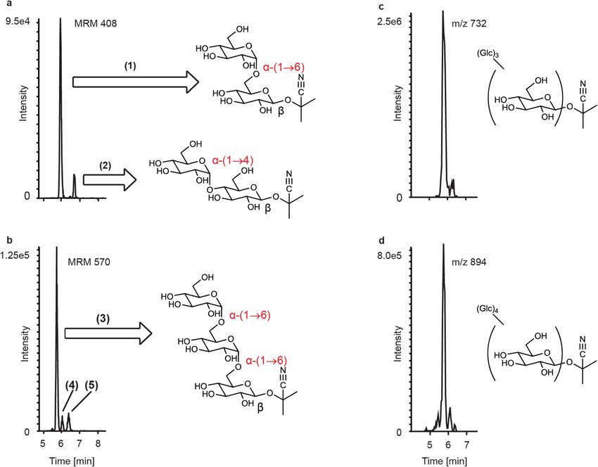
www.nature.com/scientificreports/
give an unstable hydroxynitrile that rearranges to produce hydrogen cyanide and acetone. Rearrangement of
the hydroxynitrile occurs spontaneously but can be accelerated by an enzyme known as hydroxynitrile lyase16.
As a crop, cassava displays resistance to drought, but is challenged by several viral diseases (e.g. cassava mosaic,
cassava brown streak, and cassava vein mosaic viruses)17, many of which are vectored by the whitefly Bemisia
tabaci when feeding on cassava phloem tissue18. Viral epidemics have become more severe in Africa in recent
years due to large outbreaks of whiteflies12.
The whitefly B. tabaci is a complex of cryptic species that are morphologically indistinguishable19. Collectively
they are polyphagous phloem feeders able to feed on over 600 species of p lants20, and are an important world-
wide crop pest in part because of their ability to vector over 300 plant viruses21. As a consequence, discovery of
the mechanisms by which species in the B. tabaci complex feed so successfully on crops could lead to new control
measures for B. tabaci on cassava and other crops12,21,22.
For whiteflies and other phloem-feeding herbivores, two-component defenses are being increasingly impli-
cated in their interactions with host plants23–29. Glucosinolates have been shown to affect aphid feeding, with
certain indolic glucosinolate hydrolysis products and downstream metabolites being detected after Myzus per-
sicae ingestion27. Glucosinolates were also observed to alter B. tabaci performance and host selection29, and this
insect has been shown to detoxify glucosinolates via the formation of desulpho-glucosinolates30, mercapturic
acid pathway metabolites and the transfer of additional glucose residues28. Moreover, the activities of cyanide
detoxification enzymes in B. tabaci are increased when feeding on cassava in comparison to sweet potato (a
non-cyanogenic plant)23.
Here we provide evidence concerning the interaction of cassava cyanogenic glycosides with the phloem feeder
B. tabaci. We first report the activation of cyanogenic glycosides upon feeding as seen by the formation of the
hydrogen cyanide detoxification product beta-cyanoalanine31. Then we describe the detoxification of cyanogenic
glycosides via both transglucosidation and phosphorylation of their sugar moieties. Since the glucosylated and
phosphorylated derivatives are resistant to hydrolysis by the plant activating enzyme linamarase, they therefore
represent non-toxic derivatives.
Results
Cassava cyanogenic glycosides are activated by B. tabaci feeding. The hydrolysis of cyanogenic
glycosides during whitefly feeding leads to the release of the notorious respiratory toxin hydrogen cyanide. To
assess the extent of this activation, we investigated the levels of a well-known cyanide detoxification product,
beta-cyanoalanine (Fig. 1a), in B. tabaci species SSA1-SG3 (BtSSA1-SG3), a species with an extended host range
that feeds on cassava32. BtSSA1-SG3 adults feeding on leaves of the cyanogenic plant cassava (M. esculenta)
were compared to those feeding on a non-cyanogenic plant, eggplant (Solanum melongena). Since all plants
produce beta-cyanoalanine as a natural by-product of ethylene biosynthesis31,33 the concentrations of this amino
acid were also measured in the tissues of both of these plant species to determine the endogenous levels of this
compound in fresh plants . The levels of beta-cyanoalanine in cassava and eggplant were not different from each
other (p = 0.11, N = 3) (Fig. 1b); however, BtSSA1-SG3 adults produced much higher levels of this compound
while feeding on cassava than while feeding on eggplant (p < 0.0001, N = 3) (Fig. 1c). The accumulation of beta-
cyanoalanine in the BtSSA1-SG3 whiteflies during feeding on cassava provides strong evidence for the activation
of cyanogenic glycosides.
Cassava cyanogenic glycosides are glucosylated by B. tabaci. Honeydew from BtSSA1-SG3 adults
feeding on cassava was collected and chemically analyzed via an untargeted LC–MS approach for the presence
of the cyanogenic glycoside linamarin and possible derivatives. A peak corresponding to the native glycoside
was easily observed, confirming that this insect was exposed to linamarin while feeding on cassava phloem sap.
Additionally, MS signals consistent with glycosylated linamarin derivatives were also detected. The molecular
masses of these putative products presented a characteristic serial mass addition of + 162 Da units up to + 648 Da,
suggestive of glucosylation, with elution times being slightly shorter with each addition. These peaks were also
detected in honeydew from the B. tabaci species MEAM1 (BtMEAM1, a broad generalist not naturally found
feeding on cassava)32 when these whiteflies were fed artificial diets consisting of sucrose and linamarin, but not
when fed sucrose alone, confirming they were linamarin derivatives produced by the insects.
In order to elucidate the structure of these whitefly metabolites of linamarin, the compounds resulting from
the addition of one and two apparent glucose moieties, which corresponded to the most intense MS signals in
the series of glucosylated derivatives, were purified using HPLC fractionation. NMR analyses revealed that the
structures of these compounds were indeed glucosylated linamarin conjugates. Sugar addition to the original
glucose of linamarin occurred in either an α-(1→6) or α-(1→4) orientation (compounds 1 and 2, respectively)
with the latter having slightly greater retention. The diglucose derivative showed the serial addition of two
α-(1→6) linked glucose moieties to the previously existing β-linked glucose (compound 3) (Fig. 2a,b and NMR
Supplementary Note). Quantitative 1H-NMR using sucrose as an external standard was utilized to estimate the
amount of the monoglucose derivative 1 and diglucose derivative 3 purified (5.88 µg and 3.38 µg respectively).
LC–MS standard curves for these purified metabolites were then constructed, allowing for the calculation of
molar ratios for these compounds in comparison to intact linamarin in the honeydew of BtSSA1-SG3 adults feed-
ing on cassava. It was found that glycosylated linamarin derivatives corresponding to 1 and 2 glucose additions
were present in the honeydew in a combined 5.62:1 (SE = 0.89, N = 3) ratio to intact linamarin. Additional peaks
with mass spectra consistent with two glucose additions were also observed (Compounds 4 and 5); however the
quantities and purities of the fractions obtained after chromatographic separation were not sufficient for adequate
structure elucidation. Glycosides with masses corresponding to three and four glucose additions to the original
β-linked glucose were also detected (Fig. 2c,d).
Scientific Reports | (2021) 11:13244 | https://doi.org/10.1038/s41598-021-92553-w 2
Vol:.(1234567890)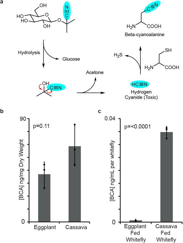
www.nature.com/scientificreports/
Figure 1. Beta-cyanoalanine levels support the activation of cassava cyanogenic glycosides during B. tabaci
feeding. (a) Scheme showing the hydrolysis of the cyanogenic glycoside linamarin and the detoxification of
the hydrogen cyanide released via beta-cyanoalanine formation. (b) Endogenous beta-cyanoalanine (BCA)
concentrations in both eggplant and cassava leaves were not different (amounts depicted have been normalized
per plant dry weight, P-value from unpaired t-test with N = 3). (c) Extracts from groups of 200 B. tabaci
SSA1-SG3 adults feeding on cassava contained elevated levels of BCA in comparison to extracts from eggplant-
fed insects (amounts depicted have been normalized per insect, P-value from unpaired t-test with N = 3).
Cassava cyanogenic glycosides are also phosphorylated by B. tabaci. In addition to glucosides,
additional unknown metabolites were detected in the honeydew of BtSSA1-SG3 whiteflies that fed on cassava
and BtMEAM1 whiteflies that fed on linamarin-containing artificial diets, but these metabolites were absent
in diets not containing linamarin. These unknown compounds again showed an MS pattern suggesting serial
glucose additions (+ 162n), with progressively earlier eluting peaks. The smallest of these metabolites displayed
a mass of 326 Da (Fig. 3a) (6), which is 80 mass units greater than linamarin, but eluting much later. Two earlier
eluting metabolites consistent with + 162 and + 324 Da additions to 6 (Fig. 3b,c, compounds 7 and 8 respectively)
were also observed. We hypothesized that the addition of 80 Da could correspond to either a sulphate or phos-
phate group linked to linamarin, with the earlier eluting peaks being a result of subsequent glycosylation. The
addition of a phosphate was supported by the accurate mass data (Supplemental Figure S1 and Supplementary
Table S1) and the disappearance of these metabolites upon incubation with alkaline phosphatase (Supplemental
Figure S2). Purification via HPLC fractionation followed by NMR analysis revealed the addition of a phosphate
moiety bound to linamarin at position 3 of the β-linked sugar (6) based on the deshielding of the 1H and 13C sig-
nals at this position (NMR Supplementary Note). Due to the low abundance of the purified products, no NMR
spectroscopic evidence could be obtained for the glucosylated phosphate derivatives.
Linamarin metabolites produced by B. tabaci resist enzymatic activation. In order to determine
whether the linamarin glycosides and phosphorylated metabolites can be activated by plant enzymes to form
hydrogen cyanide similarly to linamarin, extracts of cassava leaves containing linamarase activity, as well as
linseed extracts containing linustatinase8,34 (a disaccharidase) activity were incubated with pure linamarin and
with the honeydew of cassava-fed B. tabaci. Linamarin was degraded in the presence of both cassava enzyme
extracts and linseed enzyme extracts, while the disaccharide linustatin was only hydrolyzed in the presence of
the linseed enzyme extract. The insect-derived glycosides, however, remained unhydrolyzed in the presence of
both enzyme extracts, resisting both plant monosaccharidase and disaccharidase activities (Fig. 4). Phosphoryl-
ated linamarin and phosphorylated linamarin glycosides were also stable to enzymatic activation in the presence
of the cassava extracts (Supplemental Figure S3). Therefore, formation of these derivatives likely serves as a true
Scientific Reports | (2021) 11:13244 | https://doi.org/10.1038/s41598-021-92553-w 3
Vol.:(0123456789)www.nature.com/scientificreports/
Figure 2. Novel linamarin-derived metabolites identified in B. tabaci honeydew. Linamarin-derived
metabolites in the honeydew contain additional alpha-linked glucose moieties. (a) shows metabolites
corresponding to 1 glucose addition (compounds (1) and (2)) and (b) shows metabolite peaks corresponding
to 2 glucose additions (compounds (3), (4) and (5)). Metabolites with up to 4 further hexose additions were
detected via untargeted analysis (c and d). Compounds (1), (2) and (3) were purified from honeydew and their
structures determined by MS and NMR (Supplementary Note 1).
Figure 3. Novel phosphorylated linamarin derivatives in B. tabaci honeydew. Linamarin and the whitefly-
produced linamarin glycosides were also phosphorylated by B. tabaci. (a) The position for phosphorylation
of linamarin was elucidated as C3 (6) by NMR (Supplementary Note 1). The structures of both the
monoglucosylated (7, b) and diglucosylated (8, c) phosphorylated derivatives were supported by mass spectral
data, but NMR analysis was not possible due to low abundance.
Scientific Reports | (2021) 11:13244 | https://doi.org/10.1038/s41598-021-92553-w 4
Vol:.(1234567890)www.nature.com/scientificreports/
Figure 4. Resistance of glucosylated linamarin derivatives to activation by plant enzymes with release of
hydrogen cyanide. The cyanogenic monoglucoside linamarin, the cyanogenic diglucoside linustatin and the
glucosylated linamarin derivatives found in the honeydew of whiteflies fed on cassava were tested. Honeydew
and standards were incubated with crude enzyme extracts from cassava and linseed or with water alone.
Linamarin was readily hydrolyzed by both cassava and linseed extracts, and linustatin was hydrolyzed by linseed
enzymes. Conversely, the insect-derived glycosides were not substrates for either of the enzyme extracts.
pre-emptive detoxification of linamarin rendering products that can no longer be hydrolyzed with the release of
hydrogen cyanide.
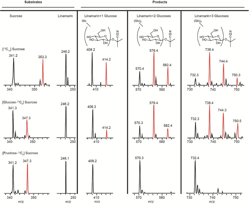
Mechanism of glucose addition to the cyanogenic glycoside linamarin. Glucosylation reactions
are most often catalyzed by either of two enzyme classes, UDP-glucosyl-transferases and transglucosidases. The
latter of these two classes utilizes a mechanism which transfers a glucose unit from a donor disaccharide directly
to an acceptor molecule, while the former utilizes an activated form of glucose (UDP-glucose). In order to eluci-
date the mechanism by which glucose units are added to linamarin by whiteflies, experiments were carried out to
feed 13C sucrose isotopologues in artificial diet. Labeled glucose was incorporated into the glycosides of linama-
rin when diets contained [13C12]sucrose and [glucose-13C6]sucrose, but not when diets contained [fructose-13C6]
sucrose, [13C6]glucose or [13C6]fructose (Fig. 5 and Supplemental Figure S4). Label was also incorporated into
phosphorylated glycosides in the same manner (Supplemental Figure S5). This mechanism is consistent with
a transglucosidase activity, which is typically carried out by enzymes of the glycoside hydrolase (GH) f amily35.
Expression profiles of the transglucosidase genes BtSUC2 and BtSUC5 in B. tabaci adults
feeding on various host plants. BtSUC2 and BtSUC5 were previously identified in the whitefly spe-
cies BtMEAM1 via phylogenetic analyses followed by heterologous production and biochemical characteriza-
tion as transglucosidases that glucosylate glucosinolates, thereby preventing their hydrolysis and release of toxic
products28. Thus we investigated their potential to participate in the glucosylation of cassava cyanogenic glyco-
sides using BtSSA1-SG3 that feeds on cassava and BtMEAM1, which is not naturally found on cassava. First the
genomic sequences of BtSUC2 and BtSUC5 in the cassava-feeding BtSSA1-SG3 were found to be approximately
95% identical to their counterparts in BtMEAM1 (Fig. 6a). Next, the expression of the BtSUC2 and BtSUC5
genes in terms of reads mapping to their exons was compared in the two whitefly species feeding on two dif-
ferent plant species (using raw RNA-Seq data from SRP12775732,36). When feeding on eggplant (no cyanogenic
glycosides), BtSUC2 expression did not differ between the species (p = 0.187, N = 3), but BtSUC5 expression was
significantly higher in BtSSA1-SG3 than in BtMEAM1 (p = 0.011, N = 3) (Fig. 6b). When feeding on cassava
(containing cyanogenic glycosides), BtSUC2 expression was significantly higher in BtMEAM1 than in BtSSA1-
SG3 (p = 0.017, N = 3), but the pattern for BtSUC5 was almost identical to that on eggplant, with expression in
BtSSA1-SG3 significantly higher than in BtMEAM1 (p = 0.013, N = 3) (Fig. 6c).
In vitro activities of the transglucosidases BtSUC2 and BtSUC5. Drosophila S2 cells producing
BtSUC2 and BtSUC5 were utilized in enzyme assays with the cyanogenic glycoside linamarin and the donor
disaccharide sucrose. BtSUC2 produced the α-(1→4) glycoside of linamarin (2) approximately 50 times more
efficiently than S2 control cells, and smaller amounts of the α-(1→6) derivative 1 (approximately 8 times more
efficiently than control cells) (Fig. 7a). No higher order glycosides were formed by BtSUC2 (Fig. 7b). BtSUC5
on the other hand produced much larger amounts of 2 (more than 50,000 times more efficient than S2 control
cells) (Fig. 7a), as well as a glycoside corresponding to a further glucose addition (5) (Fig. 7b) with a similar
efficiency. BtSUC5 also produced a smaller amount of 1 (approximately 7 times more active than controls) and
a peak corresponding to an unknown derivative (4) (Fig. 7b). BtSUC5 displayed apparent Michaelis–Menten
kinetics, with an estimated KM of ~ 0.5 mM for linamarin and ~ 0.13 M for sucrose (Supplemental Figure S6).
For BtSUC2, however, the low activity towards linamarin prevented an estimation of KM values. BtSUC2 and
BtSUC5 activities towards the purified phosphorylated linamarin derivatives 6 and 7 were also investigated, and
both enzymes glucosylated 6 forming a product with very slightly different retention time compared to 7 (Sup-
Scientific Reports | (2021) 11:13244 | https://doi.org/10.1038/s41598-021-92553-w 5
Vol.:(0123456789)www.nature.com/scientificreports/
Figure 5. Evidence for enzymatic mechanism of linamarin glucosylation by B. tabaci. Glucosylation of
cyanogenic glycosides in B. tabaci is catalyzed by a transglucosidase activity based on feeding of [ 13C] sucrose
isotopologues and linamarin to insects in artificial diet. Shown are MS regions from LC–MS analyses of
substrates and products of feeding experiments on three different diets. Feeding fully 13C-labeled sucrose and
sucrose 13C-labeled in the glucose moiety gave labeling in the newly added glucose of glucosylated linamarin
metabolites. However, feeding sucrose 13C-labeled in the fructose moiety gave no label in the products,
demonstrating incorporation of only the glucose of sucrose into the products. Feeding of free 13C-labeled
glucose and fructose with linamarin also resulted in no incorporation into glucosylated derivatives of
linamarin (Supplementary Figure S4), showing that the glucose must originate from sucrose as expected in
transglucosidase catalysis.
plemental Figure S7a), suggesting a positional isomer. While BtSUC2 did not use 7 as substrate, BtSUC5 pro-
duced a compound corresponding by mass to 8, but with a different retention time (Supplemental Figure S7b).
The precise amounts of the phosphorylated derivatives used in these assays as substrates were not determined
due to their low abundance.
BtSUC2 and BtSUC5 were also assayed with other plant-derived glucosides as potential substrates. Specialized
plant metabolites from various two-component defensive compound classes were tested including benzoxazinoid,
cyanogenic, phenolic, iridoid, flavonoid and other cyanogenic glycosides, as well as glucosinolates. BtSUC2 and
5 had detectable transglucosidase activity with all substrates (Supplemental Figure S8), with BtSUC5 having
the greatest activity of the two proteins and the flavonoid rutin having the least activity of the nine substrates.
Discussion
Phloem-feeding insects were once thought to be largely unaffected by two-component plant defenses such as
cyanogenic glycosides and glucosinolates, as their feeding mode does not appear to cause sufficient tissue damage
to initiate hydrolytic activation of these compounds37. However, increasing evidence suggests that phloem feed-
ing can indeed cause such activation23,24,26,29. We recently demonstrated the activation of aliphatic glucosinolates
by B. tabaci feeding on plants of the Brassicaceae after finding isothiocyanate hydrolysis products and their
detoxified glutathione conjugates in the h oneydew28. Here, we report that B. tabaci feeding on the cyanogenic
glycoside-producing plant cassava accumulates beta-cyanoalanine, a cysteine conjugate of hydrogen cyanide
that is considered to be a detoxification product38, in their bodies. Whiteflies fed on cassava contained 50 times
greater amounts of beta-cyanoalanine than their counterparts fed on a non-cyanogenic plant, eggplant (Fig. 1).
Scientific Reports | (2021) 11:13244 | https://doi.org/10.1038/s41598-021-92553-w 6
Vol:.(1234567890)www.nature.com/scientificreports/
Figure 6. Comparison of transglucosidase BtSUC2 and BtSUC5 sequences in B. tabaci species and relative
gene expression in different species on different food plants. (a) The percent sequence identities of SSA1-SG3
homologues of BtSUC2 and BtSUC5 to the originally isolated genes from BtMEAM1 were determined at
the DNA and protein levels. (b,c) Mean numbers of reads mapped to BtSUC2 and BtSUC5 were plotted in
the species BtMEAM1 and BtSSA1-SG3 feeding on two host plants, eggplant and cassava. Asterisks denote
statistically significant differences (p value < 0.05) between means of biological replicates (N = 3) with individual
data overlaid as points on the histogram.
The residual beta-cyanoalanine found after eggplant feeding is likely of plant and not insect origin since this
amino acid is also produced by plants upon detoxification of the cyanide formed during the biosynthesis of the
hormone ethylene31,33. Undamaged cassava and eggplant tissues contained similar amounts of beta-cyanoalanine.
Therefore the drastically higher levels of beta-cyanoalanine in B. tabaci when feeding on cassava instead of egg-
plant suggest an enzymatic conversion of liberated cyanide from the cyanogenic glycosides of cassava. While
beta-cyanoalanine synthase activity has been reported in whitefly enzyme extracts via the production of H 2S23,
no specific B. tabaci enzymes have been identified that could catalyze this transformation. Activation of cassava
cyanogenic glycosides would be presumed to happen during whitefly feeding, upon stylet penetration or naviga-
tion to the phloem, or in the gut of the whitefly post ingestion.
Since cyanogenic glycosides are activated by hydrolysis upon whitefly feeding, we explored whether this insect
possessed the ability to metabolize the chief cassava cyanogenic glycoside linamarin to a non-hydrolyzable prod-
uct prior to its potential activation. We found glucosylated linamarin derivatives in the honeydew of whiteflies
feeding on cassava with the additions of α-glucosides in either an α-(1→6) or α-(1→4) fashion. Additional sugar
additions occurred that are hypothesized to be combinations of various linkage modes (α-(1→6) or α-(1→4)) of
glucose. For example, the most abundant peak from two glucose additions (3) was determined to be α-(1→6);
α-(1→6). Two other peaks from two glucose additions to linamarin have later elution times consistent with
Scientific Reports | (2021) 11:13244 | https://doi.org/10.1038/s41598-021-92553-w 7
Vol.:(0123456789)www.nature.com/scientificreports/
Figure 7. Chromatographic analyses of glucosylated products catalyzed by the B. tabaci transglucosidases
BtSUC2 and 5. Medium of Drosophila S2 cells expressing these enzymes was assayed with linamarin and
sucrose. Depicted are extracted multiple reaction monitoring (MRM) LC–MS chromatograms for mono-
and di-glucosylated linamarin derivatives. (a) BtSUC2 and 5 showed transglucosidation activity, producing
α-(1→4)-linked glucose derivatives (2) of linamarin with BtSUC2 also producing small amounts of the
α-(1→6)-linked derivative (1) above control levels. (b) BtSUC5 additionally produced the diglycosylated
product (5). The S2 control assay was performed on medium extracts of untransformed S2 cells. (1: α-(1→6)-
linked glucose derivative of linamarin; 2: α-(1→4)-linked glucose derivative of linamarin; 3: α-(1→6), α-(1→6)-
linked diglucosylated derivative of linamarin; 4, 5: unknown diglucosylated derivatives of linamarin).
α-(1→4)-linked sugars, as the α-(1→6)-linked product (1) shows earlier elution than the α-(1→4)-linked product
(2). The additional two peaks may therefore correspond to a compound with both α-(1→6) and α-(1→4)-linked
sugars (4), and a compound with two α-(1→4) additions (5) as the last eluting peak. This is supported by the fact
that the enzyme BtSUC5, which produces almost exclusively α-(1→4) glucosylated linamarin (2) in incubations
with sucrose and linamarin, also produced 5 (Fig. 7b). Interestingly, the glucosylation of cyanogenic glycosides by
B. tabaci resulted in the formation of many more peaks than the glucosylation of glucosinolates reported in our
previous study28. However, regardless of whether linamarin had one or two α-glucose additions, these metabolites
were resistant to activation by linamarase and thus represent detoxification products.
Phosphorylated linamarin glycosides were also detected in B. tabaci honeydew. Phosphorylation is hypoth-
esized to be somewhat independent from the glucosylation of linamarin due to the capability of BtSUC2 and
BtSUC5 to glucosylate both phosphorylated and non-phosphorylated linamarin (Supplemental Figure S7). Yet the
biological role of phosphorylation is likely detoxification, similarly to glucosylation, as phosphorylated linamarin
derivatives were not activated by linamarase. The formation of phosphorylated phenolic glycosides in Lymantria
dispar (the gypsy moth) was also proposed to represent a detoxification r eaction39. Although phosphorylation
is an atypical mammalian phase II detoxification process, it may be more widespread in insects based on recent
reports40–42. Phosphorylation of sugars occurs elsewhere in metabolism as a marker for breakdown, as in the
case of glycolysis43,44, or in the synthesis of sugar polymers44,45. For example, C-3 phosphorylation as described
Scientific Reports | (2021) 11:13244 | https://doi.org/10.1038/s41598-021-92553-w 8
Vol:.(1234567890)www.nature.com/scientificreports/
here occurs in the process of glycogen synthesis46,47. The phosphorylation of plant glycosides in L. dispar and its
close relative Orgyia antigua also occurred at C-339.
The glucosylation of plant defense compounds is a logical detoxification reaction for phloem-feeding her-
bivores since phloem sap is rich in sugars. Moreover, phloem-feeding insects also possess transglucosidase
enzymes that produce higher order sugar oligomers from sucrose in order to reduce the high osmotic pressures
that may occur in the gut when feeding on phloem sap48. These enzymes can also glucosylate other substrates28,
and were shown here in experiments with 13C-labeled sucrose isotopologues (Fig. 5) to be the enzymes involved
in forming glucosylated cyanogenic glycoside derivatives. Two B. tabaci transglucosidases, BtSUC2 and BtSUC5,
previously demonstrated to form glucosylated glucosinolates were shown to transglucosylate the cyanogenic
glycoside linamarin in vitro. BtSUC2 produced the α-(1→6) (1) and α-(1→4) (2) linked monoglycosylated
derivatives of linamarin identified in honeydew. In previous in vitro assays with these enzymes, derivatives with
α-(1→6) linkages were not previously observed with glucosinolates as substrates28. BtSUC5 also displayed other
products including a compound with a second glucose addition (5) that was not fully characterized. BtSUC5 is
likely to also carry out the transglucosidation of linamarin in vivo since the KM values for BtSUC5 were within
physiologically expected levels for both linamarin and sucrose.
Further incubations of BtSUC2 and BtSUC5 with various plant defense glycosides resulted in the transglucosi-
dation of virtually all substrates tested despite the great variety of aglycones present in the compounds offered,
which included cyanogenic glycosides, glucosinolates, iridoid glycosides, benzoxazinoids, phenolic glycosides
and flavonoid glycosides (Supplemental Figure S8). This substrate promiscuity suggests a very flexible catalytic
pocket that accommodated all substrates, with the flavonoid glycoside rutin, the largest substrate tested, pre-
sumably fitting less well. A similar size limit on catalysis in transglucosideases was observed in studies with a
dextran-sucrase where the velocity of reaction was reduced when switching the acceptor from maltose to the
larger maltotriose49. The catalytic versatility of these enzymes in vitro was not observed in vivo when some of
these substrates were fed directly to w hiteflies28. Thus, more work needs to be carried out to determine the ability
of these transglucosidases to react with plant defense compounds.
The glycoside hydrolase 13 family, which contains the transglucosidases, is greatly expanded in B. tabaci and
other phloem-feeding insects28,50. The capacity of these enzymes to metabolize a wide range of plant defenses
may be responsible for the broad host range of B. tabaci. In comparing the two B. tabaci species studied, the
expression of the glycoside hydrolase gene BtSUC5 was higher in BtSSA1-SG3 (which can feed on cassava but
also on a wide range of alternative plant hosts)32,36 than in BtMEAM1 (a broad generalist not naturally found
feeding on cassava) regardless of the host plant tested. Since at the protein level, BtSUC5 had a much higher
transglucosidase activity than BtSUC2 with linamarin in vitro, the higher expression of the encoded gene BtSUC5
in BtSSA1-SG3 vs. BtMEAM1 (Fig. 6c) may be important for the ability of this whitefly species to utilize cassava.
The tissue-specific expression of BtSUC2 and BtSUC5 may also play an important role in this detoxification route.
The expression of BtSUC5 has been previously shown to be higher in the whitefly gut relative to its whole b ody28,
however it is not currently known whether such enzymes are also produced in other tissues or might be secreted
in the insect saliva to prevent hydrolysis before ingestion.
In conclusion, our investigations into B. tabaci whitefly metabolism of cyanogenic glycosides illustrate the
diverse chemical transformations carried out by this phloem-feeding insect. Although cyanogenic glycosides
and other two-component defenses were previously believed not to be activated by phloem-feeders, our results
suggest that B. tabaci are indeed susceptible to two-component defenses and this may have selected for their
potential to pre-emptively detoxify them by glucosylation and phosphorylation. Knowledge of how B. tabaci
detoxifies cyanogenic glycosides could be employed to reduce its infestation of cassava, a cyanogenic glycoside-
containing crop that is a staple food for millions of Africans. For example, RNA interference targeting the
transglucosidases involved in detoxification could be applied via the cassava plant or by direct spraying51 to
reduce whitefly feeding by increasing their susceptibility to cyanogenic glycosides. As this approach targets the
whitefly specifically, it should not harm beneficial insects that come into contact with cassava. A parallel strategy
could be to design specific inhibitors of the transglucosidase for application to the crop. Further information
on the metabolic adaptations of whiteflies to cassava could also be exploited in crop protection and contribute
additionally to basic knowledge on how phloem-feeding insects survive on plants with two-component defenses.
Materials and methods
Plants. Eggplant (Solanum melongena, cv. Black Beauty), and cassava (Manihot esculenta, cv. MCol22) plants
were grown under standard greenhouse conditions at 26 ± 2 °C with supplemental lighting and a photoperiod
of 14:10 h (light:dark).
Insects. Bemisia tabaci (Hemiptera: Aleyrodidae) species MEAM1 (Middle East-Asia Minor 1) was collected
in southern Israel in 2003 and from Sudan in the late 1990s, and reared continuously on cotton as separate popu-
lations. An additional MEAM1 population was collected in Peru in 2012 and maintained on eggplant. B. tabaci
species SSA1-SG3 (sub-Saharan Africa 1—species group 3) was collected on Bagamoyo Road, Tanzania in 2013
and reared on cassava plants.
Bemisia tabaci feeding on cassava and eggplant for beta‑cyanoalanine analysis. Groups of
adult B. tabaci (200 individuals, BtSSA1-SG3), were reared on eggplant or cassava, then collected 1-3 days after
emergence and kept frozen prior to extraction and LC-MS analysis of beta-cyanoalanine.
Bemisia tabaci feeding on cassava and eggplant for analysis of BtSUC2 and 5 expres-
sion. Groups of adult B. tabaci (50 individuals of BtSSA1-SG3 and BtMEAM1), were reared on sucrose-based
Scientific Reports | (2021) 11:13244 | https://doi.org/10.1038/s41598-021-92553-w 9
Vol.:(0123456789)www.nature.com/scientificreports/
artificial diets for 72 h and transferred to cassava or eggplant for 24 h. Further RNA isolation and analysis was
conducted as in32, and gene expression analyses were performed as described below.
Honeydew collection. Honeydew was collected from three different sources:
Bemisia tabaci feeding on cassava for honeydew metabolite analysis. Groups of adult B. tabaci
(50 individuals, BtSSA1-SG3), were reared on eggplant and transferred 1–3 days after emergence to fresh cas-
sava. Insects were enclosed within glass clip cages while feeding After 96 h, the honeydew deposited on the glass
tubes was washed off with water:methanol (20:80, v:v), dried under nitrogen gas, and resuspended in water prior
to LC–MS analysis of phosphorylated and glucosylated linamarin derivatives.
Bemisia tabaci feeding on artificial diets with linamarin. Feeding of artificial was replicated
as described in28 with changes to diet composition and collection methods as described below. Groups of
150 B. tabaci (MEAM1) adults were collected from eggplant and switched to artificial diet feeders consisting of a
glass tube (3 cm height × 2 cm diameter) with a liquid diet covered with a double layer of Parafilm. Insects were
allowed to feed through the Parafilm on a 10% sucrose solution containing no additives (control) or the cyano-
genic glycoside linamarin (Sigma-Aldrich) at a concentration of 5 mM. After 96 h, the honeydew deposited on
the glass tubes was washed off with water:methanol (20:80, v:v), dried under nitrogen gas, and resuspended in
water prior to LC–MS analysis of phosphorylated and glucosylated linamarin derivatives.
Bemisia tabaci feeding on artificial diets containing isotopically‑labeled sugars and linama-
rin. Feeding of isotopically labelled sugars was performed as in28 with diet composition modified accordingly.
Four different sucrose isotopologues were added to artificial diets: [ 12C12]sucrose, [13C12]sucrose, [glucose-13C6]
sucrose, and [fructose-13C6]sucrose. The monosaccharides [13C6]fructose and [13C6]glucose were also fed. The
artificial feeding devices consisted of a glass tube (5 cm high × 2.5 cm diameter) with the liquid diet (50 µL) held
within a double layer of Parafilm. About 50 B. tabaci MEAM1 adults were placed in each tube. Feeding assays
were performed for 72 h on diets that contained 5 mM linamarin and 0.29 M of the labeled sugars. The honey-
dew deposited on the glass tubes was washed with water: methanol (20:80, v:v) and stored at − 20 °C until pro-
cessing and further MS analysis of 13C isotope incorporation into linamarin-derived glucosylated metabolites. A
full summary of artificial diet constituents is outlined in Supplementary Table S2.
Purification and LC–MS analysis of glucosylated and phosphorylated cyanogenic glyco-
sides. Purification of glucosylated or phosphorylated cyanogenic glycosides was performed similarly to28,
via fractionation on a Nucleodur Sphinx RP column (250 × 4.6 mm, 5 µm, Macherey–Nagel, Düren, Germany)
using an HP 1200 HPLC (Agilent Technologies, Santa Clara, CA, USA) coupled to a fraction collector (Advan-
tec, Dublin, CA, USA), with methods modified accordingly. Chromatographic separation was attained using a
gradient of 0.05% aqueous formic acid (Solvent A) and acetonitrile (Solvent B) at a flow rate of 1 mL min−1 at
20 °C as follows: 5–29% B (12 min), 29–100% B (0.1 min), a 2.9 min hold at 100% B, 100–5% B (0.1 min), and
a 3.9 min hold at 5% B. Linustatin was purified from linseed by crushing 10 g of seeds in liquid nitrogen and
extracting with 80% methanol prior to centrifugation at 10,000×g. The methanol supernatant was then dried
down and concentrated before resuspension in water for purification on a reverse phase column as described
above.
Qualitative analysis of glucosylated cyanogenic glycosides in feces and honeydew extracts was performed on
an HP 1100 series HPLC as in28 using instrument parameters modified accordingly, as described below. Separa-
tion was achieved on a Nucleodur Sphinx RP column (250 × 4.6 mm, 5 µm, Macherey–Nagel) with a gradient
of 0.2% aqueous formic acid (solvent A) and acetonitrile (solvent B) with a flow rate of 1 mL min−1 at 25 °C as
follows: 5–55% B (25 min), 55–100% B (0.1 min), 100% B 0.9 min hold, 100–5% B (0.1 min), 5% B 3.9 min hold.
The HPLC was coupled to an Esquire 6000 ESI-Ion Trap mass spectrometer (Bruker Daltonics, Bremen, Ger-
many) operated in both positive and negative modes in the range of m/z 60–1500 with skimmer voltage − 40 V;
capillary exit voltage − 146.7 V; capillary voltage 4000 V; nebulizer pressure 35 psi; drying gas 11 L min−1; and
gas temperature 330 °C. DataAnalysis software V4 (Bruker Daltonics) was used for chromatogram analysis.
Qualitative analysis of isotopically-labeled glucosylated cyanogenic glycosides in feces and honeydew extracts was
performed by LC–MS as described above for feces and honeydew except that capillary exit volatage was − 113.5 V.
High resolution mass spectrometry of phosphorylated compounds was achieved on an Thermo Scientific Ulti-
Mate 3000 UHPLC coupled to a Bruker TIMS-TOF mass spectrometer. Separation was achieved on a Nucleodur
Sphinx RP column (250 × 4.6 mm, 5 µm, Macherey–Nagel, Germany) with a gradient of 0.2% aqueous formic
acid (solvent A) and acetonitrile (solvent B) with a flow rate of 1 mL min−1 (split 1:3 source: waste) at 25 °C as
follows: 5–55% B (25 min), 55–100% B (0.1 min), 100% B 0.9 min hold, 100–5% B (0.1 min), 5% B 3.9 min hold.
The MS was operated in negative mode scanning from m/z 50–1500 with the following parameters. Source End
in−1, Dry temperature 330 °C. Tune
plate offset: 500 V, capillary: 3500 V, Neubilizer: 3.5 bar, Dry gas: 11.0 L m
General Funnel 1RF: 150 Vpp, Funnel 2 RF: 200 Vpp, isCID energy: 0.0 eV, Multipole RF: 50 Vpp, Deflection
Delta: − 70 V, Quadrupole energy: 4.0 eV, Low mass: 90 m/z Collision energy: 7.0 eV, Collision RF: 400 Vpp,
Transfer time: 80.0 µs, Pre-pulse storage: 5.0 µs. Calibration took place externally immediately before the sam-
ples were run using Agilent ESI-L Low Concentration Tune Mix and an enhanced quadratic calibration curve.
Quantification of the glucosylated cyanogenic glycosides in transglucosidase and linamarase/linustatinase
assays and in honeydew was accomplished via an HP 1260 series HPLC coupled to an AB Sciex API 5000 mass
spectrometer (Applied Biosystems, Darmstadt, Germany). The column utilized was a Nucleodur Sphinx RP
column (250 × 4.6 mm, 5 µm, Macherey–Nagel) using a chromatographic gradient of 0.05% aqueous formic
Scientific Reports | (2021) 11:13244 | https://doi.org/10.1038/s41598-021-92553-w 10
Vol:.(1234567890)www.nature.com/scientificreports/
acid (Solvent A) and acetonitrile (Solvent B) at a flow rate of 1 mL min−1 at 20 °C as follows: 5–29% B (12 min),
29–100% B (0.1 min), a 2.9 min hold at 100% B, 100–5% B (0.1 min), and a 3.9 min hold at 5% B.The MS was
operated in the negative mode with collision gas value 7, curtain gas pressure 35 psi, spray gas pressures 60 psi,
ion spray voltage − 4500 V, and turbogas temperature 600 °C. Compounds were detected using multiple reac-
tion monitoring (MRM) detection with the parameters outlined in Supplementary Table S3. Quantification
was achieved using external calibration curves constructed from solutions of purified glucosylated cyanogenic
glycosides of known concentrations (determined in solution via NMR as described below). Analyst 1.5 software
(Applied Biosystems) was used for data acquisition and processing. All averages and standard errors were calcu-
lated from three independent biological replicates. No other statistical tests were performed.
Extraction and LC–MS analysis of beta‑cyanolalanine. Groups of 200 B. tabaci adults (BtSSA1-SG3)
were collected from eggplant and cassava plants. The bodies were crushed using liquid nitrogen and extracted in
1 mL of water:methanol (20:80, v:v). The supernatant was evaporated using nitrogen and resuspended in 50 µL
water before LC–MS analysis (see below).
For analysis of beta-cyanoalanine in plants, 3 young leaves of cassava and eggplant each were crushed using
liquid nitrogen and a mortar and pestle and extracted in 0.5 mL water:methanol (20:80, v:v) per mg of leaf tis-
sue. Aliquots of the supernatants were then evaporated and resuspended in identical volumes of water before
LC–MS analysis.
Quantification of beta-cyanoalanine levels was performed on an HP 1260 HPLC coupled to an AB Sciex API
5000 mass spectrometer. The column utilized was a Agilent XDB-C18 column (50 × 4.6 mm, 1.8 µm, Agilent
Technologies, Boeblingen, Germany) using a chromatographic gradient of 0.05% aqueous formic acid (Solvent A)
and acetonitrile (Solvent B) with a flow rate of 1.1 mL min−1 at 25 °C as follows: 0.5 min hold at 10% B, 10–45%
B (3.5 min), 45–100% B (0.02 min), 0.98 min hold at 100% B, 100–10% B (0.02 min), 1.98 min hold at 10% B.
The mass spectrometer was operated in the negative mode with collision gas value 8, curtain gas pressure 25
psi, spray gas pressures 60 psi, ion spray voltage − 4500 V, and turbogas temperature 700 °C. Compounds were
detected using scheduled multiple reaction monitoring (MRM) detection with the parameters outlined in Sup-
plementary Table S3. Analyst 1.5 software was used for data acquisition and processing.
NMR spectroscopy. NMR analyses were performed as i n28, with spectra (1H, 1H-1H COSY, 1H-13C HSQC,
1
H-13C HMBC and 1H-1H SELTOCSY) acquired on a 700 MHz Avance III HD spectrometer equipped with a
1.7 mm cryoprobe (Bruker Biospin, Rheinstetten, Germany). Further information is available in the supplemen-
tary information file. Data acquisition and processing was accomplished using TopSpin ver. 3.2 (Bruker Biospin,
Rheinstetten, Germany). Samples were measured in MeOH-d3 or D2O as indicated at 293 K. For quantification
via 1H NMR (10 s delay between scans), purified compounds were dried under N 2 flow and resuspended in D
2O,
and a sucrose solution (3.13 mM in D2O) was used as an external quantification standard.
Cloning and heterologous expression of B. tabaci transglucosidases in Drosophila S2
cells. Vectors, cells and cell media were obtained from ThermoFisher Scientific (Waltham, MA, USA). Full-
length ORFs from previously characterized B. tabaci Glucohydrolase Family 13 genes (BtSUC2 and BtSUC5)
were amplified as previously described28 using the primer sets outlined in Supplemental Table S4.
Determination of BtSUC2 and BtSUC5 gene expression. The number of raw reads mapping to
BtSUC2 and BtSUC5 were determined from S RP12775732,36 using the STAR s oftware52, based on the genome
and gtf files available at NCBI. Sequence alignment between SSA1 and MEAM1 sequences was performed using
the BtSSA1 genome (unpublished, provided by EBI).
Transglucosidase enzyme assays. Enzyme assays were performed as in28 with modifications listed
below. Drosophila S2 cells expressing BtSUC2 and BtSUC5 enzymes as well as non-transfected control cells were
centrifuged at 100×g for 5 min. The resulting supernatant was utilized as the secreted protein fraction for enzyme
assays. For determination of linamarin transglucosidation activity, 5 µL of supernatant from each culture con-
taining an equivalent total protein content (verified by Bradford assays) was mixed with a 5 µL 50 mM phosphate
buffer at pH 7.0 containing 2 M sucrose and 5 mM linamarin, and reacted for 6 h at 25 °C with no stirring. These
assay conditions were also repeated for other potential substrates: I3M-GSL (Phytoplan Diehm & Neuberger
GmbH), pOHBz-GSL(Phytoplan), DIMBOA-Glc, amygdalin (Roth), dhurrin (Roth), salicin (Sigma), arbutin
(Roth), aucubin (Roth), and rutin (Sigma). Assay conditions were chosen after screening a range of substrate
concentrations and pH values that reflect previous work on glucohydrolases and phloem-feeding insects. The
reaction was stopped using 15 μL of methanol and immediately stored at − 20 °C. For LC–MS analysis, enzyme
assays were centrifuged at 5200×g for 5 min and the supernatant obtained was diluted 1:10 in water for the analy-
sis of glucosylated linamarin, and diluted 1:1000 in water.
For KM estimation, enzyme preparations of control cells were first checked and found to catalyze less than
1% of the supplied linamarin to transglucosylated products also to catalyze only low levels of sucrose hydrolysis.
Enzyme assays were performed in the same manner as described above, except incubation times were reduced
to 30 min. Peak areas were integrated and compared based on duplicate analysis. The sucrose KM determination
was performed at a constant linamarin concentration of 2.5 mM with sucrose concentrations ranging from 0.05
to 1 M. The linamarin KM determination utilized sucrose at a final concentration of 1 M and a range of linamarin
concentrations from 25 µM to 20 mM. All assays were carried out under linear reaction conditions with respect
to time and protein concentration. Substrate concentration was never reduced below 95% of the initial level
during the 30 min assay period.
Scientific Reports | (2021) 11:13244 | https://doi.org/10.1038/s41598-021-92553-w 11
Vol.:(0123456789)www.nature.com/scientificreports/
For tests on the reactivity of glucosylated cyanogenic glycosides with plant linamarase, linamarin (Sigma)
(10 µL of a 5 mM solution), linustatin purified from linseed extracts (see above section on cyanogenic glycoside
purification) and honeydew from cassava-reared adult SSA1-SG3 whiteflies containing glucosylated linamarin
glycosides were mixed with a crude leaf enzyme extract from cassava with native linamarase activity or crude
linseed extracts with native linustatinase activity in 20 mM phosphate buffer solution (10 μL), pH 7.0. The
cassava linamarase extract was obtained from 10 g of leaves and the linseed linustatinase extract was obtained
from 10 g of seeds flash frozen in liquid nitrogen, crushed using a mortar and pestle, extracted with phosphate
buffer (20 mM pH 7.0) and filtered using vacuum filtration. Control reactions were supplemented with 5 µL of
water instead of the cassava or linseed extract solutions. Reactions were incubated at room temperature with
no stirring, stopped after 1 h with 20 µL acetic acid and stored frozen until LC–MS analysis. Phosphorylated
derivatives were tested for stability to cassava crude enzyme extract (but not to linseed extract) under the same
reaction conditions.
Received: 30 January 2021; Accepted: 10 June 2021
References
1. Morant, A. V. et al. β-Glucosidases as detonators of plant chemical defense. Phytochemistry 69, 1795–1813 (2008).
2. Pentzold, S., Zagrobelny, M., Rook, F. & Bak, S. How insects overcome two-component plant chemical defence: Plant β-glucosidases
as the main target for herbivore adaptation. Biol. Rev. 89, 531–551 (2014).
3. Wang, X. W., Li, P. & Liu, S. S. Whitefly interactions with plants. Curr. Opin. Insect Sci. 19, 70–75. https://doi.org/10.1016/j.cois.
2017.02.001 (2017).
4. Bak, S. et al. Cyanogenic glycosides: A case study for evolution and application of cytochromes P450. Phytochem. Rev. 5, 309–329
(2006).
5. Hansen, C. H. et al. CYP83b1 is the oxime-metabolizing enzyme in the glucosinolate pathway in Arabidopsis. J. Biol. Chem. 276,
24790–24796. https://doi.org/10.1074/jbc.M102637200 (2001).
6. Ratzka, A., Vogel, H., Kliebenstein, D. J., Mitchell-Olds, T. & Kroymann, J. Disarming the mustard oil bomb. Proc. Natl. Acad. Sci.
USA 99, 11223–11228. https://doi.org/10.1073/pnas.172112899 (2002).
7. Poulton, J. E. Cyanogenesis in plants. Plant Physiol. 94, 401–405. https://doi.org/10.1104/pp.94.2.401 (1990).
8. Niedzwiedz-Siegien, I. Cyanogenic glucosides in Linum usitatissimum. Phytochemistry 49, 59–63. https://doi.org/10.1016/S0031-
9422(97)00953-9 (1998).
9. Selmar, D. Transport of cyanogenic glucosides—Linustatin uptake by hevea cotyledons. Planta 191, 191–199 (1993).
10. Olsen, K. M. & Schaal, B. A. Evidence on the origin of cassava: Phylogeography of Manihot esculenta. Proc. Natl. Acad. Sci. USA
96, 5586–5591. https://doi.org/10.1073/pnas.96.10.5586 (1999).
11. Cooperation, O. f. E., Development, Food & Organization, A. Agriculture in sub-Saharan Africa: Prospects and challenges for the
next decade. OECD-FAO agricultural outlook 2016–2025 (2016).
12. Jacobson, A. L., Duffy, S. & Sseruwagi, P. Whitefly-transmitted viruses threatening cassava production in Africa. Curr. Opin. Virol.
33, 167–176. https://doi.org/10.1016/j.coviro.2018.08.016 (2018).
13. Mkpong, O. E., Yan, H., Chism, G. & Sayre, R. T. Purification, characterization, and localization of linamarase in cassava. Plant
Physiol. 93, 176–181. https://doi.org/10.1104/pp.93.1.176 (1990).
14. Nambisan, B. & Sundaresan, S. Distribution of linamarin and its metabolizing enzymes in cassava tissues. J. Sci. Food Agric. 66,
503–507. https://doi.org/10.1002/jsfa.2740660413 (1994).
15. Cooke, R. D., Blake, G. G. & Battershill, J. M. Purification of cassava linamarase. Phytochemistry 17, 381–383. https://doi.org/10.
1016/S0031-9422(00)89320-6 (1978).
16. Hughes, J., Carvalho, F. J. & Hughes, M. A. Purification, characterization, and cloning of alpha-hydroxynitrile lyase from cassava
(Manihot esculenta Crantz). Arch. Biochem. Biophys. 311, 496–502. https://doi.org/10.1006/abbi.1994.1267 (1994).
17. Calvert, L. & Thresh, J. M. The viruses and virus diseases of cassava. in: Cassava: Biology, Production and Utilization (ed. Hillocks,
R.J., Thresh, J.M., Bellotti, A.C.) 237–260 (CABI Publishing, 2002).
18. Legg, J. P. et al. Cassava virus diseases: Biology, epidemiology, and management. Adv. Virus Res. 91, 85–142. https://doi.org/10.
1016/bs.aivir.2014.10.001 (2015).
19. Boykin, L. M. Bemisia tabaci nomenclature: Lessons learned. Pest Manag. Sci. 70, 1454–1459. https://doi.org/10.1002/ps.3709
(2014).
20. Oliveira, M. R. V., Henneberry, T. J. & Anderson, P. History, current status, and collaborative research projects for Bemisia tabaci.
Crop Prot. 20, 709–723. https://doi.org/10.1016/S0261-2194(01)00108-9 (2001).
21. Chen, W. B. et al. The draft genome of whitefly Bemisia tabaci MEAM1, a global crop pest, provides novel insights into virus
transmission, host adaptation, and insecticide resistance. BMC Biol. 14, 1–15. https://doi.org/10.1186/s12915-016-0321-y (2016).
22. Perez-Fons, L. et al. A metabolomics characterisation of natural variation in the resistance of cassava to whitefly. BMC Plant Biol.
19, 518. https://doi.org/10.1186/s12870-019-2107-1 (2019).
23. Antony, B. et al. Bemisia tabaci (Homoptera: Aleyrodidae) and Indian cassava mosaic virus transmission. Int. J. Trop. Insect Sci.
26, 176–182 (2006).
24. Danner, H., Desurmont, G. A., Cristescu, S. M. & van Dam, N. M. Herbivore-induced plant volatiles accurately predict history of
coexistence, diet breadth, and feeding mode of herbivores. New Phytol. 220, 726–738. https://doi.org/10.1111/nph.14428 (2018).
25. Kempema, L. A., Cui, X., Holzer, F. M. & Walling, L. L. Arabidopsis transcriptome changes in response to phloem-feeding silverleaf
whitefly nymphs. Similarities and distinctions in responses to aphids. Plant Physiol. 143, 849–865. https://doi.org/10.1104/pp.106.
090662 (2007).
26. Kim, J. H. & Jander, G. Myzus persicae (green peach aphid) feeding on Arabidopsis induces the formation of a deterrent indole
glucosinolate. Plant J. 49, 1008–1019. https://doi.org/10.1111/j.1365-313X.2006.03019.x (2007).
27. Kim, J. H., Lee, B. W., Schroeder, F. C. & Jander, G. Identification of indole glucosinolate breakdown products with antifeedant
effects on Myzus persicae (green peach aphid). Plant J. 54, 1015–1026. https://doi.org/10.1111/j.1365-313X.2008.03476.x (2008).
28. Malka, O. et al. Glucosylation prevents plant defense activation in phloem-feeding insects. Nat. Chem. Biol. 16, 1420–1426. https://
doi.org/10.1038/s41589-020-00658-6 (2020).
29. Markovich, O. et al. Arabidopsis thaliana plants with different levels of aliphatic- and indolyl-glucosinolates affect host selection
and performance of Bemisia tabaci. J. Chem. Ecol. 39, 1361–1372. https://doi.org/10.1007/s10886-013-0358-0 (2013).
30. Malka, O. et al. Glucosinolate desulfation by the phloem-feeding insect Bemisia tabaci. J. Chem. Ecol. 42, 230–235. https://d oi.o
rg/
10.1007/s10886-016-0675-1 (2016).
Scientific Reports | (2021) 11:13244 | https://doi.org/10.1038/s41598-021-92553-w 12
Vol:.(1234567890)You can also read