A nth rah ydr oqu ino ne 2 6 di sulfonate is a novel, powerful antidote for paraquat poisoning
←
→
Page content transcription
If your browser does not render page correctly, please read the page content below
www.nature.com/scientificreports
OPEN Anthrahydroquinone‑2‑6‑di
sulfonate is a novel, powerful
antidote for paraquat poisoning
Jin Qian1,4, Chun‑Yuan Wu2,4, Dong‑Ming Wu2, Li‑Hua Li1, Qi Li1, Tang Deng1,
Qi‑Feng Huang1, Shuang‑Qin Xu1, Hang‑Fei Wang1, Xin‑Xin Wu1, Zi‑Yi Cheng1*,
Chuan‑Zhu Lv3* & Xiao‑Ran Liu1*
Paraquat (PQ) is a widely used fast-acting pyridine herbicide. Accidental ingestion or self-
administration via various routes can cause severe organ damage. Currently, no effective antidote is
available commercially, and the mortality rate of poisoned patients is exceptionally high. Here, the
efficacy of anthrahydroquinone-2-6-disulfonate (AH2QDS) was observed in treating PQ poisoning by
constructing in vivo and ex vivo models. We then explored the detoxification mechanism of AH2QDS.
We demonstrated that, in a rat model, the PQ concentration in the PQ + AH2QDS group significantly
decreased compared to the PQ only group. Additionally, AH2QDS protected the mitochondria of
rats and A549 cells and decreased oxidative stress damage, thus improving animal survival and cell
viability. Finally, the differentially expressed genes were analysed in the PQ + AH2QDS group and the
PQ group by NextGen sequencing, and we verified that Nrf2’s expression in the PQ + AH2QDS group
was significantly higher than that in the PQ group. Our work identified that AH2QDS can detoxify PQ
by reducing PQ uptake and protecting mitochondria while enhancing the body’s antioxidant activity.
Paraquat (1,1’-dimethyl-4,4’-bipyridinium, PQ) is a fast-acting herbicide widely used for chemical weed con-
trol worldwide1. PQ is exceptionally toxic to the human body and can cause acute poisoning by accidental or
spontaneous ingestion. The vast majority of these poisonings are oral ingestions, and the adult lethal dose is
5–15 mL (20–40 mg/kg) of a 20% (w/v) aqueous solution. When PQ enters the body by various means (such
as oral, local contact and injection), it is rapidly absorbed and enriched, causing an acute poisoning reaction
that damages the digestive tract, kidneys, liver, lungs and other organs, resulting in multi-organ failure, with a
mortality rate of 50–80%2–5.
Clinically, activated carbon and montmorillonite powder are commonly used via gastric administration, and
20% mannitol is used as a cathartic (“white and black” scheme)6–8. The above method is mainly based on the
physical adsorption of PQ to accelerate excretion and prevent its further absorption. In addition, many chemi-
cal methods for the treatment of PQ poisoning have also been developed, such as vitamin C and glutathione,
which are also used to combat peroxidation damage caused by PQ9,10. Current anti-PQ therapies include oxygen
therapy, immunosuppressants, chemotherapy drugs, antifibrotic drugs, and even lung transplant surgery to
manage of PQ poisoning11–14. Unfortunately, the clinical benefits of these technologies are insufficient, and the
mortality of affected patients remains high. It is generally believed that PQ causes abundant reactive oxygen
species (ROS) after absorption into the blood15. Once an imbalance of the redox system begins to occur, it will
destroy mitochondria, causing the activity decline of various antioxidant enzymes, which are continuously
stimulated by oxidation in biological s ystems16. In summary, we believe that blocking the absorption of PQ and
antioxidant capacity may be key to the treatment for paraquat poisoning. Therefore, there is an urgent need for
a new antidote that meets all these requirements.
Recently, we developed an antidote that can directly bind to PQ. This antidote, called anthrahydroquinone-
2-6-disulfonate (AH2QDS)17,18, has strong redox properties and can quickly reduce PQ to nontoxic substances
in vitro. Since ionic PQ contains a dibasic pyridinium ion structure and A H2QDS contains a dibasic sulfonic
acid structure, both planar structures provide low steric hindrance and strong molecular attraction interactions
1
Key Laboratory of Emergency and Trauma of Ministry of Education, The First Affiliated Hospital of Hainan Medical
University, Hainan Medical University, Haikou 571199, China. 2Institute of Environment and Plant Protection,
Chinese Academy of Tropical Agricultural Sciences, Haikou 571101, China. 3Emergency Medicine Center,
Sichuan Provincial People’s Hospital, University of Electronic Science and Technology of China, Chengdu 610072,
China. 4These authors contributed equally: Jin Qian and Chun-Yuan Wu. *email: chengziyi@hainmc.edu.cn;
lvchuanzhu677@126.com; hy0203049@hainmc.edu.cn
Scientific Reports | (2021) 11:20159 | https://doi.org/10.1038/s41598-021-99591-4 1
Vol.:(0123456789)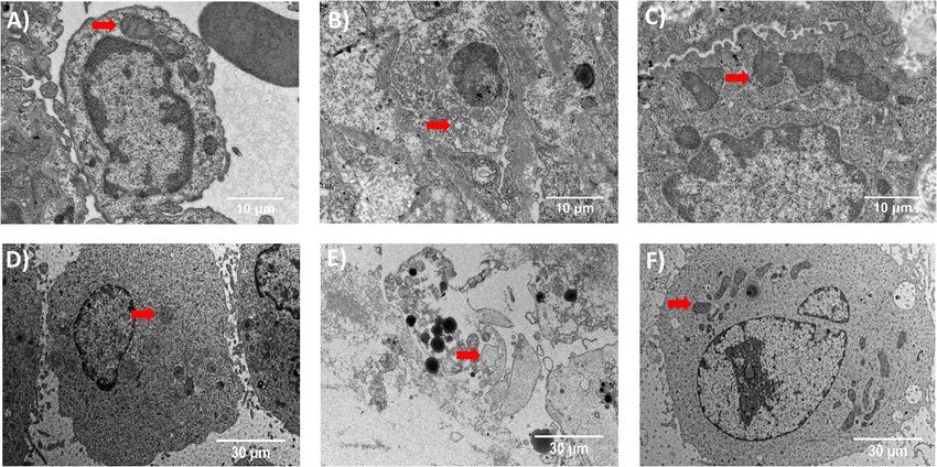
www.nature.com/scientificreports/
Figure 1. Complexation of PQ by A
H2QDS. (A,B) Structures of PQ (A) and AH2QDS (B). (C,D) Complexes of
PQ and AH2QDS by AutoDock Vina. (C) is the side view and (D) is the top view.
lead to the formation of a chain-like structure, with a needle-like structure under the scanning electron micro-
scope (Figure S5B). Early in vitro experiments found that after mixing PQ with A H2QDS, a green precipitate
was formed immediately. If detoxification is performed at a 1:1 mol ratio, the concentration of PQ in the mixed
solution will drop below the limit of detection in 60 min. Accordingly, PQ can be transformed into a nontoxic
substance in the system (Figure S5A).
We hypothesised that AH2QDS would be able to achieve detoxification of PQ in the organism. To test this
hypothesis, we constructed an in vitro toxicity model of A549 cells19and then intervened with AH2QDS, con-
firming that compared with the PQ group, the AH2QDS intervention group showed lessened functional damage
mitochondria and significantly improved cell activity. After that, we established a gavage PQ poisoning SD rat
model20 and found a significant decrease in PQ concentration in plasma detoxified with AH2QDS. Furthermore,
compared with the PQ-treated rats, the tissue suffered only minor damage in the AH2QDS intervention group.
The 30-day survival rate was also improved. We found A H2QDS restored the level of antioxidants and diminished
PQ-induced oxidative stress by lowering the level of oxidative stress factors. To further explore the detoxification
mechanism of A H2QDS, we analysed the differentially expressed genes by NextGen sequencing, and we found
that oxidative stress plays an essential role in A
H2QDS treatment of PQ poisoning and that the nuclear factor
Nrf2 plays a vital role in this process.
In conclusion, A H2QDS can rapidly neutralize PQ to prevent the absorption of poison and remove the
oxidative stress products produced by PQ, thus suggesting great clinical promise as a specific antidote for PQ
poisoning.
Results
Binding of PQ and AH2QDS. Firstly, we used AutoDock V ina21 to simulate the binding conformation
between AH2QDS and PQ (Fig. 1A,B). A grid map of dimensions 26 Å × 26 Å × 26 Å with a grid space of 0.375 Å
was set. The search space’s center was set to − 0.014 Å, − 0.008 Å and − 0.037 Å (x, y, z). One hundred GA (genetic
algorithm) runs was placed, and all other parameters were the default option values by AutoDock Vina. Molecu-
lar docking results indicate that the crystal structure of the PQ + AH2QDS complex contains three intermolecu-
lar interactions, with π π stacking between the two benzene rings of A H2QDS and the PQ molecule, hydrophobic
interactions between the middle of A H2QDS and PQ, and the two sides of A H2QDS forming a salt bridge with
PQ (Fig. 1C,D). The above docking simulation studies demonstrate at a theoretical level that AH2QDS is able to
bind to PQ to form a complex, thereby eliminating the toxicity of PQ. Next, we constructed in vivo and in vitro
models to validate the detoxification of PQ by AH2QDS.
AH2QDS for the treatment of PQ poisoning in vitro. In the in vitro experiment, we first used CCK8
to determine the effects of different concentrations of PQ on the viability of A549 cells22. As shown in Fig-
ure S1A, the cell viability decreased gradually in a time-dependent manner starting 24 h after the PQ interven-
tion. Interestingly, a significant difference in cell viability was caused by different concentrations of PQ at 48 h.
At 72 h, under the 200 μM PQ intervention, the cell viability decreased to 50% (Figure S1A). According to the
Scientific Reports | (2021) 11:20159 | https://doi.org/10.1038/s41598-021-99591-4 2
Vol:.(1234567890)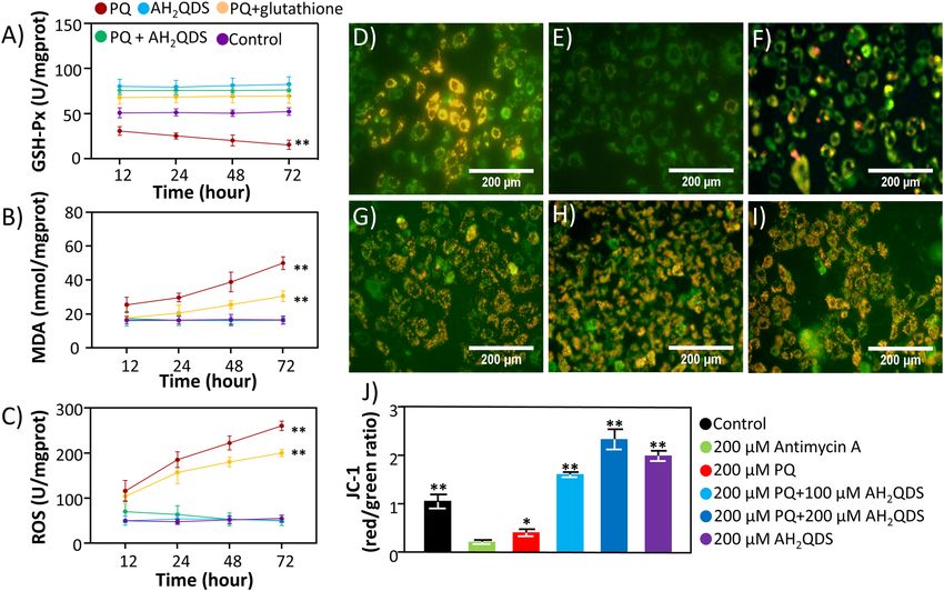
www.nature.com/scientificreports/
Figure 2. Antidotal effects of AH2QDS on PQ poisoning in vitro. (A) A549 cells in different treatment groups
were incubated at different drug concentrations. (B) Viability of A549 cells co-cultured with PQ (200 μM) and
various concentrations of A H2QDS/Glutathione for 72 h. Data are presented as means ± SEM, n = 3, NS = not
significant, *P < 0.05, **P < 0.001.
data, we used this condition in subsequent experiments. At the same time, we measured the effects of different
concentrations of AH2QDS on the viability of A549 cells (Figure S1B). It is worth noting that when the concen-
tration of AH2QDS is greater than 200 μM, it will also have a toxic effect on cells, so we chose 200 μM A H2QDS
as the concentration for the follow-up experiment. Given the oxidative damage-related mechanism of PQ, we
also used glutathione, which is often used to resist the damage caused by oxidative stress10. Here, we chose dif-
ferent concentrations of glutathione to determine its effect on the viability of A549 cells (Figure S1C). The results
showed that glutathione had no toxic effect on cells.
Next, Fig. 2A,B showed that PQ could significantly damage the viability of A549 cells, while glutathione and
AH2QDS intervention raised the activity of A549 cells. In addition, we pre-intervened A H2QDS and glutathione
and then administered PQ staining after different treatment times to assay the cell activity for 72 h. The results
showed that the Glutathione/AH2QDS pretreatment + PQ group still showed a significant rise in cell viability
compared to the PQ group (Figure S2). However, the Glutathione pretreatment group only showed good results
at 1 h, while the A H2QDS pretreatment group could still exert excellent cytoprotective effects until 12 h. This
result reflects the ability of both AH2QDS and Glutathione to induce intrinsic cellular protective effects, however,
AH2QDS is more protective than Glutathione.
AH2QDS improve antioxidation in the treatment of PQ poisoning. PQ poisoning often causes
oxidative stress damage. Consistent with the literature, the results in Fig. 3A–C showed that the level of GSH-
Px in the PQ group decreased, indicating that its antioxidant capacity decreased23. In contrast, ROS and MDA
levels increased in the PQ group, indicating that oxidative damage was a ggravated24,25. In this context, the level
of GSH-Px in the group treated with A H2QDS was significantly higher, and the levels of ROS and MDA were
significantly lower than those in the PQ group. The same trend was also confirmed in vivo (Fig. 7D–F). In sum-
mary, AH2QDS plays an antioxidant role in the treatment of severe PQ.
Protective effect of AH2QDS on cell mitochondria. Many studies have reported that PQ poison-
ing often causes damage to mitochondria26–28. To explore whether AH2QDS can protect mitochondria, we
used a transmission electron microscope to observe the mitochondrial structure under a microscope. From
the Fig. 8D–F, we can see that after PQ intervention, the mitochondrial structure of A549 cells was destroyed,
vacuoles appeared in the cell body, the cell wall was broken, and a large number of organelles were extruded.
However, the morphology of cells in the untreated control group and PQ + AH2QDS group was normal, the
chromatin was evenly distributed, the morphology of the cells was as expected, and the morphology of the mito-
chondria was normal. Furthermore, through detection of the mitochondrial membrane potential, we found that
the membrane potential of the PQ + AH2QDS high-dose group was the highest, followed by the high-dose group
and PQ + AH2QDS low-dose group, and the mitochondrial membrane potential of the PQ group was the lowest
(Fig. 3D–J). The above results indicate the protective effect of A
H2QDS on cell mitochondria in vitro.
The survival rate in a rat model of PQ poisoning. According to the literature, the single-dose oral
LD50 for PQ was 100 mg/kg in rats29. Therefore, to determine PQ toxicity, we gavaged PQ at doses of 100, 200,
300, 400, and 500 mg/kg in vivo (Fig. 4A). We found that when the concentration of PQ was more than 300 mg/
kg (3X LD50), the rats showed obvious poisoning symptoms and died within two weeks. When the concentra-
tion of PQ was more than 400 mg/kg (4X LD50), the animals died in approximately three days. According to
these data, to show the superior detoxification ability of AH2QDS, we chose 400 mg/kg (4X LD50) as the dose
Scientific Reports | (2021) 11:20159 | https://doi.org/10.1038/s41598-021-99591-4 3
Vol.:(0123456789)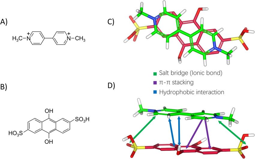
www.nature.com/scientificreports/
Figure 3. AH2QDS can protect the function of mitochondria and improve antioxidation in the treatment of PQ
poisoning in vitro. (A–C) The levels of GSH-Px, MDA, and ROS were detected in different treatment groups.
(D–I) The mitochondrial membrane potential of A549 cells in different treatment groups was detected. When
the mitochondrial membrane potential is high, it can produce red fluorescence, whereas, when the level of
mitochondrial membrane potential is low, it can show green fluorescence. (D) The control group without any
handling. (E) For the 200 μM Antimycin A group, A549 cells were incubated with 200 μMAntimycin A. (F) is
the 200 μM PQ group, A549 cells were incubated with 200 μM PQ. (G) shows 200 μM PQ + 100 μM AH2QDS
group, A549 cells were incubated with 200 μM PQ and 100 μM AH2QDS. (H) represents 200 μM PQ + 200 μM
AH2QDS group, A549 cells were incubated with 200 μM PQ and 200 μM AH2QDS. (I) means that in the
200 μM AH2QDS group, A549 cells were incubated with only 200 μM AH2QDS. (J) Mitochondrial membrane
potential was determined using Mitochondrial Membrane Potential Assay Kit with JC-1. Mitochondrial JC-1
monomers (green) and aggregates (red) were observed under a fluorescence microscope. The mitochondrial
membrane potential was presented as the ratio of J-aggregates to monomers. Data are presented as
means ± SEM, n = 3, *P < 0.05, **P < 0.001.
of PQ to evaluate the detoxification effect of AH2QDS. Subsequently, we used AH2QDS to detoxify the animals
at different times after exposure. As shown in Fig. 3A, the 30-day survival rate of rats exposed to 400 mg/kg
PQ could reach 100% when they were detoxified with A H2QDS within 2 h. However, as the time window for
AH2QDS treatment was extended, the 30-day survival rate of SD rats gradually decreased (Fig. 4B). According to
the above results, we chose 2 h as the detoxification time of AH2QDS. Next, we designed different experimental
groups to verify the detoxification effect of AH2QDS (Fig. 4C). We discovered that the untreated control group’s
30-day survival rates, the A H2QDS group, and the PQ + AH2QDS group were all 100%. The untreated control
group’s 30-day survival rates and the PQ + "white and black" group were zero, and all of the rats died within one
week. The detoxification effect of AH2QDS is better than that of the “white and black” scheme.
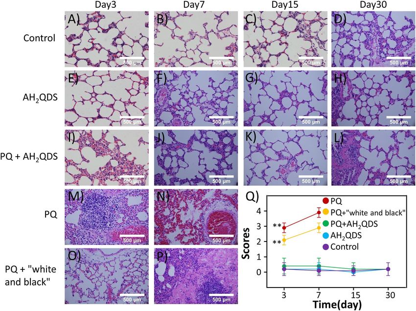
AH2QDS mitigates organ damage in a rat model of PQ poisoning. The lung is the main target
organ after PQ p oisoning30. Patients often die of acute lung injury in the early stage, and pulmonary fibrosis
often occurs later31,32. According to Fig. 5A–P, we also observed alveolar inflammation in the lung tissue of rats
in the PQ and PQ + c "white and black" groups at all time points, including destruction of the alveolar structure,
oedema in the alveolar cavity, intracapillary hyperaemia, and inflammatory cell infiltration, indicating acute
lung injury. On the seventh day, alveolar fusion, alveolar septum thickening, and fibrous tissue hyperplasia were
found in the lungs of the two groups, indicating pulmonary fibrosis. However, the lung tissues of rats in the
untreated control, AH2QDS, and PQ + AH2QDS groups were as expected at all periods, with very little infiltra-
tion of inflammatory cells, no collapse of the alveolar walls, no thickening of the alveolar septa, no exudation in
the alveoli, and no capillary dilation, hyperaemia or other manifestations. The pathological injury score of the
lung tissue showed that lung injury in the PQ group and PQ + "white and black" group was significantly worse
Scientific Reports | (2021) 11:20159 | https://doi.org/10.1038/s41598-021-99591-4 4
Vol:.(1234567890)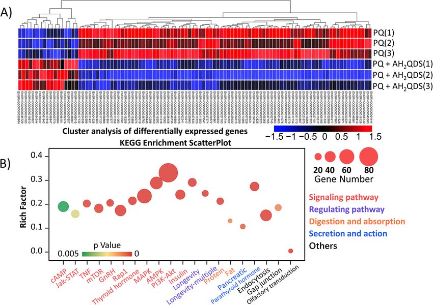
www.nature.com/scientificreports/
Figure 4. AH2QDS can improve the survival rate of rats with PQ poisoning. (A) The survival of rats with
different concentrations of PQ. (B) Prolong the time of AH2QDS intervention in rats with PQ poisoning and
observe the changes in survival rate. (C) The survival curve of rats in different treatment groups. The untreated
control group did not make any interventions. In the A H2QDS group, only 400 mg/kg AH2QDS antidote
was given by gavage. PQ was given by gavage only at a concentration of 400 mg/kg in the PQ group. In the
PQ + "white and black" group, 400 mg/kg of PQ was given by gavage first, and 500 mg/kg was given by gavage
2 h later, with a "white and black" scheme. In the PQ + AH2QDS group, 400 mg/kg of PQ was given to the
stomach first, and 400 mg/kg of A H2QDS antidote was given 2 h later. Kaplan–Meier survival analysis was used
to analyze the survival rate of rats in different treatment groups, n = 7, *P < 0.05, **P < 0.001.
than that in the untreated control, A H2QDS, and PQ + AH2QDS groups, and the difference was statistically sig-
nificant (p < 0.001) (Fig. 5Q).
In addition to lung injury, PQ poisoning can also cause severe functional damage to multiple organs, so we
measured the liver and kidney function and blood gas of the SD rats in each group33–35. As shown in Fig. 6A,B,
the ALT, AST, CREA, and UREA levels in the PQ and PQ + "white and black" groups increased from the 3rd
day and peaked on the 7th day. The ALT, AST, CREA, and UREA levels in the untreated control, A H2QDS, and
PQ + AH2QDS groups were significantly lower than those in the PQ and PQ + "white and black" groups in the
first seven days, and the difference was statistically significant (P < 0.001). In addition, we compared the PQ group
with the PQ + "white and black" group and found that the ALT, AST, CREA, and UREA levels in the PQ + "white
and black" group were significantly lower than those in the PQ group (P < 0.001). However, the hepatic and
renal function of the untreated control, A H2QDS, and PQ + AH2QDS groups was in the normal range during
each period, and there was no significant difference between them (P > 0.05). PQ poisoning has been verified
to cause functional damage to multiple organs, and A H2QDS treatment of PQ poisoning can alleviate liver and
kidney function damage. The blood gas analysis results showed (Fig. 6 C-D) that the pH and PaO2 values in the
PQ and PQ + "white and black" groups were significantly lower than those in the untreated control, A H2QDS,
and PQ + AH2QDS groups (P < 0.001). Compared with the PQ group and PQ + "white and black" group, the
pH and PaO2 values in the PQ + "white and black" group were significantly higher than those in the PQ group.
The PaO2 values in the untreated control, A H2QDS, and PQ + AH2QDS groups were in the normal range dur-
ing each period, and there was no significant difference between the three groups (P > 0.05). Contrary to this
trend, the PaCO2 values in the PQ and PQ + "white and black" groups showed an increasing trend, indicating
that hypoxaemia and carbon dioxide retention occurred in PQ-poisoned rats, which eventually led to type II
respiratory failure. In summary, these findings suggest that A H2QDS can lower the damage to organ function
caused by PQ poisoning.
AH2QDS can rapidly decrease the concentration of PQ in vivo. As shown in Fig. 7A–C, the
PQ concentration in the PQ group peaked at 4 h and decreased to 0 at 96 h. The concentration of PQ in the
Scientific Reports | (2021) 11:20159 | https://doi.org/10.1038/s41598-021-99591-4 5
Vol.:(0123456789)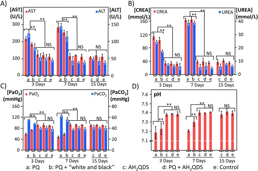
www.nature.com/scientificreports/
Figure 5. AH2QDS decreases lung injury. (A–P) H&E staining in lung tissue of rats in the diverse groups at
different time points. (A–D) The untreated control group. (E–H) The AH2QDS group. (I–L) The PQ + AH2QDS
group. (M,N) The PQ group. (O,P) The PQ + "white and black" group. Since a 4X LD50 dose of PQ was used,
rats in the PQ group and PQ + "white and black" group died within one week, so there is no data for subsequent
time points. (Q) Lung injury scores of different treatment groups. Data are presented as means ± SEM, n = 3,
*P < 0.05, **P < 0.001.
PQ + AH2QDS group and PQ + "white and black" group decreased immediately after 2 h and was significantly
lower than that in the PQ group (P < 0.001). The difference between 2 and 24 h was significantly smaller in the
PQ + AH2QDS group than in the PQ + "white and black" group, and the difference was statistically significant.
Similarly, the concentration of PQ in the lung tissue and urine decreased significantly in the PQ + AH2QDS
group. The decrease in PQ drug concentration may have occurred because A H2QDS neutralizes PQ in the gas-
trointestinal tract.
Protection of mitochondria by AH2QDS in vivo. The induction of mitochondrial damage by PQ has
been confirmed in in vitro experiments, and we also observed the same phenomenon in vivo experiments.
Figure 8B showed that the PQ group’s mitochondria were swollen, structurally damaged, vacuolated and empty.
Under the electron microscope, the mitochondrial structure was as expected in the rat lung tissues in the
untreated control group and PQ + AH2QDS group (Fig. 8A/C). These pictures illustrated that A
H2QDS protects
the structure of mitochondria.
NextGen sequencing. To better understand the detoxification mechanism of A
H2QDS, we used RNAseq
to investigate the differential gene expression patterns of rat lung tissue in the PQ and PQ + AH2QDS groups.
Firstly, we performed data quality control (Figure S4A), after which we used principal component analysis (PCA)
to identify outlier samples and high similarity samples. As illustrated in the Figure S4B, in this experiment, dif-
ferent samples from the same experimental group are arranged compactly and aggregated into clusters, showing
good repeatability. In contrast, different experimental groups are clearly separated from each other, showing
reasonable specificity. We can see from Fig. 9A that there were 3325 gene changes in the PQ group compared
with the PQ + AH2QDS group, including 1455 upregulated genes and 1870 downregulated genes. As shown in
Fig. 9B, the most differentially regulated pathways in these two samples are the PI3K-AKT pathway, MAPK
pathway, AMPK pathway, etc. Consistent with our previous findings, these pathways are mainly oxidative stress-
related pathways. We investigated the most significant pathway, namely, the PI3K-AKT pathway, to identify the
genes with significant changes, and the results showed that Nrf2, Foxo3, Rxra, Itga4, Creb3l2, Angpt1, Egfr, Tnc,
Lamc1, and Met were significantly upregulated. Nrf2 is significantly upregulated in tissues, and its function is
Scientific Reports | (2021) 11:20159 | https://doi.org/10.1038/s41598-021-99591-4 6
Vol:.(1234567890)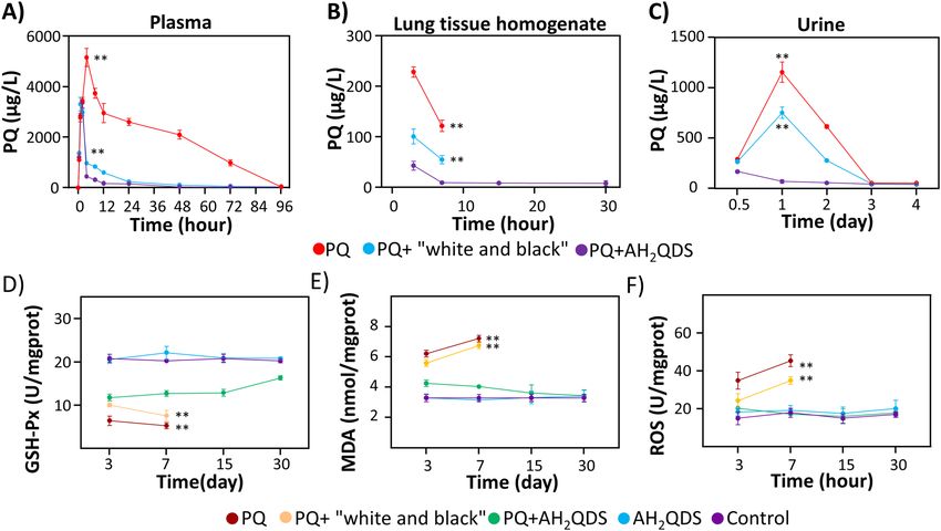
www.nature.com/scientificreports/
Figure 6. AH2QDS can minimize the damage of multiple organs and individual functions. (A,B) Changes in
liver and kidney function of rats in each group. (C,D) Changes in blood gas analysis results of rats in each group.
Due to a 4X LD50 dose of PQ intervention, animals in the PQ group and PQ + "white and black" group died
within one week, so there is no subsequent time point data. At 30 days, all groups’ liver and kidney function
and blood gas results were in the normal range. Data are presented as means ± SEM, n = 3, NS = not significant,
*P < 0.05, **P < 0.001.
closely related to oxidative stress, so we speculate that Nrf2 may be an essential gene for A
H2QDS treatment of
PQ poisoning.
We further verified by western blot and RT-qPCR experiments that in in vitro experiments, as illustrated in
the Fig. 10A–C, compared with PQ treatment, glutathione and A H2QDS could significantly increase the expres-
sion of Nrf2, while the Nrf2 level of PQ + AH2QDS group was significantly higher than that of the PQ + glu-
tathione group. The in vivo experiment showed that the levels of Nrf2 in the AH2QDS, PQ, and PQ + "white and
black" groups were higher than that in the untreated control group, while the Nrf2 level in the PQ + AH2QDS
group was significantly higher than those in the other groups (Fig. 10D–F). The results indicated that “white
and black” scheme did not activate Nrf2. In contrast, glutathione could increase the expression of Nrf2, but its
effect was weaker than that of A H2QDS, indicating that our antidote, A H2QDS, could significantly increase the
expression of Nrf2, thus exerting its detoxification effect.
Discussion
In this study, we used A
H2QDS as an intervention in a rat model of PQ poisoning. Compared with those of
the PQ group, the poisoning symptoms of the PQ + AH2QDS group were significantly improved, with a lower
blood drug concentration, less organ function damage, and a higher survival rate. In the PQ + AH2QDS group,
mitochondrial damage in lung tissue was alleviated, and a similar phenomenon was found in the cell test. The
structure of the mitochondria was intact, the damage was significantly alleviated, and the expression of Nrf2
was significantly increased. These studies have proven for the first time that A
H2QDS is an effective treatment
for PQ poisoning, and Nrf2 plays a crucial role in its detoxification process.
Previous studies have shown that activated carbon or the “white and black” scheme can effectively treat PQ
poisoning6–8. In contrast, our experiments only confirmed that the conventional “white and black” scheme can
quickly and effectively degrade the PQ blood concentration but does not affect the survival rate of rats. Specifi-
cally, a 4X LD50 dose of PQ was used to construct the poisoning model, and the drug intervention time was as
long as 2 h, during which most of the PQ may have been absorbed into the blood, while the “white and black”
scheme could only absorb the residual poison in the stomach and accelerate its excretion but had no effect on
Scientific Reports | (2021) 11:20159 | https://doi.org/10.1038/s41598-021-99591-4 7
Vol.:(0123456789)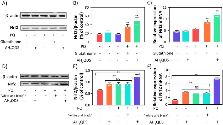
www.nature.com/scientificreports/
Figure 7. AH2QDS can decrease PQ drug concentration in vivo and improve the antioxidant reaction to
oxidative stress. (A–C) The concentration of PQ in plasma, tissue and urine was detected by Ultra high-
performance liquid chromatography-tandem mass spectrometry. (D–F) The levels of GSH-Px, MDA, and ROS
were detected in different treatment groups. Due to a 4X LD50 dose of PQ intervention, animals in the PQ
group and PQ + "white and black" group died within one week, so there is no subsequent time point data. Data
are presented as means ± SEM, n = 3, *P < 0.05, **P < 0.001.
Figure 8. AH2QDS can protect mitochondria’s structural integrity. (A–C) The transmission electron
microscope observed mitochondria’s structure in lung tissue. (A) the untreated control group did not receive
any interventions. (B) PQ was given by gavage only at a concentration of 400 mg/kg in the PQ group. (C) in
the PQ + AH2QDS group, 400 mg/kg of PQ was given to the stomach first, 2 h later, and 400 mg/kg of A H2QDS
antidote was given. (D–F) The transmission electron microscope observed A549 cells’ mitochondrial structure.
(D) the untreated control group, without any treatment. (E) in the PQ group, A549 cells were incubated with
200 μm PQ. (F) A549 cells were incubated with 200 μM AH2QDS and 200 μM PQ in the PQ + AH2QDS group.
The mitochondria have been marked with red arrows in the picture.
Scientific Reports | (2021) 11:20159 | https://doi.org/10.1038/s41598-021-99591-4 8
Vol:.(1234567890)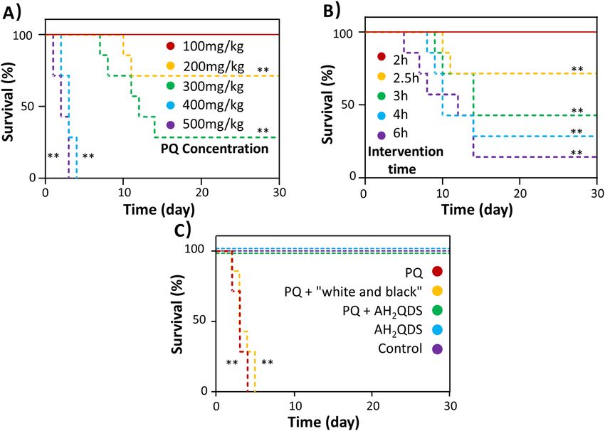
www.nature.com/scientificreports/
Figure 9. Differentially expressed genes by using NextGen sequencing method. (A) Heat map of differentially
expressed gene expression profiles of the PQ group and the PQ + AH2QDS group. Gene enrichment analysis
was performed based on differentially expressed genes, and the blue to red color indicates the expression level
from low to high. (B) Regulatory biological pathways under AH2QDS treatment were analyzed using the
KEGG database. Rich Factor indicates the proportion of differential genes annotated to this KEGG Pathway
as a percentage of genome-wide genes. The horizontal coordinate indicates the description bar of the KEGG
Pathway. GeneNumber, the number of genes annotated to this KEGG Pathway among the differential genes used
for enrichment. The size of the dot indicates the number of genes on the enrichment, and the colour indicates
the Qvalue, the lower, the more significant.
the PQ already in the blood. Additionally, the results also showed that A H2QDS is not only faster than the “white
and black” scheme in removing toxins but also plays a specific role in the blood-related effects of PQ.
The toxic effect of PQ on mitochondria was proposed as early as 1 96836. Since then, a large number of studies
on the damage of PQ to mitochondria have been p ublished26–28. Some scholars indicated that PQ could cause
accumulation of the hMn-SOD precursor of human manganese-dependent peroxidase and diminish Mn-SOD
activity. The conversion of GSH to GSSG leads to a decrease in GSH levels and weakens its antioxidant a ctivity37.
Other studies have shown that PQ can cause the production of H2O2 and lessen the activity of catalase38. H2O2 can
induce changes in mitochondrial permeability and affect the mitochondrial membrane potential, resulting in the
movement of cytochrome C from the mitochondria into the cytoplasm, and then induce apoptosis by activating
caspase939. In our study, the microstructure of the lung tissue and A549 cells in the PQ group was observed under
a projection electron microscope. It was found that the structure of the mitochondria was destroyed, vacuoles
appeared in the cells, the cell walls were broken, and a large number of organelles were extruded. In contrast, the
morphology of the mitochondria in the PQ + AH2QDS group was as expected, and the lamellar structure was
normal. The cell membrane potential of the PQ + AH2QDS high-dose group was the highest, and the membrane
potential was positively correlated with the concentration of A H2QDS, while the mitochondrial membrane
potential of cells treated with only PQ was the lowest. In summary, PQ can destroy the tissue structure of the
mitochondria, affect the membrane potential, and eventually lead to cell rupture and death, while A H2QDS can
prevent this process and protect the function and structure of the mitochondria.
The data show that after PQ is absorbed into the blood, it causes the formation of excess reactive oxygen spe-
cies (ROS), which leads to imbalance of the redox system, the consumption of NADPH, damage to mitochondria,
the destruction of lipids, proteins and DNA, and a decrease in the activity of various antioxidant enzymes24. After
continuous oxidative stimulation, the body eventually sustains tissue damage. A large amount of ROS produced
by PQ may be the leading cause of acute lung injury caused by PQ poisoning. In this study, it was found that after
PQ exposure, the levels of ROS and MDA in the PQ group and conventional treatment group increased, while
the level of GSH-Px decreased. In the PQ + AH2QDS group, the ROS and MDA levels decreased, and the level
of GSH-Px increased. The results show that PQ can produce a large amount of ROS to cause lipid peroxidation
and oxidative stress injury. A H2QDS can inhibit PQ’s effect, improve antioxidant ability, and decrease the level
of lipid peroxidation.
Scientific Reports | (2021) 11:20159 | https://doi.org/10.1038/s41598-021-99591-4 9
Vol.:(0123456789)www.nature.com/scientificreports/
Figure 10. AH2QDS up regulated the expression of Nrf2. (A–C) After AH2QDS intervention, the expression of
Nrf2 in A549 cells increased significantly. (D–F) The expression of Nrf2 in the lung tissue of rats detoxified by
AH2QDS enhanced significantly. The grouping of gels/blots cropped from different parts of the same gel. Full-
length gels and blots are included in Figure S6. Data are presented as means ± SEM, n = 3, NS = not significant,
*P < 0.05, **P < 0.001.
After further investigation of the detoxification mechanism of A H2QDS, we found that the main differentially
regulated pathway was the oxidative stress pathway, in which we found that the nuclear factor Nrf2 was signifi-
cantly upregulated. Many studies have shown that Nrf2 can be used as a "guard" to protect the body against a
variety of toxic effects40–42. Nrf2 can be activated in a variety of processes involving oxidative stress. Nrf2 was
expressed in epithelial cells, macrophages and vascular endothelial cells of normal rat lung tissue43–45. MDA in
the serum of rats poisoned by PQ increased significantly with the prolongation of poisoning time, while the
activity of SOD decreased significantly. Nrf2 protein increased significantly in lung tissue injury induced by PQ.
It has been found that the Nrf2-ARE pathway protects the lungs against dibutyl hydroxytoluene-induced acute
respiratory distress syndrome (ARDS) and hyperoxia-induced lung injury by activating antioxidant e nzymes46,47.
In our experiment, PQ, as a potent stressor, could activate the Nrf2 signalling pathway. Nrf2 was expressed at
low levels in normal rat lung tissue and A549 cells, but the expression of Nrf2 was significantly increased after
AH2QDS treatment. These results show that Nrf2 plays a vital role in the treatment of PQ poisoning by A H2QDS.
The direct mechanism of AH2QDS in the treatment of PQ poisoning is that AH2QDS enters the gastroin-
testinal tract and comes into contact with the PQ solution. Through a rapid redox reaction, PQ is reduced to
a nontoxic green needle-like solid. Thus, detoxification is realized. Energy spectrum analysis showed that the
acicular substance was stable and could not be dissolved in strong acids, strong bases, or organic solvents and
was extremely stable at room temperature and pressure. At the same time, it was also found in the faeces of SD
rats. We were concerned that after administration of AH2QDS, a green needle-like solid will be formed in the
blood, leading to the formation insoluble thrombi and resulting in thrombotic disease and a series of clinical
symptoms. Therefore, we tested the blood and tissues of experimental animal SD rats but did not find this sub-
stance. So, after administering AH2QDS, green needle-like solids would not be formed in the blood, tissues and
organs to cause thrombotic disease.
To prove whether there is an indirect mechanism of AH2QDS in PQ poisoning treatment, we constructed
animal and cell models. ELISA, WB, and qPCR were performed to detect the levels of GSH-Px, MDA, ROS, and
Nrf2, and transmission electron microscopy was performed to observe the microstructure of the mitochondria.
The same trend was observed in vivo and in vitro. After the intervention with AH2QDS, the expression of nuclear
factor Nrf2 was enhanced, mitochondrial damage was relieved, and antioxidant reaction to oxidative stress was
improved. Unfortunately, our experiment cannot determine whether the mechanism of A H2QDS in the treatment
of PQ poisoning is the direct mechanism or the indirect mechanism. Further research is needed.
In this paper, AH2QDS was used as an antidote in the treatment of PQ poisoning for the first time and
achieved excellent results, but this was verified only in SD rats, and it has not been tested in more advanced mam-
mals; thus, a long and strict clinical study is needed to investigate the use of AH2QDS in humans. Additionally,
a 4X LD50 dose of PQ was given to SD rats in the poisoning model, and A H2QDS was given for detoxification
2 h later. The 30-day survival rate of SD rats in the treatment group reached 100%, but if the time window of
Scientific Reports | (2021) 11:20159 | https://doi.org/10.1038/s41598-021-99591-4 10
Vol:.(1234567890)www.nature.com/scientificreports/
treatment with AH2QDS were prolonged (2.5 h, 3 h, 4 h, or 6 h), the 30-day survival rate of SD rats in the treat-
ment group decreases with the prolongation of intervention time. This may be because 2 h after ingestion of PQ,
the rats have rapidly absorbed it into the blood and transported it to various organs through the blood flow. Even
if AH2QDS can detoxify the absorbed PQ, too high a concentration of PQ causes irreversible toxic damage to
the organs in this time. In the follow-up studies, the sequencing results will be further analysed, and mechanistic
research will be performed to elucidate the molecular functions of the gene, the cell location, and the biologi-
cal process involved. At the same time, experiments were carried out on the Nrf2-ARE pathway through gene
silencing/overexpression of related proteins to demonstrate the profound relationship between the Nrf2-ARE
pathway and AH2QDS in the treatment of PQ poisoning.
Conclusion
In summary, paraquat poisoning is still an extremely high clinical mortality disease, and conventional treat-
ments are clinically ineffective. The new antidote we developed, AH2QDS, can lower the concentration of PQ by
binding it and protect the mitochondria and reduce the oxidative stress damage caused by PQ. The relationship
between mitochondrial damage, the expression changes upstream and downstream of the Nrf2-ARE pathway,
and AH2QDS in PQ poisoning treatment must be further explored.
Methods
Animals and cell lines. All animal experiments were performed as per the protocols approved by the Ani-
mal Care and Use Committee of Hainan Medical University. All methods were performed in accordance with the
guidelines and regulations of the Animal Care and Use Committee of Hainan Medical University and as per the
ARRIVE guidelines 2.0. Human type II alveolar lung epithelial cells (A549) were purchased from the Shanghai
Institute for Biological Sciences. The cells were maintained in a 5% CO2 incubator at 37 °C in medium (F12K)
supplemented with 10% FBS and penicillin/streptomycin (100 U/ml). Sprague–Dawley (SD) rats (8 weeks, male,
SPF grade) were purchased from Changsha Tianqn Biotechnology Co., Ltd., and were maintained in specific
pathogen-free (SPF) facilities.
Main reagent. Paraquat solution, purchased from Nanjing Red Sun Co., Ltd., was given at a 400 mg/kg
concentration. Twenty percent PQ solution was diluted into 1 mL of PQ solution with PBS. For the “white and
black” scheme, 500 mg/kg activated carbon, 500 mg/kg montmorillonite powder and 5 mL mannitol were used
for gastric cancer. Activated carbon was purchased from National Pharmaceutical Group Chemical Reagent
Co., Ltd. Montmorillonite powder was purchased from Xiansheng Pharmaceutical Co., Ltd. Mannitol (20%)
was purchased from Jiangsu Zhengda Tianqing Pharmaceutical Co., Ltd. Anthrahydroquinone-2-6-disulfonate
(AH2QDS) was synthesized by the Chinese Academy of Tropical Agricultural Sciences. The method is patented
(Patent No: 2016103413306). Chemical name: anthraquinone-2-dioxo-6-disodium disulfonate, chemical for-
mula: C14H8O8S2.2Na, molecular weight: 368.33. We prepared the A H2QDS solution at a concentration of
40 mmol/L.
Modeling studies of PQ and AH2QDS binding. Chemical structures of PQ and A H2QDS were drawn
with ChemDraw Pro 16.0 software. The binding conformations between PQ and A
H2QDS were simulated with
AutoDock Vina20.
Cell counting kit‑8 (CCK8). A549 cells were incubated with different concentrations of PQ, A
H2QDS and
glutathione for 12 h. After 12, 24, 48 and 72 h, 10 μL of CCK8 solution (Dojindo, Japan) was added, and the cells
were incubated in the incubator for 2 h. An enzyme labelling instrument was used to measure the absorbance at
450 nm, and a formula was used to calculate the cell viability.
Mitochondrial membrane potential. The cell culture medium was removed, the cells were washed with
PBS, 1 ml of medium was added, and 1 mL of JC-1 staining working solution was added and mixed well. After
incubating the cells for 20 min in the incubator at 37 °C, the supernatant was removed, the cells were washed
with diluted staining buffer (1x), 2 mL of medium was added, and images were captured under the fluorescence
microscope.
Animal experiments. SD rats (~ 300 g) were subjected to gavage 400 mg/kg PQ, and 500 mg/kg "white and
black" scheme and 400 mg/kg A H2QDS intervention treatment were administered 2 h later. The specific meth-
ods used to establish the model is shown in Figure S3. We selected rats without collecting blood after establish-
ing the model and observed and recorded the survival of each group over 30 days. The occurrence of death was
recorded as 1, and no death was recorded as 0. Finally, a survival curve was drawn. The animal protocol passed
the ethical review of the ethics committee of The First Affiliated Hospital of Hainan Medical University. (Issue
number: 2020 (Research) No. (97); Review category: A Quick Review; Decision: Approval; Decision Date: July
8, 2020).
Sample collection. Blood was collected from the rats at different time points in anticoagulant tubes treated
with heparin, and the plasma was separated and stored at − 80 °C for the detection of drug concentrations in
the blood. The urine was left in the centrifuge tube for the detection of drug concentrations in urine. Rats were
anaesthetized by intraperitoneal injection of 10% chloral hydrate (300 mg/kg), and the blood from the abdomi-
nal aorta was collected for the detection of liver, kidney and lung function. Finally, the rats were killed by exsan-
Scientific Reports | (2021) 11:20159 | https://doi.org/10.1038/s41598-021-99591-4 11
Vol.:(0123456789)www.nature.com/scientificreports/
guination, and the lung tissue was collected, washed with PBS and stored at − 80 °C for follow-up analysis. The
bodies of the animals were then incinerated.
Histopathology. SD rats were sacrificed at different times, and the lungs of the rats were harvested, fixed in
4% formalin, embedded in paraffin, sectioned, and stained with haematoxylin and eosin (H&E). The lung injury
score was determined according to methods that were previously reported in the literature48. A score of 0 means
there is no alveolitis. 1 point means mild alveolitis, the lesions are limited to local and pleural lesions, accounting
for less than 20% of the lung, and the alveolar structure is sound. A score of 2 indicates moderate alveolitis, and
the lesion area accounts for 20–50% of the lung. Finally, a score of 3 means severe alveolitis, with diffuse alveolitis
involving more than 50% of the lung.
Blood analysis. The collected venous blood samples were placed into a test tube with a coagulant and cen-
trifuged at 3000 r/min for 5 min. Rat serum was obtained and placed into an automatic biochemical function
analyser for analysis. After collecting blood from the abdominal aorta with an arterial blood gas sampler and
rubbing with both hands for 1 min, 0.1 mL was injected into the blood gas analyser for analysis.
Transmission electron microscopy. Lung tissue and A549 cells were collected and placed overnight in
2.5% glutaraldehyde fixed solution that was prechilled at 4 °C, cleaned with PBS, fixed with 1 ml of 1% osmic
acid for 1.5 h, dehydrated with alcohol and acetone, and impregnated with resin, and ultrathin sections were
stained with uranium acetate and lead citrate. The ultrastructure was observed under a transmission electron
microscope.
Ultra‑high‑performance liquid chromatography‑tandem mass spectrometry. The concen-
trations of PQ and A H2QDS were determined by ultra-high-performance liquid chromatography-tandem
mass spectrometry (UPLC/Xevo TQ-S, Waters). The mobile phase was acetonitrile/100 mM ammonium
formate (pH = 3.7) = 50 × 50, and the flow rate was 0.3 mL/min. An ACQUITY UPLC BEH HILIC column
(100 mm × 2.1 mm, 1.7 μm) was used. PQ was quantified in the MRM mode of positive ion multireaction moni-
toring with an electrospray ion source. Negative ion SIR mode was used to quantify A H2QDS. The parameters
were as follows: capillary voltage: 3.2 kV, ion source temperature: 150 °C, cone hole back blowing gas flow rate:
30 L/hr, dissolvent temperature: 350 °C, and dissolvent gas flow rate: 800 L/hr.
Cytokine detection. The GSH-Px, MDA and ROS kits purchased from Nanjing Jiancheng Company were
used according to the instructions to detect the levels of GSH-Px, MDA and ROS, respectively.
Western blotting. Proteins were extracted from tissues and cells with a BCA kit (Biyuntian Biotechnol-
ogy Co., Ltd.), separated in SDS-PAGE gels, and transferred to cellulose membranes. After sealing, the mem-
branes were incubated with the primary antibody overnight, then incubated with the secondary antibody for
1 h (Table S1), and finally developed by exposure.
Quantitative real‑time polymerase chain reaction (RT‑qPCR). TRIzol (Biyuntian Biotechnology
Co., Ltd.) was used to extract RNA, and a cDNA reverse transcription kit (Applied Biosystems, cat. no. 4368814)
was used to reverse-transcribe the extracted RNA into cDNA. PCR was performed on an ABI Prism 7900HT
system (Applied Biosystems, Foster City, CA, USA) using SYBR GREEN PCR Master Mix (Applied Biosystems).
Primers were purchased from Sangon Biotech (Shanghai) Co., Ltd. The primer sequences are listed in Table S2.
NextGen sequencing. Total RNA was extracted from rat lung tissue in the PQ and PQ + AH2QDS groups
and enriched with eukaryotic mRNA using magnetic beads with Oligo(dT). The second cDNA strand was then
purified by QiaQuick PCR kit and eluted with EB buffer, followed by end repair, the addition of poly(A) and
ligation of the sequencing junction, then agarose gel electrophoresis for fragment size selection, and finally PCR
amplification. After that, the library was sequenced on the Illumina NovaSeq6000 platform.
To make sure reads reliable, Illumina paired-ended sequenced Raw reads were filtered using the fastp to
remove low quality reads (https://github.com/OpenGene/fastp). The filtered data is then compared to the ref-
erence sequence. Reference genome and gene model annotation files were downloaded from genome website
directly. (https://www.ncbi.nlm.nih.gov/assembly/GCF_000001895.5#/def). The sequenced data were imported
into Partek Flow (Partek Inc., St. Louis, MO) and principal component analysis (PCA) images were generated
to visualise distribution differences.
Differential expression analysis was performed using the D ESeq249. Based on the Kyoto Encyclopedia of
Genes and Genomes (KEGG)50, we used the R package cluster P rofiler51 to perform KEGG functional enrich-
ment analysis of differentially expressed genes.
Statistical analysis. Statistical analyses were performed using GraphPad Prism 8.0 or SPSS 20.0 software.
Measurement data are expressed as the mean ± SEM, and significance was tested by single-factor analysis of
variance (ANOVA). Kaplan–Meier survival analysis was used to analyse the survival rate of rats in different
treatment groups. P < 0.05 indicates that a difference is statistically significant.
Ethical approval. The experiment was carried out according to the guiding principles for animal experi-
ments at Hainan Medical University.
Scientific Reports | (2021) 11:20159 | https://doi.org/10.1038/s41598-021-99591-4 12
Vol:.(1234567890)www.nature.com/scientificreports/
Received: 28 April 2021; Accepted: 29 September 2021
References
1. Cicchetti, F., Drouin-Ouellet, J. & Gross, R. E. Environmental toxins and Parkinson’s disease: What have we learned from pesticide-
induced animal models?. Trends Pharmacol. Sci. 30, 475–483. https://doi.org/10.1016/j.tips.2009.06.005 (2009).
2. Gawarammana, I. B. & Buckley, N. A. Medical management of paraquat ingestion. Br. J. Clin. Pharmacol. 72, 745–757 (2011).
3. Hong, S. Y., Lee, J. S., Sun, I. O., Lee, K. Y. & Gil, H. W. Prediction of patient survival in cases of acute paraquat poisoning. PLoS
ONE 9, e111674 (2014).
4. Wu, W. P. et al. Addition of immunosuppressive treatment to hemoperfusion is associated with improved survival after paraquat
poisoning: A nationwide study. PLoS ONE 9, 2 (2014).
5. Xiangjun, et al. A synthetic receptor as a specific antidote for paraquat poisoning. Theranostics 2, 2 (2019).
6. Feng, E., Cheng, Y., Tan, Z. & Wang, H. Experience of continuous fluid therapy in successfully rescuing a patient with acute severe
paraquat poisoning. Zhonghua wei zhong bing ji jiu yi xue 31, 1043–1044. https://doi.org/10.3760/cma.j.issn.2095-4352.2019.08.
027 (2019).
7. Wang, W. et al. Effect of rhubarb as the main composition of sequential treatment in patients with acute paraquat poisoning: a
prospective clinical research. Zhonghua wei zhong bing ji jiu yi xue 27, 254–258. https://doi.org/10.3760/cma.j.issn.2095-4352.
2015.04.006 (2015).
8. Zhao, B. et al. Clinical study on the treatment of acute paraquat poisoning with sequential whole gastric and bowel irrigation.
Zhonghua Lao Dong Wei Sheng Zhi Ye Bing Za Zhi 33, 213–215 (2015).
9. Rodrigues da Silva, M. et al. Beneficial effects of ascorbic acid to treat lung fibrosis induced by paraquat. PLoS ONE 13, e0205535.
https://doi.org/10.1371/journal.pone.0205535 (2018).
10. Kobayashi, S. et al. Enhanced expression of cystine/glutamate transporter in the lung caused by the oxidative-stress-inducing agent
paraquat. Free Radical Biol. Med. 53, 2197–2203. https://doi.org/10.1016/j.freeradbiomed.2012.09.040 (2012).
11. Lin, X. et al. Association between liberal oxygen therapy and mortality in patients with paraquat poisoning: A multi-center retro-
spective cohort study. PLoS ONE 16, e0245363. https://doi.org/10.1371/journal.pone.0245363 (2021).
12. Li, L., Sydenham, E., Chaudhary, B., Beecher, D. & You, C. Glucocorticoid with cyclophosphamide for paraquat-induced lung
fibrosis. Cochrane Database Syst. Rev. https://doi.org/10.1002/14651858.CD008084.pub4 (2014).
13. Gawarammana, I. et al. High-dose immunosuppression to prevent death after paraquat self-poisoning—a randomised controlled
trial. Clin. Toxicol. 56, 633–639. https://doi.org/10.1080/15563650.2017.1394465 (2018).
14. Pourgholamhossein, F. et al. Pirfenidone protects against paraquat-induced lung injury and fibrosis in mice by modulation of
inflammation, oxidative stress, and gene expression. Food Chem. Toxicol. 112, 39–46. https://doi.org/10.1016/j.fct.2017.12.034
(2018).
15. Dinis-Oliveira, R. J. et al. Paraquat poisonings: Mechanisms of lung toxicity, clinical features, and treatment. Crit. Rev. Toxicol. 38,
13–71. https://doi.org/10.1080/10408440701669959 (2008).
16. Tsukamoto, M., Tampo, Y., Sawada, M. & Yonaha, M. Paraquat-induced oxidative stress and dysfunction of the glutathione redox
cycle in pulmonary microvascular endothelial cells. Toxicol. Appl. Pharmacol. 178, 82–92. https://doi.org/10.1006/taap.2001.9325
(2002).
17. Wu, C.-Y., Li, Q.-F. & Wu, D.-M. A rapid detoxification solution for paraquat and method. CN105944279A (2016).
18. Wu, C.-Y., Wu, D.-M., Liu, X.-R., Qian, J. & Li, Q.-F. A specific antidote for acute paraquat poisoning. CN110585180A (2019).
19. Ariyama, J., Shimada, H., Aono, M., Tsuchida, H. & Hirai, K. Propofol improves recovery from paraquat acute toxicity in vitro
and in vivo. Intensive Care Med. 26, 981–987. https://doi.org/10.1007/s001340051291 (2000).
20. Zhang, X. et al. A synthetic receptor as a specific antidote for paraquat poisoning. Theranostics 9, 633–645. https://d oi.o
rg/1 0.7 150/
thno.31485 (2019).
21. Trott, O. & Olson, A. J. AutoDock Vina: Improving the speed and accuracy of docking with a new scoring function, efficient
optimization, and multithreading. J. Comput. Chem. 31, 455–461. https://doi.org/10.1002/jcc.21334 (2010).
22. Zhang, D. et al. Role of AP-2alpha and MAPK7 in the regulation of autocrine TGF-beta/miR-200b signals to maintain epithelial-
mesenchymal transition in cholangiocarcinoma. J. Hematol. Oncol. 10, 170. https://doi.org/10.1186/s13045-017-0528-6 (2017).
23. Liu, Z., Wang, X., Li, L., Wei, G. & Zhao, M. Hydrogen sulfide protects against paraquat-induced acute liver injury in rats by
regulating oxidative stress, mitochondrial function, and inflammation. Oxid. Med. Cell. Longev. 2020, 6325378. https://doi.org/
10.1155/2020/6325378 (2020).
24. Zhang, Z. et al. Klotho alleviates lung injury caused by paraquat via suppressing ROS/P38 MAPK-regulated inflammatory responses
and apoptosis. Oxid. Med. Cell. Longev. 2020, 1854206. https://doi.org/10.1155/2020/1854206 (2020).
25. Ma, J., Li, Y., Li, W. & Li, X. Hepatotoxicity of paraquat on common carp (Cyprinus carpio L.). Sci. Total Environ. https://doi.org/
10.1016/j.scitotenv.2017.10.231 (2018).
26. Xie, L. et al. SIRT3 mediates the antioxidant effect of hydrogen sulfide in endothelial cells. Antioxid. Redox Signal. 24, 329–343.
https://doi.org/10.1089/ars.2015.6331 (2016).
27. Bora, S. et al. Paraquat exposure over generation affects lifespan and reproduction through mitochondrial disruption in C. elegans.
Toxicology 447, 152632. https://doi.org/10.1016/j.tox.2020.152632 (2021).
28. Wang, X., Souders, C., Zhao, Y. & Martyniuk, C. Paraquat affects mitochondrial bioenergetics, dopamine system expression, and
locomotor activity in zebrafish (Danio rerio). Chemosphere 191, 106–117. https://doi.org/10.1016/j.chemosphere.2017.10.032
(2018).
29. Kimbrough, R. D. & Gaines, T. B. Toxicity of paraquat to rats and its effect on rat lungs. Toxicol. Appl. Pharmacol. 17, 679–690
(1970).
30. Jin, Y. et al. Transplantation of endothelial progenitor cells attenuated paraquat-induced acute lung injury via miR-141-3p-Notch-
Nrf2 axis. Cell Biosci. 8, 21. https://doi.org/10.1186/s13578-018-0219-1 (2018).
31. Wu, L. et al. Metformin activates the protective effects of the AMPK pathway in acute lung injury caused by paraquat poisoning.
Oxid. Med. Cell. Longev. 2019, 1709718. https://doi.org/10.1155/2019/1709718 (2019).
32. Tai, W. et al. Rapamycin attenuates the paraquat-induced pulmonary fibrosis through activating Nrf2 pathway. J. Cell. Physiol. 235,
1759–1768. https://doi.org/10.1002/jcp.29094 (2020).
33. Yang, C. et al. Spectrum of toxic hepatitis following intentional paraquat ingestion: Analysis of 187 cases. Liver Int. 32, 1400–1406.
https://doi.org/10.1111/j.1478-3231.2012.02829.x (2012).
34. Chen, G., Lin, J. & Huang, Y. Combined methylprednisolone and dexamethasone therapy for paraquat poisoning. Crit. Care Med.
30, 2584–2587. https://doi.org/10.1097/00003246-200211000-00030 (2002).
35. Huang, C. & Zhang, X. Prognostic significance of arterial blood gas analysis in the early evaluation of paraquat poisoning patients.
Clin. Toxicol. 49, 734–738. https://doi.org/10.3109/15563650.2011.607459 (2011).
36. Gage, J. C. The action of paraquat and diquat on the respiration of liver cell fractions. Biochem. J. 109, 757–761. https://doi.org/
10.1042/bj1090757 (1968).
Scientific Reports | (2021) 11:20159 | https://doi.org/10.1038/s41598-021-99591-4 13
Vol.:(0123456789)You can also read