Porphyromonas gingivalis Tyrosine Phosphatase Php1 Promotes Community Development and Pathogenicity - mBio
←
→
Page content transcription
If your browser does not render page correctly, please read the page content below
RESEARCH ARTICLE
                                                                                                                                           Host-Microbe Biology

Porphyromonas gingivalis Tyrosine Phosphatase Php1
Promotes Community Development and Pathogenicity
Young-Jung Jung,a* Daniel P. Miller,a John D. Perpich,a Zackary R. Fitzsimonds,a Daonan Shen,a Jun Ohshima,a*
  Richard J. Lamonta

                                                                                                                                                                           Downloaded from http://mbio.asm.org/ on March 13, 2021 by guest
a
Department of Oral Immunology and Infectious Diseases, University of Louisville School of Dentistry, Louisville, Kentucky, USA

ABSTRACT      Protein-tyrosine phosphorylation in bacteria plays a significant role in
multiple cellular functions, including those related to community development and
virulence. Metal-dependent protein tyrosine phosphatases that belong to the poly-
merase and histindinol phosphatase (PHP) family are widespread in Gram-positive
bacteria. Here, we show that Porphyromonas gingivalis, a Gram-negative periodontal
pathogen, expresses a PHP protein, Php1, with divalent metal ion-dependent ty-
rosine phosphatase activity. Php1 tyrosine phosphatase activity was attenuated by
mutation of conserved histidine residues that are important for the coordination of
metal ions and by mutation of a conserved arginine residue, a key residue for catal-
ysis in other bacterial PHPs. The php1 gene is located immediately downstream of
the gene encoding the bacterial tyrosine (BY) kinase Ptk1, which was a substrate for
Php1 in vitro. Php1 rapidly caused the conversion of Ptk1 to a state of low tyrosine
phosphorylation in the absence of discernible intermediate phosphoforms. Active
Php1 was required for P. gingivalis exopolysaccharide production and for community
development with the antecedent oral biofilm constituent Streptococcus gordonii un-
der nutrient-depleted conditions. In contrast, the absence of Php1 had no effect on
the ability of P. gingivalis to form monospecies biofilms. In vitro, Php1 enzymatic ac-
tivity was resistant to the effects of the streptococcal secreted metabolites pABA
and H2O2, which inhibited Ltp1, an enzyme in the low-molecular-weight (LMW)
phosphotyrosine phosphatase family. Ptk1 reciprocally phosphorylated Php1 on ty-
rosine residues 159 and 161, which independently impacted phosphatase activity.
Loss of Php1 rendered P. gingivalis nonvirulent in an animal model of periodontal                                      Citation Jung Y-J, Miller DP, Perpich JD,
                                                                                                                       Fitzsimonds ZR, Shen D, Ohshima J, Lamont RJ.
disease. Collectively, these results demonstrate that P. gingivalis possesses active
                                                                                                                       2019. Porphyromonas gingivalis tyrosine
PHP and LMW tyrosine phosphatases, a unique configuration in Gram-negatives                                             phosphatase Php1 promotes community
which may allow P. gingivalis to maintain phosphorylation/dephosphorylation ho-                                        development and pathogenicity. mBio
                                                                                                                       10:e02004-19. https://doi.org/10.1128/mBio
meostasis in multispecies communities. Moreover, Php1 contributes to the patho-
                                                                                                                       .02004-19.
genic potential of the organism.                                                                                       Editor Indranil Biswas, KUMC
IMPORTANCE Periodontal diseases are among the most common infections of hu-                                            Copyright © 2019 Jung et al. This is an open-
                                                                                                                       access article distributed under the terms of
mans and are also associated with systemic inflammatory conditions. Colonization
                                                                                                                       the Creative Commons Attribution 4.0
and pathogenicity of P. gingivalis are regulated by signal transduction pathways                                       International license.
based on protein tyrosine phosphorylation and dephosphorylation. Here, we identify                                     Address correspondence to Richard J. Lamont,
and characterize a novel component of the tyrosine (de)phosphorylation axis: a poly-                                   rich.lamont@louisville.edu.
                                                                                                                       * Present address: Young-Jung Jung,
merase and histindinol phosphatase (PHP) family enzyme. This tyrosine phosphatase,
                                                                                                                       Department of Oral Microbiology and
designated Php1, was required for P. gingivalis community development with other                                       Immunology, School of Dentistry, Seoul
oral bacteria, and in the absence of Php1 activity P. gingivalis was unable to cause                                   National University, Seoul, Republic of Korea;
                                                                                                                       Jun Ohshima, Department of Restorative
disease in a mouse model of periodontitis. This work provides significant insights                                      Dentistry and Endodontology, Graduate School
into the protein tyrosine (de)phosphorylation network in P. gingivalis, its adaptation                                 of Dentistry, Osaka University, Osaka, Japan.
to heterotypic communities, and its contribution to colonization and virulence.                                        Received 30 July 2019
                                                                                                                       Accepted 23 August 2019
                                                                                                                       Published 24 September 2019
KEYWORDS microbial communities, periodontitis, tyrosine phosphatase

                                                                                                                                                      ®
September/October 2019 Volume 10 Issue 5 e02004-19                                                                                                        mbio.asm.org 1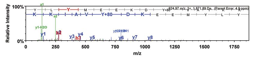
®
Jung et al.

P    eriodontal diseases, which involve inflammatory-based destruction of the tissues
     that surround and support the teeth, are initiated by multispecies communities in
the gingival compartment. Periodontal diseases and periodontal organisms are also
epidemiologically and mechanistically associated with serious systemic conditions,
such as coronary artery disease and Alzheimer’s disease (1–3). In the gingival compart-
ment, Porphyromonas gingivalis resides within multispecies communities, and interac-
tions among community participants define spatial organization and overall metabolic
and pathogenic properties. Such interactions are based on coadhesion and small-
molecule-based communication, and these processes are extensively studied (4–6).
Less is known, however, regarding the signal transduction pathways within bacterial
cells that relay information derived from the microenvironment and from community
partner species.

                                                                                                               Downloaded from http://mbio.asm.org/ on March 13, 2021 by guest
    While not a numerically dominant member of the periodontal microbiome, the
keystone pathogen P. gingivalis can elevate the nososymbiocity (or pathogenic poten-
tial) of periodontal communities through interactions with accessory pathogens and
pathobionts (7, 8). Streptococcus gordonii is a major partner species of P. gingivalis, and
in the murine oral periodontitis model coinfection with both species results in greater
alveolar bone loss than infection with either species alone (9, 10). Community devel-
opment between the two bacterial species is mediated by coadhesion and chemical
communication based on streptococcal metabolites such as pABA (11). Our previous
studies have shown that virulence and community development of P. gingivalis are
controlled by Ptk1, a BY kinase of P. gingivalis (12). Ptk1 is a substrate of Ltp1, a
low-molecular-weight protein tyrosine phosphatase (LMW-PTP) (12–14), forming a
cognate kinase-phosphatase pair, but their genes are remotely located (ptk1
[PGN_1524 and PGN_RS07275] and ltp1 [PGN_0491 and PGN_RS02345]), an uncommon
arrangement in Gram-negative bacteria, indicating the possibility of additional phos-
phatases or kinases encoded by genes adjacent to the kinase or phosphatase (15).
    The operon in which ptk1 is located in P. gingivalis consists of three genes
(PGN_1523 [PGN_RS07270], ptk1, and PGN_1525 [PGN_RS07280]) (12). Bioinformati-
cally, PGN_1525 has histidinol phosphatase-like motifs belonging to the polymerase
and histidinol phosphatase family of protein tyrosine phosphatases (PHP-PTP). Most
PHP-PTPs are found in Gram-positive bacteria, such as Streptococcus pneumoniae,
Bacillus subtilis, and Lactobacillus rhamnosus, and they are involved in the control of
capsular polysaccharide production, DNA metabolism, cell division, and bacterial inva-
siveness (16–20). Thus far, the only Gram-negative organism that has been demon-
strated to have an active tyrosine phosphatase in the PHP family is Myxococcus xanthus,
in which PhpA is responsible for negative control of extracellular polysaccharide (EPS)
production and spore formation (21). In this study, we demonstrate that PGN_1525
encodes an active PHP family tyrosine phosphatase, designated here Php1, which can
act on Ptk1 and participate in the control of community development along with
virulence in an animal model of periodontal disease.

RESULTS
    Comparative sequence analysis and enzymatic characterization of Php1. In P.
gingivalis 33277, PGN_1525, a gene immediately downstream of ptk1, encodes a
predicted 28-kDa protein containing a PHP domain (PF02811). BLASTP analysis revealed
that it shares significant homology with PHP-PTP proteins of other bacterial species,
including all the invariant histidine, aspartate, and arginine residues in four conserved
motifs (see Fig. S2A in the supplemental material). Moreover, homology modeling
(Fig. S2B) suggests strong structural conservancy with YwqE, a PHP-PTP of Bacillus
subtilis. The tertiary conformation is highly homologous along with conserved residues
H27, H64, H155, and R158, which form a catalytic pocket. Thus, here the PGN_1525
protein is designated Php1. Previous studies have shown that divalent metal ions are
essential cofactors for PHP tyrosine phosphatase enzyme activity (21–23). The active
site of the prototypic PHP-PTP enzymes YwqE and CpsB (from Streptococcus pneu-
moniae) contains a cluster of three metal ions bound adjacent to each other between

September/October 2019 Volume 10 Issue 5 e02004-19                                            mbio.asm.org 2
®
P. gingivalis Tyrosine Phosphatase
A B
pNP (nmole/min/μg)
0.5
Activity (% of MnCl2 )
120 ****
100 0.4
80 0.3
60 0.2
40 ****
20 0.1
0
C +
u 2+
n 2+
C +
a 2+
-20
2+
i +
o2
g2
-
N 2
3 0
25
5
4
16
0. 125
0. 5
1
2
8
32
0.125
0.
Zn
2
C
M
M
06
0.0
Mn 2+ (mM)
Downloaded from http://mbio.asm.org/ on March 13, 2021 by guest
C
pNP (nmol/min/μg)
1.5 ****
****
1.0 ****
****
0.5 **
7 8 9 10
pH
FIG 1 Characterization of Php1 phosphatase activity. (A) Recombinant Php1 was incubated with 50 mM
pNPP in a reaction mixture (pH 8.0) containing 1 mM (final concentration) the indicated ions. (B) Effect
of Mn2⫹ concentration on Php1 activity against pNPP. (C) Php1 activity against pNPP measured at
different pH values of the reaction mixture. Reactions were performed in triplicate, and data are
expressed as means ⫾ standard errors (SE). **, P ⬍ 0.01; ****, P ⬍ 0.001.
-sheets (22, 24). The phosphatase activity of recombinant Php1 also was dependent
on the presence of Mn2⫹ or, to a lesser degree, Co2⫹ (Fig. 1A), as described for CpsB
(25), and PhpA from M. xanthus (21). YwqE is also Mn2⫹ dependent but is also
stimulated by Cu or Zn ions, and different members of the enzyme family differ in their
pattern of sensitivity to cations other than Mn2⫹ (25, 26). Php1 activity was enhanced
by increasing concentrations of Mn2⫹ up to 4 mM but was decreased at higher Mn2⫹
concentrations (Fig. 1B). When the effect of pH on the phosphatase activity of Php1 was
examined, activity was enhanced by increasing pH within a range of pH 8.0 to 10.0,
while activity was not significantly above background levels at pH 7.0 (Fig. 1C). Hence,
in vitro, Php1 is only active under basic conditions, as described for this family of
enzymes (26).
Php1 phosphatase activity on p-nitrophenyl phosphate (pNPP) was inhibited by the
tyrosine phosphatase inhibitor Na3VO4 (Fig. S3). Inhibition of Php1 was also observed
with NaF, which, although a classic serine/threonine phosphatase inhibitor, is a feature
of Php enzymes (27) (Fig. S3). Inhibition by EDTA (Fig. S3) corroborated the importance
of divalent cations for enzyme activity.
Substrate specificity of Php1. To define the substrate specificity of Php1, phos-
phatase activity was tested against phospho-amino acids and phosphopeptides
using a malachite green phosphate assay. When phospho-amino acids were used as
substrates, Php1 was most active against phosphotyrosine, with significantly less
phosphate release from phosphoserine and phosphothreonine (Fig. 2A). Php1
activity against phosphotyrosine was significantly inhibited by Na3VO4, NaF, and
EDTA. When phosphopeptides were tested as substrates, Php1 was only active
against a tyrosine phosphopeptide, and phosphate release from a serine phospho-
peptide was not significantly above background levels (Fig. 2B). Php1 activity
against the tyrosine phosphopeptide was also significantly inhibited by Na3VO4,
NaF, and EDTA. The Km for tyrosine phosphopeptide was 15.56 ⫾ 2.21 M, with a
September/October 2019 Volume 10 Issue 5 e02004-19 mbio.asm.org 3®
Jung et al.
A B
2.5
PO4 (pmol/min/μg)
PO4 (pmol/min/μg)
3
2.0
**
1.5 2
*** **
1.0 *** ***
*** 1 ***
0.5 *** ***
*** ***
TA
aF
TA
aF
VO
4
VO
4
N
N
ED
ED
a
3
a
3
N
N
Downloaded from http://mbio.asm.org/ on March 13, 2021 by guest
pY pS pT pY pS
Phospho-amino acid Phosphopeptide
C 40
Velocity ( mol /min )
30
20
10
0
0 200 400 600
Tyr phosphopeptide ( M)
FIG 2 Substrate specificity of Php1. Recombinant Php1 was incubated with 200 M phospho-amino
acids (A) or 100 M phosphopeptides (B) in the presence or absence of the indicated inhibitors. (C)
Saturation kinetics of Php1 with (circles, 4 mM) or without (triangles) Mn2⫹ and tyrosine phosphopeptide
substrate at pH 8.0. The graph is a nonlinear fit of the experimental data to the Michaelis-Menten
equation. The experiments were performed three times in triplicate, and representative data are shown
as means ⫾ SE. **, P ⬍ 0.01; ***, P ⬍ 0.005.
Vmax of 31.07 ⫾ 1.11 M/min, a kcat of 0.518 ⫾ 0.019 s⫺1, and an enzyme efficiency
of 33.29 s⫺1 M⫺1 when determined using the Michaelis-Menten equation (Fig. 2C).
As a control, Php1 enzyme with no divalent metals was assayed and found to have no
activity. These results establish Php1 as an active tyrosine phosphatase in vitro with little
activity against serine and threonine.
Dephosphorylation of the Ptk1 tyrosine kinase by Php1. Because of the juxta-
position of the Ptk1- and Php1-encoding genes, we tested the possibility that Ptk1 and
Php1 can function as a cognate kinase-phosphatase pair. Recombinant FPtk1 (the
C-terminal catalytically active region of Ptk1) purified from Escherichia coli is highly
tyrosine phosphorylated, and we have established that residues Y775, Y782, Y784,
Y786, Y788, and Y790 in the tyrosine-rich C-terminal cluster are all phosphorylated (13).
Tyrosine phosphorylation of Ptk1 decreased after incubation with Php1 in a time- and
dose-dependent manner (Fig. 3A and C). Dephosphorylation of Ptk1 required phos-
phatase enzyme, as the phosphorylation level of Ptk1 was stable in the absence of Php1
(Fig. 3B). Phos-tag electrophoresis showed a rapid transition of Ptk1 from a maximally
phosphorylated form to a minimally phosphorylated form in the absence of discrete
intermediates (Fig. 3D).
Mutation of conserved residues of Php1. Histidine, cysteine, and arginine residues
in conserved motifs of the PHP domain of Php1 (depicted in Fig. 4A) were selected for
site-directed mutagenesis to determine their roles in Php1 catalytic activity. Among
conserved histidine residues, replacement of H27, H64, and H155 with alanine elimi-
nated Php1 phosphatase activity with substrates of pNPP, phosphotyrosine, or tyrosine
phosphopeptide (Fig. 4B to D), indicating an important role for these residues. An
September/October 2019 Volume 10 Issue 5 e02004-19 mbio.asm.org 4®
P. gingivalis Tyrosine Phosphatase
A B time (min)
Ptk1 + + -
Php1 - + + 0 30 60 120 240
time (min) 30 60 120 240
Ptk1Immunoblot
Ptk1 Immunoblot
C D Ptk1 + + -
Ptk1 +
Php1 - + +
Php1 (μg) 0 1 2 4 8 time (min) 30 60 120 240
More
Downloaded from http://mbio.asm.org/ on March 13, 2021 by guest
phosphorylated
Ptk1 Immunoblot
Phos-tag
Less
phosphorylated
FIG 3 Dephosphorylation of Ptk1 by Php1. Recombinant Ptk1 was incubated with (A) or without (B)
recombinant Php1 (5 g) for the indicated times, and tyrosine phosphorylation of Ptk1 was analyzed by
immunoblotting with phosphotyrosine antibodies. (C) Recombinant Ptk1 was incubated with the indi-
cated concentrations of Php1 for 240 min, and tyrosine phosphorylation of Ptk1 was analyzed by
immunoblotting. (D) Ptk1 reacted with Php1 as described for panel A and phosphorylation analyzed by
Mn2⫹-Phos-tag–SDS-PAGE.
H213A-substituted protein retained 30 to 50% of native enzyme activity with all
substrates (Fig. 4B to D). Crystal structure analyses of S. pneumoniae CpsB and B. subtilis
YwqE (22, 24) showed that histidine residues in motifs II and IV (corresponding to H64
and H213 in P. gingivalis) are involved in coordination of metal ions. Incomplete
inactivation of Php1 by H213A mutation suggests that the role of H213 in metal ion
binding is less critical than that of H64. A C28S mutation in Php1 reduced enzyme
activity by 30 to 40%. In the structural model (Fig. S2B), C28 faces away from the
catalytic site, which may explain why the C28S mutant has less impact on enzyme
activity than mutation of the conserved histidine residues. Further, the corresponding
substitution in motif I of L. rhamnosus Wzb augmented enzyme activity (23), indicating
that the role of the conserved cysteine residue in catalytic activity can vary among
different bacteria. In addition to the essential histidine residues, the arginine residue at
position 158 in motif III was also required for function, as Php1 with an R158A
substitution lost all enzyme activity (Fig. 4B to D). The corresponding arginine residue
in S. pneumoniae CpsB also has been reported to be essential for enzyme activity by
participating in electron transfer (22, 25).
To investigate the role of these residues in Php1 activity against a P. gingivalis
substrate, Ptk1 was incubated with native Php1 or with mutant derivatives. Php1
derivatives C28S, H64, and H213 dephosphorylated Ptk1 to a level similar to that of
wild-type Php1 (Fig. 4E), indicating that the phosphatase activity against a cognate
kinase is less dependent on these residues than is activity against synthetic substrates.
However, mutant derivatives H27A, H155A, and R158A were unable to dephosphorylate
Ptk1, supporting an import role for these residues in the Php1-Ptk1 axis.
Role of Php1 in P. gingivalis single- and dual-species community development
and in extracellular polysaccharide production. Ptk1 is required for development of
dual-species P. gingivalis-S. gordonii communities in a nutrient-depleted model that
allows bacterial signaling-dependent accretion to be distinguished from an increase in
biomass due to growth and division (28). Hence, we examined the role of Php1 tyrosine
September/October 2019 Volume 10 Issue 5 e02004-19 mbio.asm.org 5®
Jung et al.
A
Motif I Motif II Motif III Motif IV
DXHCH H HXER DXH
H27A C28S H64A H155A R158A H213A
B C D
12 Phosphotyrosine
PO4(pmol/min/μg)
0.4
pNP (nmol/min/μg)
Tyrosine phosphopeptide
10
PO4(pmol/min/μg)
*** ***
0.3 8 8
*** *** ***
0.2 6
*** ***
Downloaded from http://mbio.asm.org/ on March 13, 2021 by guest
4 *** 4
0.1 ***
*** 2
0.0 *** *** *** *** *** *** *** ***
*** *** ***
0
A
A
5A
8A
3A
T
S
A
H A
R A
8A
3A
T
S
R A
H A
A
A
S
3A
T
W
28
27
64
-2
W
28
27
64
5
5
8
28
27
-0.1 64
W
15
15
21
15
15
21
15
15
21
C
H
H
C
H
H
C
H
H
H
R
H
H
H
E
Php1 - WT H27A C28S H64A H155A R158A H213A WT
EDTA - - - - - - - - +
α-pY
Ptk1
α-His
FIG 4 Mutations of conserved His, Cys, and Arg residues in Php1 attenuate phosphatase activity. (A) Schematic representation
of Php1 domain structure. Amino acid residues in the four conserved motifs that were mutated are indicated. (B to D)
Phosphatase activity of recombinant Php1 wild type (WT) and its mutant proteins was measured using pNPP (B), phospho-
tyrosine (C), or tyrosine phosphopeptide (D) as substrates. The experiments were performed three times in triplicate, and
representative data are shown as means ⫾ standard deviations (SD). ***, P ⬍ 0.001. (E) Phosphorylation of Ptk1. Recombinant
His-tag Ptk1 was incubated with Php1 WT and its mutant derivatives, and tyrosine phosphorylation of Ptk1 was examined by
immunoblotting using antiphosphotyrosine antibody. EDTA was used as a control to inhibit Php1 phosphatase activity. His
antibodies were used as a loading control in a duplicate experiment.
phosphatase activity in community development with S. gordonii. While this assay
system did not include saliva or serum, our previous results showed that these biofluids
do not have a significant effect on P. gingivalis-S. gordonii community formation (28). As
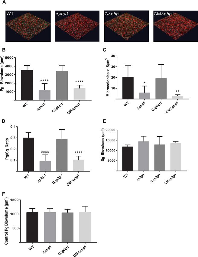
shown in Fig. 5, the Δphp1 strain demonstrated reduced accumulation on an S. gordonii
substrate compared to that of the parental strain. Complementation of the Δphp1
mutant with the wild-type php1 allele in trans restored the levels of P. gingivalis
biovolume in the dual-species accumulations. In contrast, complementation of the
Δphp1 mutant with the R158A mutant php1 allele, which produces catalytically inactive
Php1 protein, failed to restore the ability of P. gingivalis to develop into heterotypic
communities with S. gordonii. These results suggest that the Php1 phosphatase activity
is required for the development of P. gingivalis microcolonies on a streptococcal
substratum. Monospecies biofilm formation by P. gingivalis, however, was unaffected
by the loss of Php1 (Fig. 6A). Hence, Php1 is a component of signaling pathways in P.
gingivalis that show specificity for mixed-species community development. Ptk1 is also
required for maximal extracellular polysaccharide production by P. gingivalis (12), and
consistent with this we found that the Php1 mutant strain produced significantly less
exopolysaccharide than the parental strain (Fig. 6B). Polysaccharide production was
restored in the mutant strain complemented with the wild-type php1 allele but not in
the strain complemented with the R158A mutant php1 allele. While loss of exopoly-
saccharide in the encapsulated strain W83 enhances monospecies biofilm production
(29), the current data indicate that exopolysaccharide in the nonencapsulated 33277
strain does not play a similar role. Collectively, these results show that a functional
Php1-Ptk1 axis is required for both community development with S. gordonii and
extracellular polysaccharide production.
September/October 2019 Volume 10 Issue 5 e02004-19 mbio.asm.org 6®
P. gingivalis Tyrosine Phosphatase
Downloaded from http://mbio.asm.org/ on March 13, 2021 by guest
FIG 5 Php1 is required for maximal Porphyromonas gingivalis (Pg)–Streptococcus gordonii (Sg) community development. (A)
Confocal scanning laser microscopy visualization of P. gingivalis 33277 (WT), Δphp1, Δphp1 ⫹ pTphp1 (CΔphp1), or Δphp1⫹
pTphp1R158A (CMΔphp1) (green) strain reacted with a substrate of S. gordonii (red) for 24 h. A series of 0.2-m-deep optical
fluorescent x-y sections (213 by 213 m) were collected using a Leica SP8 confocal microscope and digitally reconstructed into a
three-dimensional (3D) image. (B) Total P. gingivalis biovolume was obtained with the 3D Find Objects function of Volocity. (C)
Numbers of microcolonies using an object size cutoff algorithm of greater than 15 m3 for the total 3D volume for P. gingivalis
fluorescence. (D) Ratio of P. gingivalis to S. gordonii biovolume. (E) Total S. gordonii biovolume was obtained with the 3D Find Objects
function of Volocity. (F) Biovolume of P. gingivalis in a parallel experiment without the S. gordonii substratum as a control for
nonspecific adherence to the glass coverslip. Error bars are SD, n ⫽ 3. *, P ⬍ 0.05; **, P ⬍ 0.01; ****, P ⬍ 0.001, each compared to the WT.
In contrast to Php1, the Ltp1 phosphatase of P. gingivalis restricts community
development with S. gordonii (14). The question then arises as to how the two
phosphatases are differentially utilized in P. gingivalis in the context of a dual-species
community. Previous studies found that para-amino benzoic acid (pABA), secreted by
September/October 2019 Volume 10 Issue 5 e02004-19 mbio.asm.org 7®
Jung et al.
A B
0.6
0.5
0.4
**** ****
OD 595nm
0.4
EPS/Pg
0.3
0.2
0.2
0.1
**** ****
T
p1
p1
p1
p1
T
W
W
ph
ph
ph
ph
' ' ' '
C
M
C
FIG 6 (A) Microtiter plate monospecies biofilm production by P. gingivalis 33277 (WT) and the Δphp1
Downloaded from http://mbio.asm.org/ on March 13, 2021 by guest
mutant at 48 h determined by optical density (OD) of crystal violet staining at 595 nm. (B) Exopolysac-
charide produced by P. gingivalis 33277 (WT), Δphp1, Δphp1 ⫹ pTphp1 (CΔphp1), or Δphp1⫹
pTphp1R158A (CMΔphp1) strain was stained with FITC᎑labeled concanavalin A and wheat germ agglutinin.
Bacterial cells were stained with Syto 17. Data are ratios of lectin binding (green) to whole-cell staining
(red). Error bars are SD, n ⫽ 3. ****, P ⬍ 0.001.
S. gordonii, downregulates the expression of ltp1 but not php1, consequently stimulat-
ing community development (11). Additionally, LMW-PTPs such as Ltp1 can also act as
redox sensors and are highly sensitive to inhibition by H2O2 (30), and analogues of
amino-benzoic acid are competitive inhibitors of tyrosine phosphatases (31, 32). S.
gordonii enzymes Cbe and SpxB, which synthesize pABA and H2O2, respectively, are
known to influence community development with P. gingivalis (11, 28). Hence, we
tested the effect of pABA and H2O2 on the activity of Ltp1 and Php1. Both H2O2 and
pABA inhibited Ltp1 but not Php1 (Fig. 7), indicating that the action of S. gordonii-
A 150
Ltp1
% Activity Remaining
Php1
100 * *
* * * *
*
50 *
*
0
0 2 4 6 8 12 16 24 DMSO
pABA (mM)
B 150
Ltp1
% Activity Remaining
Php1
100
*
50 *
*
*
0
0 100 250 500 1000
H2 O 2 (μ M)
FIG 7 Inhibition of Php1 activity by streptococcal metabolites. Recombinant Php1 was reacted with
pABA (A) or H2O2 (B) at the concentrations indicated, and activity against pNPP was measured. Data are
expressed relative to activity in the absence of inhibitor. Dimethyl sulfoxide (DMSO) is the vehicle control
for pABA. Data are means from three biological replicates ⫾ SE. *, P ⬍ 0.001.
September/October 2019 Volume 10 Issue 5 e02004-19 mbio.asm.org 8®
P. gingivalis Tyrosine Phosphatase
secreted compounds will favor the development of communities with P. gingivalis
through selective inhibition of Ltp1.
Phosphorylation of Php1 by Ptk1. We have reported previously that Ptk1 can
phosphorylate Ltp1 (13); thus, we tested whether Php1 also acts as a substrate of Ptk1.
Tyrosine phosphorylation of Php1 increased after incubation with FPtk1 in the presence
of ATP, whereas catalytically inactive FPk1 with a D717N mutation in the Walker B
domain failed to phosphorylate Php1 (Fig. 8A). To identify the tyrosine phosphorylation
sites, Php1 phosphorylated by Ptk1 was analyzed by mass spectrometry, which iden-
tified five tyrosine residues as targets of Ptk1 (Fig. 8B). As phosphorylated Y159 and
Y161 residues are close to R158 in motif III, which is required for enzyme activity, we
examined whether phosphorylation at these sites affects the phosphatase activity of
Php1. Y159 and Y161 were replaced with glutamate or phenylalanine to mimic a
Downloaded from http://mbio.asm.org/ on March 13, 2021 by guest
phosphorylated or unphosphorylated status, respectively. Php1 Y159F showed 2-fold
higher phosphatase activity with pNPP substrate than the wild-type Php1, whereas
Php1 Y159E had activity similar to that of the wild type (Fig. 8C). In contrast, the Y161F
mutation slightly decreased the Php1 activity while Php1 Y161E exhibited increased
activity. Similar results were obtained with Ptk1 as a substrate (Fig. 8D). Phosphoryla-
tion/dephosphorylation of Php1 at Y159 and Y161 may, therefore, represent a means
to fine-tune activity and suggests a reciprocal regulatory relationship with Ptk1.
Role of Php1 in P. gingivalis in vivo pathogenicity. The contribution of Php1 to
the virulence of P. gingivalis was evaluated by measuring alveolar bone loss in a murine
model. Mice infected with the parental P. gingivalis developed alveolar bone loss not
seen in the sham-treated animals (Fig. 9). Deletion of php1 significantly reduced the
ability of P. gingivalis to induce bone loss, demonstrating Php1 is essential for optimal
virulence in a murine model of infection.
DISCUSSION
Posttranslational modifications of proteins rapidly and reversibly impact conforma-
tion, localization, activity, and interaction with other macromolecules, thereby provid-
ing a flexible interface connecting intracellular pathways with extracellular stimuli (33).
Regulation of tyrosine phosphorylation in bacteria is mediated mainly by bacterial
protein-tyrosine (BY) kinases in conjunction with three families of protein-tyrosine
phosphatases: the low-molecular-weight phosphatases (LMW-PTPs), a family of small
acidic enzymes also found in eukaryotes; the eukaryotic-like phosphatases (PTPs),
which are dual-specific phosphatases that also display activity against phosphoserine
and phosphothreonine; and the DNA polymerase and histidinol phosphate phosphoe-
sterase (PHP) family (15, 34). In addition to tyrosine phosphatases, PHP proteins also
include other types of enzymes, such as histidinol phosphatases, DNA polymerases, and
families of uncharacterized proteins in bacteria and eukaryotes (35). Bacterial protein
tyrosine phosphatase-encoding genes are often arranged in operons next to BY
kinase-encoding genes, which constitute their substrates (15, 36, 37). In the operon of
Gram-negative bacteria, a gene immediately upstream of a BY kinase gene usually
encodes an LMW-PTP (15). On the other hand, in Gram-positive bacteria, a PHP family
tyrosine phosphatase is encoded by a gene immediately upstream or downstream of a
BY kinase gene, and LMW-PTPs are often encoded by distantly located genes (19, 36,
38). While Myxococcus xanthus contains a PHP enzyme, P. gingivalis thus far is the first
Gram-negative organism possessing both functional LMW and PHP tyrosine phospha-
tases and with the same operon arrangement as that found in Gram-positive bacteria.
Crystal structures of CpsB, an S. pneumoniae PHP-PTP, and YwqE, a B. subtilis
PHP-PTP, have revealed distorted triosephosphate isomerase (TIM) barrel structures
composed of several ␣-helices and parallel -strands with three metal ions in their
active sites (22, 24). Consistent with other PHP family enzymes (21, 25, 26), the Php1
tyrosine phosphatase in P. gingivalis displayed metal ion dependence, primarily for
manganese. Attenuation of Php1 activity by mutations of the conserved histidine
residues also supports the essentiality of divalent ions, as the corresponding residues
in CpsB and YwqE are involved in the coordination of metal ions in their active sites (22,
September/October 2019 Volume 10 Issue 5 e02004-19 mbio.asm.org 9®
Jung et al.
A Php1 + + +
Ptk1 - WT D717N
α-pY
Php1
α-GST
B
01 MFSIFKRktkqvnpfeqgwltdlidihchllpavddgsksieetlslidlleeigvkQHI
61 LTPHIMEEYPSNDAIFLRARFEELLAAITPDKASRlrLAAEYMLDAAFLDRLAEPLLTLG
121 DRYILVETSYMAPPIGLIGLLADLRFKGLSPVLAHPERYLYMEEKDYVAIKKQGVMFQLN
Downloaded from http://mbio.asm.org/ on March 13, 2021 by guest
181 LFSLFGAYNSSASEKAYALLEAGYYDLIGTDIHHLQPIARLLSEASLPPDLEGKIKSLVE
241 NNTRlfs
C
300 ***
***
Activity (% of WT)
250
200
***
150
100
***
50
T
E
F
E
F
W
59
61
59
61
Y1
Y1
Y1
Y1
D
Php1
WT Y159E Y159F Y161E Y161F
Ptk1 α-pY
Ptk1 α-His
Php1
FIG 8 Phosphorylation of Php1 by Ptk1 modulates activity. (A) Recombinant Php1 was incubated with
recombinant Ptk1 wild type (WT) or Ptk1 with a mutation in its Walker B domain (D717N) in the presence
of 5 mM ATP for 1 h. Tyrosine phosphorylation of Php1 was analyzed by immunoblotting using
phosphotyrosine antibodies and GST-tag antibodies as a loading control. (B, upper) Mass spectroscopy
sequence coverage for Php1. High-confidence MS/MS data for 20 unique peptides comprising 61
exclusive unique and 119 total spectra were used to assign sequence coverage for 185 (uppercase)
of 247 amino acids, achieving 75% coverage. Nondetected residues are in lowercase. Phosphorylated
residues are shown in boldface. (Lower) Example of MS/MS spectra used to assign site-specific
phosphorylation to MS/MS. (C) Phosphatase activity of recombinant Php1 wild-type (WT) and mutant
derivatives was measured against pNPP, and the activity of Php1 mutants is shown relative to that
of the WT. Data are means ⫾ SD from three biological replicates. ***, P ⬍ 0.001. (D) Recombinant
Ptk1 was incubated with/without Php1 WT and mutant derivatives, and tyrosine phosphorylation of
Ptk1 was examined by immunoblotting using phosphotyrosine antibodies and His-tag antibodies as
a loading control.
September/October 2019 Volume 10 Issue 5 e02004-19 mbio.asm.org 10®
P. gingivalis Tyrosine Phosphatase
0.25
ABC-CEJ Distance (mm)
**
0.20
0.15
0.10
0.05
0.00
Sham 33277 'php1
FIG 9 Php1 is required for virulence in vivo. Alveolar bone loss in mice following infection with parental
(33277) and Δphp1 mutant P. gingivalis strains was determined by CT analysis. Reconstructed images
Downloaded from http://mbio.asm.org/ on March 13, 2021 by guest
of the maxillary molars were analyzed along the sagittal slice to determine the distance between the ABC
(alveolar bone crest) and the CEJ (cementoenamel junction) relative to the root length of the tooth. The
data are the averages and scatter for each mouse from 12 measurements across both the first and second
molars relative to the sham-treated mice and are expressed as means ⫾ SE. **, P ⬍ 0.01 by one-way
analysis of variance relative to sham infection.
24). These results suggest that conditions affecting the concentration of divalent ions
in saliva and gingival crevicular fluid influence Php1 activity in P. gingivalis. Additionally,
Php1 was optimally active at alkaline pH, and the alkaline pH (7.5 to 8.0) of gingival
crevicular fluid (39) may therefore provide a favorable environment for Php1 activity.
Php1 activity of P. gingivalis in inflamed gingival tissues may be further enhanced as
gingival inflammation raises the pH of subgingival pockets up to pH 8.7 (39).
Php1 was capable of dephosphorylation of the chromosomally adjacent tyrosine
kinase Ptk1. Hence, P. gingivalis has a unique configuration whereby two tyrosine
phosphatases (Php1 and Ltp1) can act on a BY kinase (Ptk1). BY kinases such as Ptk1
possess a cluster of tyrosine residues in the C-terminal domain which are autophos-
phorylated (13, 40). Phosphorylation in this region is required for kinase function (40),
and it is thought that no single tyrosine residue is essential for activity (41). However,
it is unclear whether the overall level of tyrosine phosphorylation or a specific combi-
nation of tyrosine residue phosphorylation controls kinase activity. The results of our
Phos-tag electrophoresis analysis indicate that tyrosine phosphatases switch Ptk1
between high and low phosphorylated states, more consistent with a model where a
threshold level of phosphorylation is required for activity.
Although both Php1 and Ltp1 can dephosphorylate recombinant Ptk1 in vitro, the
(de)phosphorylation axes controlled by the two phosphatases are functionally distinct. Ltp1
participates in a regulatory pathway that constrains P. gingivalis-S. gordonii community
development as well as extracellular polysaccharide production (12, 14). In the current
study, we found that loss of Php1 diminished the ability of P. gingivalis to accumulate with
S. gordonii. In addition, although not organized into a discrete capsule in strain 33277, active
Php1 enzyme was required for maximal exopolysaccharide production. The activity of Ltp1,
but not Php1, was inhibited by pABA and H2O2, suggesting that as Php1 is found mainly
in Gram-positive organisms, it has evolved resistance to common Gram-positive metabo-
lites. Hence, metabolic cues produced by S. gordonii and sensed by P. gingivalis serve to
stimulate exopolysaccharide production and the development of dual-species communi-
ties, within which stress responses are reduced, indicative of a mutualistic interaction (42,
43). In dental biofilms, P. gingivalis is frequently in close association with oral streptococci,
which may have provided the evolutionary driver for a PHP family phosphatase in order to
maintain tyrosine phosphorylation/dephosphorylation ratios in the presence of streptococ-
cal metabolites. Differential properties of the two phosphatases may result from dephos-
phorylation of different P. gingivalis substrates involved in community development and
exopolysaccharide production.
Php1 can be phosphorylated by Ptk1 on two tyrosine residues, 159 and 161. While
tyrosine phosphorylation in the DPY or DPYY motif of LMW-PTPs has been reported in
E. coli, Mycobacterium tuberculosis, Staphylococcus aureus, and eukaryotic cells (44–47),
September/October 2019 Volume 10 Issue 5 e02004-19 mbio.asm.org 11®
Jung et al.
to our knowledge, this is the first report of phosphorylation of a PHP family phospha-
tase. The phosphatase activity of Php1 was differentially affected by phosphorylation.
Increased activity of Php1 Y159F and decreased activity of Php1 Y161F suggest that the
phosphorylation of tyrosine 159 is inhibitory, and the phosphorylation of tyrosine 161
might stimulate activity. Tyrosine 159 is conserved in PHP-PTPs of Streptococcus species
or altered to asparagine in PHP-PTPs of other Gram-positive bacteria (see Fig. S2 in the
supplemental material), whereas tyrosine 161 is not a conserved residue among
PHP-PTPs, indicating a potential P. gingivalis-specific regulatory role.
P. gingivalis is a well-recognized pathogen in periodontal diseases (48, 49); however,
virulence is expressed in the context of heterotypic communities (5, 6). In the murine
alveolar bone loss model, the endogenous mouse microbiota can provide the com-
munity context for P. gingivalis-induced disease (50). P. gingivalis exopolysaccharide can
Downloaded from http://mbio.asm.org/ on March 13, 2021 by guest
also be involved in heterotypic community development and is necessary for virulence
of the organism in murine models of disease (51–53). Here, we found that loss of Php1
rendered P. gingivalis unable to induce bone loss in a murine model. These results
substantiate the importance of Php1 for the pathophysiology of the organism, effec-
tuated, at least in part, through control of community development and exopolysac-
charide production by the Php1-Ptk1 signaling axis. Additionally, in a recent study
using a random transposon insertion library, we found that insertional inactivation of
either php1 or ptk1 significantly reduced fitness in an epithelial colonization model (54),
indicating that the P. gingivalis Php1-Ptk1 axis also plays an important role in the
interaction with host epithelial barriers and may represent a master regulator of
properties important in colonization and virulence.
MATERIALS AND METHODS
Bacterial culture. Porphyromonas gingivalis ATCC 33277 (33277) was cultured anaerobically at 37°C
in Trypticase soy broth (TSB) supplemented with 1 mg/ml yeast extract, 5 g/ml hemin, and 1 g/ml
menadione. When appropriate, tetracycline (1 g/ml) and/or erythromycin (10 g/ml) were added to the
medium. Streptococcus gordonii DL-1 was cultured anaerobically at 37°C in brain heart infusion broth
supplemented with 5 mg/ml yeast extract. Escherichia coli strains were grown aerobically at 37°C with
shaking in Luria-Bertani broth containing 100 g/ml ampicillin or 50 g/ml kanamycin when required.
Construction of mutant and complemented strains. Allelic exchange Δphp1 mutants were gen-
erated as previously described (55). Briefly, upstream and downstream fragments of php1 (PGN_1525)
were amplified by PCR using the primers listed in Table S1 in the supplemental material, and the
fragments were fused to ermF using the PCR fusion technique. The resulting constructs were electro-
porated into electrocompetent P. gingivalis, and transformants were selected on TSB plates supple-
mented with erythromycin. The expression of the upstream and downstream genes PGN_1524 and
PGN_1526 in the Δphp1 strain was comparable to that in the parent strain when analyzed with reverse
transcription-PCR (RT-PCR) (data not shown), and there was no difference in growth rate between parent
and mutant strains in TSB (data not shown).
To generate the CΔphp1 complemented strain, the promoter region upstream of PGN_1523 (12) and DNA
sequence containing the coding region of php1 were amplified by PCR and fused together using the PCR
fusion technique. The construct was confirmed by sequencing and cloned into pT-COW (56), and the resulting
plasmid, pTphp1, was transferred into the Δphp1 strain through conjugation with E. coli S17-1 containing
pTphp1. Transconjugants were selected with gentamicin (50 g/ml), erythromycin, and tetracycline. To
generate a Δphp1 strain that expresses Php1 with an R158A mutation in the motif III of the PHP domain (the
CMΔphp1 strain), a site-specific mutation was introduced into php1 of pTphp1 using a Q5 site-directed
mutagenesis kit (New England Biolabs) and the primers listed in Table S1. The resulting plasmid, pTphp1R158A,
was confirmed by sequencing and transferred by conjugation into the Δphp1 strain, and transconjugants
were selected with gentamicin, erythromycin, and tetracycline. Expression of php1 in the wild type (WT) and
in the CΔphp1 and CMΔphp mutant strains was confirmed by RT-PCR (Fig. S1).
Expression and purification of recombinant proteins. The 33277 php1 coding region was amplified
by PCR using primers listed in Table S1 and cloned into pGEX-4T-1 (GE Healthcare). The resulting plasmid,
pGEX-php1, was transformed into E. coli BL21(DE3) Star, and glutathione S-transferase (GST)-tagged protein
was purified using glutathione resin (GenScript). Recombinant Php1 with a His tag, after cloning of php1 into
pLATE51 (Thermo Fisher), was expressed in E. coli BL21(DE3) Star and purified using nickel-nitrilotriacetic acid
agarose (Qiagen). Site-specific mutations were introduced into php1 of pGEX-php1 or pLATE51-php1 using a
Q5 site-directed mutagenesis kit with the primers listed in Table S1. The following mutations in the PHP
domain of Php1 were created: H27A (motif I), C28S (motif I), H64A (motif II), H155A (motif III), R158A (motif
III), Y159E or Y159F (motif III), Y161E or Y161F (motif III), and H213A (motif IV). All constructs were confirmed
by sequencing. Ltp1 and the active domain of Ptk1 (FPtk1; amino acid residues 541 to 821) were produced
as described previously (12, 14). Recombinant Php1 and its derivatives were soluble and purified under native
conditions. After buffer exchange into Tris-buffered saline (TBS; pH 7.4), the purity of the recombinant proteins
was assessed using SDS-PAGE and Coomassie staining.
September/October 2019 Volume 10 Issue 5 e02004-19 mbio.asm.org 12®
P. gingivalis Tyrosine Phosphatase
Phosphatase activity assay. To measure phosphatase activity of recombinant Php1, p-nitrophenyl
phosphate (pNPP; New England Biolabs), phospho-amino acids (phosphotyrosine [Sigma], phosphoser-
ine [Sigma], and phosphothreonine [Sigma]), or phosphopeptides (tyrosine phosphopeptide ENDpYI-
NASL [Promega] and serine phosphopeptide DLDVPIPGRFDRRVpSVAAE [Ser/Thr phosphatase substrate
I; R&D Systems]) were used as substrates. The phosphatase assays using pNPP as a substrate were carried
out at 37°C using 2 g Php1 and 50 mM pNPP in a total volume of 50 l containing 100 mM Tris-HCl (pH
8.0), 150 mM NaCl, 5 mM dithiothreitol (DTT), and 1 mM MnCl2 unless otherwise stated. After adding 50
l of 3 M NaOH to stop the reaction, the release of p-nitrophenol (pNP) was determined by reading the
optical density at 405 nm. When phosphatase activity was measured using phospho-amino acids or
phosphopeptides, the reaction was performed at 37°C using 2 g Php1 and 200 M phospho-amino
acids or 100 M phosphopeptides in 80 l of the reaction buffer described above. Phosphate release was
determined with a malachite green phosphate assay kit (Sigma) according to the manufacturer’s
instructions. Where indicated, sodium orthovanadate (Na3VO4; Sigma), sodium fluoride (NaF; Sigma), or
ethylenediaminetetraacetic acid (EDTA) was added to the reaction buffer. Kinetic parameters were
determined from a nonlinear fit of the Michaelis-Menten equation using Prism 6 software (GraphPad),
Downloaded from http://mbio.asm.org/ on March 13, 2021 by guest
and a standard curve of inorganic phosphate was used.
Substrate dephosphorylation. Ptk1 (5 g) was incubated with 5 g Php1 in a total volume of 50 l
containing 100 mM Tris-HCl (pH 8.0), 150 mM NaCl, 5 mM DTT, and 1 mM MnCl2 at 37°C for 2 h unless
otherwise indicated. The reactions were stopped by adding 5⫻ sample buffer and boiling for 10 min. Samples
were separated by SDS-PAGE or Mn2⫹-Phos-tag (Wako)–SDS-PAGE and transferred to polyvinylidene difluo-
ride membranes. The membranes were blocked (TBS containing 0.1% Tween 20 [TBST] and 5% bovine serum
albumin) at room temperature for 1 h and washed with TBST. Membranes were probed with mouse
antiphosphotyrosine antibody (clone PY20; 1:1,000; Sigma), mouse anti-His-tag antibody (Cell Signaling), or
rabbit anti-GST-tag antibody (Cell Signaling) at 4°C overnight. After washing (3⫻ TBST), membranes were
incubated with a horseradish peroxidase-conjugated anti-mouse IgG or anti-rabbit IgG secondary antibody
(1:1,000; Cell Signaling) at room temperature for 1 h. Immunoreactive bands were detected with Pierce ECL
Western blotting substrate (Thermo Fisher) and a ChemiDoc XRS⫹ imaging system (Bio-Rad).
Identification of phosphorylated residues of Php1. Php1 (5 g) was incubated with Ptk1 (5 g)
and 5 mM ATP with PhosSTOP proprietary broad-spectrum phosphatase inhibitor cocktail (Sigma) for
30 min. The proteins were separated by SDS-PAGE and stained with Coomassie brilliant blue, and the
Php1 protein band was cut from the gel and stored in water with PhosStop. At the University of Michigan
Proteomics and Peptide Synthesis core facility, in-gel digestion of Php1 with trypsin was performed using
a ProGest robot (Digilab). The gel slice first was washed with 25 mM ammonium bicarbonate followed
by acetonitrile. The slice was then reduced with 10 mM DTT at 60°C, followed by alkylation with 50 mM
iodoacetamide at room temperature. The protein was then digested with trypsin (Promega) at 37°C for
4 h followed by inactivation using formic acid, and the supernatant was analyzed directly without further
processing. The gel digest was analyzed by nano-liquid chromatography-tandem mass spectrometry
(LC-MS/MS) with a Waters NanoAcquity high-performance liquid chromatography system interfaced to
a ThermoFisher Q Exactive. Peptides were loaded on a trapping column and eluted over a 75-m
analytical column at 350 nl/min; both columns were packed with Luna C18 resin (Phenomenex). The mass
spectrometer was operated in data-dependent mode, with the Orbitrap operating at 60,000 full width at
half maximum (FWHM) and 17,500 FWHM for MS and MS/MS, respectively. The fifteen most abundant
ions were selected for MS/MS. Peptide spectra were analyzed using Scaffold 4 (Proteome Software).
P. gingivalis-S. gordonii dual-species community development. Heterotypic P. gingivalis-S. gor-
donii communities were generated as previously described (28). Briefly, hexidium iodide (15 g/ml;
Molecular Probes)-labeled S. gordonii (2 ⫻ 108 cells) was deposited on a glass coverslip anaerobically at
37°C for 16 h in phosphate-buffered saline (PBS). After removing unattached S. gordonii, 5 (and
6)-carboxyfluorescein succinimidyl ester (CFSE; 4 g/ml; Molecular Probes)-labeled P. gingivalis parental
or mutant strains (5 ⫻ 107 cells) were reacted with S. gordonii in prereduced PBS anaerobically at 37°C
for 24 h. After washing with PBS, communities were fixed with 4% paraformaldehyde and examined on
a confocal microscope (SP8; Leica) using 488-nm and 552-nm lasers for CFSE and hexidium iodide,
respectively. Three-dimensional reconstruction and quantification of the volume of P. gingivalis and S.
gordonii fluorescence were carried out using Volocity software (Perkin Elmer).
P. gingivalis monospecies biofilm formation. P. gingivalis cells (5 ⫻ 107) in TSB were incubated at
37°C anaerobically for 48 h in individual wells of 96-well plates. The resulting biofilms were washed,
stained with 1% crystal violet, and destained with 95% ethanol, and absorbance at 595 nm was
determined.
Extracellular polysaccharide production. Exopolysaccharide production was quantified with fluo-
rescent lectins as described previously (14). In brief, P. gingivalis cells were labeled with Syto 17
(Invitrogen) and deposited in individual wells of 96-well plates. Polysaccharide was labeled with
fluorescein isothiocyanate (FITC)᎑conjugated concanavalin A and wheat germ agglutinin᎑FITC (100 g
ml⫺1) for 30 min at room temperature. After washing, fluorescent levels at 488 nm (FITC) and 648 nm
(Syto 17) were determined.
Murine alveolar bone loss. C57BL/6 mice, 10 to 12 weeks old, were obtained from Jackson
Laboratory. Female mice were used, as studies have shown there to be no gender-dependent differences
in alveolar bone loss in a periodontal disease model (57). Mice were fed a standard diet with water ad
libitum. The University of Louisville Institutional Animal Care and Use Committee approved all animal
procedures in this study. P. gingivalis 33277 and Δphp1 strains (109 CFU were suspended in 0.1 ml of
sterile PBS with 2% carboxymethylcellulose [CMC]) were orally inoculated into mice at 2-day intervals
over a 12-day period. A control group of mice were mock inoculated with PBS and CMC alone. Forty-two
September/October 2019 Volume 10 Issue 5 e02004-19 mbio.asm.org 13®
Jung et al.
days after the last infection, mice were euthanized and skulls were subjected to micro-computed
tomography (CT) scanning (SKYSCAN 1174; Bruker). Bone loss was assessed by measuring the distance
between the alveolar bone crest and the cementoenamel junction at 14 interdental points between the
first and second maxillary molars.
Statistics. Experiments were conducted in triplicate, and the data presented are representative of at
least three independent experiments. Prism 6 software was used for statistical analyses. Analysis of
variance with Tukey’s post hoc test was used to compare more than two groups.
SUPPLEMENTAL MATERIAL
Supplemental material for this article may be found at https://doi.org/10.1128/mBio
.02004-19.
FIG S1, EPS file, 0.6 MB.
FIG S2, EPS file, 2.2 MB.
FIG S3, EPS file, 0.4 MB.
Downloaded from http://mbio.asm.org/ on March 13, 2021 by guest
TABLE S1, DOCX file, 0.02 MB.
ACKNOWLEDGMENTS
This work was supported by the Research Resettlement Fund for the new faculty of
Seoul National University. We also thank the NIH/NIDCR for support through DE012505,
DE023193, DE011111 (R.J.L.), DE026939, DE028346 (D.P.M.), and DE028166 (Z.R.F.).
REFERENCES
1. Kumar PS. 2013. Oral microbiota and systemic disease. Anaerobe 24: function aspects of the Porphyromonas gingivalis tyrosine kinase Ptk1.
90 –93. https://doi.org/10.1016/j.anaerobe.2013.09.010. Mol Oral Microbiol 32:314 –323. https://doi.org/10.1111/omi.12173.
2. Maddi A, Scannapieco FA. 2013. Oral biofilms, oral and periodontal 14. Maeda K, Tribble GD, Tucker CM, Anaya C, Shizukuishi S, Lewis JP,
infections, and systemic disease. Am J Dent 26:249 –254. Demuth DR, Lamont RJ. 2008. A Porphyromonas gingivalis tyrosine phos-
3. Dominy SS, Lynch C, Ermini F, Benedyk M, Marczyk A, Konradi A, Nguyen phatase is a multifunctional regulator of virulence attributes. Mol Micro-
M, Haditsch U, Raha D, Griffin C, Holsinger LJ, Arastu-Kapur S, Kaba S, Lee biol 69:1153–1164. https://doi.org/10.1111/j.1365-2958.2008.06338.x.
A, Ryder MI, Potempa B, Mydel P, Hellvard A, Adamowicz K, Hasturk H, 15. Grangeasse C, Cozzone AJ, Deutscher J, Mijakovic I. 2007. Tyrosine
Walker GD, Reynolds EC, Faull RLM, Curtis MA, Dragunow M, Potempa J. phosphorylation: an emerging regulatory device of bacterial physiology.
2019. Porphyromonas gingivalis in Alzheimer’s disease brains: evidence Trends Biochem Sci 32:86 –94. https://doi.org/10.1016/j.tibs.2006.12.004.
for disease causation and treatment with small-molecule inhibitors. Sci 16. Morona JK, Paton JC, Miller DC, Morona R. 2000. Tyrosine phosphoryla-
Adv 5:eaau3333. https://doi.org/10.1126/sciadv.aau3333. tion of CpsD negatively regulates capsular polysaccharide biosynthesis
4. Murray JL, Connell JL, Stacy A, Turner KH, Whiteley M. 2014. Mechanisms in Streptococcus pneumoniae. Mol Microbiol 35:1431–1442. https://doi
of synergy in polymicrobial infections. J Microbiol 52:188 –199. https:// .org/10.1046/j.1365-2958.2000.01808.x.
doi.org/10.1007/s12275-014-4067-3. 17. Bender MH, Cartee RT, Yother J. 2003. Positive correlation between
5. Lamont RJ, Hajishengallis G. 2015. Polymicrobial synergy and dysbiosis tyrosine phosphorylation of CpsD and capsular polysaccharide produc-
in inflammatory disease. Trends Mol Med 21:172–183. https://doi.org/10 tion in Streptococcus pneumoniae. J Bacteriol 185:6057– 6066. https://doi
.1016/j.molmed.2014.11.004. .org/10.1128/jb.185.20.6057-6066.2003.
6. Lamont RJ, Koo H, Hajishengallis G. 2018. The oral microbiota: dynamic 18. Mijakovic I, Petranovic D, Macek B, Cepo T, Mann M, Davies J, Jensen PR,
communities and host interactions. Nat Rev Microbiol 16:745–759. Vujaklija D. 2006. Bacterial single-stranded DNA-binding proteins are
https://doi.org/10.1038/s41579-018-0089-x. phosphorylated on tyrosine. Nucleic Acids Res 34:1588 –1596. https://
7. Hajishengallis G, Lamont RJ. 2016. Dancing with the stars: how choreo- doi.org/10.1093/nar/gkj514.
graphed bacterial interactions dictate nososymbiocity and give rise to 19. Morona JK, Miller DC, Morona R, Paton JC. 2004. The effect that muta-
keystone pathogens, accessory pathogens, and pathobionts. Trends tions in the conserved capsular polysaccharide biosynthesis genes cpsA,
Microbiol 24:477– 489. https://doi.org/10.1016/j.tim.2016.02.010. cpsB, and cpsD have on virulence of Streptococcus pneumoniae. J Infect
8. Hajishengallis G, Darveau RP, Curtis MA. 2012. The keystone- Dis 189:1905–1913. https://doi.org/10.1086/383352.
pathogen hypothesis. Nat Rev Microbiol 10:717–725. https://doi.org/ 20. Nourikyan J, Kjos M, Mercy C, Cluzel C, Morlot C, Noirot-Gros MF, Guiral
10.1038/nrmicro2873. S, Lavergne JP, Veening JW, Grangeasse C. 2015. Autophosphorylation of
9. Daep CA, Novak EA, Lamont RJ, Demuth DR. 2011. Structural dissection the bacterial tyrosine-kinase CpsD connects capsule synthesis with the
and in vivo effectiveness of a peptide inhibitor of Porphyromonas gin- cell cycle in Streptococcus pneumoniae. PLoS Genet 11:e1005518. https://
givalis adherence to Streptococcus gordonii. Infect Immun 79:67–74. doi.org/10.1371/journal.pgen.1005518.
https://doi.org/10.1128/IAI.00361-10. 21. Mori Y, Maeda M, Takegawa K, Kimura Y. 2012. PhpA, a tyrosine phos-
10. Mahmoud MY, Steinbach-Rankins JM, Demuth DR. 2019. Functional phatase of Myxococcus xanthus, is involved in the production of exopo-
assessment of peptide-modified PLGA nanoparticles against oral bio- lysaccharide. Microbiology 158:2546 –2555. https://doi.org/10.1099/mic
films in a murine model of periodontitis. J Control Release 297:3–13. .0.059824-0.
https://doi.org/10.1016/j.jconrel.2019.01.036. 22. Hagelueken G, Huang H, Mainprize IL, Whitfield C, Naismith JH. 2009.
11. Kuboniwa M, Houser JR, Hendrickson EL, Wang Q, Alghamdi SA, Saka- Crystal structures of Wzb of Escherichia coli and CpsB of Streptococcus
naka A, Miller DP, Hutcherson JA, Wang T, Beck DAC, Whiteley M, Amano pneumoniae, representatives of two families of tyrosine phosphatases
A, Wang H, Marcotte EM, Hackett M, Lamont RJ. 2017. Metabolic cross- that regulate capsule assembly. J Mol Biol 392:678 – 688. https://doi.org/
talk regulates Porphyromonas gingivalis colonization and virulence dur- 10.1016/j.jmb.2009.07.026.
ing oral polymicrobial infection. Nat Microbiol 2:1493–1499. https://doi 23. LaPointe G, Atlan D, Gilbert C. 2008. Characterization and site-directed
.org/10.1038/s41564-017-0021-6. mutagenesis of Wzb, an O-phosphatase from Lactobacillus rhamnosus.
12. Wright CJ, Xue P, Hirano T, Liu C, Whitmore SE, Hackett M, Lamont RJ. BMC Biochem 9:10. https://doi.org/10.1186/1471-2091-9-10.
2014. Characterization of a bacterial tyrosine kinase in Porphyromonas 24. Kim HS, Lee SJ, Yoon HJ, An DR, Kim DJ, Kim SJ, Suh SW. 2011. Crystal
gingivalis involved in polymicrobial synergy. Microbiologyopen structures of YwqE from Bacillus subtilis and CpsB from Streptococcus
3:383–394. https://doi.org/10.1002/mbo3.177. pneumoniae, unique metal-dependent tyrosine phosphatases. J Struct
13. Liu C, Miller DP, Wang Y, Merchant M, Lamont RJ. 2017. Structure- Biol 175:442– 450. https://doi.org/10.1016/j.jsb.2011.05.007.
September/October 2019 Volume 10 Issue 5 e02004-19 mbio.asm.org 14®
P. gingivalis Tyrosine Phosphatase
25. Morona JK, Morona R, Miller DC, Paton JC. 2002. Streptococcus pneu- RJ, Hackett M. 2012. Proteomics of Streptococcus gordonii within a model
moniae capsule biosynthesis protein CpsB is a novel manganese- developing oral microbial community. BMC Microbiol 12:211. https://doi
dependent phosphotyrosine-protein phosphatase. J Bacteriol 184: .org/10.1186/1471-2180-12-211.
577–583. https://doi.org/10.1128/jb.184.2.577-583.2002. 44. Brelle S, Baronian G, Huc-Brandt S, Zaki LG, Cohen-Gonsaud M, Bischoff
26. Mijakovic I, Musumeci L, Tautz L, Petranovic D, Edwards RA, Jensen PR, M, Molle V. 2016. Phosphorylation-mediated regulation of the Staphy-
Mustelin T, Deutscher J, Bottini N. 2005. In vitro characterization of the lococcus aureus secreted tyrosine phosphatase PtpA. Biochem Biophys
Bacillus subtilis protein tyrosine phosphatase YwqE. J Bacteriol 187: Res Commun 469:619 – 625. https://doi.org/10.1016/j.bbrc.2015.11.123.
3384 –3390. https://doi.org/10.1128/JB.187.10.3384-3390.2005. 45. Nadler C, Koby S, Peleg A, Johnson AC, Suddala KC, Sathiyamoorthy K,
27. Lapenta F, Monton Silva A, Brandimarti R, Lanzi M, Gratani FL, Vellosillo Smith BE, Saper MA, Rosenshine I. 2012. Cycling of Etk and Etp phos-
Gonzalez P, Perticarari S, Hochkoeppler A. 2016. Escherichia coli DnaE phorylation states is involved in formation of group 4 capsule by Esch-
polymerase couples pyrophosphatase activity to DNA replication. PLoS erichia coli. PLoS One 7:e37984. https://doi.org/10.1371/journal.pone
One 11:e0152915. https://doi.org/10.1371/journal.pone.0152915. .0037984.
28. Kuboniwa M, Tribble GD, James CE, Kilic AO, Tao L, Herzberg MC, 46. Zhou P, Li W, Wong D, Xie J, Av-Gay Y. 2015. Phosphorylation control of
Shizukuishi S, Lamont RJ. 2006. Streptococcus gordonii utilizes several protein tyrosine phosphatase A activity in Mycobacterium tuberculosis.
distinct gene functions to recruit Porphyromonas gingivalis into a mixed FEBS Lett 589:326 –331. https://doi.org/10.1016/j.febslet.2014.12.015.
community. Mol Microbiol 60:121–139. https://doi.org/10.1111/j.1365 47. Bucciantini M, Chiarugi P, Cirri P, Taddei L, Stefani M, Raugei G, Nordlund
Downloaded from http://mbio.asm.org/ on March 13, 2021 by guest
-2958.2006.05099.x. P, Ramponi G. 1999. The low Mr phosphotyrosine protein phosphatase
29. Davey ME, Duncan MJ. 2006. Enhanced biofilm formation and loss of behaves differently when phosphorylated at Tyr131 or Tyr132 by Src
capsule synthesis: deletion of a putative glycosyltransferase in Porphy- kinase. FEBS Lett 456:73–78. https://doi.org/10.1016/s0014-5793(99)
romonas gingivalis. J Bacteriol 188:5510 –5523. https://doi.org/10.1128/ 00828-5.
JB.01685-05. 48. Hajishengallis G. 2015. Periodontitis: from microbial immune subversion
30. Xing K, Raza A, Lofgren S, Fernando MR, Ho YS, Lou MF. 2007. Low to systemic inflammation. Nat Rev Immunol 15:30 – 44. https://doi.org/
molecular weight protein tyrosine phosphatase (LMW-PTP) and its possible 10.1038/nri3785.
physiological functions of redox signaling in the eye lens. Biochim Biophys 49. Byrne SJ, Dashper SG, Darby IB, Adams GG, Hoffmann B, Reynolds EC.
Acta 1774:545–555. https://doi.org/10.1016/j.bbapap.2007.03.001. 2009. Progression of chronic periodontitis can be predicted by the levels
31. Zhou M, Ji M. 2005. Molecular docking and 3D-QSAR on 2-(oxalylamino) of Porphyromonas gingivalis and Treponema denticola in subgingival
benzoic acid and its analogues as protein tyrosine phosphatase 1B plaque. Oral Microbiol Immunol 24:469 – 477. https://doi.org/10.1111/j
inhibitors. Bioorg Med Chem Lett 15:5521–5525. https://doi.org/10 .1399-302X.2009.00544.x.
.1016/j.bmcl.2005.08.078. 50. Hajishengallis G, Liang S, Payne MA, Hashim A, Jotwani R, Eskan MA,
32. Iversen LF, Andersen HS, Branner S, Mortensen SB, Peters GH, Norris K, McIntosh ML, Alsam A, Kirkwood KL, Lambris JD, Darveau RP, Curtis MA.
Olsen OH, Jeppesen CB, Lundt BF, Ripka W, Moller KB, Moller NP. 2000. 2011. Low-abundance biofilm species orchestrates inflammatory peri-
Structure-based design of a low molecular weight, nonphosphorus, odontal disease through the commensal microbiota and complement.
nonpeptide, and highly selective inhibitor of protein-tyrosine phospha- Cell Host Microbe 10:497–506. https://doi.org/10.1016/j.chom.2011.10
tase 1B. J Biol Chem 275:10300 –10307. https://doi.org/10.1074/jbc.275 .006.
.14.10300. 51. Singh A, Wyant T, Anaya-Bergman C, Aduse-Opoku J, Brunner J, Laine
33. Cousin C, Derouiche A, Shi L, Pagot Y, Poncet S, Mijakovic I. 2013. ML, Curtis MA, Lewis JP. 2011. The capsule of Porphyromonas gingivalis
Protein-serine/threonine/tyrosine kinases in bacterial signaling and reg- leads to a reduction in the host inflammatory response, evasion of
ulation. FEMS Microbiol Lett 346:11–19. https://doi.org/10.1111/1574 phagocytosis, and increase in virulence. Infect Immun 79:4533– 4542.
-6968.12189. https://doi.org/10.1128/IAI.05016-11.
34. Mijakovic I, Grangeasse C, Turgay K. 2016. Exploring the diversity of 52. Polak D, Ferdman O, Houri-Haddad Y. 2017. Porphyromonas gingivalis
protein modifications: special bacterial phosphorylation systems. FEMS capsule-mediated coaggregation as a virulence factor in mixed infection
Microbiol Rev 40:398 – 417. https://doi.org/10.1093/femsre/fuw003. with Fusobacterium nucleatum. J Periodontol 88:502–510. https://doi
35. Kalinina OV, Gelfand MS, Russell RB. 2009. Combining specificity deter- .org/10.1902/jop.2016.160397.
mining and conserved residues improves functional site prediction. BMC 53. Li C, Kurniyati Hu B, Bian J, Sun J, Zhang W, Liu J, Pan Y, Li C. 2012.
Bioinformatics 10:174. https://doi.org/10.1186/1471-2105-10-174. Abrogation of neuraminidase reduces biofilm formation, capsule bio-
36. Toniolo C, Balducci E, Romano MR, Proietti D, Ferlenghi I, Grandi G, Berti synthesis, and virulence of Porphyromonas gingivalis. Infect Immun 80:
F, Ros IM, Janulczyk R. 2015. Streptococcus agalactiae capsule polymer 3–13. https://doi.org/10.1128/IAI.05773-11.
length and attachment is determined by the proteins CpsABCD. J Biol 54. Miller DP, Hutcherson JA, Wang Y, Nowakowska ZM, Potempa J, Yoder-
Chem 290:9521–9532. https://doi.org/10.1074/jbc.M114.631499. Himes DR, Scott DA, Whiteley M, Lamont RJ. 2017. Genes contributing to
37. Nierop Groot MN, Kleerebezem M. 2007. Mutational analysis of the Porphyromonas gingivalis fitness in abscess and epithelial cell coloniza-
Lactococcus lactis NIZO B40 exopolysaccharide (EPS) gene cluster: EPS tion environments. Front Cell Infect Microbiol 7:378. https://doi.org/10
biosynthesis correlates with unphosphorylated EpsB. J Appl Microbiol .3389/fcimb.2017.00378.
103:2645–2656. https://doi.org/10.1111/j.1365-2672.2007.03516.x. 55. Simionato MR, Tucker CM, Kuboniwa M, Lamont G, Demuth DR, Tribble
38. Mijakovic I, Poncet S, Boel G, Maze A, Gillet S, Jamet E, Decottignies P, GD, Lamont RJ. 2006. Porphyromonas gingivalis genes involved in com-
Grangeasse C, Doublet P, Le Marechal P, Deutscher J. 2003. Transmem- munity development with Streptococcus gordonii. Infect Immun 74:
brane modulator-dependent bacterial tyrosine kinase activates UDP- 6419 – 6428. https://doi.org/10.1128/IAI.00639-06.
glucose dehydrogenases. EMBO J 22:4709 – 4718. https://doi.org/10 56. Gardner RG, Russell JB, Wilson DB, Wang GR, Shoemaker NB. 1996. Use
.1093/emboj/cdg458. of a modified Bacteroides-Prevotella shuttle vector to transfer a recon-
39. Bickel M, Cimasoni G. 1985. The pH of human crevicular fluid measured structed beta-1, 4-D-endoglucanase gene into Bacteroides uniformis and
by a new microanalytical technique. J Periodontal Res 20:35– 40. https:// Prevotella ruminicola B(1)4. Appl Environ Microbiol 62:196 –202.
doi.org/10.1111/j.1600-0765.1985.tb00408.x. 57. Shusterman A, Salyma Y, Nashef A, Soller M, Wilensky A, Mott R, Weiss
40. Grangeasse C, Nessler S, Mijakovic I. 2012. Bacterial tyrosine kinases: EI, Houri-Haddad Y, Iraqi FA. 2013. Genotype is an important determi-
evolution, biological function and structural insights. Philos Trans R Soc nant factor of host susceptibility to periodontitis in the Collaborative
Lond B Biol Sci 367:2640 –2655. https://doi.org/10.1098/rstb.2011.0424. Cross and inbred mouse populations. BMC Genet 14:68. https://doi.org/
41. Paiment A, Hocking J, Whitfield C. 2002. Impact of phosphorylation of 10.1186/1471-2156-14-68.
specific residues in the tyrosine autokinase, Wzc, on its activity in 58. Dabour N, LaPointe G. 2005. Identification and molecular characteriza-
assembly of group 1 capsules in Escherichia coli. J Bacteriol 184: tion of the chromosomal exopolysaccharide biosynthesis gene cluster
6437– 6447. https://doi.org/10.1128/jb.184.23.6437-6447.2002. from Lactococcus lactis subsp. cremoris SMQ-461. Appl Environ Microbiol
42. Hendrickson EL, Beck DA, Miller DP, Wang Q, Whiteley M, Lamont RJ, 71:7414 –7425. https://doi.org/10.1128/AEM.71.11.7414-7425.2005.
Hackett M. 2017. Insights into dynamic polymicrobial synergy revealed 59. Minic Z, Marie C, Delorme C, Faurie JM, Mercier G, Ehrlich D, Renault P
by time-coursed RNA-Seq. Front Microbiol 8:261. https://doi.org/10 2007. Control of EpsE, the phosphoglycosyltransferase initiating exopo-
.3389/fmicb.2017.00261. lysaccharide synthesis in Streptococcus thermophilus, by EpsD tyrosine
43. Hendrickson EL, Wang T, Dickinson BC, Whitmore SE, Wright CJ, Lamont kinase. J Bacteriol 189:1351–1357. https://doi.org/10.1128/JB.01122-06.
September/October 2019 Volume 10 Issue 5 e02004-19 mbio.asm.org 15You can also read