STRUCTURAL IMPACT ON SARS-COV-2 SPIKE PROTEIN BY D614G SUBSTITUTION - SCIENCE
←
→
Page content transcription
If your browser does not render page correctly, please read the page content below
REPORTS

                                                                                                                                    Cite as: J. Zhang et al., Science
                                                                                                                                   10.1126/science.abf2303 (2021).

Structural impact on SARS-CoV-2 spike protein by D614G
substitution
Jun Zhang1,2†, Yongfei Cai1,2†, Tianshu Xiao1,2, Jianming Lu3, Hanqin Peng1, Sarah M. Sterling4,5, Richard M.
Walsh Jr.4,5, Sophia Rits-Volloch1, Haisun Zhu6, Alec N. Woosley6, Wei Yang6, Piotr Sliz1,2,5, Bing Chen1,2*
1
  Division of Molecular Medicine, Boston Children’s Hospital, Boston, MA 02115, USA. 2Department of Pediatrics, Harvard Medical School, Boston, MA 02115, USA. 3Codex
BioSolutions, Inc., Gaithersburg, MD 20879, USA. 4The Harvard Cryo-EM Center for Structural Biology, Harvard Medical School, Boston, MA 02115, USA. 5Department of
Biological Chemistry and Molecular Pharmacology, Blavatnik Institute, Harvard Medical School, Boston, MA 02115, USA. 6Institute for Protein Innovation, Harvard Institutes
of Medicine, Boston, MA 02115, USA.
†These authors contributed equally to this work.
*Corresponding author. Email: bchen@crystal.harvard.edu

                                                                                                                                                                             Downloaded from http://science.sciencemag.org/ on May 18, 2021
Substitution for aspartic acid by glycine at position 614 in the spike (S) protein of severe acute respiratory
syndrome coronavirus 2 appears to facilitate rapid viral spread. The G614 strain and its recent variants are
now the dominant circulating forms. We report here cryo-EM structures of a full-length G614 S trimer,
which adopts three distinct prefusion conformations differing primarily by the position of one receptor-
binding domain. A loop disordered in the D614 S trimer wedges between domains within a protomer in the
G614 spike. This added interaction appears to prevent premature dissociation of the G614 trimer,
effectively increasing the number of functional spikes and enhancing infectivity, and to modulate structural
rearrangements for membrane fusion. These findings extend our understanding of viral entry and suggest
an improved immunogen for vaccine development.

Severe acute respiratory syndrome coronavirus 2 (SARS-CoV-                              RBD-ACE2 complexes (14–17), and segments of S2 in the
2), an enveloped positive-stranded RNA virus, is the cause of                           postfusion state (18). In the prefusion ectodomain structure,
the COVID-19 pandemic (1). Although the viral evolution is                              S1 folds into four domains - NTD (N-terminal domain), RBD,
slowed by the RNA proofreading capability of its replication                            and two CTDs (C-terminal domains), and wraps around the
machinery (2), a variant with a single-residue substitution                             prefusion S2, with the RBD sampling two distinct confor-
(D614G) in its spike (S) protein rapidly became the dominant                            mations – “up” for a receptor-accessible state and “down” for
strain throughout the world (3). It has since further evolved                           a receptor-inaccessible state. We and others have reported
to give several variants of concern (VOCs) (4–6). The trimeric                          structures of a purified, full-length D614 S protein in both
S protein decorates the viral surface and is an important tar-                          prefusion and postfusion conformations (19, 20). Studies by
get for development of diagnostics, therapeutics and vac-                               cryo-electron tomography, with chemically inactivated SARS-
cines, therefore understanding the effect of key mutations                              CoV-2 preparations, using both D614 and G614 variants have
may guide intervention strategies. Here we focus on D614G                               revealed additional structural details of S proteins present on
mutation that is in all currently circulating strains. The S pro-                       the surface of virion (21–24).
tein is produced as a single-chain precursor and subsequently                               Epidemiological surveillance indicated that the SARS-
processed by a furin-like protease into the receptor-binding                            CoV-2 carrying G614 outcompeted the original virus and be-
fragment S1 and the fusion fragment S2 (7). After engagement                            came the globally dominant form within a month (3, 25, 26).
of the receptor-binding domain (RBD) in S1 with the viral re-                           This single-residue substitution appears to correlate with
ceptor angiotensin converting enzyme 2 (ACE2) on the host                               high viral loads in infected patients and high infectivity of
cell surface, followed by a second proteolytic cleavage within                          pseudotyped viruses, but not with disease severity (3). The
S2 (S2’ site) (8), the S protein undergoes large conformational                         G614 virus has comparable sensitivity to neutralization by
changes, resulting in dissociation of S1 and irreversible re-                           convalescent human sera or vaccinated hamster sera (3, 27–
folding of S2 into a postfusion structure (9, 10). This induces                         30), suggesting that vaccines containing D614 remain effec-
fusion of the virus and host cell membranes to initiate infec-                          tive against the G614 virus. Moreover, S1 dissociates more
tion. Rapid advances in structural biology of the SARS-CoV-2                            readily from the D614 virus than from G614 virus (31), indi-
S protein include structures of its soluble fragments: the ec-                          cating that the D614 viral spike is less stable than the G614
todomain stabilized in its prefusion conformation (11–13),                              variant. The G614 ectodomain trimer is reported to sample

First release: 16 March 2021                                             www.sciencemag.org                (Page numbers not final at time of first release) 1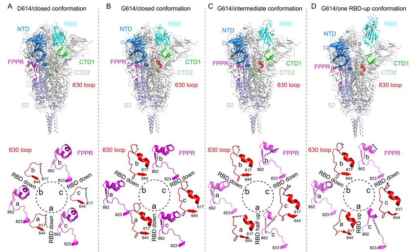
the RBD-up conformations more frequently than does the                three distinct classes each containing a similar number of
D614 trimer (13, 29, 32), but it is puzzling why the former           particles. The three classes represent a closed, three RBD-
binds more weakly to recombinant ACE2 than the latter (32).           down conformation, a one RBD-up conformation and an in-
The known S trimer structures indicate that the D614G                 termediate conformation with one RBD flipped up only half-
change breaks a salt bridge between D614 and a lysine resi-           way. All structures were refined to 3.1-3.5 Å resolution (figs.
due (K854) in the fusion peptide proximal region (FPPR) (19,          S3 to S8 and table S2). The overall structure of the G614 S
33, 34), which may help clamp the RBD in the prefusion con-           protein in the closed, three RBD-down prefusion confor-
formation. This observation can explain why the G614 trimer           mation is very similar to that of our published D614 S trimer
favors the RBD-up conformations, but does not account for             (Fig. 2) (19). In the three RBD-down structure, the four do-
its increased stability. To resolve these issues, we report here      mains in each S1, including NTD, RBD, CTD1 and CTD2, wrap
the structural consequences of the D614G substitution in the          around the three-fold axis of the trimer, protecting the pre-
context of the full-length S protein.                                 fusion S2. The furin cleavage site is disordered, making it un-
    We compared the membrane fusion activity of the full-             certain whether this structure represents the uncleaved or
length G614 S protein (fig. S1) with that of the D614 S con-          cleaved trimer, although the preparation contains primarily
struct in a cell-cell fusion assay (19). All the cells expressing S   the cleaved forms (Fig. 1A). The S2 fragment folds around a

                                                                                                                                           Downloaded from http://science.sciencemag.org/ on May 18, 2021
fused efficiently with cells transfected with a human ACE2            central three-stranded coiled coil that forms the most stable
construct (fig. S2A), demonstrating that the S proteins ex-           part of the structure; it is also the least variable region among
pressed on the cell surfaces are fully functional. At low trans-      all the known S trimer structures. The S2 structure is identi-
fection levels, G614 S had higher fusion activity than the D614       cal in the two G614 structures with one RBD projecting up-
S, but the difference diminished with the increased amount            wards, either completely or partially (fig. S9). In the
of transfected DNA, suggesting that the high expression lev-          conformation with one RBD fully up, the two neighboring
els can compensate for lower fusion efficiency of the D614 S          NTDs, including the one from the same protomer, shift away
protein. The G614 trimer remains sensitive to inhibition by           from the three-fold axis (fig. S9). In the RBD intermediate
an engineered trimeric ACE2-based inhibitor that competes             conformation, only the NTD from the adjacent protomer
with the receptor on the target cells (35) (fig. S2B). For pro-       packing directly against the moving RBD shifts. The D614G
tein purification, we used a construct fused with a C-terminal        substitution eliminates a salt bridge between D614 in CTD2
strep-tag, which was equally active in cell-cell fusion as the        of one subunit and K854 in the FPPR of the adjacent subunit
untagged version (fig. S2A), and purified both G614 and D614          (19, 34), but the FPPR in the three RBD-down conformation
proteins under identical conditions. The D614 protein eluted          of the G614 trimer remains structured.
in three peaks characterized previously as the prefusion S tri-           To examine the structural changes resulted from the
mer, the postfusion S2 trimer and the dissociated monomeric           D614G substitution, we superposed the structures of the G614
S1 (19). The G614 protein eluted as a single major peak, cor-         trimer onto the D614 trimer in the closed conformation align-
responding to the prefusion trimer (Fig. 1A). This suggests           ing them by the invariant S2 (Fig. 2B). A shift by a clockwise,
that D614G has a striking effect on the stability of the SARS-        outward rotation of all three S1 subunits, relative to the D614
CoV-2 S trimer. Coomassie-stained SDS-PAGE analysis shows             structure, is evident even for the G614 trimer in the closed
that G614 elutes mainly as the prefusion trimer, comprising           conformation. A similar shift was also observed in the RBD-
the cleaved S1/S2 complex (~90%) and a small amount of the            intermediate and RBD-up G614 structures. Thus, the D614G
uncleaved S precursor (~10%). We next measured binding of             substitution has led to a slightly more open conformation
the prefusion trimer fractions of the full-length proteins to         than that of the D614 trimer, even when all three RBDs are
recombinant soluble ACE2 by bio-layer interferometry (BLI)            down. The D614G change has apparently also rigidified a
(Fig. 1B). The S trimers bound more strongly to a dimeric             neighboring segment of CTD2, residues 620-640, which we
ACE2 than to a monomeric ACE2, as expected. The G614 pro-             designate the “630 loop”. This loop inserts into a gap, slightly
tein bound ACE2 less tightly than did the D614 protein, con-          wider in the G614 than in the D614 trimer, between the NTD
sistent with the measurements reported by others using                and CTD1 of the same protomer (Figs. 3 and 4). The 630 loop
soluble constructs (32). This observation appears incon-              is disordered in the closed D614 trimer (fig. S10), because the
sistent with reports that the G614 trimer has a more exposed          gap is too narrow for it to insert. The closed D614 trimer thus
RBD than the D614 trimer (13, 21, 22, 29, 32). We note that           has three ordered FPPRs and three disordered 630 loops,
the second binding event between dimeric ACE2 and a G614              while the closed G614 trimer has three structured 630 loops
trimer has both a slower on-rate and a slower off-rate than           along with three ordered FPPRs. In the two conformers with
for a D614 trimer (table S1).                                         one partly or fully open RBD, the two segments are disor-
    We determined the cryo-EM structures of the full-length           dered in the RBD-shifted subunit, and their central parts
G614 S trimer using RELION (36). 3D classification identified         have difficult-to-model density in one other subunit. The

First release: 16 March 2021                             www.sciencemag.org        (Page numbers not final at time of first release)   2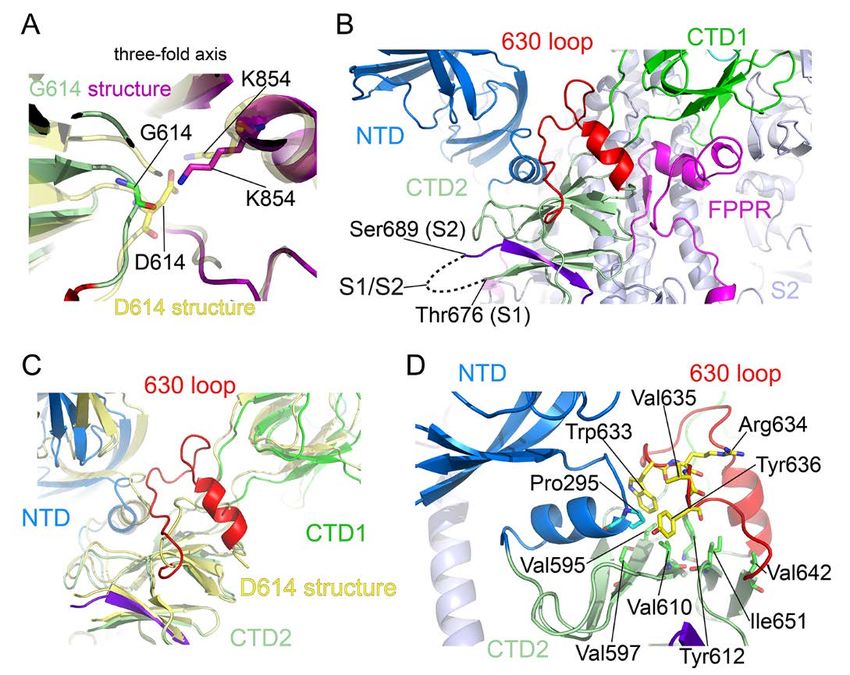
third pair appears well ordered throughout (Fig. 3). Thus,         cleaved S1/S2 complex and preventing S1 dissociation. The
opening of the RBD in the full-length G614 trimer correlates       mutant V635K had wildtype phenotypes in these assays,
with a displacement of the 630 loop, and the FPPR away from        likely because V635 does not make any direct contact with
its position in the D614 trimer. The D614G change did not          the CTD2. The mutants R634E and Y636A showed interme-
cause any large local structural rearrangements except for         diate levels of antibody binding because Y636 appears to con-
loss of the D614-K854 salt bridge (19, 34), and a small shift of   tribute less to the 630 loop-CTD2 interaction than W633 and
residue 614 toward the three-fold axis (Fig. 4A). The position     R634 may help maintain the loop’s overall shape for inserting
of the FPPR and the conformation of K854 may allow a hy-           between domains. Likewise, a similar pattern was observed
drogen bond between the K854 amino group and the main-             with these mutants in the cell-cell fusion assay except that
chain carbonyl of G614, perhaps accounting for the subtlety        Y636A showed substantially weaker fusion activity than
of the structural difference (fig. S11, A and B). Although the     R634E (fig. S12C). Thus key residues important for stabilizing
loss of the salt bridge involving D614 does not destabilize the    the S trimer structure appear critical for membrane fusion
packing of the FPPR against the rest of the trimer, it does        activity, as premature dissociation of S1 would lead to inacti-
weaken the FPPR density, especially between residues 842-          vation of the S trimer. To further confirm the folding of the
846. The 630 loop, which packs directly against the NTD,           630 loop in the G614 trimer, we collected additional data un-

                                                                                                                                        Downloaded from http://science.sciencemag.org/ on May 18, 2021
CTD1 and CTD2 of the same protomer, lies close to the S1/S2        der slightly different conditions and found the same three
boundary of the same protomer and the FPPR of an adjacent          classes representing the closed, RBD-intermediate and one
protomer (Fig. 4B). Inserting this wedge-like loop between         RBD-up conformations (fig. S13A). There is relatively strong
the NTD and CTD1 (Fig. 4C) may help secure the positions of        density in both 2D class averages and 3D reconstructions for
the NTD and CTDs. CTD2 is formed by two stacked, four-             the heptad repeat 2 (HR2) region (fig. S1) and detergent mi-
strand β-sheets, with a fifth strand in one sheet contributed      celle, invisible in all our previous cryo-EM analysis. The in-
by the connector between the NTD and RBD. In the other             creased length and lack of symmetry limited the resolution of
sheet, an interstrand loop contains the S1/S2 cleavage site,       these 3D reconstructions to 4.3-4.7Å. Nevertheless, the den-
and thus one strand is the N-terminal segment of S2 (Fig. 4B).     sity for the 630 loop was evident in the closed trimer even at
In the G614 trimer, one side of the 630 loop packs along a         this resolution (fig. S13B). We note that although S1 in the
long hydrophobic surface, largely solvent-exposed in the           G614 trimer moves outwards from its position in the D614
D614 trimer, formed by residues on the “upward” facing sur-        trimer, the extent of the shift is still appreciably smaller than
face of the CTD2 along with Pro295 from the NTD (Fig. 4D).         the shift seen in soluble S trimers stabilized by a trimeriza-
Trp633 and Tyr636 of the 630 loop appear to contribute to          tion foldon tag and two proline mutations (fig. S14).
this interaction. S1 dissociation from S2 requires breaking the        Our structures provide an explanation for why the G614
S2 strand from the second β-sheet. An ordered 630 loop that        virus, with a more stable S trimer, is more infectious than the
stabilizes the CTD2 by closing off an exposed, hydrophobic         original strain (fig. S15). The transition from the closed to one
surface may retard S1 shedding, thereby enhancing the sta-         RBD-up conformation in a G614 trimer requires an order-dis-
bility of a cleaved S trimer. We note that the density for a       order transition in one 630 loop and partial disordering of a
fatty acid ligand making contacts with the neighboring RBDs        second. Thus, kinetic barriers will probably make both the
in the D614 trimer is absent in all the G614 reconstructions       forward and reverse transitions slower than in a D614 trimer,
(fig. S11C) (37), suggesting that the ligand is not required for   in which all three 630 loops are unstructured in both confor-
three RBDs to adopt the down conformation. To test the im-         mations. In the one RBD-up conformation, S will also shed S1
pact of the 630 loop on S1 shedding and membrane fusion,           much more slowly from a G614 trimer than from D614, be-
we generated five S mutants each containing a single residue       cause the remaining two RBDs are stabilized by the ordered
change either in the 630 loop (W633A, R634E, V635K and             and partially ordered 630 loops, and a return of the first RBD
Y636A) or the CTD2 hydrophobic surface (V610K) in the G614         to the down configuration can occur unless locked in place
sequence. These mutants expressed the same level of S, with        by ACE2 receptor binding. This picture can account both for
a similar extent of cleavage between S1 and S2, as expected        the greater prevalence of a one-up RBD conformation and a
(fig. S12A). When detected by monoclonal antibodies using          lower overall ACE2 affinity, because the other two RBDs will
flow cytometry, mutants V610K and W633A showed mark-               remain inaccessible. It can also explain why we captured very
edly lower binding of RBD-specific antibodies [REGN10933           few trimers in the RBD-up conformation in our previous
and REGN10987; (38)], and of an NTD-specific antibody              cryo-EM study of the D614 trimer but instead saw abundant
[4A8; (39)] than the parental G614 S, while binding to an S2-      postfusion S2 (19), because any one RBD-up conformation
specific antibody [0304-3H3; (39)] was slightly higher (fig.       would proceed to two RBD-up and three RBD-up and shed
S12B). These results are consistent with the hydrophobic in-       very quickly, allowing S2 to convert to the postfusion form.
teractions between the 630 loop and CTD2 stabilizing the           Our interpretation of the structural differences is also

First release: 16 March 2021                           www.sciencemag.org       (Page numbers not final at time of first release)   3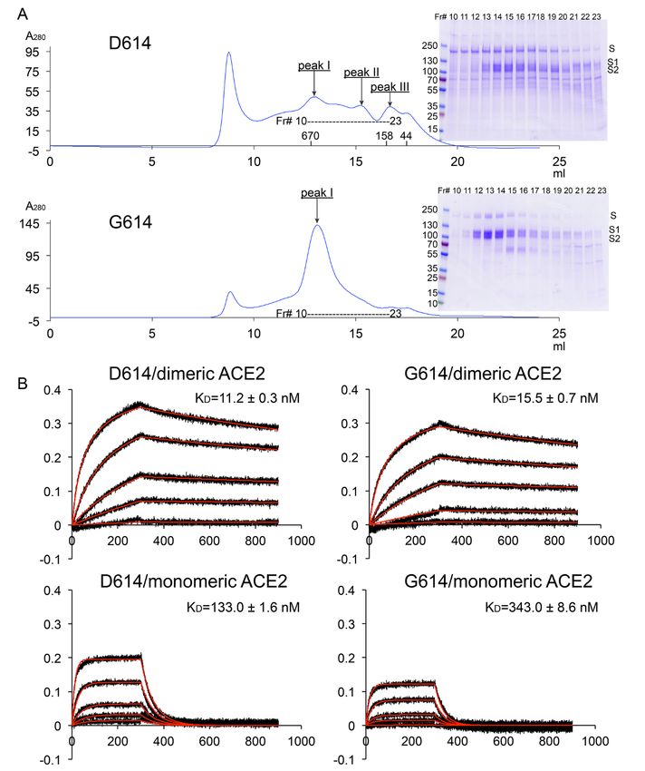
consistent with the spike conformational distribution on the      D614G substitution (table S3), suggesting that the increased
virions in cryo-ET studies of chemically inactivated SARS-        transmissibility of the G614 virus has led to a great number
CoV-2. The D614 preparation contains primarily postfusion         of replication events and to greater genetic diversity, despite
S2 spikes (24). One study of a G614 virus that had lost the       a lower absolute mutation rate.
furin cleavage site showed almost no postfusion spikes, and       REFERENCES AND NOTES
a 50:50 distribution of prefusion spikes between fully closed     1. P. Zhou, X.-L. Yang, X.-G. Wang, B. Hu, L. Zhang, W. Zhang, H.-R. Si, Y. Zhu, B. Li, C.-
and one RBD-up (21); and another showed 3% postfusion                  L. Huang, H.-D. Chen, J. Chen, Y. Luo, H. Guo, R.-D. Jiang, M.-Q. Liu, Y. Chen, X.-R.
spikes and 97% in the prefusion form (~31%, fully closed;              Shen, X. Wang, X.-S. Zheng, K. Zhao, Q.-J. Chen, F. Deng, L.-L. Liu, B. Yan, F.-X.
                                                                       Zhan, Y.-Y. Wang, G.-F. Xiao, Z.-L. Shi, A pneumonia outbreak associated with a
~55%, one RBD-up; ~14% two RBD-up) (22). The structured                new coronavirus of probable bat origin. Nature 579, 270–273 (2020).
630 loop in the G614 trimer not only reinforces the packing            doi:10.1038/s41586-020-2012-7 Medline
among three protomers, but also stabilizes the CTD2 to in-        2. F. Robson, K. S. Khan, T. K. Le, C. Paris, S. Demirbag, P. Barfuss, P. Rocchi, W.-L.
hibit release of the N-terminal segment of S2, effectively             Ng, Coronavirus RNA Proofreading: Molecular Basis and Therapeutic Targeting.
                                                                       Mol. Cell 79, 710–727 (2020). doi:10.1016/j.molcel.2020.07.027 Medline
blocking S1 dissociation. This property can account for the       3. B. Korber, W. M. Fischer, S. Gnanakaran, H. Yoon, J. Theiler, W. Abfalterer, N.
paucity of postfusion spikes on the G614 variant. In addition          Hengartner, E. E. Giorgi, T. Bhattacharya, B. Foley, K. M. Hastie, M. D. Parker, D.
to the FPPR that might modulate the fusogenic structural re-           G. Partridge, C. M. Evans, T. M. Freeman, T. I. de Silva, Sheffield COVID-19

                                                                                                                                                                  Downloaded from http://science.sciencemag.org/ on May 18, 2021
arrangements of S protein (19), CTD2 and the 630 loop within           Genomics Group, C. McDanal, L. G. Perez, H. Tang, A. Moon-Walker, S. P. Whelan,
                                                                       C. C. LaBranche, E. O. Saphire, D. C. Montefiori, Tracking Changes in SARS-CoV-
it, are probably also the key components of the S fusion ma-           2 Spike: Evidence that D614G Increases Infectivity of the COVID-19 Virus. Cell 182,
chinery. If ACE2 captures the RBD-up conformation (40), ex-            812–827.e19 (2020). doi:10.1016/j.cell.2020.06.043 Medline
pelling both the 630 loop and the FPPR from their positions       4. H. Tegally, E. Wilkinson, M. Giovanetti, A. Iranzadeh, V. Fonseca, J. Giandhari, D.
in the closed S trimer conformation, the FPPR shift may help           Doolabh, S. Pillay, E. J. San, N. Msomi, K. Mlisana, A. von Gottberg, S. Walaza, M.
                                                                       Allam, A. Ismail, T. Mohale, A. J. Glass, S. Engelbrecht, G. Van Zyl, W. Preiser, F.
expose the S2’ site near the fusion peptide for proteolytic            Petruccione, A. Sigal, D. Hardie, G. Marais, M. Hsiao, S. Korsman, M.-A. Davies, L.
cleavage, while departure of the 630 loop from the hydropho-           Tyers, I. Mudau, D. York, C. Maslo, D. Goedhals, S. Abrahams, O. Laguda-Akingba,
bic surface of the CTD2 can destabilize this domain and free           A. Alisoltani-Dehkordi, A. Godzik, C. K. Wibmer, B. T. Sewell, J. Lourenço, L. C. J.
the N-terminal segment of S2 to dissociate from S1, if the             Alcantara, S. L. Kosakovsky Pond, S. Weaver, D. Martin, R. J. Lessells, J. N.
                                                                       Bhiman, C. Williamson, T. de Oliveira, Emergence and rapid spread of a new severe
furin site has already been cleaved, and release S1 altogether.        acute respiratory syndrome-related coronavirus 2 (SARS-CoV-2) lineage with
Dissociation of S1 would then initiate a cascade of refolding          multiple spike mutations in South Africa. medRxiv 2020.12.21.20248640
events in the metastable prefusion S2, allowing the fusogenic          [Preprint]. 22 December 2020. https://doi.org/10.1101/2020.12.21.20248640.
transition to a stable postfusion structure. This model is sim-   5. F. Grabowski, G. Preibisch, S. Giziński, M. Kochańczyk, T. Lipniacki, SARS-CoV-2
                                                                       Variant of Concern 202012/01 has about twofold replicative advantage and
ilar to that proposed for membrane fusion catalyzed by HIV             acquires concerning mutations. medRxiv 2020.12.28.20248906 [Preprint]. 21
envelope protein (41). The SARS-CoV-2 S protein is the cen-            February 2021. https://doi.org/10.1101/2020.12.28.20248906.
terpiece of the first-generation vaccines that almost all used    6. C. M. Voloch, R. da Silva F Jr., L. G. P. de Almeida, C. C. Cardoso, O. J. Brustolini, A.
the D614 sequence. The G614 S trimer is naturally con-                 L. Gerber, A. P. de C Guimarães, D. Mariani, R. M. da Costa, O. C. Ferreira Jr.,
                                                                       Covid19-UFRJ Workgroup, LNCC-Workgroup, A. C. Cavalcanti, T. S. Frauches, C.
strained in a prefusion state that presents both the RBD-              M. B. de Mello, R. M. Galliez, D. S. Faffe, T. M. P. P. Castiñeiras, A. Tanuri, A. T. R.
down and RBD-up conformations with great stability. It is              de Vasconcelos, Genomic characterization of a novel SARS-CoV-2 lineage from
therefore likely to be a superior immunogen for eliciting pro-         Rio de Janeiro, Brazil. medRxiv 2020.12.23.20248598 [Preprint]. 26 December
tective neutralizing antibody responses, which appear largely          2020. https://doi.org/10.1101/2020.12.23.20248598.
                                                                  7. B. J. Bosch, R. van der Zee, C. A. de Haan, P. J. Rottier, The coronavirus spike protein
to target the RBD and NTD (39, 42). It may also be an excel-           is a class I virus fusion protein: Structural and functional characterization of the
lent scaffold for designing next-generation vaccines against           fusion core complex. J. Virol. 77, 8801–8811 (2003). doi:10.1128/JVI.77.16.8801-
new variants that have become resistant to protections of-             8811.2003 Medline
fered by the existing vaccines (43–46). In summary, we sug-       8. M. Hoffmann, H. Kleine-Weber, S. Schroeder, N. Krüger, T. Herrler, S. Erichsen, T.
                                                                       S. Schiergens, G. Herrler, N.-H. Wu, A. Nitsche, M. A. Müller, C. Drosten, S.
gest that the enhanced infectivity of the G614 virus largely           Pöhlmann, SARS-CoV-2 Cell Entry Depends on ACE2 and TMPRSS2 and Is
results from the increased stability of the S trimer, rather           Blocked by a Clinically Proven Protease Inhibitor. Cell 181, 271–280.e8 (2020).
than the better exposed RBDs. Indeed, if the virus that passed         doi:10.1016/j.cell.2020.02.052 Medline
from bats to humans or to an intermediate vector contained        9. J. K. Millet, G. R. Whittaker, Host cell entry of Middle East respiratory syndrome
                                                                       coronavirus after two-step, furin-mediated activation of the spike protein. Proc.
D614 [also present in the bat coronavirus BatCoV RaTG13                Natl. Acad. Sci. U.S.A. 111, 15214–15219 (2014). doi:10.1073/pnas.1407087111
(1)], then it could have gained fitness in the new host by ac-         Medline
quiring changes such as G614 for greater stability and infec-     10. M. A. Tortorici, D. Veesler, Structural insights into coronavirus entry. Adv. Virus
tivity than the parental form. Not surprisingly, the recent            Res. 105, 93–116 (2019). doi:10.1016/bs.aivir.2019.08.002 Medline
                                                                  11. D. Wrapp, N. Wang, K. S. Corbett, J. A. Goldsmith, C.-L. Hsieh, O. Abiona, B. S.
fast-spreading variants, including the B.1.1.7 (VUI202012/01;          Graham, J. S. McLellan, Cryo-EM structure of the 2019-nCoV spike in the
501Y.V1) lineage from the United Kingdom, the B.1.351                  prefusion        conformation.       Science        367,      1260–1263        (2020).
(501Y.V2) lineage from South Africa and the B.1.1.28                   doi:10.1126/science.abb2507 Medline
(484K.V2; P.1) lineage from Brazil (4–6), all contain the         12. A. C. Walls, Y.-J. Park, M. A. Tortorici, A. Wall, A. T. McGuire, D. Veesler, Structure,
                                                                       function, and antigenicity of the SARS-CoV-2 spike glycoprotein. Cell 181, 281–

First release: 16 March 2021                          www.sciencemag.org             (Page numbers not final at time of first release)                       4
292.e6 (2020). doi:10.1016/j.cell.2020.02.058 Medline Weaver, P.-Y. Shi, Spike mutation D614G alters SARS-CoV-2 fitness. Nature
13. S. M. Gobeil, K. Janowska, S. McDowell, K. Mansouri, R. Parks, K. Manne, V. Stalls, 10.1038/s41586-020-2895-3 (2020). doi:10.1038/s41586-020-2895-3 Medline
M. F. Kopp, R. Henderson, R. J. Edwards, B. F. Haynes, P. Acharya, D614G Mutation 28. J. Hu, C.-L. He, Q.-Z. Gao, G.-J. Zhang, X.-X. Cao, Q.-X. Long, H.-J. Deng, L.-Y.
Alters SARS-CoV-2 Spike Conformation and Enhances Protease Cleavage at the Huang, J. Chen, K. Wang, N. Tang, A.-L. Huang, D614G mutation of SARS-CoV-2
S1/S2 Junction. Cell Rep. 34, 108630 (2021). doi:10.1016/j.celrep.2020.108630 spike protein enhances viral infectivity. bioRxiv 2020.06.20.161323 [Preprint]. 6
Medline July 2020. https://doi.org/10.1101/2020.06.20.161323.
14. J. Lan, J. Ge, J. Yu, S. Shan, H. Zhou, S. Fan, Q. Zhang, X. Shi, Q. Wang, L. Zhang, X. 29. D. Weissman, M.-G. Alameh, T. de Silva, P. Collini, H. Hornsby, R. Brown, C. C.
Wang, Structure of the SARS-CoV-2 spike receptor-binding domain bound to the LaBranche, R. J. Edwards, L. Sutherland, S. Santra, K. Mansouri, S. Gobeil, C.
ACE2 receptor. Nature 581, 215–220 (2020). doi:10.1038/s41586-020-2180-5 McDanal, N. Pardi, N. Hengartner, P. J. C. Lin, Y. Tam, P. A. Shaw, M. G. Lewis, C.
Medline Boesler, U. Şahin, P. Acharya, B. F. Haynes, B. Korber, D. C. Montefiori, D614G
15. R. Yan, Y. Zhang, Y. Li, L. Xia, Y. Guo, Q. Zhou, Structural basis for the recognition Spike Mutation Increases SARS CoV-2 Susceptibility to Neutralization. Cell Host
of SARS-CoV-2 by full-length human ACE2. Science 367, 1444–1448 (2020). Microbe 29, 23–31.e4 (2021). doi:10.1016/j.chom.2020.11.012 Medline
doi:10.1126/science.abb2762 Medline 30. E. J. Anderson, N. G. Rouphael, A. T. Widge, L. A. Jackson, P. C. Roberts, M.
16. J. Shang, G. Ye, K. Shi, Y. Wan, C. Luo, H. Aihara, Q. Geng, A. Auerbach, F. Li, Makhene, J. D. Chappell, M. R. Denison, L. J. Stevens, A. J. Pruijssers, A. B.
Structural basis of receptor recognition by SARS-CoV-2. Nature 581, 221–224 McDermott, B. Flach, B. C. Lin, N. A. Doria-Rose, S. O’Dell, S. D. Schmidt, K. S.
(2020). doi:10.1038/s41586-020-2179-y Medline Corbett, P. A. Swanson 2nd, M. Padilla, K. M. Neuzil, H. Bennett, B. Leav, M.
17. Q. Wang, Y. Zhang, L. Wu, S. Niu, C. Song, Z. Zhang, G. Lu, C. Qiao, Y. Hu, K.-Y. Yuen, Makowski, J. Albert, K. Cross, V. V. Edara, K. Floyd, M. S. Suthar, D. R. Martinez, R.
Q. Wang, H. Zhou, J. Yan, J. Qi, Structural and Functional Basis of SARS-CoV-2 Baric, W. Buchanan, C. J. Luke, V. K. Phadke, C. A. Rostad, J. E. Ledgerwood, B. S.
Entry by Using Human ACE2. Cell 181, 894–904.e9 (2020). Graham, J. H. Beigel, mRNA-1273 Study Group, Safety and Immunogenicity of
Downloaded from http://science.sciencemag.org/ on May 18, 2021
doi:10.1016/j.cell.2020.03.045 Medline SARS-CoV-2 mRNA-1273 Vaccine in Older Adults. N. Engl. J. Med. 383, 2427–
18. S. Xia, M. Liu, C. Wang, W. Xu, Q. Lan, S. Feng, F. Qi, L. Bao, L. Du, S. Liu, C. Qin, F. 2438 (2020). doi:10.1056/NEJMoa2028436 Medline
Sun, Z. Shi, Y. Zhu, S. Jiang, L. Lu, Inhibition of SARS-CoV-2 (previously 2019- 31. L. Zhang, C. B. Jackson, H. Mou, A. Ojha, H. Peng, B. D. Quinlan, E. S. Rangarajan,
nCoV) infection by a highly potent pan-coronavirus fusion inhibitor targeting its A. Pan, A. Vanderheiden, M. S. Suthar, W. Li, T. Izard, C. Rader, M. Farzan, H. Choe,
spike protein that harbors a high capacity to mediate membrane fusion. Cell Res. SARS-CoV-2 spike-protein D614G mutation increases virion spike density and
30, 343–355 (2020). doi:10.1038/s41422-020-0305-x Medline infectivity. Nat. Commun. 11, 6013 (2020). doi:10.1038/s41467-020-19808-4
19. Y. Cai, J. Zhang, T. Xiao, H. Peng, S. M. Sterling, R. M. Walsh Jr., S. Rawson, S. Rits- Medline
Volloch, B. Chen, Distinct conformational states of SARS-CoV-2 spike protein. 32. L. Yurkovetskiy, X. Wang, K. E. Pascal, C. Tomkins-Tinch, T. P. Nyalile, Y. Wang, A.
Science 369, 1586–1592 (2020). doi:10.1126/science.abd4251 Medline Baum, W. E. Diehl, A. Dauphin, C. Carbone, K. Veinotte, S. B. Egri, S. F. Schaffner,
20. S. Bangaru, G. Ozorowski, H. L. Turner, A. Antanasijevic, D. Huang, X. Wang, J. L. J. E. Lemieux, J. B. Munro, A. Rafique, A. Barve, P. C. Sabeti, C. A. Kyratsous, N. V.
Torres, J. K. Diedrich, J.-H. Tian, A. D. Portnoff, N. Patel, M. J. Massare, J. R. Yates Dudkina, K. Shen, J. Luban, Structural and Functional Analysis of the D614G
3rd, D. Nemazee, J. C. Paulson, G. Glenn, G. Smith, A. B. Ward, Structural analysis SARS-CoV-2 Spike Protein Variant. Cell 183, 739–751.e8 (2020).
of full-length SARS-CoV-2 spike protein from an advanced vaccine candidate. doi:10.1016/j.cell.2020.09.032
Science 370, 1089–1094 (2020). doi:10.1126/science.abe1502 Medline 33. T. Zhou, Y. Tsybovsky, J. Gorman, M. Rapp, G. Cerutti, G.-Y. Chuang, P. S.
21. B. Turoňová, M. Sikora, C. Schürmann, W. J. H. Hagen, S. Welsch, F. E. C. Blanc, S. Katsamba, J. M. Sampson, A. Schön, J. Bimela, J. C. Boyington, A. Nazzari, A. S.
von Bülow, M. Gecht, K. Bagola, C. Hörner, G. van Zandbergen, J. Landry, N. T. D. Olia, W. Shi, M. Sastry, T. Stephens, J. Stuckey, I-T. Teng, P. Wang, S. Wang, B.
de Azevedo, S. Mosalaganti, A. Schwarz, R. Covino, M. D. Mühlebach, G. Hummer, Zhang, R. A. Friesner, D. D. Ho, J. R. Mascola, L. Shapiro, P. D. Kwong, Cryo-EM
J. Krijnse Locker, M. Beck, In situ structural analysis of SARS-CoV-2 spike reveals Structures of SARS-CoV-2 Spike without and with ACE2 Reveal a pH-Dependent
flexibility mediated by three hinges. Science 370, 203–208 (2020). Switch to Mediate Endosomal Positioning of Receptor-Binding Domains. Cell Host
doi:10.1126/science.abd5223 Medline Microbe 28, 867–879.e5 (2020). doi:10.1016/j.chom.2020.11.004 Medline
22. Z. Ke, J. Oton, K. Qu, M. Cortese, V. Zila, L. McKeane, T. Nakane, J. Zivanov, C. J. 34. X. Xiong, K. Qu, K. A. Ciazynska, M. Hosmillo, A. P. Carter, S. Ebrahimi, Z. Ke, S. H.
Neufeldt, B. Cerikan, J. M. Lu, J. Peukes, X. Xiong, H.-G. Kräusslich, S. H. W. W. Scheres, L. Bergamaschi, G. L. Grice, Y. Zhang, CITIID-NIHR COVID-19
Scheres, R. Bartenschlager, J. A. G. Briggs, Structures and distributions of SARS- BioResource Collaboration, J. A. Nathan, S. Baker, L. C. James, H. E. Baxendale, I.
CoV-2 spike proteins on intact virions. Nature 588, 498–502 (2020). Goodfellow, R. Doffinger, J. A. G. Briggs, A thermostable, closed SARS-CoV-2
doi:10.1038/s41586-020-2665-2 Medline spike protein trimer. Nat. Struct. Mol. Biol. 27, 934–941 (2020).
23. H. Yao, Y. Song, Y. Chen, N. Wu, J. Xu, C. Sun, J. Zhang, T. Weng, Z. Zhang, Z. Wu, doi:10.1038/s41594-020-0478-5 Medline
L. Cheng, D. Shi, X. Lu, J. Lei, M. Crispin, Y. Shi, L. Li, S. Li, Molecular Architecture 35. T. Xiao, J. Lu, J. Zhang, R. I. Johnson, L. G. A. McKay, N. Storm, C. L. Lavine, H.
of the SARS-CoV-2 Virus. Cell 183, 730–738.e13 (2020). Peng, Y. Cai, S. Rits-Volloch, S. Lu, B. D. Quinlan, M. Farzan, M. S. Seaman, A.
doi:10.1016/j.cell.2020.09.018 Griffiths, B. Chen, A trimeric human angiotensin-converting enzyme 2 as an anti-
24. C. Liu, C. Liu, L. Mendonça, Y. Yang, Y. Gao, C. Shen, J. Liu, T. Ni, B. Ju, C. Liu, X. SARS-CoV-2 agent. Nat. Struct. Mol. Biol. 28, 202–209 (2021).
Tang, J. Wei, X. Ma, Y. Zhu, W. Liu, S. Xu, Y. Liu, J. Yuan, J. Wu, Z. Liu, Z. Zhang, L. doi:10.1038/s41594-020-00549-3 Medline
Liu, P. Wang, P. Zhang, The Architecture of Inactivated SARS-CoV-2 with 36. S. H. Scheres, RELION: Implementation of a Bayesian approach to cryo-EM
Postfusion Spikes Revealed by Cryo-EM and Cryo-ET. Structure 28, 1218–1224.e4 structure determination. J. Struct. Biol. 180, 519–530 (2012).
(2020). doi:10.1016/j.str.2020.10.001 Medline doi:10.1016/j.jsb.2012.09.006 Medline
25. Q. Li, J. Wu, J. Nie, L. Zhang, H. Hao, S. Liu, C. Zhao, Q. Zhang, H. Liu, L. Nie, H. Qin, 37. C. Toelzer, K. Gupta, S. K. N. Yadav, U. Borucu, A. D. Davidson, M. Kavanagh
M. Wang, Q. Lu, X. Li, Q. Sun, J. Liu, L. Zhang, X. Li, W. Huang, Y. Wang, The Impact Williamson, D. K. Shoemark, F. Garzoni, O. Staufer, R. Milligan, J. Capin, A. J.
of Mutations in SARS-CoV-2 Spike on Viral Infectivity and Antigenicity. Cell 182, Mulholland, J. Spatz, D. Fitzgerald, I. Berger, C. Schaffitzel, Free fatty acid binding
1284–1294.e9 (2020). doi:10.1016/j.cell.2020.07.012 Medline pocket in the locked structure of SARS-CoV-2 spike protein. Science 370, 725–
26. S. Isabel, L. Graña-Miraglia, J. M. Gutierrez, C. Bundalovic-Torma, H. E. Groves, M. 730 (2020). doi:10.1126/science.abd3255 Medline
R. Isabel, A. Eshaghi, S. N. Patel, J. B. Gubbay, T. Poutanen, D. S. Guttman, S. M. 38. J. Hansen, A. Baum, K. E. Pascal, V. Russo, S. Giordano, E. Wloga, B. O. Fulton, Y.
Poutanen, Evolutionary and structural analyses of SARS-CoV-2 D614G spike Yan, K. Koon, K. Patel, K. M. Chung, A. Hermann, E. Ullman, J. Cruz, A. Rafique, T.
protein mutation now documented worldwide. Sci. Rep. 10, 14031 (2020). Huang, J. Fairhurst, C. Libertiny, M. Malbec, W. Y. Lee, R. Welsh, G. Farr, S.
doi:10.1038/s41598-020-70827-z Medline Pennington, D. Deshpande, J. Cheng, A. Watty, P. Bouffard, R. Babb, N.
27. J. A. Plante, Y. Liu, J. Liu, H. Xia, B. A. Johnson, K. G. Lokugamage, X. Zhang, A. E. Levenkova, C. Chen, B. Zhang, A. Romero Hernandez, K. Saotome, Y. Zhou, M.
Muruato, J. Zou, C. R. Fontes-Garfias, D. Mirchandani, D. Scharton, J. P. Bilello, Z. Franklin, S. Sivapalasingam, D. C. Lye, S. Weston, J. Logue, R. Haupt, M. Frieman,
Ku, Z. An, B. Kalveram, A. N. Freiberg, V. D. Menachery, X. Xie, K. S. Plante, S. C. G. Chen, W. Olson, A. J. Murphy, N. Stahl, G. D. Yancopoulos, C. A. Kyratsous,
First release: 16 March 2021 www.sciencemag.org (Page numbers not final at time of first release) 5Studies in humanized mice and convalescent humans yield a SARS-CoV-2 doi:10.1016/j.jsb.2015.08.008 Medline
antibody cocktail. Science 369, 1010–1014 (2020). doi:10.1126/science.abd0827 53. P. Emsley, B. Lohkamp, W. G. Scott, K. Cowtan, Features and development of Coot.
Medline Acta Cryst. D66, 486–501 (2010). doi:10.1107/S0907444910007493 Medline
39. X. Chi, R. Yan, J. Zhang, G. Zhang, Y. Zhang, M. Hao, Z. Zhang, P. Fan, Y. Dong, Y. 54. P. D. Adams, P. V. Afonine, G. Bunkóczi, V. B. Chen, I. W. Davis, N. Echols, J. J.
Yang, Z. Chen, Y. Guo, J. Zhang, Y. Li, X. Song, Y. Chen, L. Xia, L. Fu, L. Hou, J. Xu, Headd, L.-W. Hung, G. J. Kapral, R. W. Grosse-Kunstleve, A. J. McCoy, N. W.
C. Yu, J. Li, Q. Zhou, W. Chen, A neutralizing human antibody binds to the N- Moriarty, R. Oeffner, R. J. Read, D. C. Richardson, J. S. Richardson, T. C.
terminal domain of the Spike protein of SARS-CoV-2. Science 369, 650–655 Terwilliger, P. H. Zwart, PHENIX: A comprehensive Python-based system for
(2020). doi:10.1126/science.abc6952 Medline macromolecular structure solution. Acta Cryst. D66, 213–221 (2010).
40. D. J. Benton, A. G. Wrobel, P. Xu, C. Roustan, S. R. Martin, P. B. Rosenthal, J. J. doi:10.1107/S0907444909052925 Medline
Skehel, S. J. Gamblin, Receptor binding and priming of the spike protein of SARS- 55. T. I. Croll, ISOLDE: A physically realistic environment for model building into low-
CoV-2 for membrane fusion. Nature 588, 327–330 (2020). doi:10.1038/s41586- resolution electron-density maps. Acta Cryst. D74, 519–530 (2018).
020-2772-0 Medline doi:10.1107/S2059798318002425 Medline
41. M. M. Shaik, H. Peng, J. Lu, S. Rits-Volloch, C. Xu, M. Liao, B. Chen, Structural basis 56. A. Morin, B. Eisenbraun, J. Key, P. C. Sanschagrin, M. A. Timony, M. Ottaviano, P.
of coreceptor recognition by HIV-1 envelope spike. Nature 565, 318–323 (2019). Sliz, Collaboration gets the most out of software. eLife 2, e01456 (2013).
doi:10.1038/s41586-018-0804-9 Medline doi:10.7554/eLife.01456 Medline
42. L. Liu, P. Wang, M. S. Nair, J. Yu, M. Rapp, Q. Wang, Y. Luo, J. F.-W. Chan, V. Sahi,
A. Figueroa, X. V. Guo, G. Cerutti, J. Bimela, J. Gorman, T. Zhou, Z. Chen, K.-Y. ACKNOWLEDGMENTS
Yuen, P. D. Kwong, J. G. Sodroski, M. T. Yin, Z. Sheng, Y. Huang, L. Shapiro, D. D. We thank the SBGrid team for technical assistance, K. Arnett for support and advice
Ho, Potent neutralizing antibodies against multiple epitopes on SARS-CoV-2 on the BLI experiments, and S. Harrison, M. Liao, A. Carfi and D. Barouch, A.
Downloaded from http://science.sciencemag.org/ on May 18, 2021
spike. Nature 584, 450–456 (2020). doi:10.1038/s41586-020-2571-7 Medline Burgin and R. Meijers for critical reading of the manuscript. EM data were
43. K. Wu, A. P. Werner, J. I. Moliva, M. Koch, A. Choi, G. B. E. Stewart-Jones, H. collected at the Harvard Cryo-EM Center for Structural Biology of Harvard
Bennett, S. Boyoglu-Barnum, W. Shi, B. S. Graham, A. Carfi, K. S. Corbett, R. A. Medical School. We acknowledge support for COVID-19 related structural biology
Seder, D. K. Edwards, mRNA-1273 vaccine induces neutralizing antibodies against research at Harvard from the Nancy Lurie Marks Family Foundation and the
spike mutants from global SARS-CoV-2 variants. bioRxiv 2021.01.25.427948 Massachusetts Consortium on Pathogen Readiness (MassCPR). Funding: This
[Preprint]. 25 January 2021. https://doi.org/10.1101/2021.01.25.427948. work was supported by NIH grants AI147884 (to B.C.), AI147884-01A1S1 (to
44. C. K. Wibmer, F. Ayres, T. Hermanus, M. Madzivhandila, P. Kgagudi, B. B.C), AI141002 (to B.C.), AI127193 (to B.C. and James Chou), a COVID-19 Award
Oosthuysen, B. E. Lambson, T. de Oliveira, M. Vermeulen, K. van der Berg, T. by MassCPR (to B.C.), as well as a Fast grant by Emergent Ventures (to B.C.).
Rossouw, M. Boswell, V. Ueckermann, S. Meiring, A. von Gottberg, C. Cohen, L. Author contributions: B.C. J.Z., Y.C. and T.X. conceived the project. Y.C. and
Morris, J. N. Bhiman, P. L. Moore, SARS-CoV-2 501Y.V2 escapes neutralization by H.P. expressed and purified the full-length S proteins. T.X. expressed and
South African COVID-19 donor plasma. Nat. Med. 10.1038/s41591-021-01285-x purified soluble ACE2 constructs with the help from H.P. T.X. performed BLI and
(2021). doi:10.1038/s41591-021-01285-x Medline cell-cell fusion experiments. J.Z. prepared cryo grids and performed EM data
45. Z. Wang, F. Schmidt, Y. Weisblum, F. Muecksch, C. O. Barnes, S. Finkin, D. collection with contributions from S.M.S. and R.M.W. J.Z. processed the cryo-EM
Schaefer-Babajew, M. Cipolla, C. Gaebler, J. A. Lieberman, T. Y. Oliveira, Z. Yang, data, built and refined the atomic models. J.L. created the G614 expression
M. E. Abernathy, K. E. Huey-Tubman, A. Hurley, M. Turroja, K. A. West, K. Gordon, construct. S.R.V. contributed to cell culture and protein production. H.Z., A.N.W.
K. G. Millard, V. Ramos, J. Da Silva, J. Xu, R. A. Colbert, R. Patel, J. Dizon, C. Unson- and W.Y. produced antibodies and performed the flow cytometry experiments.
O’Brien, I. Shimeliovich, A. Gazumyan, M. Caskey, P. J. Bjorkman, R. Casellas, T. P.S. provided computational support. All authors analyzed the data. B.C., J.Z.,
Hatziioannou, P. D. Bieniasz, M. C. Nussenzweig, mRNA vaccine-elicited Y.C. and T.X. wrote the manuscript with input from all other authors. Competing
antibodies to SARS-CoV-2 and circulating variants. Nature 10.1038/s41586-021- interests: W.Y. serves on the scientific advisory boards of Hummingbird
03324-6 (2021). doi:10.1038/s41586-021-03324-6 Medline Bioscience and GO Therapeutics and is a consultant to GV20 Oncotherapy. All
46. P. Wang, M. S. Nair, L. Liu, S. Iketani, Y. Luo, Y. Guo, M. Wang, J. Yu, B. Zhang, P. other authors declare no competing interests. Data and materials availability:
D. Kwong, B. S. Graham, J. R. Mascola, J. Y. Chang, M. T. Yin, M. Sobieszczyk, C. The atomic structure coordinates are deposited in the RCSB Protein Data Bank
A. Kyratsous, L. Shapiro, Z. Sheng, Y. Huang, D. D. Ho, Antibody Resistance of (PDB) under the accession numbers 7KRQ, 7KRR and 7KRS; and the electron
SARS-CoV-2 Variants B.1.351 and B.1.1.7. bioRxiv 2021.01.25.428137 [Preprint]. microscopy maps have been deposited in the Electron Microscopy Data Bank
12 February 2021. https://doi.org/10.1101/2021.01.25.428137. (EMDB) under the accession numbers EMD-23010, EMD-23011 and EMD-23012.
47. G. Frey, H. Peng, S. Rits-Volloch, M. Morelli, Y. Cheng, B. Chen, A fusion- All materials generated during the current study are available from the
intermediate state of HIV-1 gp41 targeted by broadly neutralizing antibodies. Proc. corresponding author under an MTA with Boston Children’s Hospital. This work
Natl. Acad. Sci. U.S.A. 105, 3739–3744 (2008). doi:10.1073/pnas.0800255105 is licensed under a Creative Commons Attribution 4.0 International (CC BY 4.0)
Medline license, which permits unrestricted use, distribution, and reproduction in any
48. J. M. Kovacs, J. P. Nkolola, H. Peng, A. Cheung, J. Perry, C. A. Miller, M. S. Seaman, medium, provided the original work is properly cited. To view a copy of this
D. H. Barouch, B. Chen, HIV-1 envelope trimer elicits more potent neutralizing license, visit https://creativecommons.org/licenses/by/4.0/. This license does
antibody responses than monomeric gp120. Proc. Natl. Acad. Sci. U.S.A. 109, not apply to figures/photos/artwork or other content included in the article that
12111–12116 (2012). doi:10.1073/pnas.1204533109 Medline is credited to a third party; obtain authorization from the rights holder before
49. J. Chen, J. M. Kovacs, H. Peng, S. Rits-Volloch, J. Lu, D. Park, E. Zablowsky, M. S. using such material.
Seaman, B. Chen, Effect of the cytoplasmic domain on antigenic characteristics
of HIV-1 envelope glycoprotein. Science 349, 191–195 (2015). SUPPLEMENTARY MATERIALS
doi:10.1126/science.aaa9804 Medline science.sciencemag.org/cgi/content/full/science.abf2303/DC1
50. D. N. Mastronarde, Automated electron microscope tomography using robust Materials and Methods
prediction of specimen movements. J. Struct. Biol. 152, 36–51 (2005). Figs. S1 to S15
doi:10.1016/j.jsb.2005.07.007 Medline Tables S1 to S3
51. S. Q. Zheng, E. Palovcak, J.-P. Armache, K. A. Verba, Y. Cheng, D. A. Agard, References (47–56)
MotionCor2: Anisotropic correction of beam-induced motion for improved cryo- MDAR Reproducibility Checklist
electron microscopy. Nat. Methods 14, 331–332 (2017). doi:10.1038/nmeth.4193
Medline 12 October 2020; accepted 10 March 2021
52. A. Rohou, N. Grigorieff, CTFFIND4: Fast and accurate defocus estimation from Published online 16 March 2021
electron micrographs. J. Struct. Biol. 192, 216–221 (2015). 10.1126/science.abf2303
First release: 16 March 2021 www.sciencemag.org (Page numbers not final at time of first release) 6Downloaded from http://science.sciencemag.org/ on May 18, 2021 Fig. 1. Characterization of the purified full-length SARS-CoV-2 S proteins. (A) The full-length SARS-CoV-2 S protein carrying either D614 or G614 was extracted and purified in detergent DDM, and further resolved by gel- filtration chromatography on a Superose 6 column. The molecular weight standards include thyoglobulin (670 kDa), γ-globulin (158 kDa) and ovalbumin (44 kDa). Peak I, the prefusion S trimer; peak II, the postfusion S2 trimer; and peak III, the dissociated monomeric S1. Inset, peak fractions were analyzed by Coomassie stained SDS-PAGE. Labeled bands are S, S1 and S2. Fr#, fraction number. (B) Binding analysis of fractions of Peak 1 in (A) with soluble ACE2 constructs by bio-layer interferometry (BLI). The purified S proteins were immobilized to AR2G biosensors and dipped into the wells containing ACE2 at various concentrations (5.56-450 nM for monomeric ACE2, 2.78-225 nM for dimeric ACE2). Binding kinetics was evaluated using a 1:1 Langmuir binding model for the monomeric ACE2 and a bivalent model for dimeric ACE2. The sensorgrams are in black and the fits in red. Binding constants are also summarized here and in table S1. All experiments were repeated at least twice with essentially identical results. First release: 16 March 2021 www.sciencemag.org (Page numbers not final at time of first release) 7
Downloaded from http://science.sciencemag.org/ on May 18, 2021
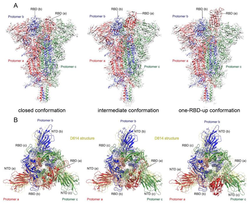
Fig. 2. Cryo-EM structures of the full-length SARS-CoV-2 S protein carrying G614. (A) Three structures of
the G614 S trimer, representing a closed, three RBD-down conformation, an RBD-intermediate conformation
and a one RBD-up conformation, were modeled based on corresponding cryo-EM density maps at 3.1-3.5Å
resolution. Three protomers (a, b, c) are colored in red, blue and green, respectively. RBD locations are
indicated. (B) Top views of superposition of three structures of the G614 S in (A) in ribbon representation with
the structure of the prefusion trimer of the D614 S (PDB ID: 6XR8), shown in yellow. NTD and RBD of each
protomer are indicated. Side views of the superposition are shown in fig. S8.
First release: 16 March 2021 www.sciencemag.org (Page numbers not final at time of first release) 8Downloaded from http://science.sciencemag.org/ on May 18, 2021 Fig. 3. Cryo-EM structures of the full-length SARS-CoV-2 S protein carrying G614. (A) Top, the structure of the closed, three RBD-down conformation of the D614 S trimer is shown in ribbon diagram with one protomer colored as NTD in blue, RBD in cyan, CTD1 in green, CTD2 in light green, S2 in light blue, the 630 loop in red and the FPPR in magenta. Bottom, structures of three segments (residues 617-644) containing the 630 loop in red and three segments (residues 823-862) containing the FPPR in magenta from all three protomers (a, b and c) are shown. Position of each RBD is indicated. (B to D) Structures of the G614 trimer in the closed, three RBD-down conformation, the RBD-intermediate conformation and the one RBD-up conformation, respectively, are shown as in (A). Dash lines indicate gaps. First release: 16 March 2021 www.sciencemag.org (Page numbers not final at time of first release) 9
Downloaded from http://science.sciencemag.org/ on May 18, 2021
                        Fig. 4. Close-up views of the D614G substitution. (A) A close-up view of
                        the region near the residue 614 with superposition of the G614 trimer
                        structure in green (CTD2) and magenta (FPPR) and the D614 trimer in
                        yellow, both in the closed prefusion conformation. Residues G614, D614 and
                        two K854 from both structures are shown in stick model. The direction of
                        the three-fold axis of the trimer is indicated. (B) Location of the 630 loop in
                        the S trimer. The 630 loop is highlighted in red, NTD in blue, CTD1 in green,
                        CTD2 in light green, S2 in light blue, and the FPPR from a neighboring
                        protomer in magenta. The S1/S2 boundary and the nearest ordered
                        residues Thr676 from S1 and S689 from S2 are all indicated. A strand from
                        the N-terminal end of S2, packed in the CTD2, is highlighted in purple. (C) A
                        view showing that the 630 loop wedges between the NTD and the CTD1 and
                        pushed them apart. (D) Packing of the 630 loop against the hydrophobic
                        surface formed by residues Val595, Val597, Val610 Tyr 612, Val642 and Ile
                        651 from the CTD2 and Pro295 from the NTD. Residues Trp633 and Val635
                        from the 630 loop contribute to this interaction.

First release: 16 March 2021                         www.sciencemag.org      (Page numbers not final at time of first release) 10
Structural impact on SARS-CoV-2 spike protein by D614G substitution
Jun Zhang, Yongfei Cai, Tianshu Xiao, Jianming Lu, Hanqin Peng, Sarah M. Sterling, Richard M. Walsh Jr., Sophia Rits-Volloch,
Haisun Zhu, Alec N. Woosley, Wei Yang, Piotr Sliz and Bing Chen
published online March 16, 2021
Downloaded from http://science.sciencemag.org/ on May 18, 2021
ARTICLE TOOLS http://science.sciencemag.org/content/early/2021/03/16/science.abf2303
SUPPLEMENTARY http://science.sciencemag.org/content/suppl/2021/03/16/science.abf2303.DC1
MATERIALS
RELATED http://stm.sciencemag.org/content/scitransmed/13/590/eabf7517.full
CONTENT
http://stm.sciencemag.org/content/scitransmed/13/578/eabd6990.full
http://stm.sciencemag.org/content/scitransmed/13/577/eabd2223.full
http://stm.sciencemag.org/content/scitransmed/13/577/eabf1555.full
http://science.sciencemag.org/content/sci/372/6541/466.full
REFERENCES This article cites 56 articles, 19 of which you can access for free
http://science.sciencemag.org/content/early/2021/03/16/science.abf2303#BIBL
PERMISSIONS http://www.sciencemag.org/help/reprints-and-permissions
Use of this article is subject to the Terms of Service
Science (print ISSN 0036-8075; online ISSN 1095-9203) is published by the American Association for the Advancement of
Science, 1200 New York Avenue NW, Washington, DC 20005. The title Science is a registered trademark of AAAS.
Copyright © 2021 The Authors, some rights reserved; exclusive licensee American Association for the Advancement of Science.
No claim to original U.S. Government Works. Distributed under a Creative Commons Attribution License 4.0 (CC BY).You can also read