Intrinsically Disordered Proteins at the Nano-scale - arXiv
←
→
Page content transcription
If your browser does not render page correctly, please read the page content below
Intrinsically Disordered Proteins at the Nano-scale
                                                                T. Ehm1,2 , H. Shinar1 , S. Meir1 , A. Sekhon1 , V. Sethi1 ,
arXiv:2101.06902v1 [physics.bio-ph] 18 Jan 2021

                                                                I. L. Morgan3 , G. Rahamim1 , O. A. Saleh3,4 and R. Beck1
                                                                1
                                                                  The School of Physics and Astronomy, The Center for Nanoscience and
                                                                Nanotechnology, and the Center for Physics and Chemistry of Living Systems, Tel
                                                                Aviv University, Israel
                                                                2
                                                                  Faculty of Physics and Center for NanoScience, Ludwig-Maximilians-Universität,
                                                                München, Germany
                                                                3
                                                                  BMSE Program, University of California, Santa Barbara CA 93110
                                                                4
                                                                  Materials Department, University of California, Santa Barbara CA 93110
                                                                E-mail: roy@tauex.tau.ac.il

                                                                Abstract.
                                                                    The human proteome is enriched in proteins that do not fold into a stable 3D
                                                                structure. These intrinsically disordered proteins (IDPs) spontaneously fluctuate
                                                                between a large number of configurations in their native form. Remarkably, the
                                                                disorder does not lead to dysfunction as with denatured folded proteins. In fact, unlike
                                                                denatured proteins, recent evidences strongly suggest that multiple biological functions
                                                                stem from such structural plasticity. Here, focusing on the nanoscopic length-scale, we
                                                                review the latest advances in IDP research and discuss some of the future directions
                                                                in this highly promising field.

                                                  Keywords: Intrinsically disordered proteins, nanoscopic characterization, single-
                                                  molecule force spectroscopy, SAXS, FRET, NMR, FCS

                                                  1. Introduction

                                                  The conventional paradigm in structural biology draws a direct connection between the
                                                  amino-acid sequence of a protein to a singular 3D structure. The unique structure
                                                  is considered essential to the protein’s biological function by permitting only specific
                                                  interactions.
                                                       In the last two decades it has been recognized that up to 40-50% of the proteome
                                                  does not fit this simplified convention (Fig. 1). Instead, intrinsically disordered proteins
                                                  and regions (IDP/IDR) fluctuate between a large number of conformations while still
                                                  retaining their biological functions [1–5]. An IDR is usually defined as an unstructured
                                                  amino-acid stretch as part of a (folded) protein, and an IDP as a complete protein that
                                                  does not fold to a stable 3D structure [2–5]. For brevity, we will also use the IDP term
                                                  to describe proteins having IDRs.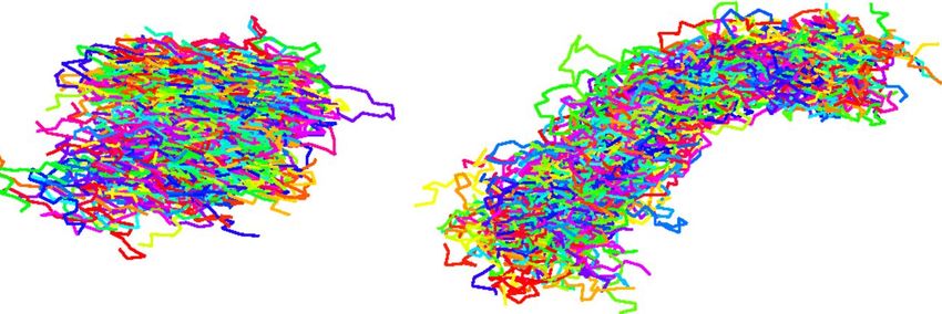
Intrinsically disordered proteins                                                                      2

      Sequence

                                    Folded                               Disordered

                                      Order-to-Disorder

     (un)Structure
                                         Transition

       Function
                                                          Liquid Phase                  Passivation
                       Lock & Key        Master-key                       Aggregation
                                                           Separation                   & Filtration

              Figure 1. Contemporary sequence-function paradigm. The folded and disordered
              conformations, and the transition between the two, lead to biological function.

     Typically, most IDP sequences are rich in structure-breaking charged and polar
amino acids, and depleted in order-promoting hydrophobic residues (Fig. 2, and
Refs. [2, 4–8]). Indeed, computer-based methods exploit the amino-acid propensity and
sequence as a sign of a disorder [9]. Generally, IDPs fall into three distinct compositional
classes that reflect the fraction of charged versus polar residues: polar, polyampholytes,
and polyelectrolytes [10, 11]. In addition, the balance between solvent mediated
intra-chain attraction and repulsion directly influences the IDP’s compactness. The
compactness, in-turn, determines the accessibility to interact with other biomolecules.
Therefore, modeling the inter- and intra-molecular IDP interactions, as well as the
resulting ensemble of conformations, is an extremely complex and relevant problem in
the research of nano-scale systems.
     It is possible to characterize the IDP ensemble average parameters such as the
end-to-end distance (Ree ) or the radius of gyration (RG ) distributions. In addition,
the IDP length, charged amino-acid distribution, and propensity to form transient
bridges, dictate the ensemble physical properties and can be connected to polymer
physics theories. This could lead to new language that relates the biological function
to basic physical parameters. Thus, IDP research has the opportunity to gain a novel
insight into of the biological sequence-to-function problem (Fig. 1).

2. Biological function

The structural plasticity of IDPs is the key to their function. This plasticity, almost
by definition, limits the strength of the IDPs’ interactions with other biomolecules and
the environment. Interactions larger than the thermal energy would ultimately lead to
a specific configuration, which conflicts with the IDPs’ large ensemble of conformations
[13, 14]. Therefore, we must ask ourselves: does structural plasticity compete with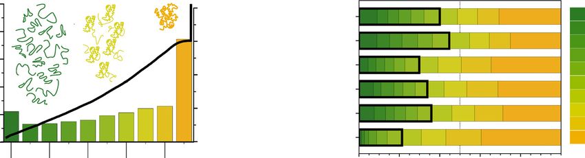
Intrinsically disordered proteins 3
0.6 1.00
Disordered Disordered
0.75 Hydrophilic
0.5
Mean absolute change
Disorder Propensity
Hydrophobic
0.50 Negative
0.4 Positive
0.25
0.3
0.0
0.2
-0.25
0.1
-0.50
(A) (B)
-0.75
0.2 0.3 0.4 0.5 0.6
Mean hydrophobicity Amino Acids
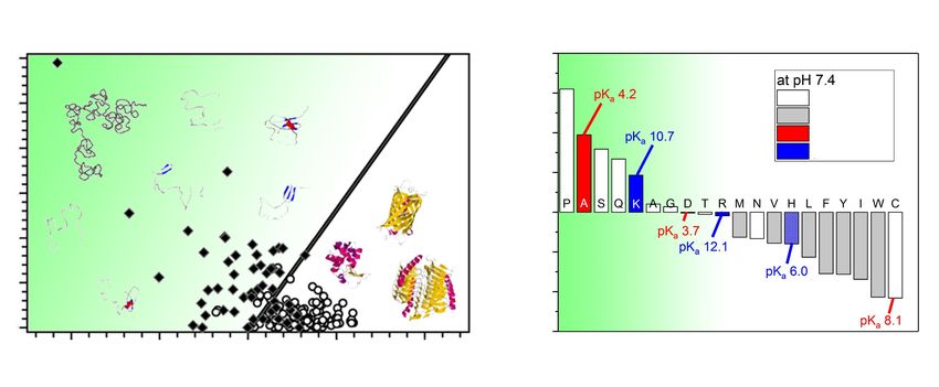
Figure 2. The role of charge, hydrophobicity and amino acid residue in IDPs.
(A) Nearly 250 folded (open circles) and nearly 90 natively unfolded proteins (black
diamonds) demonstrate that IDPs are charged and hydrophilic. The solid line
empirically separates between IDPs and compact globular proteins. Panel is adapted
from [5]. (B) The contribution of each amino acid in promoting disorder. Disordered
propensity is evaluated from the fractional difference of amino acids composition of
IDPs in the DisProt database and a completely ordered proteins from the protein-
database (PDB). Panel is adapted from [12].
.
specificity?
Distinct from many structural proteins, IDPs can interact with multiple different
partners. In analogy to the lock-and-key concept for structural proteins, IDPs serve as
“master-keys”, each capable of openning many different “locks” [1,3,7]. However, given
IDP’s prevalence, several other functions have been reported including cellular signaling
pathways, a regulator for protein-protein and protein-DNA networks or folding into
ordered structures upon binding to other cellular counterparts, to name a few (Fig.
1) [15–18].
In several cases disorder-to-order transition of IDPs is the ensemble’s conforma-
tional response to various stimuli. Such response can lead to collapse or expansion
of the IDP, e.g. globule-to-coil or collapse transitions [7, 10, 19]. Recently, high-speed
AFM measurements demonstrated constantly folded and disordered regions as well as
disorder-to-order transitions in IDPs [19].
Unraveling the factors governing the IDPs’ ensemble conformation and their
disorder-to-order transition is crucial to understand their biological functionalities [20].
An illustrative example is the regulation of glucose homeostasis by human pancreatic
glucokinase (GCK) enzyme. GCK catalyzes glucose catabolism in the pancreas. At low
glucose concentration, IDR of GCK associates with glucose and undergoes a disordered-
to-ordered transition. Following glucose release, the IDR undergoes order-to-disorder
transition on millisecond time-scale. This “time delay loop” allows slow turnover and
kinetic cooperativity of the enzyme. At high glucose concentration, the delay loop is
bypassed and excess glucose is catabolized [21].Opinion Trends in Biochemical Sciences September 2015, Vol. 40, No. 9
Intrinsically disordered proteins 4
(A) (B) (C) Disordered Structured
Cumula!ve distribu!on func!on
1.0 0.4 100
Druggable Disease* Human genome
Frac!on of human genome
(3734 ) (7508) 90
0.8
Disorder content (%)
3208 0.3 Cancer 80
70
1751 1187 0.6 Cardiovascular 60
487 2626 0.2 diseases
50
0.4 Diabetes 40
309
0.1 30
4639 0.2 Neurodegenera!on 20
10
0.0 0.0 Druggable genome 0
Disordered 80–90 60–70 40–50 20–30 0–10
(8061) 90–100 70–80 50–60 30–40 10–20 0 0.2 0.4 0.6 0.8 1
Disorder content (%) Cumula!ve protein frac!on
Ti BS
Figure 3. Disordered proteins links to human diseases. (A) Venn diagram of
Figure 1. Prevalence of protein disorder in some common human diseases. (A) Venn diagram of three subsets of the human proteome. Proteins are defined as ‘disordered’
if they contain more than 40% of their residues in regions of at least 40 consecutive disordered amino acids, as ‘druggable’ if they are known or predicted to interact with
three types of proteins: Those that interact with drugs; those that are related to
drugs [1], and as ‘disease-related’ or ‘disease-modifying’ (disease*) if they are involved in cancer, diabetes, neurodegeneration, or cardiovascular diseases (proteins in
these groups were determined with a keyword method adapted from [55,56]). (B) Fraction of proteins encoded by the human genome (right axis) binned according to their
disease; and those that are disordered. (B) Fraction of humans proteins (right axis)
content of structural disorder (x-axis). Green bins represent highly disordered proteins, and orange bins structured ones. The black line is the cumulative distribution
binned according to their content of structural disorder. Black line is the cumulative
function (left axis). Cartoons illustrate ensembles of three proteins with varying disorder content. (C) Comparison of the amount of protein disorder encoded by the human
genome, by the druggable genome, and in disease-related proteins. Proteins are binned horizontally by disordered content (colour bar). Black boxes represent the fraction
distribution function (left axis). (C) Amount of disorder in different protein categories.
of disordered proteins as defined in (A). The analysis of disorder was performed using the s2D method [10]; an individual residue was considered disordered if its a-helical
and b-strand populations are smaller than 0.5.
Figure adapted from Ref. [22].
has entered clinical use [17–19]. A recently proposed ap- difference can be expressed as the sum of enthalpic and
proach to obtain drugs targeting disordered regions relies on entropic contributions (Equation 1):
Obviously,
the computational the offunction
docking of IDP
small-molecule fragmentsis driven by their amino-acid sequence with
DG ¼ DH " TDS [1]
against an ensemble
mutations resultingof representative
in numerous conformations
diseasesof(Fig. 3). Known examples are the disordered
the protein of interest [20]. Its application to a-synuclein, where the change in enthalpy (DH) is determined by a
a regions
disorderedfound in the inAlzheimer
protein involved and Parkinson
Parkinson’s disease, iden- associated
variety of interatomicproteins Amyloid-β
forces, including and α-
electrostatic, van
tified a compound that inhibits the aggregation of a-synu- der Waals, and hydrogen-bonding interactions, and the
synuclein, that can form toxic oligomers, amyloid
clein [20]. However, it is still poorly understood whether this
fibrils, and other types of aggregates
entropic contribution DS represents the change in the size
[23,24].binds
compound Othermorediseases,
preferentiallysuch as the recently
the monomeric protein emerged novel coronavirus
of the conformational space available(SARS-CoV-19),
to the overall system,
than its aggregated species. A clearer example of direct including the protein, ligand, and solvent molecules.
are intimately
targeting of monomeric related to protein
disordered proteins isdisorder.
the case of The SARS-CoV-19
Enthalpic and entropic genome sequences
factors can either encode
contribute
the oncoprotein c-Myc [21–23]. A recent high-throughput favourably
an IDR in its nucleocapsid, an essential structural component that binds or unfavourably to DG, resulting
to RNA in the
andfour
screening yielded a series of compounds, which interact with possible modes: (i) DH>0, DSIntrinsically disordered proteins 5
[39, 40].
Recently, much attention has been drawn to membraneless organelles, which are
liquid-like condensates formed reversibly by dynamic self-assembly of proteins, mostly
IDPs, and RNA through a liquid-liquid phase separation (LLPS) [14, 16, 41]. This
behavior has raised many questions: Does the high density within such condensates
enable certain protein functions? Is it the proteins’ way of defending themselves against
degradation? Or is LLPS just an unwanted outcome of the IDPs’ sequence, which leads
to transient interactions with multiple partners? The answers to this questions are most
likely different for alternative organelles compositions.
There is little understanding of the IDP’s sequence-encoded mechanism that drives
this phase separation. Regardless, membraneless organelles constituents often contain
multiple repetitive sequences, facilitating multivalent, weak interactions with their
partners to form the condensates. As mentioned, such interactions are frequent for
IDPs. The size of membranelss organelles are in the range of sub µm to 10 µm and
thus can be observed with optical microscopy. For example, the disordered regions of
Ddx4 [42], LAF-1 [43], FUS [44], which are rich in glycine, form droplets of 1µm.
Even folded proteins that have IDRs, such as hnRNPA1 [45] and TDP-43 [46],
form droplets, designated as stress granules [47]. Assembly and disassembly of these
granules is a highly regulated process involving the IDR interactions with multiple
proteins and mRNAs. Recently, using multi-bait engineered ascorbate peroxidase
(APEX) proximity labeling technique, over a hundred proteins were discovered that
either promote assembly or induced disassembly of stress granules [16]. Importantly,
APEX revealed that most of these proteins are indeed disordered. Such studies are
extremely relevant for in-depth understanding of the compositional changes in stress
granules’ proteins in neurodegenerative disorders.
3. Artificial IDPs and polymer physics
Disordered proteins’ ability to engage in a variety of manners often results in a rich
ensemble of phase transition behaviors [48, 49]. These phase behaviors can be easily
manipulated by slightly changing the physio-chemical properties of the IDPs. Recently,
the Chilkoti group studied a simplified version of artificial IDPs, all made of a repetitive
8-mer peptide sequence originated from Drosophilia melanogaster Rec-1 resilin [50].
Interestingly, changing a single amino acid in the repeat, or increasing the number
of repeats, results in a drastic and robust change in the LLPS temperature. The
IDP sequence also controls the temperature-ramp hysteresis phenomena. Notably, this
hysteresis changes when reversing the amino acid sequence originating from N- to C-
terminal [51]. These findings demonstrate the ability to fine-tune the phase behaviors
and indicates to the significance of IDP’s sequence, as in structural proteins.
Given IDPs’ structural plasticity, it is tempting to investigate them as polymers,
at least to first order (Fig. 4). By doing so, the power of statistical-mechanics can be
harnessed to quantify the volumetric dimension of IDPs. Then, coarse-grained structuralIntrinsically disordered proteins 6
A B NUS Native C NUS Denatured
Rg
Ree
Figure 4. (A) Ensemble structural characterization of IDP that includes the end
to end distance (Ree ), typically measured by FRET, and radius of gyration (Rg ),
measured by SAXS. Representative ensembles for NUS protein in its (B) native
condition and (C) in denatured condition. Both ensembles recapture FRET and SAXS
data at these conditions and show larger asphericity in denatured conditions. Panels
(B) and (C) are adapted from Ref. [52].
descriptors are obtained from the scaling laws of polymer physics [53]. Specifically, the
degree of IDP collapse or expansion can be quantified in terms of chain’s length and
its relation to the ensemble’s radius of gyration: Rg ∝ N ν . Here, N is the number of
monomers, and ν is the Flory scaling exponent [6, 53].
Qualitatively, ν determines the balance between intra-chain and solvent-chain
interactions, also referred to as solvent quality. For good (ν ≈ 0.6), θ (ν=0.5), or poor
(ν ≈ 0.3) solvents the conditions are respectively less, equal or favorable intra-chain
interactions respective to solvent/chain ones. Thus, the value of the scaling exponent
ν can act as a physical descriptor for the collapse transition as a function of different
environmental conditions [54]. In the following, we will introduce several techniques
targeting the nanoscopic dimensions of IDPs.
4. Nanoscopic techniques
The detection and characterization of disordered proteins and regions are of
great interest in order to determine their function. IDPs’ structural plasticity
dictate techniques and analyses targeting representative parameters of the protein’s
conformational ensemble [55]. Naturally, we will not be able to discuss the entire breadth
of techniques; instead, we will focus on some of those targeting the nano-scale regime,
which is, in most cases, the relevant length-scale.
4.1. Simulations
To further understand the conformational dynamics of IDPs, advanced molecular
dynamics (MD) simulations are used. However, the inherently large number of degrees-
of-freedom, the inaccuracies in the simulation models, and the need for long simulations
have stymied progress. To overcome these limitations, alternative approaches haveIntrinsically disordered proteins 7
been developed. For example, a hierarchical approach [56] based on the “flexible-
meccano” model [57] is generating representative and meaningful starting ensembles.
Alternatively, in the ”sample-and-select” approach, sub ensembles are derived from a
broader distribution of molecular conformations based on experimental NMR and small-
angle X-ray scattering (SAXS) data [58–61].
A recent computational work [62] developed a related strategy: experimental SAXS
data on a few select IDPs was used, through a fitting approach, to develop a set of
sequence-specific residue interaction parameters in a coarse-grained simulation. These
parameters were then used to investigate the conformational ensembles of a range
of IDPs. The authors found the IDPs displayed apparent polymeric (swollen chain,
ν = 0.6) behavior when considering Rg vs. N . Yet, the simulated structural ensembles
showed significant heterogeneity: certain IDPs showed elevated intra-segment contacts
and transient sub-segment compactions. However, this was not universal: other IDPs
in the study behaved as homogeneous random-walk chains. The authors concluded that
sequence-specific structure could be hidden behind polymeric exponents [62].
4.2. SAXS
A common and powerful technique suited for IDP structural characterization is SAXS.
The technique’s key advantage is measuring the ensemble conformations while kept in
solution without the need for crystallization or external tagging. A simple SAXS analysis
immediately provides Rg and the pair distance distribution [63, 64]. Additionally,
traditional Kratky plots (q 2 I(q) vs. q), directly obtained from the SAXS profiles of
IDPs, provide a qualitative picture of the presence of globular or unfolded conformations
[65, 66]. Here, I is the scattering intensity, and q is the scattered wave-vector
proportional to the scattering angle.
Moreover, the scattering profile can be used to extract the Flory exponent (ν)
mentioned above [52, 67]. More advanced analysis techniques are regularly used to
extract the ensemble’s dominant conformations from the SAXS signal (Fig. 4). The
ensemble optimization method [68, 69], and molecular form-factor [54] are just a few
examples for such techniques resulting in representative conformations that fit the
experimental data. We note that the combination of SAXS with other complementary
methods (Fig. 4) such as simulations, NMR, and Förster resonance energy transfer
(FRET) provides a more comprehensive picture of IDPs, and their conformations in
solution [6, 37, 52, 55, 63, 70, 71].
4.3. FRET
The distance-dependent dipole-dipole interactions between probes bound to the side
chains of IDPs provide the basis for determining long-range intra-molecular distances
between selected sites. Experiments based on single-molecule [72] or ensemble [73]
measurements are characterized by 1 − 10 nm distance resolution range and an ability
to recover distributions of intra-molecular distances in the transient ensembles of IDP.Intrinsically disordered proteins 8
For example, it was shown that α-synuclein deviates from ideal chain behavior at
segments labeled in the NAC domain and N-termini using the ensemble method. It was
suggested that those conformations bias might be related to the initiation of amyloid
transition [73]. Another example, using a single-molecule method, showed that the
previously mentioned scaling exponents (ν) in water strongly depend on the sequence
composition. Two of the total examined IDPs did not reach the θ-point under any
solvent conditions. This may reflect their biological functional need for an expanded
state optimized for interactions with cellular partners [72].
4.4. NMR
NMR spectroscopy offers a unique platform in deciphering IDP dynamics and structure
[74]. The traditional NMR hydrogen chemical shift signal is, in many cases, insufficient
for IDP characterization. Fortunately, new tools have been developed to overcome low
signal to noise ratio difficulties such as paramagnetic relaxation enhancement (PRE) [75]
and proline based 2D H-N residue correlations [76].
In PRE, the introduction of paramagnetic spin labels in proteins affects the chemical
shift and transverse relaxation rate signal between the unpaired electron and NMR active
nuclei on the basis of distance between them [75]. For example, the PRE signal and 15 N
relaxation data was analyzed to quantify the interaction between the IDP osteopontin
and heparin [77]. There, it was found that on binding with heparin, osteopontin largely
remains in a disordered state and undergoes structural/dynamical adaption which is
mainly mediated by electrostatic interactions.
A recent development using in-cell NMR spectroscopy provides the opportunity
to explore the structural plasticity of an IDP in its native environment [78, 79]. Both
eukaryotic and prokaryotic IDPs have been investigated using in-cell NMR such as α-
synuclein, prokaryotic ubiquitin-like protein, Pup, tubulin-related neuronal protein, tau,
FG-Nups in nucleoporins, and the negative regulator of flagellin synthesis, FlgM. These
studies revealed that the cellular conformational dynamics may differ significantly from
these observed in-vitro [80].
4.5. FCS
Due to diffusion and the protein structural plasticity, the emission of fluorescently-
labeled molecules fluctuate. In fluorescence correlation spectroscopy (FCS), these
fluctuations are recorded within an illuminated confocal volume [81–83]. The timescale
for conformational fluctuation (i.e., chemical kinetics within the biomolecule) lies in the
nano- to microseconds range, whereas for translational diffusion from microsecond to
milliseconds. Information about the diffusion coefficient and chemical kinetics can be
inferred from the fluorescence fluctuations autocorrelation and cross-correlation signal.
Furthermore, from the diffusion coefficient, it is possible to determine the molecular size
and hydrodynamic radius of the investigated biomolecules.Intrinsically disordered proteins 9
A f B C
Single
NFL tail
Foldons
L NFL tail
polymer
Figure 5. Mechanical perturbation reveals subtle internal IDP structure. (A) Under
constant tension f , the length L of a polymer of NFL tails is measured. (B) A single
NFL tail contains multiple, independent regions with internal structure (foldons).
(C) An initial large tension breaks apart internal structure in the NFL tails. After
rapidly lowering the tension, at constant tension, the NFL polymer shows a logarithmic
decrease in length over time t, indicating multiple regions of internal structure are
reforming. Panel (C) is adapted from Ref. [86].
FCS is also sensitive to fluorophore quenching reactions. These measurements
can provide information about the internal dynamics of the IDPs [84]. Fluorescence
self-quenching of tetramethyl rhodamine, which is chemically labelled at two different
residual positions, was analyzed to study the conformational kinetics of unfolded
intestinal fatty acid binding protein [85] and amyloid forming yeast prion protein,
Sup35 [84].
4.6. Single-molecule force spectroscopy
Single-molecule force spectroscopy (SMFS) uses the thermodynamic effects of applied
tension to gain insight into the nanoscopic conformation of IDPs. SMFS consists of
tethering an IDP between a static surface and a force probe and measuring the chain
extension, with nanometer accuracy, as a function of force. This nanomechanical assay is
a high-precision differential measurement of the effect of varying perturbations (tensions)
on IDP interactions and conformations. For example, AFM-based experiments on
several amyloid precursor IDPs show a sawtooth pattern that indicates the mechanical
unfolding of multiple different conformations [87]. Similarly, optical tweezer experiments
on α-synuclein show it has several marginally stable and rapidly fluctuating subsegments
[88].
A noteworthy recent work demonstrated the power of SMFS-based perturbation
in revealing more subtle IDP structural behaviors [86]. In the work, a polyprotein
of the disordered Neurofilament light-chain tail (NFLt) domain was subjected to force-
quench experiments, in which a large tension pulled the chain straight, and was followed
by a sudden jump to a small tension that permitted structure in the IDP to re-form
(Fig. 5). The experiment was carried out in a magnetic tweezer, an SMFS instrumentIntrinsically disordered proteins 10
that offers high-stability in applied force, allowing the tracking of NFLt dynamics over
long periods of time. The experiment revealed a glassy, non-exponential relaxation in
which the chain extension decreased logarithmically with time for many minutes. This
behavior, extraordinary for its slow speed, was attributed to multiple, small collapse
events within a single NFLt domain, with each collapse changing chain extension by
only ≈ 1 nm. This experiment highlights both the high sensitivity of SMFS, as well as
the rich and physically-complex structural dynamics of IDPs.
4.7. Quantifying IDP’s interactions
Intermolecular interactions of biomolecules play an essential role in the proper function
and growth of biological cells [89–91]. These interactions can be specific or non-specific.
Non-specific interactions are transient and weak, mainly governed by steric repulsion or
ionic bridging. IDPs’ function is largely dominated by these weak interactions. This
was demonstrated, for example, in a recent AFM-based SMFS study, in which the
interactions between nuclear transport factors and disordered FG repeats was found
to display unexpected complexities due to weak, multivalent contacts between the
interacting partners [92].
Improved protein-protein interaction resolution can be achieved using bulk, low-
throughput spectroscopic methods such as FRET, surface plasmon resonance (SPR),
and nuclear magnetic resonance (NMR), to name only a few. For example, the
interaction between the disordered region in transcription factors Sp1 and TAF4 was
estimated by NMR to be Kd = 69µM [93]. IDP Ntr2 modulates the RNA Helicase
Brr2 with Kd = 14µM and 7µM determined by SPR and NMR, respectively [94]. SPR
was also used to detect and characterize IDP–ligand interactions with tau protein as a
model IDP [95].
Another recently introduced technique aiming to overcome the technical difficulties
to measure weak and transient protein-protein interaction is the nanoparticle mobility
assay [13]. There, nanoparticles grafted with IDPs were imaged while diffusing over a
surface grafted with a second set of IDPs. A similar approach was used to study strong
DNA-DNA interactions [96], delocalized long-range polymer-surface interactions [97]
and bulk-mediated diffusion on supported lipid-bilayers [98]. By carefully analyzing the
particles’ diffusive nature, the authors detected an altered interaction caused by a single
mutation on the polypeptide sequence [13].
5. Summary and perspective
Two decades of IDP research place them at the forefront of proteomics [1]. Apparently,
structural plasticity is valuable primarily because it enables the IDPs to adjust to their
environment. In addition, IDPs are also used in various nano-biomedical technologies.
For example, PASylation technology involves conjugating pharmacologically active
compounds with natively disordered biosynthetic polymers made of the small L-aminoIntrinsically disordered proteins 11
acids Proline, Alanine and/or Serine [99]. This technology may serve as an alternative to
PEGylation, which is currently widely used to sterically stabilized therapeutic proteins
and peptides but is often accompanied by immunogenicity and lack of biodegradability
[100, 101]. IDPs can also serve to modify the adhesion properties of surfaces. For
example, a study on barnacle adhesive IDPs on silica surfaces showed modified protein-
surface affinities, adhesive activities and kinetic adsorption rates [102].
IDPs’ large repertoire has been implicated in numerous diseases, making them
potential targets for therapeutic intervention. Disease-associated IDPs can serve
as potential targets for drugs modulating protein-protein interaction networks that
participate in both the “one to many” and “many to one” interaction [103].
The relation between IDPs and polymer- and statistical-physics is continuously
revisited. Naı̈vely, IDP function could have been understood from coarse metrics such
as the IDP’s length, total charge, net hydrophobicity, etc. However, as with folded
proteins, the sequence of amino acids, and not just the net composition, does dictate
the resulting function, at least to some extent. Yet building a coarse-grained model for
IDPs is a highly complicated and fascinating problem due to their unique properties.
The most fundamental difficulty is somewhat technical - the large ensemble of IDP
conformations is often impossible to sample with modern computers [104]. Another
problem evolves from the extended amount of electrostatic interactions that frequently
exist in IDP systems. Those interactions tend to span over a broad range of length and
time scales, and hence are hard to quantify by a finite force field [105], although new
IDP-specific force fields have been suggested [106–109]. Nevertheless, further research
is highly needed, especially since many phenomena involving IDPs are still not fully
understood [110].
Acknowledgments
This work was supported by the National Science Foundation under Grant No. 1715627,
the United States-Israel Bi-national Science Foundation under Grant No. 2016696, and
the Israel Science Foundation under Grants No. 1454/19.
References
[1] Wright P E and Dyson H J 1999 Journal of Molecular Biology 293 321–331
[2] Uversky V N 2014 Chemical Reviews 114 6557–6560
[3] Dunker A K, Lawson J D, Brown C J, Williams R M, Romero P, Oh J S, Oldfield C J, Campen
A M, Ratliff C M, Hipps K W et al. 2001 Journal of Molecular Graphics and Modelling 19
26–59
[4] Uversky V N and Dunker A K 2010 Biochimica Et Biophysica Acta-Proteins and Proteomics
1804 1231–1264
[5] Uversky V N 2011 The International Journal of Biochemistry & Cell Biology 43 1090–1103
[6] Kornreich M, Avinery R, Malka-Gibor E, Laser-Azogui A and Beck R 2015 FEBS letters 589
2464–76
[7] Uversky V N 2019 Frontiers in Physics 7 10Intrinsically disordered proteins 12
[8] Uversky V N, Gillespie J R and Fink A L 2000 Proteins: Structure, Function, and Bioinformatics
41 415–427
[9] Ferron F, Longhi S, Canard B and Karlin D 2006 Proteins: Structure, Function, and
Bioinformatics 65 1–14
[10] Das R K, Ruff K M and Pappu R V 2015 Current opinion in structural biology 32 102–112
[11] Mao A H, Lyle N and Pappu R V 2013 Biochemical Journal 449 307–318
[12] Radivojac P, Iakoucheva L M, Oldfield C J, Obradovic Z, Uversky V N and Dunker A K 2007
Biophysical Journal 92 1439–1456
[13] Chakraborty I, Rahamim G, Avinery R, Roichman Y and Beck R 2019 Nano Letters 19 6524–
6534
[14] Shin Y and Brangwynne C P 2017 Science 357
[15] Wright P E and Dyson H J 2015 Nature Reviews Molecular Cell Biology 16 18–29
[16] Marmor Kollet H, Siany A, Kedersha N, Knafo N, Rivkin N, Danino Y M, Olender T, Cohen N,
Moens T, Higginbottom A et al. 2020 Molecular Cell
[17] Dunker A K, Cortese M S, Romero P, Iakoucheva L M and Uversky V N 2005 The FEBS journal
272 5129–5148
[18] Gianni S, Dogan J and Jemth P 2016 Current Ppinion in Structural Biology 36 18–24
[19] Kodera N, Noshiro D, Dora S K, Mori T, Habchi J, Blocquel D, Gruet A, Dosnon M, Salladini
E, Bignon C et al. 2020 Nature Nanotechnology 1–9
[20] Zhou J, Oldfield C J, Yan W, Shen B and Dunker A K 2019 Protein Science 28 1652–1663
[21] Larion M, Salinas R K, Bruschweiler-Li L, Miller B G and Brüschweiler R 2012 PLoS Biology 10
e1001452
[22] Heller G T, Sormanni P and Vendruscolo M 2015 Trends in Biochemical Sciences 40 491–496
[23] Carballo-Pacheco M and Strodel B 2017 Protein Science 26 174–185
[24] Coskuner-Weber O and Uversky V N 2018 International journal of molecular sciences 19 336
[25] Ceraolo C and Giorgi F M 2020 Journal of Medical Virology 92 522–528
[26] Cubuk J, Alston J J, Incicco J J, Singh S, Stuchell-Brereton M D, Ward M D, Zimmerman M I,
Vithani N, Griffith D, Wagoner J A, Bowman G R, Hall K B, Soranno A and Holehouse A S
2020 bioRxiv
[27] Richardson L G, Jelokhani-Niaraki M and Smith M D 2009 BMC Biochemistry 10 1–8
[28] Chino H, Hatta T, Natsume T and Mizushima N 2019 Molecular Cell 74 909–921
[29] Basu S and Bahadur R P 2016 Cellular and Molecular Life Sciences 73 4075–4084
[30] Pregent S, Lichtenstein A, Avinery R, Laser-Azogui A, Patolsky F and Beck R 2015 Nano letters
15 3080–7
[31] Laser-Azogui A, Kornreich M, Malka-Gibor E and Beck R 2015 Current Opinion in Cell Biology
32 92–101
[32] Safinya C R, Deek J, Beck R, Jones J B, Leal C, Ewert K K and Li Y 2013 Liquid crystals 40
1748–1758
[33] Guharoy M, Szabo B, Martos S C, Kosol S and Tompa P 2013 Cytoskeleton 70 550–571
[34] Chung P J, Song C, Deek J, Miller H P, Li Y, Choi M C, Wilson L, Feinstein S C and Safinya
C R 2016 Nature Communications 7 12278
[35] Kornreich M, Malka-Gibor E, Zuker B, Laser-Azogui A and Beck R 2016 Physical Review Letters
117 148101
[36] Chang L and Goldman R D 2004 Nature Reviews Molecular Cell Biology 5 601–613
[37] Malka-Gibor E, Kornreich M, Laser-Azogui A, Doron O, Zingerman-Koladko I, Harapin J,
Medalia O and Beck R 2017 Biophysical Journal 112 892–900 (Preprint 1609.05546)
[38] Beck R, Deek J, Jones J B and Safinya C R 2010 Nature Materials 9 40–46
[39] Schwartz T U 2016 Journal of Molecular Biology 428 1986–2000
[40] Lemke E A 2016 Journal of Molecular Biology 428 2011–2024
[41] Brangwynne C P, Eckmann C R, Courson D S, Rybarska A, Hoege C, Gharakhani J, Julicher F
and Hyman A A 2009 Science 324 1729–1732Intrinsically disordered proteins 13
[42] Nott T J, Petsalaki E, Farber P, Jervis D, Fussner E, Plochowietz A, Craggs T D, Bazett-Jones
D P, Pawson T, Forman-Kay J D and Baldwin A J 2015 Molecular Cell 57 936–947
[43] Elbaum-Garfinkle S, Kim Y, Szczepaniak K, Chen C C H, Eckmann C R, Myong S and
Brangwynne C P 2015 Proceedings of the National Academy of Sciences 112 7189–7194
[44] Patel A, Lee H O, Jawerth L, Maharana S, Jahnel M, Hein M Y, Stoynov S, Mahamid J, Saha
S, Franzmann T M, Pozniakovski A, Poser I, Maghelli N, Royer L A, Weigert M, Myers E W,
Grill S, Drechsel D, Hyman A A and Alberti S 2015 Cell 162 1066–1077
[45] Molliex A, Temirov J, Lee J, Coughlin M, Kanagaraj A P, Kim H J, Mittag T and Taylor J P
2015 Cell 163 123–133
[46] Conicella A E, Zerze G H, Mittal J and Fawzi N L 2016 Structure 24 1537–1549
[47] Buchan J R and Parker R 2009 Molecular Cell 36 932–941
[48] Dignon G L, Zheng W, Kim Y C, Best R B and Mittal J 2018 PLoS Computational Biology 14
e1005941
[49] Zhou H X, Nguemaha V, Mazarakos K and Qin S 2018 Trends in Biochemical Sciences 43 499–516
[50] Dzuricky M, Rogers B A, Shahid A, Cremer P S and Chilkoti A 2020 Nature Chemistry 12
814–825
[51] Quiroz F G, Li N K, Roberts S, Weber P, Dzuricky M, Weitzhandler I, Yingling Y G and Chilkoti
A 2019 Science Advances 5 eaax5177
[52] Fuertes G, Banterle N, Ruff K M, Chowdhury A, Mercadante D, Koehler C, Kachala M, Girona
G E, Milles S, Mishra A et al. 2017 Proceedings of the National Academy of Sciences 114
E6342–E6351
[53] Wang Z G 2017 Macromolecules 50 9073–9114
[54] Riback J A, Bowman M A, Zmyslowski A M, Knoverek C R, Jumper J M, Hinshaw J R, Kaye
E B, Freed K F, Clark P L and Sosnick T R 2017 Science 358 238–241
[55] Lazar T, Martı́nez-Pérez E, Quaglia F, Hatos A, Chemes L B, Iserte J A, Méndez N A, Garrone
N A, Saldaño T E, Marchetti J, Rueda A J V, Bernadó P, Blackledge M, Cordeiro T N,
Fagerberg E, Forman-Kay J D, Fornasari M S, Gibson T J, Gomes G N W, Gradinaru C C,
Head-Gordon T, Jensen M R, Lemke E A, Longhi S, Marino-Buslje C, Minervini G, Mittag T,
Monzon A M, Pappu R V, Parisi G, Ricard-Blum S, Ruff K M, Salladini E, Skepö M, Svergun
D, Vallet S D, Varadi M, Tompa P, Tosatto S C E and Piovesan D 2020 Nucleic Acids Research
[56] Pietrek L M, Stelzl L S and Hummer G 2019 Journal of Chemical Theory and Computation 16
725–737
[57] Bernadó P, Bertoncini C W, Griesinger C, Zweckstetter M and Blackledge M 2005 Journal of the
American Chemical Society 127 17968–17969
[58] Jensen M R, Zweckstetter M, Huang J r and Blackledge M 2014 Chemical Reviews 114 6632–6660
[59] Fisher C K and Stultz C M 2011 Current Opinion in Structural Biology 21 426–431
[60] Mittag T and Forman-Kay J D 2007 Current Opinion in Structural Biology 17 3–14
[61] Allison J R, Varnai P, Dobson C M and Vendruscolo M 2009 Journal of the American Chemical
Society 131 18314–18326
[62] Baul U, Chakraborty D, Mugnai M L, Straub J E and Thirumalai D 2019 The Journal of Physical
Chemistry B 123 3462–3474
[63] Kornreich M, Avinery R and Beck R 2013 Current Opinion in Biotechnology 24 716–723
[64] Manalastas-Cantos K, Konarev P, Hajizadeh N R, Kikhney A, Petoukhov M, Molodenskiy D,
Panjkovich A, Mertens H, Gruzinov A, Borges C et al. 2020 Journal of Applied Crystallography
54
[65] Bernadó P and Svergun D I 2012 Molecular Biosystems 8 151–167
[66] Almagor L, Avinery R, Hirsch J A and Beck R 2013 Biophysical Journal 104 2392–2400
[67] Hammouda B 2012 Macromolecular Theory and Simulations 21 372–381
[68] Bernadó P, Mylonas E, Petoukhov M V, Blackledge M and Svergun D I 2007 Journal of the
American Chemical Society 129 5656–5664
[69] Tria G, Mertens H D, Kachala M and Svergun D I 2015 International Union of CrystallographyIntrinsically disordered proteins 14
Journal 2 207–217
[70] Storm I M, Kornreich M, Hernandez-Garcia A, Voets I K, Beck R, Cohen Stuart M A, Leermakers
F A and De Vries R 2015 The Journal of Physical Chemistry B 119 4084–4092
[71] Storm I M, Kornreich M, Voets I K, Beck R, de Vries R, Stuart M A C and Leermakers F A 2016
Soft Matter 12 8004–8014
[72] Hofmann H, Soranno A, Borgia A, Gast K, Nettels D and Schuler B 2012 Proceedings of the
National Academy of Sciences of the United States of America 109 16155–60
[73] Grupi A and Haas E 2011 Journal of Molecular Biology 405 1267–1283
[74] Konrat R 2014 Journal of Magnetic Resonance 241 74–85
[75] Otting G 2010 Annual review of biophysics 39 387–405
[76] Solyom Z, Schwarten M, Geist L, Konrat R, Willbold D and Brutscher B 2013 Journal of
Biomolecular NMR 55 311–321
[77] Platzer G, Schedlbauer A, Chemelli A, Ozdowy P, Coudevylle N, Auer R, Kontaxis G, Hartl M,
Miles A J, Wallace B A et al. 2011 Biochemistry 50 6113–6124
[78] Luchinat E and Banci L 2016 Journal of Biological Chemistry 291 3776–3784
[79] Barbieri L, Luchinat E and Banci L 2016 Nature Protocols 11 1101–1111
[80] Sciolino N, Burz D S and Shekhtman A 2019 Proteomics 19 1800055
[81] Magde D, Elson E and Webb W W 1972 Physical Review Letters 29 705
[82] Elson E L and Magde D 1974 Biopolymers: Original Research on Biomolecules 13 1–27
[83] Elson E L 2011 Biophysical Journal 101 2855–2870
[84] Mukhopadhyay S, Krishnan R, Lemke E A, Lindquist S and Deniz A A 2007 Proceedings of the
National Academy of Sciences 104 2649–2654
[85] Chattopadhyay K, Elson E L and Frieden C 2005 Proceedings of the National Academy of Sciences
102 2385–2389
[86] Morgan I L, Avinery R, Beck R and Saleh O A 2020 Biophysical Journal 118 337a–338a
[87] Hervas R, Oroz J, Galera-Prat A, Goni O, Valbuena A, Vera A M, Gomez-Sicilia A, Losada-
Urzaiz F, Uversky V N, Menendez M, Laurents D V, Bruix M and Carrion-Vazquez M
2012 PLoS Biol 10 e1001335 ISSN 1545-7885 (Electronic) 1544-9173 (Linking) URL https:
//www.ncbi.nlm.nih.gov/pubmed/22666178
[88] Solanki A, Neupane K and Woodside M T 2014 Phys Rev Lett 112 158103 ISSN 1079-7114
(Electronic) 0031-9007 (Linking) URL https://www.ncbi.nlm.nih.gov/pubmed/24785077
[89] Leckband D and Israelachvili J 2001 Quarterly Reviews of Biophysics 34 105
[90] Wyttenbach T and Bowers M T 2007 Annu. Rev. Phys. Chem. 58 511–533
[91] Musiani F and Giorgetti A 2017 Chapter two - protein aggregation and molecular crowding:
Perspectives from multiscale simulations Early Stage Protein Misfolding and Amyloid
Aggregation (International Review of Cell and Molecular Biology vol 329) ed Sandal M
(Academic Press) pp 49 – 77
[92] Hayama R, Sorci M, Keating IV J J, Hecht L M, Plawsky J L, Belfort G, Chait B T and Rout
M P 2019 PLOS ONE 14 e0217897
[93] Hibino E, Inoue R, Sugiyama M, Kuwahara J, Matsuzaki K and Hoshino M 2016 Protein Science
25 2006–2017
[94] Wollenhaupt J, Henning L M, Sticht J, Becke C, Freund C, Santos K F and Wahl M C 2018
Biophysical Journal 114 788–799
[95] Vagrys D, Davidson J, Chen I, Hubbard R E and Davis B 2020 International Journal of Molecular
Sciences 21 5257
[96] Xu Q, Feng L, Sha R, Seeman N C and Chaikin P M 2011 Physical Review Letters 106 228102
[97] Skaug M J, Mabry J N and Schwartz D K 2014 Journal of the American Chemical Society 136
1327–1332
[98] Yoo J, Lee T S, Choi B, Shon M J and Yoon T Y 2016 Journal of the American Chemical Society
138 14238–14241
[99] Binder U and Skerra A 2017 Current Opinion in Colloid & Interface Science 31 10–17Intrinsically disordered proteins 15
[100] Gebauer M and Skerra A 2018 Bioorganic & Medicinal Chemistry 26 2882–2887
[101] Schlapschy M, Binder U, Börger C, Theobald I, Wachinger K, Kisling S, Haller D and Skerra A
2013 Protein Engineering, Design & Selection 26 489–501
[102] Wang X, Wang C, Xu B, Wei J, Xiao Y and Huang F 2018 Applied Surface Science 427 942–949
[103] Wang J, Cao Z, Zhao L and Li S 2011 International Journal of Molecular Sciences 12 3205–3219
[104] Csizmok V, Follis A V, Kriwacki R W and Forman-Kay J D 2016 Chemical Reviews 116 6424–
6462
[105] Cragnell C, Rieloff E and Skepö M 2018 Journal of Molecular Biology 430 2478–2492
[106] Knott M and Best R B 2014 The Journal of Chemical Physics 140 05B603 1
[107] Sigalov A B 2016 Biochimie 125 112–118
[108] Zhao Y, Cortes-Huerto R, Kremer K and Rudzinski J F 2020 The Journal of Physical Chemistry
B 124 4097–4113
[109] Schuler B, Soranno A, Hofmann H and Nettels D 2016 Annual Review of Biophysics 45 207–231
[110] Bhattacharya S and Lin X 2019 Biomolecules 9 146You can also read