Naturally-occurring hotspot cancer mutations in Gα13 promote oncogenic signaling
←
→
Page content transcription
If your browser does not render page correctly, please read the page content below
JBC Papers in Press. Published on October 27, 2020 as Manuscript AC120.014698
       The latest version is at https://www.jbc.org/cgi/doi/10.1074/jbc.AC120.014698
Maziarz et al., 2020                                          GNA13 Arg‐200 oncogenic mutants

       Naturally-occurring hotspot cancer mutations in Gα13 promote
                            oncogenic signaling
     Marcin Maziarz1, Anthony Federico2, Jingyi Zhao1, Lorena Dujmusic1, Zhiming Zhao1, Stefano
                       Monti2, Xaralabos Varelas1, and Mikel Garcia-Marcos1*,
1
    Department of Biochemistry, Boston University School of Medicine, Boston, MA 02118, USA.
2
    Section of Computational Biomedicine, Boston University School of Medicine, Boston, MA 02118, USA
*
 To whom correspondence should be addressed:
Mikel Garcia-Marcos, Ph.D. Associate Professor, Department of Biochemistry, Boston University School
of Medicine; 72 E. Concord St, Silvio Conte Building (K), Room K208 (office)/ K206 (lab)
Boston, MA 02118. Tel: (617) 639-4387; Email: mgm1@bu.edu

Running Title: GNA13 Arg-200 oncogenic mutants
Keywords: G protein-coupled receptor (GPCR), heterotrimeric G protein, bladder cancer, signal

                                                                                                                Downloaded from http://www.jbc.org/ by guest on December 20, 2020
transduction, cancer biology, oncogenesis, GTPase, cell signaling.
ABSTRACT                                                RhoGEF-RhoGTPase cascade components that
        Heterotrimeric G-proteins are signaling         are upregulated in bladder cancers. Moreover,
switches broadly divided into four families             Gα13 Arg-200 mutants induced oncogenic
based on the sequence and functional similarity         transformation in vitro as determined by foci
of their Gα subunits: Gs, Gi/o, Gq/11 and G12/13.       formation assays. In summary, our findings on
Artificial mutations that activate Gα subunits of       Gα13 mutants establish that naturally-occurring
each of these families have long been known to          hotspot mutations in Gα subunits of any of the
induce      oncogenic      transformation      in       four families of heterotrimeric G-proteins are
experimental systems. With the advent of next-          putative cancer drivers.
generation sequencing, activating hotspot                        Heterotrimeric G-proteins are critical
mutations in Gs, Gi/o or Gq/11 proteins have also       transducers of signaling triggered by a large family
been identified in patient tumor samples. In            of G-protein-coupled receptors (GPCRs1).
contrast, patient tumor-associated G12/13               Essentially, GPCRs promote GTP loading on the α-
mutations characterized to date lead to                 subunits of G-proteins (1,2), which triggers
inactivation rather than activation. By using           signaling downstream. Heterotrimeric G-proteins
bioinformatic pathway analysis and signaling            are composed of a nucleotide-binding Gα subunit
assays, here we identified cancer-associated            and an obligatory Gβγ dimer, and they are classified
hotspot mutations in Arg-200 of Gα13 (encoded           into four families based on the nature of the Gα
by GNA13) as potent activators of oncogenic             subunits. These four families are Gs, Gi/o, Gq/11 and
signaling. First, we found that components of a         G12/13, and Gα subunits of each one of them have
G12/13-dependent     signaling    cascade    that       distinct actions on specific effectors. For example,
culminates in activation of the Hippo pathway           Gs members stimulate adenylyl cyclase activity,
effectors YAP and TAZ is frequently altered in          whereas Gi/o family members tend to inhibit it; Gq/11
bladder cancer. Upregulation of this signaling          members stimulate phospholipase C enzymes and a
cascade correlates with increased YAP/TAZ               subgroup of RhoGEFs; and G12/13 members
activation transcriptional signatures in this           stimulate a different subgroup of RhoGEFs (3,4).
cancer type. Among the G12/13 pathway                   Signaling is terminated upon GTP hydrolysis
alterations were mutations in Arg-200 of Gα13,          mediated by the intrinsic GTPase of Gα subunits.
which we validated to promote YAP/TAZ-                           The role of heterotrimeric G-proteins in
dependent (TEAD) and MRTF-A/B-dependent                 cancer-related signaling has been documented for
(SRE.L) transcriptional activity. We further            decades. Early studies identified cancer-associated
showed that this mechanism relies on the same           mutations in Gαs that disrupted its GTPase activity,

                                                    1
Maziarz et al., 2020                                                        GNA13 Arg‐200 oncogenic mutants

rendering the G-protein constitutively active (5).          trend. For example, mutations in Gα13 in some types
This seminal finding spurred a wave of studies              of lymphoma are frequent, but they lead to
exploring whether analogous mutations introduced            inactivation rather than activation (27,28). This
artificially in other Gα subunits would also promote        suggests that, at least in these lymphomas, Gα13
their ability to induce oncogenic transformation. It        activity is tumor suppressive. This is the opposite to
was found that GTPase-deficient mutants of most,            the oncogene function previously suggested from
if not all, Gα subunits tested led to oncogenic             experiments in vitro with a constitutively active
transformation in vitro, regardless of the G-protein        artificial Gα13 mutation (7,9).
family they belonged to. For example, Gαi2, Gαo,                      Activation of Gα12 or Gα13 proteins leads to
and Gαz (Gi/o family); Gαq (Gq/11 family); Gα12, and        activation of RhoA-dependent transcriptional
Gα13 (G12/13 family), in addition to Gαs (Gs family),       programs, including those mediated by the
all promoted oncogenic transformation as assessed           activation of the Hippo pathway effectors YAP and
by in vitro assays using fibroblasts (6-13). In most        TAZ. The cascade of events involves the direct
cases, transformation in vitro correlated well with         activation of a subgroup of RhoGEFs, composed of
tumor growth in vivo using mouse xenografts.                p115-RhoGEF, PDZ-RhoGEF and LARG, by
Thus, one theme emerging from these studies was             active, GTP-loaded Gα subunits of G12/13 proteins,
that enhancement of GPCR/G-protein signaling                which in turn activates RhoA, RhoB and RhoC

                                                                                                                     Downloaded from http://www.jbc.org/ by guest on December 20, 2020
tends to favor oncogenicity.                                GTPases (29). Through mechanisms that involve
          Despite these initial observations and the        the remodeling of the actin cytoskeleton,
identification of some mutations in G-proteins in           RhoGTPases induce transcriptional responses that
tumors (5,14), only with the recent advent of deep-         include those regulated by YAP/TAZ, which serve
sequencing techniques it has become obvious that            as co-factors for the TEA domain containing
dysregulation of the GPCR/G-protein signaling               transcription factor family (TEADs), and via
axis in cancer is highly prevalent (15-17). The             myocardin-related transcription factors A and B
mutational landscape of GPCR/G-protein signaling            (MRTF-A/B), which serve as co-activators for the
components in cancer supports the theme that G-             transcriptional factor SRF (30-36). Although the
protein hyperactivation in cancer tends to be pro-          G12/13-YAP/TAZ signaling axis has been shown to
oncogenic. There are many examples of GPCRs                 be oncogenic in some contexts (35-37), no cancer-
that are either overexpressed or that contain               associated mutation that activates Gα12 or Gα13 has
activating mutations (15,17-20), and negative               been characterized to date. Prompted by finding
regulators of G-protein activity have also been             that the G12/13-YAP/TAZ signaling axis is
shown to bear loss-of-function mutations (21). As           upregulated in bladder cancer, here we
for G-proteins themselves, it is now known that             characterized the effect of hotspot mutations in
hyperactive G-protein mutants can be very frequent          Gα13 identified in this cancer type. We found that
in certain types of cancers. The most striking              mutations in the Arg-200 of Gα13, a residue
example is uveal melanoma, in which ~90% of                 required to hydrolyze GTP, lead to activation of
tumors contain activating mutations in Gαq                  YAP/TAZ-dependent and MRTF-A/B-dependent
(encoded by GNAQ) or Gα11 (GNA11) (22,23).                  transcription through a RhoGEF-RhoGTPase
Similarly, activating mutations in Gαs (GNAS) can           cascade and that they promote oncogenic
be as frequent as 70% in certain subtypes of                transformation in vitro. This implies that naturally-
pancreatic ductal carcinomas (24,25), and                   occurring hotspot mutations in Gα subunits of any
activating mutations in Gαi2 can be as frequent as          of the four families of heterotrimeric G-proteins are
24% in epitheliotropic intestinal T-cell lymphomas          putative cancer drivers.
(26). Thus, for representative members of 3 out of
4 families of G-proteins (Gq/11, Gs and Gi/o) the
                                                            RESULTS AND DISCUSSION
oncogenic activity in vitro caused by artificially
introduced mutations has found a counterpart in             G12/13 pathway upregulation correlates with
prevalent mutations in cancer. Interestingly,               increased YAP/TAZ transcriptional activity in
findings so far suggest that the remaining family of        bladder cancer- We mined data from The Cancer
G-proteins (G12/13) might be an exception to this           Genome Atlas (TCGA) through cBioportal to
                                                            explore genomic alterations in components of a

                                                        2
Maziarz et al., 2020                                                        GNA13 Arg‐200 oncogenic mutants

G12/13-YAP/TAZ pathway (Fig. 1A). More                       transcriptional output of the downstream effectors
specifically we queried the G-proteins Gα12                  YAP/TAZ.
(GNA12) and Gα13 (GNA13), the RhoGEFs p115-
RhoGEF           (ARHGEF1),             PDZ-RhoGEF           Gα13 Arg-200 mutants induce YAP/TAZ activity
(ARHGEF11) and LARG (ARHGEF12), the                          via a RhoGEF-RhoGTPase axis- Although
RhoGTPases RhoA (RHOA), RhoB (RHOB) and                      overexpression of wild-type G12/13 family Gα
RhoC (RHOC), and the Hippo pathway effectors                 proteins has been found before to be sufficient to
YAP (YAP1) and TAZ (WWTR1). We found that                    promote transformation (7,8), a recent study also
these genes were altered in a large portion (~40%)           found that the mutation frequency of GNA13 in the
of the TCGA bladder cancers (TCGA-BLCA) (Fig.                TCGA-BLCA dataset is statistically higher than
1B). The alterations appeared to be largely mutually         background mutation frequency (q= 0.007) (42).
exclusive and trending towards upregulation. For             Moreover, the distribution of mutations across the
example, both heterotrimeric G-proteins, two of the          sequence of Gα13 suggested a hotspot at Arg-200
three RhoGEFs, and both Hippo effectors displayed            (Fig. 2A). The presence of an arginine in this
amplifications as the dominant feature. For RhoA             position is absolutely conserved across Gα subunits
(RHOA) and RhoB (RHOB) the main feature was                  (Fig. 2A) and its mutation in several other Gα
that they were mutated, and several of these                 subunits leads to increased activity and favors

                                                                                                                     Downloaded from http://www.jbc.org/ by guest on December 20, 2020
mutations are classified as putative drivers in              oncogenic transformation (5,11,13,14). From
cBioportal (38). Although not all RhoA/RhoB                  studies in other Gα proteins, it has been found that
mutations have been characterized, some of them              this arginine is crucial for GTPase activity and that
have been previously proposed to lead to signaling           it cannot be replaced by other amino acids, even if
activation, like A161 mutations in RhoA (39) or the          they preserve the positive charge of the side chain
E172K mutation in RhoB (40). Thus, although                  like in the case of lysine (5,43-45). Thus, we
LARG (ARHGEF12) and RhoC (RHOC) are                          hypothesized that Gα13 Arg-200 mutations
exceptions to the overall trend, these observations          identified in bladder cancer would similarly induce
suggest that the G12/13-YAP/TAZ pathway might be             the formation of an active G protein that increases
upregulated in bladder cancer.                               downstream signaling, including YAP/TAZ (Fig.
Motivated by these observations, we carried out a            2B). Because mutation of this arginine to any other
bioinformatic analysis of gene expression data to            residue is expected to have similar consequences
establish a potential correlation between                    (5,43), we focused our efforts on characterizing
upregulation of the G12/13 pathway and YAP/TAZ               Gα13 R200K and Gα13 R200G because these are the
activation. For this, we turned to a previously              two mutants most frequently found in bladder
characterized 24 gene signature that depends on              cancer. Before assessing the impact of these
YAP/TAZ (41), and analyzed its relationship to the           mutants in cell signaling assays, we validated that
expression levels of the rest of the upstream                they adopted an active conformation by using a
components of the proposed G12/13 pathway. We                well-validated assay that relies on protection from
used single-sample Gene Set Enrichment Analysis              trypsin hydrolysis (Fig. S1) (44,46). Next, we
(ssGSEA) to quantify relative enrichment of each             expressed Gα13 R200K and Gα13 R200G in
pathway across over 400 primary tumors in the                HEK293T cells and assessed activation of
TCGA-BLCA RNA-seq dataset. We found a strong                 YAP/TAZ using a TEAD reporter assay (Fig. 2C).
correlation between the activation scores of the             We compared the effect of expressing these two
G12/13 pathway and the activation scores for                 mutants to that of Gα13 WT as well as to that of
YAP/TAZ (Fig. 1C). We then tested the observed               Gα13 Q226L, an artificial mutant previously shown
correlation coefficient against a null distribution of       to enhance downstream signaling including
correlations between ssGSEA-quantified activity of           YAP/TAZ-dependent           TEAD      transcriptional
the G12/13 pathway and 10,000 random 24-gene                 activity (34,37). While expression of Gα13 WT led
signatures, resulting in a significant p-value of 1e-4       to a modest increase of TEAD activity, expression
(Fig. 1D). Taken together, these observations                of Gα13 R200K and Gα13 R200G led to a
indicate that upregulation of the G12/13 pathway in          significantly larger increase comparable to that
bladder cancer correlates with increased                     observed in cells expressing the control mutant
                                                             Gα13 Q226L (Fig. 2C). To determine if the

                                                         3
Maziarz et al., 2020                                                         GNA13 Arg‐200 oncogenic mutants

observed increase in TEAD activity by Gα13                   used for the vast majority of Gα oncogenic mutants
mutants was mediated by YAP/TAZ, we knocked                  reported to date as a good proxy for tumor growth
down both proteins simultaneously using a                    in mice, including for the oncogenic activity of
previously validated siRNA sequence (47,48). As              artificial activating mutations introduced in Gα13
expected, depletion of YAP and TAZ led to a large            (7). First, we assessed if Gα13 R200K and Gα13
suppression of TEAD activation by Gα13 R200K,                R200G mutants also lead to increased signaling
R200G or Q226L (Fig. 2D). To further map the                 activity in NIH3T3 cells. Surprisingly, we found
cascade of events leading to YAP/TAZ activation              that whereas Gα13 R200K and Gα13 R200G led to
by Gα13 mutants, we blocked the pathway that                 robust increases in the MRTF-A/B-dependent
putatively operates in bladder cancer at different           SRE.L reporter, they had no significant effect on
levels. First, inhibition of the RhoGTPases RhoA,            the activity of the YAP-TAZ-dependent TEAD
RhoB and RhoC by expression of Clostridium                   reporter (Fig. S2). These results confirm that Gα13
botulinum C3 toxin efficiently suppressed TEAD               R200K and Gα13 R200G behave as active G
activation by Gα13 R200K, R200G or Q226L (Fig.               proteins, but that the downstream signaling
2E). Then, we tested the effect of a fragment of             consequences are cell-type specific. Next, we
p115-RhoGEF that works as a dominant-negative                generated NIH3T3 cell lines stably expressing Gα13
by preventing the binding of active Gα13 to its target       WT, Gα13 R200K and Gα13 R200G at comparable

                                                                                                                      Downloaded from http://www.jbc.org/ by guest on December 20, 2020
RhoGEFs that operate upstream of RhoGTPases in               levels by lentiviral transduction and selection with
the pathway (49). Expression of this dominant-               the appropriate agents (Fig. 3A). Both Gα13 R200K
negative construct, consisting of p115-RhoGEF’s              and Gα13 R200G induced the formation of
RGS homology (RH) domain (p115RH), but not a                 numerous foci, whereas Gα13 WT only had a
control construct, inhibited TEAD activation by              modest effect (Fig. 3B, C).
Gα13 R200K, R200G or Q226L (Fig. 2F). To
further validate the specificity of these                    Conclusions- Recent reports have suggested that
manipulations, we tested their impact on Gα13-               mutations in Gα13 are putative oncogene drivers in
mediated activation of another transcriptional               bladder cancer based on bioinformatics predictions
output not controlled by YAP/TAZ but still                   (17,42,50), but no other experimental evidence to
dependent on RhoGTPase activation, i.e., the                 support the predictions was provided. The results
transcriptional activation of SRF via MRTF-A/B               presented here provide the missing experimental
(Fig. 2B). As expected, bladder cancer-associated            evidence that supports the idea of Gα13 hotspot
mutants Gα13 R200K and R200G led to robust                   mutations as putative drivers in bladder cancer, and
activation of the SRF reporter, comparable to that           suggest that pharmacological intervention of the
of the positive control Gα13 Q226L, which was                pathway activated downstream might be a viable
suppressed by inhibition of the activation of                therapeutic avenue. Moreover, our findings on Gα13
RhoGEFs or RhoGTPases but not upon YAP/TAZ                   mutants establish that naturally-occurring hotspot
depletion (Fig. 2G, H, I). Taken together, these             mutations in Gα subunits of any of the four families
results demonstrate that Gα13 hotspot mutations in           of heterotrimeric G-proteins, i.e., in Gs, Gi/o, Gq/11
Arg-200 found in bladder cancer are bona fide                and, now, G12/13, are putative cancer drivers,
activating mutations that lead to induction of               thereby providing closure to a long-held tenet.
YAP/TAZ-dependent transcription via a RhoGEF-
RhoGTPase cascade.                                           EXPERIMENTAL PROCEDURES
                                                             Data processing
 Gα13 Arg-200 mutants induce oncogenic                       Data for the oncoprint in Fig. 1B were obtained
transformation in vitro- Finally, we sought to               through cBioportal (38) by querying the term
determine if the Gα13 hotspot mutations in Arg-200           GNA13 on March 23rd, 2020 in the dataset “Bladder
described above would be sufficient to promote               Cancer (TCGA, Cell 2017)”. For the lollipop plot
oncogenic transformation in vitro. For this, we used         in Fig. 2A, data was obtained from all of the
foci formation assays with NIH3T3 cells. This                datasets classified as Bladder Urothelial Carcinoma
widely used system is particularly well-suited to            in cBioportal. TCGA-BLCA RNA-Seq count
analyze the putative oncogenic activity of Gα13              matrix (generated with STAR 2-Pass and HTSeq-
Arg-200 mutants because it has been previously               Counts) and available metadata was downloaded

                                                         4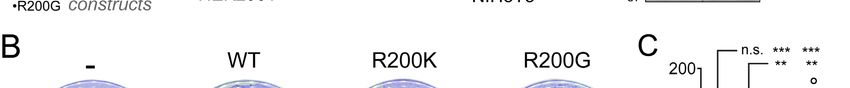
Maziarz et al., 2020                                                      GNA13 Arg‐200 oncogenic mutants

through the Genomic Data Commons gdc-client                using QuickChange II (Agilent, 200523). siRNA
(51,52). We performed a variance-stabilizing               used for knockdown of YAP and TAZ was:
transformation of the data using the R package             UGUGGAUGAGAUGGAUACA (47,48) and the
DESeq2 (v1.23.10) followed by a log-                       control siRNA was from Qiagen (1027310).
transformation (53).
                                                           Trypsin protection assays
Pathway-level correlation analysis                         This assay was carried out as previously described
Pathway activity in TCGA-BLCA was measured                 (29), except that HEK293T lysates were treated
for G12/13 and YAP/TAZ signatures – represented            with trypsin for 5 min.
by key selected genes (G12/13: GNA12, GNA13,
ARHGEF1, ARHGEF11, ARHGEF12, RHOA,                         Reporter assays in HEK293T and NIH3T3 cells
RHOB, RHOC; YAP/TAZ: YAP1, WWTR1,                          TEAD and SRE.L reporter assays in HEK293T
MYOF, AMOTL2, LATS2, CTGF, CYR61,                          cells (ATCC, CRL-3216) were performed as
ANKRD1, ASAP1, AXL, F3, IGFBP3, CRIM1,                     previously described (58) using the Dual-Glo
FJX1, FOXF2, GADD45A, CCDC80, NT5E,                        Luciferase Assay System (Promega, E2920) to
DOCK5, PTPN14, ARHGEF17, NUAK2, TGFB2,                     determine luciferase activity and fluorescein di-β-
RBMS3) – through Gene Set Variation Analysis               D-galactopyranoside (FDG, Marker Gene

                                                                                                                   Downloaded from http://www.jbc.org/ by guest on December 20, 2020
using the single sample GSEA (ssGSEA) method               Technologies) for β-galactosidase activity. Cells
and a Gaussian kernel from the R package GSVA              were transfected with the following plasmids using
(v1.34.0) (54). We then measured the Pearson               calcium phosphate: pGL3-SRE.L (0.5 µg) or
correlation between the activity of each pathway           pGL3b-8xGTIIC-luciferase (0.5 µg) and pCMV-
across all primary tumors. We further tested the           Beta (0.5 µg), along with plasmid pcDNA3.1-
significance of the observed correlation coefficient       Gα13(EE)-YFP (0.6 µg of WT or 0.15-0.2 µg of
by comparing it to a null distribution generated           mutant plasmid per well). In some experiments, 0.1
through bootstrapping 10,000 random 24-gene                µg of pEF-C3, Cherry-p115RH or mCherry, were
signatures and measuring the correlation of their          also cotrasnfected. Six hours after transfection,
ssGSEA-quantified activity with the G12/13                 media was replaced by DMEM with 0.5% FBS. 16-
pathway.                                                   24 h later, cells were washed with PBS and
                                                           harvested by gentle scraping. When using siRNA,
Plasmid constructs and siRNAs                              cells were first reverse transfected with 20 pmols of
pGL3b-8xGTIIC-luciferase (TEAD reporter) (55)              YAP/TAZ or control siRNA using Lipofectamine
was from Addgene (34615). pGL3-SRE.L (56) was              RNAiMAX (Invitrogen, 13778075) the day before
a gift from Richard Neubig (Michigan State                 calcium phosphate transfection. For NIH3T3 cells
University, MI). pCMV-Beta (Clontech, 631719)              (ATCC, CRL-1658), the same procedures were
was a gift from Matthew Layne (Boston University,          followed except that the following plasmids were
MA).       Plasmid    pCS2-Nanoluc      encoding           transfected using Turbofect (ThermoScientific,
Nanoluciferase driven by the CMV promoter, was             R0531): pGL3-SRE.L (0.25 µg) or pGL3b-
a gift from Daniel Cifuentes (Boston University,           8xGTIIC-luciferase (0.25 µg), pCS2-Nanoluc (0.05
MA). pcDNA3.1-Gα13(EE) was from the cDNA                   µg), and 0.5 µg of WT or 0.3-0.6 µg of mutant
Resource Center (GNA130EI00). pcDNA3.1-                    plasmid per well, and that firefly luciferase and
Gα13(EE)-YFP was generated as described                    nanoluciferase activities were determined using the
previously (57). Lentiviral pLVX-puro-Gα13(EE)-            Nano-Glo Dual-Luciferase reporter assay system
YFP plasmids were generated by inserting                   (Promega, N1610).
Gα13(EE)-YFP into the EcoRI site of a modified
pLVX-puro plasmid (pLVX-puro-MCS+). Plasmid                NIH3T3 foci formation
pEF-C3 was a gift from Silvio Gutkind (University          Lentivirus packaging, transduction and selection (1
of California San Diego, CA). mCherry-p115RH               µg/ml puromycin) were carried out as described
(a.k.a. Lck-mCherry-RGS, (49)) and mCherry                 previously (57). All surviving clones were pooled
(a.k.a. Lck-mCherry) plasmids were a gift from             and maintained in the presence of 0.5 µg/ml
Joachim Goedhart (University of Amsterdam,                 puromycin. For foci formation assays, NIH3T3 cell
Netherlands). All point mutations were generated           lines were seeded (200,000 cells/ plate) in 10-cm

                                                       5
Maziarz et al., 2020 GNA13 Arg‐200 oncogenic mutants
plates coated with 0.1% gelatin. Media was antibodies: GFP (1:1,000, Clontech JL-8), RFP
replaced at days 5, 7 and 9 after seeding. At day 10, (1:1,000, Rockland Immunochemicals 600-401-
cells were washed with cold PBS, fixed with cold 379), YAP/TAZ (1:1,000, Cell Signaling
100% methanol, and stained with crystal violet Technology D24E4), α-tubulin (1:2,500, Sigma
(0.05% w/v in 20% v:v methanol). After washing T6074), β-actin (1:2,000, LI-COR 926-42212), and
with 20% (v:v) methanol, plates were imaged using Glu-Glu (1:1,000, Biolegend 901801). The
a flatbed scanner, and distinct visible colonies (~1.5 secondary antibodies were goat anti-rabbit Alexa
mm2 or larger) with dark blue staining were Fluor 680 (1:10,000, Life Technologies A21077),
manually counted in the whole plate. goat anti-mouse Alexa Fluor 680 (1:10,000, Life
Technologies A28183), goat anti-mouse IRDye
Immunoblotting 800 (1:10,000, LI-COR 926-32210), and goat anti-
Cell pellets were lysed and immunoblotted as rabbit IRDye 800 (1:10,000, LI-COR 926-32211).
described previously (58) using the following
DATA AVAILABILITY STATEMENT
All data is contained in the manuscript except the raw data used for the genomics analysis, which
corresponds to the The Cancer Genome Atlas dataset named TCGA-BLCA and was accessed and/or
Downloaded from http://www.jbc.org/ by guest on December 20, 2020
downloaded directly from cBioPortal or Genomic Data Commons as indicated in Experimental Procedures.
ACKNOWLEDGEMENTS
This work was primarily supported by NIH grant R01GM130120 (to MG-M). MM was supported by
Postdoctoral Fellowship PF-19-084-01-CDD of the American Cancer Society. XV was supported by NIH
grant R01HL12439 and American Cancer Society Research Scholar Grant RSG-17-138-01-CSM.
CONFLICT OF INTEREST
The authors declare that they have no conflicts of interest with the contents of this article.
AUTHOR CONTRIBUTIONS: MM, AF, LD, JZ, ZZ, and MG-M conducted experiments. MM, AF, SM,
XV and MG-M designed experiments and analyzed results. MG-M conceived and supervised the project.
MM and MG-M wrote the manuscript.
REFERENCES
1. Oldham, W. M., and Hamm, H. E. (2008) Heterotrimeric G protein activation by G‐protein‐coupled
receptors. Nature reviews 9, 60‐71
2. Gilman, A. G. (1987) G proteins: transducers of receptor‐generated signals. Annual review of
biochemistry 56, 615‐649
3. Neves, S. R., Ram, P. T., and Iyengar, R. (2002) G protein pathways. Science 296, 1636‐1639
4. Dorsam, R. T., and Gutkind, J. S. (2007) G‐protein‐coupled receptors and cancer. Nature reviews.
Cancer 7, 79‐94
5. Landis, C. A., Masters, S. B., Spada, A., Pace, A. M., Bourne, H. R., and Vallar, L. (1989) GTPase
inhibiting mutations activate the alpha chain of Gs and stimulate adenylyl cyclase in human
pituitary tumours. Nature 340, 692‐696
6. Kalinec, G., Nazarali, A. J., Hermouet, S., Xu, N., and Gutkind, J. S. (1992) Mutated alpha subunit
of the Gq protein induces malignant transformation in NIH 3T3 cells. Molecular and cellular
biology 12, 4687‐4693
7. Xu, N., Voyno‐Yasenetskaya, T., and Gutkind, J. S. (1994) Potent transforming activity of the G13
alpha subunit defines a novel family of oncogenes. Biochemical and biophysical research
communications 201, 603‐609
6Maziarz et al., 2020 GNA13 Arg‐200 oncogenic mutants
8. Xu, N., Bradley, L., Ambdukar, I., and Gutkind, J. S. (1993) A mutant alpha subunit of G12
potentiates the eicosanoid pathway and is highly oncogenic in NIH 3T3 cells. Proceedings of the
National Academy of Sciences of the United States of America 90, 6741‐6745
9. Voyno‐Yasenetskaya, T. A., Pace, A. M., and Bourne, H. R. (1994) Mutant alpha subunits of G12
and G13 proteins induce neoplastic transformation of Rat‐1 fibroblasts. Oncogene 9, 2559‐2565
10. Wong, Y. H., Chan, J. S., Yung, L. Y., and Bourne, H. R. (1995) Mutant alpha subunit of Gz transforms
Swiss 3T3 cells. Oncogene 10, 1927‐1933
11. Pace, A. M., Wong, Y. H., and Bourne, H. R. (1991) A mutant alpha subunit of Gi2 induces
neoplastic transformation of Rat‐1 cells. Proceedings of the National Academy of Sciences of the
United States of America 88, 7031‐7035
12. Ram, P. T., Horvath, C. M., and Iyengar, R. (2000) Stat3‐mediated transformation of NIH‐3T3 cells
by the constitutively active Q205L Galphao protein. Science 287, 142‐144
13. Gupta, S. K., Gallego, C., Lowndes, J. M., Pleiman, C. M., Sable, C., Eisfelder, B. J., and Johnson, G.
L. (1992) Analysis of the fibroblast transformation potential of GTPase‐deficient gip2 oncogenes.
Molecular and cellular biology 12, 190‐197
14. Lyons, J., Landis, C. A., Harsh, G., Vallar, L., Grunewald, K., Feichtinger, H., Duh, Q. Y., Clark, O. H.,
Downloaded from http://www.jbc.org/ by guest on December 20, 2020
Kawasaki, E., Bourne, H. R., and et al. (1990) Two G protein oncogenes in human endocrine
tumors. Science 249, 655‐659
15. O'Hayre, M., Vazquez‐Prado, J., Kufareva, I., Stawiski, E. W., Handel, T. M., Seshagiri, S., and
Gutkind, J. S. (2013) The emerging mutational landscape of G proteins and G‐protein‐coupled
receptors in cancer. Nature reviews. Cancer 13, 412‐424
16. Kan, Z., Jaiswal, B. S., Stinson, J., Janakiraman, V., Bhatt, D., Stern, H. M., Yue, P., Haverty, P. M.,
Bourgon, R., Zheng, J., Moorhead, M., Chaudhuri, S., Tomsho, L. P., Peters, B. A., Pujara, K., Cordes,
S., Davis, D. P., Carlton, V. E., Yuan, W., Li, L., Wang, W., Eigenbrot, C., Kaminker, J. S., Eberhard,
D. A., Waring, P., Schuster, S. C., Modrusan, Z., Zhang, Z., Stokoe, D., de Sauvage, F. J., Faham, M.,
and Seshagiri, S. (2010) Diverse somatic mutation patterns and pathway alterations in human
cancers. Nature 466, 869‐873
17. Wu, V., Yeerna, H., Nohata, N., Chiou, J., Harismendy, O., Raimondi, F., Inoue, A., Russell, R. B.,
Tamayo, P., and Gutkind, J. S. (2019) Illuminating the Onco‐GPCRome: Novel G protein‐coupled
receptor‐driven oncocrine networks and targets for cancer immunotherapy. The Journal of
biological chemistry 294, 11062‐11086
18. Wright, S. C., Kozielewicz, P., Kowalski‐Jahn, M., Petersen, J., Bowin, C. F., Slodkowicz, G., Marti‐
Solano, M., Rodriguez, D., Hot, B., Okashah, N., Strakova, K., Valnohova, J., Babu, M. M., Lambert,
N. A., Carlsson, J., and Schulte, G. (2019) A conserved molecular switch in Class F receptors
regulates receptor activation and pathway selection. Nature communications 10, 667
19. Moore, A. R., Ceraudo, E., Sher, J. J., Guan, Y., Shoushtari, A. N., Chang, M. T., Zhang, J. Q., Walczak,
E. G., Kazmi, M. A., Taylor, B. S., Huber, T., Chi, P., Sakmar, T. P., and Chen, Y. (2016) Recurrent
activating mutations of G‐protein‐coupled receptor CYSLTR2 in uveal melanoma. Nature genetics
48, 675‐680
20. Chua, V., Lapadula, D., Randolph, C., Benovic, J. L., Wedegaertner, P. B., and Aplin, A. E. (2017)
Dysregulated GPCR Signaling and Therapeutic Options in Uveal Melanoma. Molecular cancer
research : MCR 15, 501‐506
21. DiGiacomo, V., Maziarz, M., Luebbers, A., Norris, J. M., Laksono, P., and Garcia‐Marcos, M. (2020)
Probing the mutational landscape of regulators of G protein signaling proteins in cancer. Science
signaling 13
22. Van Raamsdonk, C. D., Bezrookove, V., Green, G., Bauer, J., Gaugler, L., O'Brien, J. M., Simpson, E.
M., Barsh, G. S., and Bastian, B. C. (2009) Frequent somatic mutations of GNAQ in uveal melanoma
and blue naevi. Nature 457, 599‐602
7Maziarz et al., 2020 GNA13 Arg‐200 oncogenic mutants
23. Van Raamsdonk, C. D., Griewank, K. G., Crosby, M. B., Garrido, M. C., Vemula, S., Wiesner, T.,
Obenauf, A. C., Wackernagel, W., Green, G., Bouvier, N., Sozen, M. M., Baimukanova, G., Roy, R.,
Heguy, A., Dolgalev, I., Khanin, R., Busam, K., Speicher, M. R., O'Brien, J., and Bastian, B. C. (2010)
Mutations in GNA11 in uveal melanoma. The New England journal of medicine 363, 2191‐2199
24. Ideno, N., Yamaguchi, H., Ghosh, B., Gupta, S., Okumura, T., Steffen, D. J., Fisher, C. G., Wood, L.
D., Singhi, A. D., Nakamura, M., Gutkind, J. S., and Maitra, A. (2018) GNAS(R201C) Induces
Pancreatic Cystic Neoplasms in Mice That Express Activated KRAS by Inhibiting YAP1 Signaling.
Gastroenterology 155, 1593‐1607 e1512
25. Wu, J., Matthaei, H., Maitra, A., Dal Molin, M., Wood, L. D., Eshleman, J. R., Goggins, M., Canto,
M. I., Schulick, R. D., Edil, B. H., Wolfgang, C. L., Klein, A. P., Diaz, L. A., Jr., Allen, P. J., Schmidt, C.
M., Kinzler, K. W., Papadopoulos, N., Hruban, R. H., and Vogelstein, B. (2011) Recurrent GNAS
mutations define an unexpected pathway for pancreatic cyst development. Science translational
medicine 3, 92ra66
26. Nairismagi, M. L., Tan, J., Lim, J. Q., Nagarajan, S., Ng, C. C., Rajasegaran, V., Huang, D., Lim, W. K.,
Laurensia, Y., Wijaya, G. C., Li, Z. M., Cutcutache, I., Pang, W. L., Thangaraju, S., Ha, J., Khoo, L. P.,
Chin, S. T., Dey, S., Poore, G., Tan, L. H., Koh, H. K., Sabai, K., Rao, H. L., Chuah, K. L., Ho, Y. H., Ng,
Downloaded from http://www.jbc.org/ by guest on December 20, 2020
S. B., Chuang, S. S., Zhang, F., Liu, Y. H., Pongpruttipan, T., Ko, Y. H., Cheah, P. L., Karim, N., Chng,
W. J., Tang, T., Tao, M., Tay, K., Farid, M., Quek, R., Rozen, S. G., Tan, P., Teh, B. T., Lim, S. T., Tan,
S. Y., and Ong, C. K. (2016) JAK‐STAT and G‐protein‐coupled receptor signaling pathways are
frequently altered in epitheliotropic intestinal T‐cell lymphoma. Leukemia 30, 1311‐1319
27. O'Hayre, M., Inoue, A., Kufareva, I., Wang, Z., Mikelis, C. M., Drummond, R. A., Avino, S., Finkel,
K., Kalim, K. W., DiPasquale, G., Guo, F., Aoki, J., Zheng, Y., Lionakis, M. S., Molinolo, A. A., and
Gutkind, J. S. (2016) Inactivating mutations in GNA13 and RHOA in Burkitt's lymphoma and diffuse
large B‐cell lymphoma: a tumor suppressor function for the Galpha13/RhoA axis in B cells.
Oncogene 35, 3771‐3780
28. Muppidi, J. R., Schmitz, R., Green, J. A., Xiao, W., Larsen, A. B., Braun, S. E., An, J., Xu, Y., Rosenwald,
A., Ott, G., Gascoyne, R. D., Rimsza, L. M., Campo, E., Jaffe, E. S., Delabie, J., Smeland, E. B., Braziel,
R. M., Tubbs, R. R., Cook, J. R., Weisenburger, D. D., Chan, W. C., Vaidehi, N., Staudt, L. M., and
Cyster, J. G. (2014) Loss of signalling via Galpha13 in germinal centre B‐cell‐derived lymphoma.
Nature 516, 254‐258
29. Aittaleb, M., Boguth, C. A., and Tesmer, J. J. (2010) Structure and function of heterotrimeric G
protein‐regulated Rho guanine nucleotide exchange factors. Molecular pharmacology 77, 111‐
125
30. Miralles, F., Posern, G., Zaromytidou, A. I., and Treisman, R. (2003) Actin dynamics control SRF
activity by regulation of its coactivator MAL. Cell 113, 329‐342
31. Cen, B., Selvaraj, A., and Prywes, R. (2004) Myocardin/MKL family of SRF coactivators: key
regulators of immediate early and muscle specific gene expression. Journal of cellular
biochemistry 93, 74‐82
32. Zhao, B., Ye, X., Yu, J., Li, L., Li, W., Li, S., Lin, J. D., Wang, C. Y., Chinnaiyan, A. M., Lai, Z. C., and
Guan, K. L. (2008) TEAD mediates YAP‐dependent gene induction and growth control. Genes &
development 22, 1962‐1971
33. Yu, O. M., Miyamoto, S., and Brown, J. H. (2016) Myocardin‐Related Transcription Factor A and
Yes‐Associated Protein Exert Dual Control in G Protein‐Coupled Receptor‐ and RhoA‐Mediated
Transcriptional Regulation and Cell Proliferation. Molecular and cellular biology 36, 39‐49
34. Yu, F. X., Zhao, B., Panupinthu, N., Jewell, J. L., Lian, I., Wang, L. H., Zhao, J., Yuan, H., Tumaneng,
K., Li, H., Fu, X. D., Mills, G. B., and Guan, K. L. (2012) Regulation of the Hippo‐YAP pathway by G‐
protein‐coupled receptor signaling. Cell 150, 780‐791
8Maziarz et al., 2020 GNA13 Arg‐200 oncogenic mutants
35. Yu, O. M., Benitez, J. A., Plouffe, S. W., Ryback, D., Klein, A., Smith, J., Greenbaum, J., Delatte, B.,
Rao, A., Guan, K. L., Furnari, F. B., Chaim, O. M., Miyamoto, S., and Brown, J. H. (2018) YAP and
MRTF‐A, transcriptional co‐activators of RhoA‐mediated gene expression, are critical for
glioblastoma tumorigenicity. Oncogene 37, 5492‐5507
36. Yagi, H., Asanoma, K., Ohgami, T., Ichinoe, A., Sonoda, K., and Kato, K. (2016) GEP oncogene
promotes cell proliferation through YAP activation in ovarian cancer. Oncogene 35, 4471‐4480
37. Park, H. W., Kim, Y. C., Yu, B., Moroishi, T., Mo, J. S., Plouffe, S. W., Meng, Z., Lin, K. C., Yu, F. X.,
Alexander, C. M., Wang, C. Y., and Guan, K. L. (2015) Alternative Wnt Signaling Activates YAP/TAZ.
Cell 162, 780‐794
38. Cerami, E., Gao, J., Dogrusoz, U., Gross, B. E., Sumer, S. O., Aksoy, B. A., Jacobsen, A., Byrne, C. J.,
Heuer, M. L., Larsson, E., Antipin, Y., Reva, B., Goldberg, A. P., Sander, C., and Schultz, N. (2012)
The cBio cancer genomics portal: an open platform for exploring multidimensional cancer
genomics data. Cancer discovery 2, 401‐404
39. Nagata, Y., Kontani, K., Enami, T., Kataoka, K., Ishii, R., Totoki, Y., Kataoka, T. R., Hirata, M., Aoki,
K., Nakano, K., Kitanaka, A., Sakata‐Yanagimoto, M., Egami, S., Shiraishi, Y., Chiba, K., Tanaka, H.,
Shiozawa, Y., Yoshizato, T., Suzuki, H., Kon, A., Yoshida, K., Sato, Y., Sato‐Otsubo, A., Sanada, M.,
Downloaded from http://www.jbc.org/ by guest on December 20, 2020
Munakata, W., Nakamura, H., Hama, N., Miyano, S., Nureki, O., Shibata, T., Haga, H., Shimoda, K.,
Katada, T., Chiba, S., Watanabe, T., and Ogawa, S. (2016) Variegated RHOA mutations in adult T‐
cell leukemia/lymphoma. Blood 127, 596‐604
40. Hess, J. M., Bernards, A., Kim, J., Miller, M., Taylor‐Weiner, A., Haradhvala, N. J., Lawrence, M. S.,
and Getz, G. (2019) Passenger Hotspot Mutations in Cancer. Cancer cell 36, 288‐301 e214
41. Wang, Y., Xu, X., Maglic, D., Dill, M. T., Mojumdar, K., Ng, P. K., Jeong, K. J., Tsang, Y. H., Moreno,
D., Bhavana, V. H., Peng, X., Ge, Z., Chen, H., Li, J., Chen, Z., Zhang, H., Han, L., Du, D., Creighton,
C. J., Mills, G. B., Camargo, F., and Liang, H. (2018) Comprehensive Molecular Characterization of
the Hippo Signaling Pathway in Cancer. Cell reports 25, 1304‐1317 e1305
42. Robertson, A. G., Kim, J., Al‐Ahmadie, H., Bellmunt, J., Guo, G., Cherniack, A. D., Hinoue, T., Laird,
P. W., Hoadley, K. A., Akbani, R., Castro, M. A. A., Gibb, E. A., Kanchi, R. S., Gordenin, D. A., Shukla,
S. A., Sanchez‐Vega, F., Hansel, D. E., Czerniak, B. A., Reuter, V. E., Su, X., de Sa Carvalho, B.,
Chagas, V. S., Mungall, K. L., Sadeghi, S., Pedamallu, C. S., Lu, Y., Klimczak, L. J., Zhang, J., Choo, C.,
Ojesina, A. I., Bullman, S., Leraas, K. M., Lichtenberg, T. M., Wu, C. J., Schultz, N., Getz, G.,
Meyerson, M., Mills, G. B., McConkey, D. J., Weinstein, J. N., Kwiatkowski, D. J., and Lerner, S. P.
(2017) Comprehensive Molecular Characterization of Muscle‐Invasive Bladder Cancer. Cell 171,
540‐556 e525
43. Freissmuth, M., and Gilman, A. G. (1989) Mutations of GS alpha designed to alter the reactivity of
the protein with bacterial toxins. Substitutions at ARG187 result in loss of GTPase activity. The
Journal of biological chemistry 264, 21907‐21914
44. Kleuss, C., Raw, A. S., Lee, E., Sprang, S. R., and Gilman, A. G. (1994) Mechanism of GTP hydrolysis
by G‐protein alpha subunits. Proceedings of the National Academy of Sciences of the United States
of America 91, 9828‐9831
45. Berman, D. M., Wilkie, T. M., and Gilman, A. G. (1996) GAIP and RGS4 are GTPase‐activating
proteins for the Gi subfamily of G protein alpha subunits. Cell 86, 445‐452
46. Leyme, A., Marivin, A., Casler, J., Nguyen, L. T., and Garcia‐Marcos, M. (2014) Different
biochemical properties explain why two equivalent Galpha subunit mutants cause unrelated
diseases. The Journal of biological chemistry 289, 21818‐21827
47. Chaulk, S. G., Lattanzi, V. J., Hiemer, S. E., Fahlman, R. P., and Varelas, X. (2014) The Hippo pathway
effectors TAZ/YAP regulate dicer expression and microRNA biogenesis through Let‐7. The Journal
of biological chemistry 289, 1886‐1891
9Maziarz et al., 2020 GNA13 Arg‐200 oncogenic mutants
48. Yang, C. S., Stampouloglou, E., Kingston, N. M., Zhang, L., Monti, S., and Varelas, X. (2018)
Glutamine‐utilizing transaminases are a metabolic vulnerability of TAZ/YAP‐activated cancer cells.
EMBO reports 19
49. Reinhard, N. R., Mastop, M., Yin, T., Wu, Y., Bosma, E. K., Gadella, T. W. J., Jr., Goedhart, J., and
Hordijk, P. L. (2017) The balance between Galphai‐Cdc42/Rac and Galpha12/13‐RhoA pathways
determines endothelial barrier regulation by sphingosine‐1‐phosphate. Molecular biology of the
cell 28, 3371‐3382
50. Bailey, M. H., Tokheim, C., Porta‐Pardo, E., Sengupta, S., Bertrand, D., Weerasinghe, A., Colaprico,
A., Wendl, M. C., Kim, J., Reardon, B., Kwok‐Shing Ng, P., Jeong, K. J., Cao, S., Wang, Z., Gao, J.,
Gao, Q., Wang, F., Liu, E. M., Mularoni, L., Rubio‐Perez, C., Nagarajan, N., Cortes‐Ciriano, I., Zhou,
D. C., Liang, W. W., Hess, J. M., Yellapantula, V. D., Tamborero, D., Gonzalez‐Perez, A., Suphavilai,
C., Ko, J. Y., Khurana, E., Park, P. J., Van Allen, E. M., Liang, H., Lawrence, M. S., Godzik, A., Lopez‐
Bigas, N., Stuart, J., Wheeler, D., Getz, G., Chen, K., Lazar, A. J., Mills, G. B., Karchin, R., and Ding,
L. (2018) Comprehensive Characterization of Cancer Driver Genes and Mutations. Cell 174, 1034‐
1035
51. Jensen, M. A., Ferretti, V., Grossman, R. L., and Staudt, L. M. (2017) The NCI Genomic Data
Downloaded from http://www.jbc.org/ by guest on December 20, 2020
Commons as an engine for precision medicine. Blood 130, 453‐459
52. Wilson, S., Fitzsimons, M., Ferguson, M., Heath, A., Jensen, M., Miller, J., Murphy, M. W., Porter,
J., Sahni, H., Staudt, L., Tang, Y., Wang, Z., Yu, C., Zhang, J., Ferretti, V., and Grossman, R. L. (2017)
Developing Cancer Informatics Applications and Tools Using the NCI Genomic Data Commons API.
Cancer research 77, e15‐e18
53. Love, M. I., Huber, W., and Anders, S. (2014) Moderated estimation of fold change and dispersion
for RNA‐seq data with DESeq2. Genome biology 15, 550
54. Hanzelmann, S., Castelo, R., and Guinney, J. (2013) GSVA: gene set variation analysis for
microarray and RNA‐seq data. BMC bioinformatics 14, 7
55. Dupont, S., Morsut, L., Aragona, M., Enzo, E., Giulitti, S., Cordenonsi, M., Zanconato, F., Le Digabel,
J., Forcato, M., Bicciato, S., Elvassore, N., and Piccolo, S. (2011) Role of YAP/TAZ in
mechanotransduction. Nature 474, 179‐183
56. Wells, C. D., Gutowski, S., Bollag, G., and Sternweis, P. C. (2001) Identification of potential
mechanisms for regulation of p115 RhoGEF through analysis of endogenous and mutant forms of
the exchange factor. The Journal of biological chemistry 276, 28897‐28905
57. Maziarz, M., Park, J. C., Leyme, A., Marivin, A., Garcia‐Lopez, A., Patel, P. P., and Garcia‐Marcos,
M. (2020) Revealing the Activity of Trimeric G‐proteins in Live Cells with a Versatile Biosensor
Design. Cell 182, 770‐785 e716
58. Maziarz, M., Leyme, A., Marivin, A., Luebbers, A., Patel, P. P., Chen, Z., Sprang, S. R., and Garcia‐
Marcos, M. (2018) Atypical activation of the G protein Galphaq by the oncogenic mutation Q209P.
The Journal of biological chemistry 293, 19586‐19599
FOOTNOTES
1
The abbreviations used are: GPCR, G protein-coupled receptor; GEF, guanine nucleotide exchange factor;
TEAD, TEA domain containing transcription factor; MRTF, myocardin-related transcription factors; SRF,
serum response factor; SRE, serum response element; RH, RGS homology; YAP, yes-associated protein;
TAZ, transcriptional coactivator with PDZ-binding motif; TCGA, the cancer genome atlas; BLCA, bladder
cancer.
10Maziarz et al., 2020 GNA13 Arg‐200 oncogenic mutants
Downloaded from http://www.jbc.org/ by guest on December 20, 2020
Figure 1. G12/13-YAP/TAZ signaling axis is upregulated in many bladder cancers. (A) Diagram of a
G12/13-YAP/TAZ signaling cascade. (B) Many bladder cancers show genetic alterations in the G12/13-
YAP/TAZ signaling cascade that collectively suggest upregulation. Data mined from the TCGA-BLCA
dataset is displayed as an oncoprint representation. (C) Upregulated expression of the G12/13 pathway
correlates with YAP/TAZ activation in bladder cancers. The correlation between the expression of G12/13
pathway components and a 24-gene transcriptional signature regulated by YAP/TAZ is assessed by single-
sample Gene Set Enrichment Analysis (ssGSEA). The solid line represents the linear regression fit of the
data, with 95% confidence intervals are indicated in grey. (D) Comparison of the correlation coefficient
observed in panel C (vertical dotted line) with a null distribution generated from bootstrapping correlation
coefficients for the G12/13 pathway and 10,000 randomly selected 24-gene signatures. The statistical
significance p-value was determined as described in “Experimental procedures”.
11Downloaded from http://www.jbc.org/ by guest on December 20, 2020
GNA13 Arg‐200 oncogenic mutants
12
Maziarz et al., 2020Maziarz et al., 2020 GNA13 Arg‐200 oncogenic mutants Figure 2. Hotspot mutations in Gα13 Arg-200 cause constitutive G-protein activation and lead to enhanced YAP/TAZ-dependent and MRTF-A/B-dependent transcription. (A) Top: Lollipop plot of Gα13 residues mutated in bladder cancer. Bottom: Alignment of Gα Switch I region showing in red the fully conserved arginine that corresponds to Gα13 Arg-200. (B) Diagram of a G12/13 signaling cascade culminating in the activation of transcriptional regulators and specific luciferase-based reporters used to measure their activity. Manipulations implemented in other panels of this figure to inhibit specific steps of the pathway are indicated in red. (C) Gα13 Arg-200 mutants activate YAP/TAZ-dependent transcription. HEK293T cells were transfected with plasmids for the expression of the indicated Gα13 constructs and TEAD reporter (8xGTIIC) activity determined as described in “Experimental procedures”. Mean ± S.E.M, n= 4. *p
Maziarz et al., 2020 GNA13 Arg‐200 oncogenic mutants
Downloaded from http://www.jbc.org/ by guest on December 20, 2020
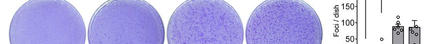
Figure 3. Gα13 Arg-200 mutants induce NIH3T3 cell transformation in vitro. (A) Generation of NIH3T3
cell lines stably expressing the indicated Gα13 proteins. Lentiviral particles for the expression of Gα13 were
generated in HEK293T cells and used to transduce NIH3T3 cells followed by antibiotic selection. Lysates
of each one of the cell lines were immunoblotted as indicated. Images were generated by splicing lanes
from the same membrane and the vertical dotted line indicates the position of the boundary between the
two segments that were merged. (B, C) Gα13 Arg-200 mutants promote foci formation in NIH3T3 cells
more efficiently than Gα13 WT. Cells were seeded on plates and stained with crystal violet 10 days later.
Images of a representative experiment are shown in panel B, whereas panel C shows the quantification of
foci. Mean ± S.E.M, n= 6. **pNaturally-occurring hotspot cancer mutations in Gα13 promote oncogenic signaling
Marcin Maziarz, Anthony Federico, Jingyi Zhao, Lorena Dujmusic, Zhiming Zhao, Stefano
Monti, Xaralabos Varelas and Mikel Garcia-Marcos
J. Biol. Chem. published online October 27, 2020
Access the most updated version of this article at doi: 10.1074/jbc.AC120.014698
Alerts:
• When this article is cited
• When a correction for this article is posted
Click here to choose from all of JBC's e-mail alerts
Downloaded from http://www.jbc.org/ by guest on December 20, 2020You can also read