A 14-Marker Multiplexed Imaging Panel for Prognostic Biomarkers and Tumor Heterogeneity in Head and Neck Squamous Cell Carcinoma
←
→
Page content transcription
If your browser does not render page correctly, please read the page content below
ORIGINAL RESEARCH
                                                                                                                                             published: 19 August 2021
                                                                                                                                        doi: 10.3389/fonc.2021.713561

                                              A 14-Marker Multiplexed Imaging
                                              Panel for Prognostic Biomarkers and
                                              Tumor Heterogeneity in Head and
                                              Neck Squamous Cell Carcinoma
                                              Junichi Mitsuda 1, Takahiro Tsujikawa 1,2*, Kanako Yoshimura 1, Sumiyo Saburi 1,
                                              Masaho Suetsugu 1, Kayo Kitamoto 1, Mari Takenaka 1, Gaku Ohmura 1, Akihito Arai 1,
                                              Hiroshi Ogi 3,4, Kyoko Itoh 3 and Shigeru Hirano 1
                                              1 Department of Otolaryngology–Head and Neck Surgery, Kyoto Prefectural University of Medicine, Kyoto, Japan,
                                              2 Department of Cell, Developmental & Cancer Biology, Oregon Health and Science University, Portland, OR, United States,
                                              3 Department of Pathology and Applied Neurobiology, Kyoto Prefectural University of Medicine, Kyoto, Japan, 4 SCREEN
                         Edited by:
                                              Holdings Co., Ltd., Kyoto, Japan
                     Makoto Tahara,
National Cancer Center Hospital East,
                              Japan
                                              Recent advances made in treatment for head and neck squamous cell carcinoma
                      Reviewed by:
                                              (HNSCC) highlight the need for new prediction tools to guide therapeutic strategies. In
                    Ryuji Yasumatsu,
             Kyushu University, Japan         this study, we aimed to develop a HNSCC-targeting multiplex immunohistochemical (IHC)
              Leandro Luongo Matos,           panel that can evaluate prognostic factors and the intratumor heterogeneity of HNSCC.
    Universidade de São Paulo, Brazil
                                              To identify IHC-based tissue biomarkers that constitute new multiplex IHC panel, a
                 *Correspondence:
                    Takahiro Tsujikawa
                                              systematic review and meta-analysis were performed to analyze reported IHC biomarkers
               tu-ji@koto.kpu-m.ac.jp         in laryngeal and pharyngeal SCC in the period of 2008–2018. The Cancer Genome Atlas
                                              (TCGA) and Reactome pathway databases were used to validate the prognostic and
                   Specialty section:
         This article was submitted to
                                              functional significance of the identified biomarkers. A 14-marker chromogenic multiplex
              Head and Neck Cancer,           IHC panel including identified biomarkers was used to analyze untreated HNSCC tissue.
               a section of the journal       Forty-five high-quality studies and thirty-one candidate tissue biomarkers were identified
                 Frontiers in Oncology
                                              (N = 7062). Prognostic validation in TCGA laryngeal and pharyngeal SCC cohort (N = 205)
           Received: 08 June 2021
           Accepted: 15 July 2021             showed that b-catenin, DKK1, PINCH1, ADAM10, and TIMP1 were significantly
         Published: 19 August 2021            associated with poor prognosis, which were related to functional categories such as
                             Citation:        immune system, cellular response, cell cycle, and developmental systems. Selected
Mitsuda J, Tsujikawa T, Yoshimura K,
  Saburi S, Suetsugu M, Kitamoto K,
                                              biomarkers were assembled to build a 14-marker panel, evaluating heterogeneity and
Takenaka M, Ohmura G, Arai A, Ogi H,          polarized expression of tumor biomarkers in the tissue structures, which was particularly
    Itoh K and Hirano S (2021) A 14-          related to activation of Wnt/b-catenin pathway. Integrated IHC analysis based on a
 Marker Multiplexed Imaging Panel for
   Prognostic Biomarkers and Tumor            systemic review and meta-analysis provides an in situ proteomics tool to assess the
     Heterogeneity in Head and Neck           aggressiveness and intratumor heterogeneity of HNSCC.
           Squamous Cell Carcinoma.
             Front. Oncol. 11:713561.         Keywords: meta-analysis, biomarker, immunohistochemistry, tumor heterogeneity, head and neck squamous cell
      doi: 10.3389/fonc.2021.713561           carcinoma (HNSCC)

Frontiers in Oncology | www.frontiersin.org                                          1                                      August 2021 | Volume 11 | Article 713561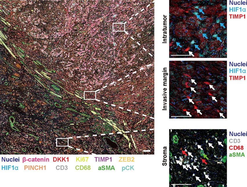
Mitsuda et al.                                                                               HNSCC-Targeting mIHC Panel Evaluating Heterogeneity

INTRODUCTION                                                            MATERIALS AND METHODS
The emergence of molecular targeting therapy and immune                 Systematic Review and
checkpoint blockade has revolutionized the clinical                     Meta-Analysis (PRISMA)
management of head and neck squamous cell carcinoma                     A literature search was performed in the PubMed database
(HNSCC), but its mortality is still high at approximately 50%,          (RRID : SCR_004846) on July 7, 2018, using the following
leading to 330,000 deaths per year worldwide (1). Increased             search criteria: (head and neck) AND (cancer OR carcinoma)
number of available therapeutic options urgently requires               AND (prognos* OR surviv*) AND (immunohistoch*) AND
optimization of the sequence and combination of treatment               (laryn* OR pharyn*). Two authors independently inspected the
modalities, thus requiring new tools for the prediction of              titles and abstracts of the retrieved articles between July 2008 and
therapeutic outcomes.                                                   July 2018. Literature written in languages other than English and
    Immunohistochemistry (IHC) has served as the gold standard          not analyzed using IHC data were excluded. To provide adequate
for histopathological evaluation of cancer. Since a large number        measures of predictability and improve the quality of the tumor
of IHC-based biomarkers have been reported, integrative                 prognostic biomarker studies, modified REMARK criteria
organization of reported biomarkers is needed to identify               containing key items selected by REMARK criteria (9) were
clinically meaningful biomarkers related to disease                     used (Supplementary Table 1).
aggressiveness of HNSCC. However, systematic reviews and                    The following information was obtained: author name, date
meta-analyses of IHC-based biomarkers have not been carried             of publication, number of patients, IHC protocols, associated
out for HNSCC, particularly for pharyngeal and laryngeal SCC,           multivariate hazard ratio (HR), 95% confidence intervals (CIs)
where decision making for treatment of these disease is directly        and corresponding P values, overall survival (OS), and evaluated
associated with organ preservation, including voice and                 biomarkers. For proteins evaluated in a single study only, the
swallowing function.                                                    summarized HR (including 95% CI) represented the value
    Intratumor heterogeneity has been increasingly acknowledged         reported in that study. For proteins that were evaluated in
since technological advances in single-cell sequencing methods          multiple studies, meta-analysis was carried out using software
were made, where the presence of subpopulations of cancer cells         “R version 3.3.3” and package “metafor”. HR and 95% CI were
with distinct genotypic, phenotypic, and morphological profiles          calculated using both the fixed-effect model and random-effect
was observed within a single tumor, including HNSCC (2–4).              model. Heterogeneity was evaluated in multiple studies by
Since intratumor heterogeneity is strongly associated with              calculating the Q statistic and I2 value. An HR greater than 1
therapeutic response and resistance (5), better treatment               indicated poor prognosis in the study group relative to the
strategies can be developed based on the characterization of            reference group and was considered statistically significant if
intratumor heterogeneity. Given that transcriptomic profiles do          the 95% CI did not overlap. The publication bias was assessed
not highly correlate with proteomic changes (6), intratumor             when there were ten or more studies through the inspection of
heterogeneity needs to be evaluated at both the transcription           funnel plots.
and protein levels, highlighting the significance of revisiting
IHC-based assessment in whole tumor tissue.                             In Silico Analysis
    Recently, we established a 14-biomarker multiplex IHC               To evaluate the prognostic significance of the extracted markers,
platform to enable comprehensive profiling of tumor and                  TCGA data (RRID : SCR_003193) was used. The data on 205
immune infiltrates in HNSCC (7). In this study, we performed             laryngeal and pharyngeal SCC cases containing information on
a systematic review and meta-analysis based on the preferred            the primary site, survival information, OS, and values of
reporting items for systematic reviews and meta-analyses                biomarker expression were downloaded from the UCSC Xena
statement (PRISMA) (8) to identify IHC-based tissue                     project (http://xena.ucsc.edu) on September 5, 2019. Based on
biomarkers associated with the prognosis of pharyngeal and              the median biomarker expression, all cases were divided into
laryngeal SCC. Modified reporting recommendations for tumor              high-expression (N = 102) and low-expression groups (N = 103).
marker prognostic studies (REMARK) criteria (9) were utilized           The 5-year OS was analyzed using the Kaplan-Meier and log-
for the selection of high-quality studies, followed by prognostic       rank tests. No statistical power analyses were used to determine
and functional validation using the Cancer Genome Atlas                 the sample size. The sample size was based on the number of
(TCGA) and the Reactome pathway database. This led to the               available and qualified tumor samples for this study. A p value of
identification of selective biomarkers including b-catenin, DKK1,        less than 0.05 was considered statistically significant. The
PINCH1, ADAM10, TIMP1, HIF1a, and ZFX. Based on the                     functional significance of the extracted biomarkers was
above results, we developed a HNSCC-targeting multiplex IHC             evaluated using the Reactome pathway database (https://
panel, enabling simultaneous assessment of 14 different tumor           reactome.org) (RRID : SCR_003485), where molecular
and stromal markers with preserved intratumor heterogeneity in          reactions are systematically described in molecular detail to
a single formalin-fixed paraffin-embedded (FFPE) tissue. In               generate a network of molecular transformations (10).
combination with other single-cell and bulk tumor analyses,
our chromogenic 14-plex tumor panel can provide spatial                 Multiplex IHC
information, contributing to the further development of tissue-         Sections (5 µm) were obtained from FFPE samples of untreated
based biomarkers.                                                       tongue, oropharyngeal, hypopharyngeal and laryngeal SCC

Frontiers in Oncology | www.frontiersin.org                         2                                   August 2021 | Volume 11 | Article 713561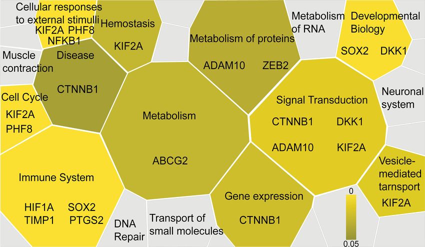
Mitsuda et al.                                                                                 HNSCC-Targeting mIHC Panel Evaluating Heterogeneity

(N = 4). Multiplex IHC was performed as described previously              management, where p16 was utilized as a highly sensitive
(7). Briefly, FFPE tumor sections were subjected to sequential             surrogate marker for prognostically favorable human
detection using 14-different antibodies for tumor and stromal             papilloma virus-related oropharyngeal cancer (16, 17).
markers. Stained slides were digitally scanned using Nanozoomer           Publication bias was not evaluable due to the limited number
S60 (Hamamatsu Photonics) at 20 × magnification. Tissue                    of studies available for funnel plot inspections (56). Following the
sections were then stripped of antibody and chromogen by                  exclusion of 5 biomarkers with favorable prognosis and 13
cleaning with alcohol and heat-mediated antigen retrieval,                biomarkers that did not meet statistical significance, our
followed by subsequent rounds of antibody staining and                    systematic review and meta-analysis identified 31 biomarkers
imaging. A complete list of antibodies and conditions used for            that were significantly associated with short OS in laryngeal and
staining are provided in Supplementary Table 2.                           pharyngeal HNSCC.
    Digital image processing was performed as previously described
(7). Digital images reflecting the antibody panel were co-registered       Functional and Prognostic Significance
and aligned. A sequential gating strategy of image cytometry was          of Identified IHC Biomarkers Reveals a
applied to identify tumor cell phenotypes based on negative cell          Broad Spectrum of Tumor Characteristics
staining by using FCS Express 6 Image Cytometry Version                   of HNSCC
6.03.0011 (De Novo Software). All studies involving human                 The biomarkers identified by the systematic review and meta-
tissues were approved by the Institutional Review Board at Kyoto          analysis were functionally classified into the hallmarks of cancer,
Prefectural University of Medicine (ERB-C-43-4), and written              such as angiogenesis, evasion of growth suppressors, genome
informed consent was obtained from all patients.                          instability and mutation, immune evasion, invasion and
                                                                          metastasis, metabolism, replicable immortality, resisting cell death,
                                                                          sustaining proliferation, and tumor promoting inflammation (57)
RESULTS                                                                   (Table 1). Details of representative markers classified by hallmarks
                                                                          are described in the Supplementary Information, showing that
A Systematic Review and Meta-Analysis                                     prognostic IHC biomarkers were associated with a wide range of
of IHC-Based Studies Identifies 31                                         hallmarks, particularly with increased invasion and metastasis as
Biomarkers Related to Poor Prognosis                                      well as sustained proliferation. Notably, multiple markers, such as
in Laryngeal and Pharyngeal Cancer                                        DKK1, ADAM10, and ZFX were related to the Wnt/b-catenin
To identify tumor biomarkers that were functionally associated            pathway (58–60).
with the prognosis of HNSCC, we performed a systematic review                 Furthermore, the functional significance of the identified IHC
and meta-analysis of previously reported IHC markers in                   biomarkers was evaluated using the Reactome pathway database
laryngeal and pharyngeal cancers. Our search of the laryngeal             (61), visualizing signaling and metabolic pathways associated
or pharyngeal SCC IHC prognostic literature yielded 431                   with reference gene datasets. A total of 31 genes corresponding to
manuscripts for consideration, as shown in Figure 1. 56                   the 31 IHC biomarkers identified by the systematic review and
studies were not related to pharyngeal or laryngeal SCC. 45               meta-analysis were utilized as a reference dataset, and the results
studies were written in other languages. 12 manuscripts were              were visualized as a Voronoi diagram based on the ReacFoam
excluded, as protein expression was not assessed using IHC. 12            function (10), which exhibited an overview of pathways highly
case reports, 9 reviews, 2 meta-analyses, and a preliminary study         related to the identified biomarkers (Figure 3 and
were excluded. The remaining 297 studies, which used IHC to               Supplementary Figure 2). The immune system, cell cycle,
measure protein expression in laryngeal and pharyngeal SCC,               cellular response to external stimuli, and developmental
were assessed in terms of study design. Modified REMARK                    systems were related to the selected 31 biomarkers. b-catenin
criteria were applied to assess the completeness of reporting             and related markers were observed in a wide range of functional
(Supplementary Table 1). Of the 297 cohort studies, 71 failed to          categories, such as signal transduction, gene expression, and
publish the HRs and 95% CIs. 67 studies did not report OS. 33             disease, suggesting the possible involvement of Wnt/b-catenin
studies did not perform a multivariable log rank analysis. 26             pathways in the progression of HNSCC (Figure 3 and
studies were unrelated to the biomarkers. 32 studies were                 Supplementary Figure 2).
excluded due to a lack of information such as age, gender.                    Since the selected IHC-based biomarkers were related to a
Finally, 45 high-quality cohort studies reporting multivariable-          broad range of functional properties, we next validated the
adjusted estimates for 45 biomarkers in laryngeal or pharyngeal           prognostic significance of these biomarkers using a TCGA cohort
SCC were extracted (Table 1. N = 7062). Since 5 out of 45                 in laryngeal and pharyngeal cancer (N = 205). OS was evaluated
biomarkers (metadherin, ZEB2, p53, Ki-67, and p16) were                   in relation to representative 31 biomarkers (Figures 4A–G,
reported by two or more studies, a meta-analysis was                      Supplementary Figures 3A–X and 4), showing that b-catenin,
performed based on the fixed-effect model (Figures 2A–E) and               DKK1, TIMP1, PINCH1, and ADAM10 were significantly
random effect model (Supplementary Figures 1A–E), which                   associated with poor prognosis in patients with laryngeal and
identified ZEB2 and metadherin as poor prognostic indicators               pharyngeal SCC (Figures 4A–E). HIF1a and ZFX tended to
(Figures 2A, B). p16 was associated with favorable OS (HR =               be associated with a poor prognosis. Interestingly, high
0.58, P = 0.017) (Figure 2E), in agreement with current clinical          expression of MCM7 or COX2 was associated with favorable

Frontiers in Oncology | www.frontiersin.org                           3                                   August 2021 | Volume 11 | Article 713561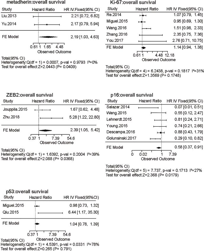
Mitsuda et al.                                                                                               HNSCC-Targeting mIHC Panel Evaluating Heterogeneity

                                    A

                                    B

 FIGURE 1 | A PRISMA flow diagram of the systematic review of IHC-based prognostic biomarkers and multiplex IHC panel selection. (A) A prisma flow diagram
 presents the PubMed searches, the number of manuscripts excluded, and the identified high-quality cohort studies. (B) Selection strategies based on prognostic
 and functional significance are applied for establishment of the 14-biomaker multiplex IHC panel.

prognosis in transcriptome analysis, potentially due to the                         analysis. Based on a chromogenic sequential IHC method
discrepancy between transcriptional and protein levels. Overall,                    enabling analysis of more than 12 proteins in one tissue
five biomarkers showed prognostic significance in IHC-based                           section (7), a 14-biomarker multiplex IHC panel was
analysis as well as during validation by TCGA analysis, which                       assembled from b-catenin, DKK1, PINCH1, ADAM10, and
served as candidates for robust predictive biomarkers for the                       TIMP1 used as prognostic markers. In addition to hematoxylin
prognosis of laryngeal and pharyngeal cancer.                                       staining, HIF1a as a hypoxia marker, ZFX as a transcriptional
                                                                                    regulator of self-renewal and stemness, ZEB2 as an epithelial to
14-Biomarker Multiplex IHC                                                          mesenchymal transformation (EMT) marker, and Ki67 as a
Panel Highlights Intratumor                                                         proliferation marker were included in the panel, considering
Heterogeneity in HNSCC                                                              the functional significance of these markers. To simultaneously
To expand our multiplex IHC platform, we established an                             analyze tumor microenvironment-related factors, the panel also
HNSCC-targeting multiplex IHC panel assembling candidate                            included pan-cytokeratin (pCK) as an epithelial marker, CD3
biomarkers derived from our systematic review and meta-                             and CD68 as immune markers, and alpha smooth muscle actin

Frontiers in Oncology | www.frontiersin.org                                     4                                        August 2021 | Volume 11 | Article 713561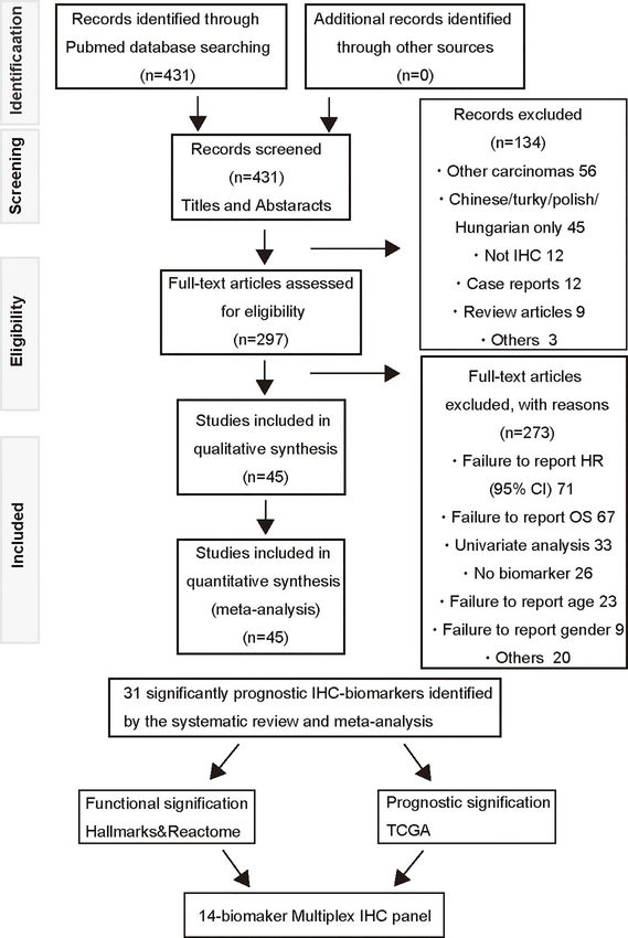
Mitsuda et al.                                                                                                   HNSCC-Targeting mIHC Panel Evaluating Heterogeneity

TABLE 1 | IHC biomarkers related to overall survival of laryngeal or pharyngeal SCC.

Function                                      Biomarker              Total No.                   HR (95% CI)                    P value                   Reference

Angiogenesis                                  EGF-like D7              116                     1.74 (1.05-2.17)                   0.012                       (11)
                                              HER1                      61                     1.34 (0.05-72.5)                   0.86                        (12)
                                              HIF1a                     91                     2.56 (1.07-6.15)                   0.036                       (13)
                                              TIP30                    105                   0.331 (0.153-0.715)                  0.005                       (14)
                                              VEGF-C                    43                   0.709 (0.221-2.274)                  0.564                       (15)
Evading growth suppressor                     p16*                     1495                    0.58 (0.37-0.91)                   0.017                     (16–21)
                                              p53*                     182                     1.04 (0.78-1.39)                   0.791                     (22, 23)
                                              p63                      108                     1.05 (1.03-1.08)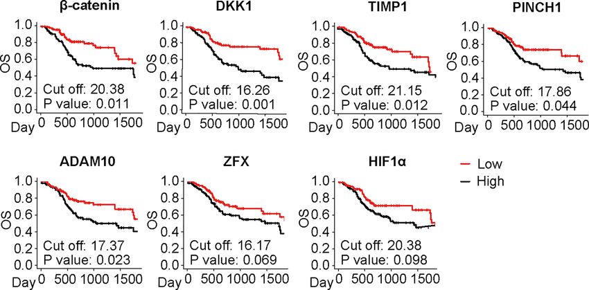
Mitsuda et al. HNSCC-Targeting mIHC Panel Evaluating Heterogeneity
A D
B E
C
FIGURE 2 | Forest plots of the effects of five biomarkers evaluated by two or more independent studies on laryngeal and pharyngeal HNSCC survival. (A–E) Forest
plots of hazard ratio (HR) for overall survival (OS) of metadherin (A), ZEB2 (B), p53 (C), Ki-67 (D), and p16 (E) are shown based on a fixed effect model. Bars
present 95% confidence intervals (CI) of HR, and the center of the lozenge gives the combined HR. Combined fixed effect HRs and tests for heterogeneity (I2) are
based on the generic inverse variance (I-V) method.
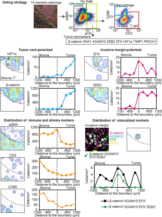
Simultaneously, spatial analysis of T cells (CD3+), tumor- aggressiveness, and ZEB2 is presumably related to metastatic
associated macrophages (CD68+), and aSMA+ cells revealed capability, multiple biomarker-positive cancer cells might be
heterogeneity of immune and stromal cell densities (Figure 6D). related to the presence of aggressive subpopulations of cancer
Furthermore, this panel revealed the potential colocalization cells, which are potentially localized at the invasive margin of
of multiple tumor cell markers such as b-catenin, ADAM10, HNSCC tumors. Taken together, these findings indicate that our
ZFX, and ZEB2 (Figure 6E). Interestingly, ZEB2, an EMT 14-biomarker multiplex IHC panel provides a platform for the
marker, was strongly colocalized with Wnt/b-catenin related quantitative assessment of intratumor heterogeneity based on
markers at the invasive margin (Figure 6E). Given that the multiple tissue biomarkers identified by a systematic review and
Wnt/b-catenin pathway is associated with cancer cell meta-analysis.
Frontiers in Oncology | www.frontiersin.org 6 August 2021 | Volume 11 | Article 713561Mitsuda et al. HNSCC-Targeting mIHC Panel Evaluating Heterogeneity
FIGURE 3 | Functional significance of the selected IHC biomarkers identified by the systematic review and meta-analysis. Functional significance of the identified
IHC biomarkers is visualized by using the Reactome pathway database (https://reactome.org), reporting on biological pathways associated with dataset of genes.
The results are visualized as a Voronoi diagram, showing an overview of the biological pathways related to the 31 biomarkers identified by the systematic review and
meta-analysis. Significantly enriched pathways are shown with a color scale from dark to light yellow. IHC biomarkers associated with each pathway are shown.
A B C D
E F G
FIGURE 4 | Prognostic significance of the selected IHC biomarkers identified by the systematic review and meta-analysis. (A–G) Kaplan-Meier analyses of overall
survival (OS) in the laryngeal and pharyngeal HNSCC cohort of the Cancer Genome Atlas (TCGA) (N = 205) stratified by gene expression of the identified biomarkers
are shown. Median is used for the cutoff values. Statistical significance is determined using log-rank test.
Frontiers in Oncology | www.frontiersin.org 7 August 2021 | Volume 11 | Article 713561Mitsuda et al. HNSCC-Targeting mIHC Panel Evaluating Heterogeneity
A B
C
FIGURE 5 | Fourteen-marker multiplex IHC panel to visualize prognostic tumor cell biomarkers and immune/stromal cells in a single FFPE tissue. A FFPE tissue of
tongue SCC is visualized by the established 14-marker multiplex IHC. (A) Representative 12-color composite images are shown. Biomarkers and colors are shown
at the bottom. Scale bar = 100 µm. (B) Top and bottom panels present expression of HIF1a and TIMP1 in the intratumor and invasive margin regions, respectively.
Blue and white arrows depict HIF1a+ cells and TIMP1+ cells, respectively. (C) Immune markers (CD3 and CD68) and aSMA+ cells in stroma region are shown. White,
red, and green arrows present CD3+, CD68+, and aSMA+ cells, respectively. Biomarkers and colors are shown at the right. Scale bars = 10 µm. Corresponding
single-marker images are shown in Supplementary Figure 3.
DISCUSSION Although the importance of intratumor heterogeneity has
been increasingly acknowledged, the vast majority of IHC-based
In this study, we developed a 14-marker multiplexed imaging studies have not considered the significance of heterogeneous
panel for prognostic biomarkers identified by a systematic review expression of tumor biomarkers. Intratumor heterogeneity can
and meta-analysis of prognostically and functionally-relevant be associated with poor clinical outcomes due to the presence of
IHC-based biomarkers in laryngeal and pharyngeal SCC. IHC subclones of cancer cells with aggressive characteristics, such as
analysis using this 14-marker multiplexed panel revealed rapid proliferation, metastatic potential, and therapeutic
intratumor heterogeneity that was especially related to an resistance (2). Recent studies based on single-cell RNA
aggressive subpopulation associated with Wnt/b-catenin sequencing have revealed that HNSCC has remarkable
pathway in HNSCC. intratumor heterogeneity in terms of clonal diversity (69) and
While some of the identified biomarkers such as p53 and Ki- locally activated partial EMT program at the leading edge of
67 are shared among many other cancer types (64–68), our primary tumors (70). In agreement with these findings, our
systematic review of laryngeal and pharyngeal HNSCC multiplex IHC analysis demonstrated the localization of
characteristically identified the prognostic significance of Wnt/ aggressive cancer cell populations expressing Wnt/b-catenin
b-catenin pathway-related markers such as b-catenin, DKK1, related markers and ZEB2 in the invasive margin (Figure 6E).
ADAM10, and ZFX (Table 1 and Figure 4) (27, 47, 58, 67, 68). Given that TIMP1 and macrophages were reported to induce the
This trend was more pronounced in laryngeal SCC as observed expression of EMT transcriptional factors, including ZEB2 (26,
in Supplementary Figure 4. These Wnt/b-catenin-related 71), increased expression of TIMP1 and CD68 in the invasive
markers are involved in a wide range of functional categories margin might be related to the regulation of EMT and metastatic
in the Reactome pathway analysis (Figure 3 and Supplementary potential. Multiplex IHC-based simultaneous detection in a
Figure 2), suggesting that activated b-catenin might be related to single tissue enables quantitative evaluation of multiple
disease aggressiveness of HNSCC, particularly in laryngeal SCC. overlapping biomarkers together with spatial distribution in
Frontiers in Oncology | www.frontiersin.org 8 August 2021 | Volume 11 | Article 713561Mitsuda et al. HNSCC-Targeting mIHC Panel Evaluating Heterogeneity
A
B C
D E
FIGURE 6 | The 14-biomarker multiplex IHC panel quantitatively assesses intratumoral heterogeneity of HNSCC. (A) The gating strategy used for single cell-based
chromogenic signal intensity is shown where cell size/area, and location are utilized for density plots similar to flow cytometry. X and y axes were shown on a
logarithmic scale. The gating strategies identified cell populations of pCK+ tumor cells with selected tumor cell biomarkers such as b-catenin, DKK1, ADAM10, ZEB2,
ZFX, HIF1a, TIMP1, and PINCH1, together with aSMA+ cells (pCK−aSMA+), T cells (pCK−aSMA−CD3+) and macrophages (pCK−aSMA−CD68+) in a single tissue.
(B–D) Left panels depict contour mapping based on cell density of each marker. Dash lines present the invasive margin between tumor cell nests and stroma. Right
panels depict cell densities of identified lineages in every 200 µm-width from stroma to the tumor side. Tumor cell biomarkers are classified into tumor core-polarized
(B), and invasive margin-polarized markers, in comparison with immune and stroma markers (D). (E) IHC image and pixel density contour graph indicate distribution
of colocalized markers at the invasive margin. Line graph show two colocalized markers. Black line shows b-catenin+ DKK1+ ADAM10+ ZFX+ cells and green line
showed b-catenin+ DKK1+ ADAM10+ ZFX+ ZEB2+ cells.
Frontiers in Oncology | www.frontiersin.org 9 August 2021 | Volume 11 | Article 713561Mitsuda et al. HNSCC-Targeting mIHC Panel Evaluating Heterogeneity
the tumor architecture, contributing to the understanding of DATA AVAILABILITY STATEMENT
intratumor heterogeneity of HNSCC.
Immune checkpoint blockade has gained increasing The original contributions presented in the study are included in
importance in the treatment of HNSCC, and many studies the article/Supplementary Material. Further inquiries can be
have investigated the association between specific immune cell directed to the corresponding author.
populations and clinical outcomes of HNSCC (72, 73). Despite
the single-case analysis in our study, increased expression of b-
catenin in the center of the tumor might be associated with the ETHICS STATEMENT
low density of CD3+ T cells (Figure 6D), which is potentially
associated with T-cell exclusion mechanisms driven by Wnt/b- The studies involving human participants were reviewed and
catenin pathway activation (74). These findings suggest that our approved by The Institutional Review Board at Kyoto Prefectural
14-biomarker multiplex IHC panel can serve as a platform to University of Medicine (ERB-C-43-4). The patients/participants
characterize the intratumor heterogeneity of cancer cell provided their written informed consent to participate in
phenotypes in association with immune cell densities. this study.
In this study, we observed discrepancies between IHC-based
results and transcriptomic analyses in terms of prognostic
significance. Notably, 15 out of 45 biomarkers exhibited poor
prognostic significance in IHC studies, but not in transcriptomic AUTHOR CONTRIBUTIONS
analysis in TCGA (Table 1 and Figure 4), leading to the notion JM performed manuscript preparation. JM and TT performed
that transcriptomic profiles did not necessarily predict protein data analysis. JM, MS, and KK collected data and performed
expression (75). These observations provide a rationale for IHC. TT conceived of the research and designed research studies.
revisiting IHC-based proteomics, which can provide a clinically TT, MT, GO, and AA obtained research funding. TT, GO, and
applicable approach for analyzing the intensity and functional AA participated in sample preparation. HO and KI provided
properties of protein expression without significant cost (76). digital analytic support for multiplex IHC. TT and SH supervised
Although extensive research was performed, a potential this study. All authors contributed to the article and approved
limitation of this study is publication bias. Due to a great the submitted version.
diversity of IHC biomarkers, identified biomarkers in our
study were based on a small number of the original studies
(Figure 2), in analogous to previous IHC meta-analyses (64, 66).
To avoid bias due to any potential heterogeneity, we utilized FUNDING
TCGA database to validate the prognostic significance of the
identified biomarkers (Figure 4). Another limitation in this This work was supported by grants from the Japanese ministry of
study is the lack of subgroup analyses stratified by the HNSCC education, culture, sports, science and technology (17H07016,
subsites such as larynx, oropharynx, and hypopharynx. Since we 19K18814, 19K18739, and 19K23893), and the public promoting
observed some interesting differences between laryngeal and association Asano foundation for studies on medicine, and by the
pharyngeal SCC (Supplementary Figure 4), further studies are research promotion award from the Oto-Rhino-Laryngological
required for exploring subsite-specific biomarkers associated Society of Japan, Inc. The funder was not involved in the study
with clinical outcomes. design, collection, analysis, interpretation of data, the writing of
In summary, the present study, based on a systematic review and this article or the decision to submit it for publication.
meta-analysis of laryngeal and pharyngeal SCC, identified IHC-
based prognostic biomarkers. Established multiplex IHC panels
enable assessment of intratumor heterogeneity of HNSCC, which SUPPLEMENTARY MATERIAL
is particularly related to activation of the Wnt/b-catenin pathway. In
combination with other single-cell and bulk tumor analyses, our The Supplementary Material for this article can be found online at:
tissue-based biomarker analysis can provide new insights into https://www.frontiersin.org/articles/10.3389/fonc.2021.713561/
diagnostic and treatment strategies for HNSCC. full#supplementary-material
4. Mroz EA, Rocco JW. Intra-Tumor Heterogeneity in Head and Neck Cancer
REFERENCES and Its Clinical Implications. World J Otorhinolaryngol Neck Surg (2016)
2:60–7. doi: 10.1016/j.wjorl.2016.05.007
1. Bray F, Ferlay J, Soerjomataram I, Siegel RL, Torre LA, Jemal A. Global Cancer Statistics 5. Easwaran H, Tsai HC, Baylin SB. Cancer Epigenetics: Tumor Heterogeneity,
2018: GLOBOCAN Estimates of Incidence and Mortality Worldwide for 36 Cancers in Plasticity of Stem-Like States, and Drug Resistance. Mol Cell (2014) 54:716–
185 Countries. CA Cancer J Clin (2018) 68:394–424. doi: 10.3322/caac.21492 27. doi: 10.1016/j.molcel.2014.05.015
2. Mroz EA, Tward AD, Pickering CR, Myers JN, Ferris RL, Rocco JW. High Intratumor 6. Vogel C, Marcotte EM. Insights Into the Regulation of Protein Abundance
Genetic Heterogeneity Is Related to Worse Outcome in Patients With Head and Neck From Proteomic and Transcriptomic Analyses. Nat Rev Genet (2012) 13:227–
Squamous Cell Carcinoma. Cancer (2013) 119:3034–42. doi: 10.1002/cncr.28150 32. doi: 10.1038/nrg3185
3. Puram SV, Rocco JW. Molecular Aspects of Head and Neck Cancer Therapy. 7. Tsujikawa T, Kumar S, Borkar RN, Azimi V, Thibault G, Chang YH, et al.
Hematol Oncol Clin North Am (2015) 29:971–92. doi: 10.1016/j.hoc.2015.07.003 Quantitative Multiplex Immunohistochemistry Reveals Myeloid-Inflamed
Frontiers in Oncology | www.frontiersin.org 10 August 2021 | Volume 11 | Article 713561Mitsuda et al. HNSCC-Targeting mIHC Panel Evaluating Heterogeneity
Tumor-Immune Complexity Associated With Poor Prognosis. Cell Rep 26. Zhu GJ, Song PP, Zhou H, Shen XH, Wang JG, Ma XF, et al. Role of Epithelial-
(2017) 19:203–17. doi: 10.1016/j.celrep.2017.03.037 Mesenchymal Transition Markers E-Cadherin, N-Cadherin, b-Catenin and
8. Moher D, Liberati A, Tetzlaff J, Altman DG, Altman D, Antes G, et al. ZEB2 in Laryngeal Squamous Cell Carcinoma. Oncol Lett (2018) 15:3472–81.
Preferred Reporting Items for Systematic Reviews and Meta-Analyses: The doi: 10.3892/ol.2018.7751
PRISMA Statement. PloS Med (2009) 6:e1000097. doi: 10.1371/ 27. Shi Y, Gong H-L, Zhou L, Tian J, Wang Y. Dickkopf-1 Is a Novel Prognostic
journal.pmed.1000097 Biomarker for Laryngeal Squamous Cell Carcinoma. Acta Otolaryngol (2014)
9. McShane LM, Altman DG, Sauerbrei W, Taube SE, Gion M, Clark GM. 134:753–9. doi: 10.3109/00016489.2014.894251
Reporting Recommendations for Tumour Marker Prognostic Studies 28. Zhang Q, Zhang W, Zhang J, Xu H, You Y. Aberrant Kif2a and Ki67
(REMARK). Br J Cancer (2005) 93:387–91. doi: 10.1038/sj.bjc.6602678 Expression Predicts Poor Survival in Laryngeal Squamous Cell Carcinoma.
10. Jassal B, Matthews L, Viteri G, Gong C, Lorente P, Fabregat A, et al. The Auris Nasus Larynx (2016) 43:433–9. doi: 10.1016/j.anl.2015.10.012
Reactome Pathway Knowledgebase. Nucleic Acids Res (2020) 48:D498–503. 29. Lu M, Zhu H, Wang X, Zhang D, Xiong L, Zhu J, et al. LAMP1 Expression Is
doi: 10.1093/nar/gkz1031 Associated With Malignant Behaviours and Predicts Unfavourable Prognosis
11. Li J-J, Yang X-M, Wang S-H, Tang Q-L. Prognostic Role of Epidermal Growth in Laryngeal Squamous Cell Carcinoma. Pathology (2016) 48:684–90.
Factor-Like Domain 7 Protein Expression in Laryngeal Squamous Cell Carcinoma. doi: 10.1016/j.pathol.2016.08.001
J Laryngol Otol (2011) 125:1152–7. doi: 10.1017/S0022215111002441 30. Ma J, Wang J, Fan W, Pu X, Zhang D, Fan C, et al. Upregulated TIMP-1
12. Almadori G, Lauriola L, Coli A, Bussu F, Gallus R, Scannone D, et al. Correlates With Poor Prognosis of Laryngeal Squamous Cell Carcinoma. Int J
Minichromosome Maintenance Protein 7 and Geminin Expression: Clin Exp Pathol (2014) 7:246–54.
Prognostic Value in Laryngeal Squamous Cell Carcinoma in Patients 31. Wu H, Xu H, Zhang S, Wang X, Zhu H, Zhang H, et al. Potential Therapeutic
Treated With Radiotherapy and Cetuximab. Head Neck (2017) 39:684–93. Target and Independent Prognostic Marker of TROP2 in Laryngeal
doi: 10.1002/hed.24670 Squamous Cell Carcinoma. Head Neck (2013) 35:1373–8. doi: 10.1002/
13. Schrijvers ML, van der laan BFAM, de bock GH, Pattje WJ, Mastik MF, hed.23138
Menkema L, et al. Overexpression of Intrinsic Hypoxia Markers HIF1a and 32. Jouppila-Mättö A, Mannermaa A, Sironen R, Kosma V-M, Soini Y, Pukkila
CA-IX Predict for Local Recurrence in Stage T1-T2 Glottic Laryngeal M. SIP1 Predicts Progression and Poor Prognosis in Pharyngeal Squamous
Carcinoma Treated With Radiotherapy. IntJRadiation Oncol BiolPhys Cell Carcinoma. Histol Histopathol (2015) 30:569–79. doi: 10.14670/HH-
(2008) 72(1):161–9. doi: 10.1016/j.ijrobp.2008.05.025 30.569
14. Zhu M, Yin F, Yang L, Chen S, Chen R, Zhou W, et al. Contribution of TIP30 33. Yu C, Liu Y, Tan H, Li G, Su Z, Ren S, et al. Metadherin Regulates Metastasis
to Chemoresistance in Laryngeal Carcinoma. Cell Death Dis (2014) 5:e1468– of Aquamous Cell Carcinoma of the Head and Neck via AKT Signaling
10. doi: 10.1038/cddis.2014.424 Pathway-Mediated Epithelial-Mesenchymal Transition. Cancer Lett (2014)
15. Baek SK, Jung KY, Lee SH, Woo JS, Kwon SY, Chung EJ, et al. Prognostic 343:258–67. doi: 10.1016/j.canlet.2013.09.033
Significance of Vascular Endothelial Growth Factor-C Expression and 34. Liu Y, Su Z, Li G, Yu C, Ren S, Huang D, et al. Increased Expression of
Lymphatic Vessel Density in Supraglottic Squamous Cell Carcinoma. Metadherin Protein Predicts Worse Disease-Free and Overall Survival in
Laryngoscope (2009) 119:1325–30. doi: 10.1002/lary.20483 Laryngeal Squamous Cell Carcinoma. Int J Cancer (2013) 133:671—679.
16. Salazar CR, Anayannis N, Smith RV, Wang Y, Jr Haigentz M, Garg M, et al. doi: 10.1002/ijc.28071
Combined P16 and Human Papillomavirus Testing Predicts Head and Neck 35. Zhang Q, Xu H, You Y, Zhang J, Chen R. High Gpx1 Expression Predicts Poor
Cancer Survival. Int J Cancer (2014) 135:2404–12. doi: 10.1002/ijc.28876 Survival in Laryngeal Squamous Cell Carcinoma. Auris Nasus Larynx (2018)
17. Golusiń ski P, Pazdrowski J, Szewczyk M, Misiołek M, Pietruszewska W, 45:13–9. doi: 10.1016/j.anl.2017.05.012
Klatka J, et al. Is Immunohistochemical Evaluation of P16 in Oropharyngeal 36. Kaira K, Toyoda M, Shimizu A, Imai H, Sakakura K, Nikkuni O, et al.
Cancer Enough to Predict the HPV Positivity? Rep Pract Oncol Radiother Decreasing Expression of Glucose-Regulated Protein GRP78/Bip as a
(2017) 22:237–42. doi: 10.1016/j.rpor.2017.01.003 Significant Prognostic Predictor in Patients With Advanced Laryngeal
18. Wang H, Zhang Z, Sun R, Lin H, Gong L, Fang M, et al. HPV Infection and Squamous Cell Carcinoma. Head Neck (2016) 38:1539–44. doi: 10.1002/
Anemia Status Stratify the Survival of Early T2 Laryngeal Squamous Cell hed.24471
Carcinoma. J Voice (2015) 29:356–62. doi: 10.1016/j.jvoice.2014.08.016 37. Garcı́a-Carracedo D, Á ngeles Villaronga M, Á lvarez-Teijeiro S, Hermida-
19. Lehnerdt GF, Bachmann HS, Adamzik M, Panic A, Köksal E, Weller P, et al. Prado F, Santamarı́a I, Allonca E, et al. Impact of PI3K/AKT/Mtor Pathway
AQP1, AQP5, Bcl-2 and P16 in Pharyngeal Squamous Cell Carcinoma. Activation on the Prognosis of Patients With Head and Neck Squamous Cell
J Laryngol Otol (2015) 129:580–6. doi: 10.1017/S002221511500119X Carcinomas. Oncotarget (2016) 7:29780–93. doi: 10.18632/oncotarget.8957
20. Young RJ, Urban D, Angel C, Corry J, Lyons B, Vallance N, et al. Frequency 38. Schrijvers ML, Pattje WJ, Slagter-Menkema L, Mastik MF, Gibcus JH,
and Prognostic Significance of P16 INK4A Protein Overexpression and Langendijk JA, et al. FADD Expression as a Prognosticator in Early-Stage
Transcriptionally Active Human Papillomavirus Infection in Laryngeal Glottic Squamous Cell Carcinoma of the Larynx Treated Primarily With
Squamous Cell Carcinoma. Br J Cancer (2015) 112:1098–104. doi: 10.1038/ Radiotherapy. Int J Radiat Oncol Biol Phys (2012) 83:1220–6. doi: 10.1016/
bjc.2015.59 j.ijrobp.2011.09.060
21. Descamps G, Karaca Y, Lechien JR, Kindt N, Decaestecker C, Remmelink M, 39. Ma J, Liu S, Zhang W, Zhang F, Wang S, Wu L, et al. High Expression of
et al. Classical Risk Factors, But Not HPV Status, Predict Survival After NDRG3 Associates With Positive Lymph Node Metastasis and Unfavourable
Chemoradiotherapy in Advanced Head and Neck Cancer Patients. J Cancer Overall Survival in Laryngeal Squamous Cell Carcinoma. Pathology (2016)
Res Clin Oncol (2016) 142:2185–96. doi: 10.1007/s00432-016-2203-7 48:691–6. doi: 10.1016/j.pathol.2016.08.005
22. Qiu X, You Y, Huang J, Wang X, Zhu H, Wang Z. LAMP3 and TP53 40. Shen B, Li D, Dong P, Gao S. Expression of ABC Transporters Is an Unfavorable
Overexpression Predicts Poor Outcome in Laryngeal Squamous Cell Prognostic Factor in Laryngeal Squamous Cell Carcinoma. Ann Otol Rhinol
Carcinoma. Int J Clin Exp Pathol (2015) 8:5519–27. Laryngol (2011) 120:820–7. doi: 10.1177/000348941112001208
23. de Miguel-Luken MJ, Chaves-Conde M, de Miguel-Luken V, Muñoz-Galvá n 41. Schrader CH, Kolb M, Zaoui K, Flechtenmacher C, Grabe N, Weber KJ, et al.
S, Ló pez-Guerra JL, Mateos JC, et al. MAP17 (PDZKIP1) as a Novel Kallikrein-Related Peptidase 6 Regulates Epithelial-to-Mesenchymal
Prognostic Biomarker for Laryngeal Cancer. Oncotarget (2015) 6:12625–36. Transition and Serves as Prognostic Biomarker for Head and Neck
doi: 10.18632/oncotarget.3470 Squamous Cell Carcinoma Patients. Mol Cancer (2015) 14:1–14.
24. Re M, Zizzi A, Ferrante L, Stramazzotti D, Goteri G, Gioacchini FM, et al. P63 doi: 10.1186/s12943-015-0381-6
and Ki-67 Immunostainings in Laryngeal Squamous Cell Carcinoma Are 42. Tsinias G, Nikou S, Papadas T, Pitsos P, Papadaki H, Bravou V. High PINCH1
Related to Survival. Eur Arch Oto-Rhino-Laryngol (2014) 271:1641–51. Expression in Human Laryngeal Carcinoma Associates With Poor Prognosis.
doi: 10.1007/s00405-013-2833-1 Anal Cell Pathol (2018) 2018:1–12. doi: 10.1155/2018/2989635
25. Han L, Jiang B, Wu H, Zhang S, Lu X. Expression and Prognostic Value of 43. Liu XK, Li Q, Xu LH, Hu LJ, Liao WG, Zhang XR, et al. Expression and
MAGE-A9 in Laryngeal Squamous Cell Carcinoma. Int J Clin Exp Pathol Clinical Significance of SIAH in Laryngeal Squamous Cell Carcinoma. Med
(2014) 7:6734–42. Oncol (2013) 30:1–8. doi: 10.1007/s12032-013-0485-z
Frontiers in Oncology | www.frontiersin.org 11 August 2021 | Volume 11 | Article 713561Mitsuda et al. HNSCC-Targeting mIHC Panel Evaluating Heterogeneity
44. Tang X, Shen X, Li L, Zhang Y, Chen G. SOX2 Overexpression Correlates 64. Oliveira LR, Ribeiro-Silva A. Prognostic Significance of
With Poor Prognosis in Laryngeal Squamous Cell Carcinoma. Auris Nasus Immunohistochemical Biomarkers in Oral Squamous Cell Carcinoma. Int
Larynx (2013) 40:481–6. doi: 10.1016/j.anl.2013.01.003 J Oral Maxillofac Surg (2011) 40:298–307. doi: 10.1016/j.ijom.2010.12.003
45. Yang F, Ma H, Feng L, Lian M, Wang R, Fan E, et al. Zinc Finger Protein X- 65. Rainsbury JW, Ahmed W, Williams HK, Roberts S, Paleri V, Mehanna H.
Linked (ZFX) Contributes to Patient Prognosis, Cell Proliferation and Prognostic Biomarkers of Survival in Oropharyngeal Squamous Cell
Apoptosis in Human Laryngeal Squamous Cell Carcinoma. Int J Clin Exp Carcinoma: Systematic Review and Meta-Analysis. Head Neck (2013)
Pathol (2015) 8:13886–99. 35:1048–55. doi: 10.1002/hed.22950
46. Ma H, Du X, Zhang S, Wang Q, Yin Y, Qiu X, et al. Achaete-Scute Complex 66. Jamieson NB, Carter CR, McKay CJ, Oien KA. Tissue Biomarkers for
Homologue-1 Promotes Development of Laryngocarcinoma via Facilitating Prognosis in Pancreatic Ductal Adenocarcinoma: A Systematic Review and
the Epithelial-Mesenchymal Transformation. Tumor Biol (2017) Meta-Analysis. Clin Cancer Res (2011) 17:3316–31. doi: 10.1158/1078-
39:1010428317705752. doi: 10.1177/1010428317705752 0432.CCR-10-3284
47. You B, Gu M, Cao X, Li X, Shi S, Shan Y, et al. Clinical Significance of 67. Bouvet M, Ellis LM, Nishizaki M, Fujiwara T, Liu W, Bucana CD, et al.
ADAM10 Expression in Laryngeal Carcinoma. Oncol Lett (2017) 13:1353–9. Adenovirus-Mediated Wild-Type P53 Gene Transfer Down-Regulates
doi: 10.3892/ol.2016.5546 Vascular Endothelial Growth Factor Expression and Inhibits Angiogenesis
48. Wang R, Guo Y, Ma H, Feng L, Wang Q, Chen X, et al. Tumor Necrosis Factor in Human Colon Cancer. Cancer Res (1998) 58(11):2288–92.
Superfamily Member 13 Is a Novel Biomarker for Diagnosis and Prognosis 68. Yerushalmi R, Woods R, Ravdin PM, Hayes MM, Gelmon KA. Ki67 in Breast
and Promotes Cancer Cell Proliferation in Laryngeal Squamous Cell Cancer: Prognostic and Predictive Potential. Lancet Oncol (2010) 11:174–83.
Carcinoma. Tumor Biol (2016) 37:2635–45. doi: 10.1007/s13277-015-4016-8 doi: 10.1016/S1470-2045(09)70262-1
49. Zhu G, Liu L, She L, Tan H, Wei M, Chen C, et al. Elevated Expression of 69. Zhang XC, Xu C, Mitchell RM, Zhang B, Zhao D, Li Y, et al. Tumor Evolution
Histone Demethylase PHF8 Associates With Adverse Prognosis in Patients of and Intratumor Heterogeneity of an Oropharyngeal Squamous Cell
Laryngeal and Hypopharyngeal Squamous Cell Carcinoma. Epigenomics Carcinoma Revealed by Whole-Genome Sequencing. Neoplasia (United
(2015) 7:143–53. doi: 10.2217/epi.14.82 States) (2013) 15:1371–8. doi: 10.1593/neo.131400
50. Yang L, Wang H, Wang Y, He Z, Chen H, Liang S, et al. Prostate Tumor 70. Puram SV, Tirosh I, Parikh AS, Patel AP, Yizhak K, Gillespie S, et al. Single-Cell
Overexpressed-1, in Conjunction With Human Papillomavirus Status, Transcriptomic Analysis of Primary and Metastatic Tumor Ecosystems in Head
Predicts Outcome in Early-Stage Human Laryngeal Squamous Cell and Neck Cancer. Cell (2017) 171:1611–24.e24. doi: 10.1016/j.cell.2017.10.044
Carcinoma. Oncotarget (2016) 7:31878–91. doi: 10.18632/oncotarget.8103 71. Lee SH, Koo BS, Kim JM, Huang S, Rho YS, Bae WJ, et al. Wnt/b-Catenin
51. Zhou X, Liu Y, Tan G. Prognostic Value of Elevated SHIP2 Expression in Signalling Maintains Self-Renewal and Tumourigenicity of Head and Neck
Laryngeal Squamous Cell Carcinoma. Arch Med Res (2011) 42:589–95. Frontsquamous Cell Carcinoma Stem-Like Cells by Activating Oct4. J Pathol
doi: 10.1016/j.arcmed.2011.10.012 (2014) 234:99–107. doi: 10.1002/path.4383
52. Yang H, Jiang WQ, Cao Y, Sun YA, Wei J, An X, et al. Elevated ZNF703 72. de Ruiter EJ, Ooft ML, Devriese LA, Willems SM. The Prognostic Role of
Protein Expression Is an Independent Unfavorable Prognostic Factor for Tumor Infiltrating T-Lymphocytes in Squamous Cell Carcinoma of the Head
Survival of the Patients With Head and Neck Squamous Cell Carcinoma. Dis and Neck: A Systematic Review and Meta-Analysis. Oncoimmunology (2017)
Markers (2015) 2015:1–178. doi: 10.1155/2015/640263 6:1–10. doi: 10.1080/2162402X.2017.1356148
53. Zhang J, Cheng L, Zhou L. Prognostic Significance of G-H2AX in Laryngeal 73. De Meulenaere A, Vermassen T, Aspeslagh S, Vandecasteele K, Rottey S,
Squamous Cell Carcinoma After Surgery. Chin (Engl) (2014) 127:2664–7. Ferdinande L. Tils in Head and Neck Cancer: Ready for Clinical
54. Chen YF, Luo RZ, Li Y, Cui BK, Song M, Yang AK, et al. High Expression Implementation and Why (Not)? Head Neck Pathol (2017) 11:354–63.
Levels of COX-2 and P300 Are Associated With Unfavorable Survival in doi: 10.1007/s12105-016-0776-8
Laryngeal Squamous Cell Carcinoma. Eur Arch Oto-Rhino-Laryngol (2013) 74. Luke JJ, Bao R, Sweis RF, Spranger S, Gajewski TF. WNT/B-Catenin Pathway
270:1009–17. doi: 10.1007/s00405-012-2275-1 Activation Correlates With Immune Exclusion Across Human Cancers. Clin
55. Yan M, Xu Q, Zhang P, Zhou XJ, Zhang ZY, Chen WT. Correlation of NF-kb Cancer Res (2019) 25:3074–83. doi: 10.1158/1078-0432.CCR-18-1942
Signal Pathway With Tumor Metastasis of Human Head and Neck Squamous 75. Akbani R, Ng PKS, Werner HMJ, Shahmoradgoli M, Zhang F, Ju Z, et al. A
Cell Carcinoma. BMC Cancer (2010) 10:1–13. doi: 10.1186/1471-2407-10-437 Pan-Cancer Proteomic Perspective on the Cancer Genome Atlas. Nat
56. Sutton AJ, Duval SJ, Tweedie RL, Abrams KR, Jones DR. Empirical Commun (2014) 5:1–15. doi: 10.1038/ncomms4887
Assessment of Effect of Publication Bias on Meta-Analyses. Br Med J (2000) 76. Uhlé n M, Björling E, Agaton C, Szigyarto CAK, Amini B, Andersen E, et al. A
320:1574–7. doi: 10.1136/bmj.320.7249.1574 Human Protein Atlas for Normal and Cancer Tissues Based on Antibody
57. Hanahan D, Weinberg RA. Hallmarks of Cancer: The Next Generation. Cell Proteomics. Mol Cell Proteomics (2005) 4:1920–32. doi: 10.1074/
(2011) 144:646–74. doi: 10.1016/j.cell.2011.02.013 mcp.M500279-MCP200
58. Chen ZY, Du Y, Wang L, Liu XH, Guo J, Weng XD. MiR-543 Promotes Cell
Proliferation and Metastasis of Renal Cell Carcinoma by Targeting Dickkopf 1 Conflict of Interest: HO is an employee for SCREEN Holdings Co., Ltd. KI has
Through the Wnt/b-Catenin Signaling Pathway. J Cancer (2018) 9:3660–8. received research funding from SCREEN Holdings Co., Ltd.
doi: 10.7150/jca.27124
59. Anders L, Mertins P, Lammich S, Murgia M, Hartmann D, Saftig P, et al. Furin-, The remaining authors declare that the research was conducted in the absence of
ADAM 10-, and G-Secretase-Mediated Cleavage of a Receptor Tyrosine any commercial or financial relationships that could be construed as a potential
Phosphatase and Regulation of b-Catenin's Transcriptional Activity. Mol Cell conflict of interest.
Biol (3917) 2006) 26:3917–34. doi: 10.1128/MCB.26.10.3917-3934.2006
60. Wang C, Fu SY, da Wang M, Yu WB, Cui QS, Wang HR, et al. Zinc Finger Publisher’s Note: All claims expressed in this article are solely those of the authors
Protein X-Linked Promotes Expansion of Epcam+ Cancer Stem-Like Cells in and do not necessarily represent those of their affiliated organizations, or those of
Hepatocellular Carcinoma. Mol Oncol (2017) 11:455–69. doi: 10.1002/1878- the publisher, the editors and the reviewers. Any product that may be evaluated in
0261.12036 this article, or claim that may be made by its manufacturer, is not guaranteed or
61. Joshi-Tope G, Gillespie M, Vastrik I, D’Eustachio P, Schmidt E, de Bono B, endorsed by the publisher.
et al. Reactome: A Knowledgebase of Biological Pathways. Nucleic Acids Res
(2005) 33:428–32. doi: 10.1093/nar/gki072 Copyright © 2021 Mitsuda, Tsujikawa, Yoshimura, Saburi, Suetsugu, Kitamoto,
62. Tsujikawa T, Thibault G, Azimi V, Sivagnanam S, Banik G, Means C, et al. Takenaka, Ohmura, Arai, Ogi, Itoh and Hirano. This is an open-access article
Robust Cell Detection and Segmentation for Image Cytometry Reveal Th17 distributed under the terms of the Creative Commons Attribution License (CC BY).
Cell Heterogeneity. Cytom Part A (2019) 95:389–98. doi: 10.1002/cyto.a.23726 The use, distribution or reproduction in other forums is permitted, provided the
63. Yin J, Jiang Y, Wu H, Wang J, Zhang S, Liu H. Overexpression of ZFX and Its original author(s) and the copyright owner(s) are credited and that the original
Involvement in Squamous Cell Carcinoma of the Tongue. Oncol Rep (2015) publication in this journal is cited, in accordance with accepted academic practice. No
33:141–8. doi: 10.3892/or.2014.3572 use, distribution or reproduction is permitted which does not comply with these terms.
Frontiers in Oncology | www.frontiersin.org 12 August 2021 | Volume 11 | Article 713561You can also read