Hailey P. Huddleston, Jorge Chahla, Brian Cole, and Adam B. Yanke
←
→
Page content transcription
If your browser does not render page correctly, please read the page content below
Management of Knee Cartilage
Injuries in Basketball
32
Hailey P. Huddleston, Jorge Chahla, Brian Cole,
and Adam B. Yanke
32.1 Introduction imaging (MRI) has shown that basketball players
have a similar level of undiagnosed, and gener-
Treatment of articular cartilage defects of the ally asymptomatic FCDs compared to athletes of
knee remains a challenging entity, particularly in other sports [11]. In the general population, the
young high-demand patients. Damaged articular number of surgical procedures to address these
cartilage has limited potential for self-healing cartilage defects is estimated to be approximately
and therefore has an increased propensity to 200,000 cases annually [4, 12].
progress to osteoarthritis [1, 2]. The prevalence Treatment options for focal chondral defects
of cartilage lesions in the general population include non-operative and surgical options. Non-
ranges from 13% to 60% and affects an estimated operative treatments are generally consid-
900,000 patients in the United States [3–6]. ered first-line, especially when no mechanical
However, the prevalence in athletes has been symptoms are present. A variety of surgical pro-
reported to be on average 36% (range 2.4–75%), cedures are available; the choice of which sur-
with 14% of these athletes being asymptomatic at gery to choose is individualized based on the
diagnosis and with the patellofemoral compart- athlete and his or her risk factors and the patient’s
ment (37%) and femoral condyle (35%) being current time in the season. If conservative mea-
the locations most likely to be affected [7, 8]. In sures, such as physical therapy or an intra-
professional basketball athletes, this number is articular injection fail, a less-invasive procedure
even higher. Three prior studies have reported such as a chondral debridement can provide sig-
that the prevalence of focal chondral defects nificant symptomatic relief, with minimal down
(FCDs) in the national basketball association time without altering the opportunity for a more
(NBA) is between 41% and 50% of players and definitive procedure. Other surgical interventions
most commonly affects the patellofemoral joint include microfracture, osteochondral autograft
(70–77% of defects) [9, 10]. Magnetic resonance transplantation (OCA), osteochondral allograft
transplantation (OAT), autologous chondrocyte
implantation (ACI), and its newer iterations
H. P. Huddleston · J. Chahla · B. Cole (matrix ACI) and newer procedures including
A. B. Yanke (*) minced cartilage procedures (DeNovo Natural
Department of Orthopaedic Surgery, Division of Tissue (NT), Zimmer Inc., Warsaw, IN), cryopre-
Sports Medicine, Rush University Medical Center,
Chicago, IL, USA
served osteochondral allografts (Cartiform,
e-mail: Jorge.chahla@rushortho.com; Athrex Inc., Naples, FL; Chondrofix, JRF,
Brian.Cole@rushortho.com Centennial, CO; Prochondrix, AlloSource,
© ESSKA 2020 379
L. Laver et al. (eds.), Basketball Sports Medicine and Science,
https://doi.org/10.1007/978-3-662-61070-1_32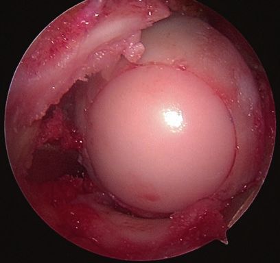
380                                                                                  H. P. Huddleston et al.

Centennial, CO), and extracellular matrix scaf-       ized over the patella, with swelling and stiffness
folds (BioCartilage, Arthrex Inc., Naplex, FL).       [14]. However, acute pain in association with
   The purpose of this chapter is to review focal     injury can also occur as approximately half of
chondral defects of the knee and their treatment,     patellofemoral FCD occurs in the setting of trau-
with special attention on the use and impact of       matic injury [15]. The location of pain can depend
these procedures in basketball players. Initially,    on the location of the FCD, and it can also be
this chapter assesses how FCDs are diagnosed          diffuse in nature. Pain can be present in addition
using patient history, physical examination, and      to intermittent effusion, crepitus, catching, and
imaging. Then, non-operative treatments, various      locking.
surgical techniques and their indications, and           A full past medical history is also essential in
postoperative rehabilitation process are investi-     diagnosing and creating a treatment plan. An
gated. Finally, outcomes of these procedures and      understanding of a patient’s comorbidities, past
their return to sport data and basketball, specifi-   surgical history, and history of physical injury is
cally, are analyzed.                                  essential. Previous injury, such as ACL injury, is
                                                      associated with chondral injury [16]. In addition,
                                                      any prior treatment such as medications, physical
32.2     Diagnosis                                   therapy, or injections should be noted.

FCDs in basketball players are diagnosed through
a combination of patient history, physical exami-     32.2.2 Physical Examination
nation, radiographs, and MRI. An early diagnosis
of FCDs is critical, especially in a young basket-    Aspects of the physical examination can suggest
ball player, as increased time from diagnosis to      the diagnosis of an FCD. On inspection, one
intervention has been shown to increase the risk      should look for evidence of effusion, deformity,
of worsening cartilage damage and development         patellar maltracking, and malalignment that may
of osteoarthritis [13].                               be present. In patients with patellofemoral FCD,
                                                      the most common type seen in basketball players,
                                                      in-toeing, valgus alignment, or hip abductor
32.2.1 Patient History                               weakness is often observed. On palpation of the
                                                      joint, tenderness is common over the femoral
The initial step of diagnosis, as with most sport     condyle or tibial plateau, depending on the loca-
injuries, is a comprehensive patient history. A       tion of the lesion. Patients usually retain full
typical presentation of an FCD would be a bas-        range of motion. A full knee examination is
ketball player who presents with continued knee       essential in ruling out other diagnoses such as
pain and swelling. The symptoms of cartilage          meniscal tears, ligamentous injury, or extensor
injury are generally non-specific, and pain is the    mechanism pathologies. While a physical exami-
most common chief complaint. A high degree of         nation must be conducted when evaluating a
suspicion is important in those who have acute        patient with cartilage injury, the findings are gen-
patellar dislocation, ligament injury, or hemar-      erally non-specific and provide little concrete
throsis [12]. Often the pain develops insidiously     evidence of the underlying diagnosis.
without an inciting event and presents as inter-
mittent pain that may be worse during specific
activity and sports play. This association should     32.2.3 Radiographic Imaging
be further explored because multiple knee pathol-
ogies can present with knee pain. For example,        Due to the lack of specificity in patient history
patellar tendonitis which commonly occurs in          and on physical examination, both radiographic
male basketball players and affects up to 11% of      and MR imaging are necessary in successfully
players can present with anterior knee pain local-    diagnosing a patient with an FCD. When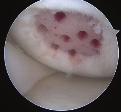
32 Management of Knee Cartilage Injuries in Basketball 381
o btaining X-rays, four views are suggested: bilat- mapping which is sensitive to cartilage proteo-
eral standing anterior posterior (AP), 45° of flex- glycan depletion [20, 21]. However, whether
ion weight-bearing posterior anterior (PA symptoms correlate with imaging findings
“Rosenburg”), non-weight-bearing 30° of flexion should always be considered. A study in basket-
true laterals, and patella sunrise view ball players found that 47.5% of the 40 knees
(“Merchant”). In addition, physicians should included in the study had asymptomatic carti-
obtain a mechanical axis X-ray. Specifically, lage lesions on MRI.
long-leg alignment views allow for the determi-
nation of the mechanical axis and to evaluate for
alignment. These images are necessary to rule 32.2.5 Diagnostic Arthroscopy
out bony defects and determine the alignment of
the joint. Radiographs should be evaluated for The most accurate test for diagnosis and grading
multiple findings such as radiolucencies, sub- of an FCD is diagnostic arthroscopy. This allows
chondral cysts, sclerosis, fragmentation, loose for visualization of the cartilage defect and allows
bodies, joint space narrowing, and physeal status for determination of lesion size, grade, and loca-
as these can affect the treatment plan. tion. There are two main grading systems for car-
tilage defects. The first is the International
Cartilage Repair Society grading system (ICRS)
32.2.4 MRI [22]. This cartilage grading system ranges from a
score of 0 to 4 based on the depth of the defect
MRI can provide more information, and is the from nearly normal to penetration beyond the
most sensitive modality, to evaluate cartilage subchondral bone. The other commonly utilized
defects. However, diagnostic accuracy based on grading system is the Outerbridge cartilage score,
MRI compared to arthroscopy has been shown which is based on the appearance of the cartilage
to be in part dependent on the severity of the defect, including the presence of swelling, frag-
cartilage defect (e.g., Outerbridge grade 3–4) mentation, and erosion [23]. The findings on
[17]. Conventional MRI methods include diagnostic arthroscopy including the severity,
T1-weighted and T2-weighted imaging and can size, depth, and location of the lesion will dictate
provide morphological and physiological infor- next steps in treatment.
mation about a patient’s knee. However, fat-sup-
pressed sequences such as T2-weighted fast
spin echo (FSE), and T1-weighted spoiled gra- 32.3 Conservative Management
dient-echo that allow for enhanced contrast
between fluid and cartilage provide improved Conservative management is generally the initial
sequences producing images with intermediate approach and is used in patients with mild symp-
cartilage signal and bright fluid signal [18]. toms or small lesions as its goal is to reduce
Newer 3D FSE and 3D multi-echo gradient- symptoms instead of reversing or fixing the
echo sequences further improve this distinction underlying lesion [24]. Types of conservative
[19]. Other novel technologies include delayed treatment include analgesics, chondroprotective
gadolinium-enhanced MRI for cartilage agents (glucosamine, chondroitin), steroid injec-
(dGEMRIC). dGEMRIC is sensitive to glycos- tions, physical therapy, and knee bracing, and
aminoglycan distribution in cartilage and allows these are especially useful mid-season to allow
visualization of areas of glycosaminoglycan for players to return to play with symptomatic
depletion; however, it requires a double-dose IV relief [25]. However, activity modification is also
contrast injection. Other techniques include T2 recommended as part of conservative manage-
relaxation time mapping, which is sensitive to ment, which may be a challenge in basketball
the cartilage–collagen matrix and water distri- players. Studies on the role of conservative man-
bution within the articular cartilage, and T1rho agement in athletes is limited, with one study382 H. P. Huddleston et al.
showing that 22 of 28 athletes had successful such as microfracture (which can be augmented
results of conservative treatment but continued to with other biological treatments or scaffolds).
have radiographic chondral abnormalities at fol- OAT is also often used for this subset of patients
low-up 14 years later [25]. Therefore, depending where the chondral lesion is small, especially in
on the size of the defect, surgery, where return to those with higher activity levels [27]. As lesions
sport has been studied, may be preferred in high- become greater than 2.5 cm, these can be treated
level athletes when conservative treatment fails. with OCA and MACI. Debridement is generally
the first-line treatment, especially in lesions32 Management of Knee Cartilage Injuries in Basketball 383
ment reconstruction or meniscal excision or repair
can occur concomitantly with the cartilage pro-
cedure. In patients with malalignment, an osteot-
omy should be considered. An osteotomy, such
as a high tibial osteotomy or a distal femoral oste-
otomy, can be utilized in patients with varus or
valgus malalignment, respectively. Furthermore,
an anteromedialization, an osteotomy of the tib-
ial tubercle, can be utilized in patients with patel-
lofemoral chondral defects [34].
32.5 Surgical Techniques
32.5.1 Abrasion Chondroplasty Fig. 32.1 Intraoperative image illustrating marrow stim-
ulation to medial femoral condyle
Chondroplasty is one of the most frequently per-
formed arthroscopic procedures. The goal of chon-
droplasty is to smooth over areas of fragmented rounding area the same way as in an abrasion
and damaged cartilage. This can be performed chondroplasty down to the subchondral bone
with a curved shaver that allows for the ability to including the calcified cartilage layer. Once this
reach most areas of the knee. The tip of the shaver is removed, a microfracture drill using Kirschner
is then used to gently remove unstable cartilage wires, fluted drill, wires, or angulated awl is used
and the calcified cartilage layer within the carti- to create holes 2–4 mm apart, releasing the under-
lage defect while care is taken to not disturb lying bone marrow cells into the cartilage defect
healthy cartilage and underlying subchondral bone which can be observed [37, 38]. In addition,
[35]. Specialized curettes, such as a D-curette or nanofracture techniques, which utilize a smaller
ring curette, can also be utilized in this situation. diameter drill are still being investigated [39].
Newer iterations of marrow stimulation are
still being investigated. These newer procedures
32.5.2 Marrow Stimulation augment the same microfracture procedure with
additional biologics, such as bone marrow aspi-
Microfracture was originally developed by rate concentrate (BMAC) or platelet-rich plasma
Steadman et al. over 20 years ago to treat small (PRP). However, whether these additions provide
chondral defects [36]. The goal of marrow stimu- any long-term benefit in patient outcomes still
lation is to stimulate cartilage defect healing with remains unclear [40, 41].
pluripotent progenitor cells, cytokine, growth
factors, and proteins from within the bone mar-
row. During this procedure, multiple small holes 32.5.3 Osteochondral Autograft
are made in the subchondral bone to stimulate the Transplantation (OAT)
cartilage (Fig. 32.1). When performing this pro-
cedure, the first step is an examination under OAT is generally indicated for patients who have
anesthesia followed by a 10-point arthroscopy to smaller higher-grade lesions and are younger and
examine all surfaces of the knee joint and to more active. OAT is performed by removing a
ensure that only a localized lesion is present. small area of healthy cartilage in an area of the
Then the next step is to prepare the osteochondral joint that is mainly non-load-bearing and placing
defect, removing any flaps, and debriding the sur- it into the chondral defect, which can be per-384 H. P. Huddleston et al.
formed either open or arthroscopically. In sur-
gery, the patient is positioned supine or the limb
can be placed in a leg holder with a tourniquet
and an examination under anesthesia is per-
formed. A small parapatellar vertical portal inci-
sion is then made, and a diagnostic arthroscopy is
performed to examine the cartilage surface.
During diagnostic arthroscopy, the cartilage
defect area is surveyed with a probe to determine
defect size and confirm no other cartilage injuries
are present.
At the cartilage defect location, a guide pin is
placed in the center of the cartilage defect, per-
pendicularly. A cannulated reamer is then placed
over the guide pin, and the guide pin is subse-
quently removed. The depth of the lesion is mea-
Fig. 32.2 Intraoperative image illustrating an osteochon-
sured with a cannulated dilator. dral allograft transplantation to lateral femoral condyle
The area of which to harvest the healthy carti-
lage from is predetermined using MRI. Graft har-
vest sizes are 6, 8, or 10 mm. Commonly, the harvest In the dowel grafting technique, a dowel of
graft is taken from the lateral trochlea and lateral similar size to the cartilage lesion is selected. A
femoral condyle. An appropriately sized harvester guidewire is positioned using sizers into the cen-
is then placed perpendicular to the graft harvest ter of the cartilage defect, and the dowel and the
location and is inserted into the subchondral bone to socket are drilled to a depth between 5 and 6 mm.
a depth of 10–15 mm with a mallet. The harvester is The allograft is harvested to the desired size
then axially loaded and turned 90° clockwise, then using a reamer from a matching zone and is
counterclockwise before being removed. A mallet is inserted into the defect [42, 43]. This press-fit
then used to fragment the graft from the surround- technique is often preferred as it eliminates the
ing cartilage, and the plug is removed. The graft is need for additional fixation (Fig. 32.2). In con-
then inspected, with any bony debris removed, and trast, in the free-shell technique, a donor graft is
shaved so that it is 1 mm shallower than the carti- matched to the defect site, inserted, and fixed
lage defect. The graft is then replaced into the joint with screws.
and is gently tapped into place. Larger defects often require the use of multi-
ple plugs in what is termed “snowman tech-
nique” or “mastercard technique.” This involves
32.5.4 Osteochondral Allograft placing and fixing the first plug, then drilling a
Transplantation (OCA) second recipient site adjacent to, or partially
over the first defect. However, based on prior
OCA is often used in patients with larger (>2 cm) studies, the snowman technique has been shown
lesions. In the operating room, the patient is posi- to provide inferior results compared to a one-
tioned supine with a tourniquet. The procedure plug technique [43, 44].
begins with a knee examination under anesthesia.
A lateral or medial parapatellar incision is then
made to access the FCD. There are two general 32.5.5 Autologous Chondrocyte
techniques that exist for OCA: cylindrical press- Implantation (ACI)
fit plugs or free-shell grafts. Whichever technique
is used, donor tissue must be size matched to Autologous chondrocyte implantation occurs
individual patients based on X-ray, CT, or MRI over the course of two procedures with ex vivo
measurement. chondrocyte expansion between procedures.32 Management of Knee Cartilage Injuries in Basketball 385
which is when a preformed biodegradable por-
cine type I/III matrix is utilized as a scaffold
for the collected and cultured chondrocyte
cells. In this procedure, the matrix is inserted
into the defect and then fixated to the surround-
ing cartilage with fibrin glue without the need
for suturing.
32.5.6 Novel Techniques
Newer techniques include autologous and allo-
genic minced cartilage (such as De Novo, biocarti-
lage, and cartiform), which are similar to an MACI
in that a collagen-chondrotoin scaffold is used to
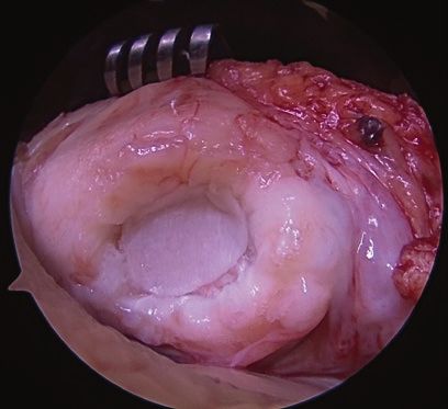
Fig. 32.3 Intraoperative image illustrating an autologous model cultured chondrocytes [49, 50]. Minced
chondrocyte implantation on the patella cartilage can be utilized instead of cultured chon-
drocytes. In this case, only one procedure is needed
as the cartilage is harvested and reimplanted in the
The initial procedure is a diagnostic arthroscopy same procedure [50]. In addition, fibrin glue is
with cartilage biopsy. During this procedure, the used to attach the minced pieces of cartilage
lesion size and grade are examined, and 200– together and attach the flap in addition to sutures to
300 mg of articular cartilage is harvested from a ensure fixation to the underlying subchondral bone
non-loading bearing surface of the knee. The [51]. A cartiform allograft is a cryopreserved
collected cartilage is then processed via an osteochondral allograft scaffold that can be used
enzymatic digestion process and is then cultured as an alternative to ACI and, similarly to minced
for 3–6 weeks. cartilage, can be implanted with fibrin glue.
In the second procedure, the harvested chon- Biocartilage is a cartilage scaffold that is hydrated
drocytes are reimplanted into the defect with PRP and can be used to fill defects after a
site (Fig. 32.3). It begins with the patient supine microfracture procedure. All of these newer tech-
with a tourniquet applied. The defect is then niques have limited data supporting their superior-
debrided with a round-eyed sharp curette to ity compared to traditional techniques. Future
expose subchondral bone. The original ACI studies are needed to evaluate the benefits and
technique involves a periosteal flap being sewn shortcomings of these newer technologies.
over the defect, followed by the injection of
cultures chondrocytes underneath the flap
(ACI-P), where the flap is harvested from the 32.6 Rehabilitation
proximal-medial tibia [45–47]. In contrast, in
ACI-C, a synthetic collagen membrane is used. 32.6.1 Patellofemoral
In either case, the flap is positioned over the
cartilage defect and sutured into place using 6-0 Rehabilitation for patients who undergo patello-
Vicryl. After the flap is checked to ensure a femoral cartilage procedures varies by institu-
watertight seal, the cultured cartilage cells are tion. However, it often includes cryotherapy,
then injected into lesion. In addition, a newer elevation, and a brace immediately after surgery.
“sandwich” technique with autologous bone Progressive passive motion and weight-bearing
grafting can also be utilized, especially in as tolerated can be implemented in the first few
patients with OCD [48]. days after surgery. Range of motion is then
A newer iteration of the ACI is an alterna- increased with a goal of 90 degrees of flexion in
tive technique called matrix ACI (MACI), the first 2 or 3 weeks [52].386 H. P. Huddleston et al.
32.6.2 Tibiofemoral microfracture compared to those who undergo
ACI at 5- and 10-years postoperatively [55, 56].
Patients who undergo cartilage repair of the tibio- In comparison to other sports, basketball play-
femoral joint undergo multiple phases of postop- ers have been shown to have inferior results
erative rehabilitation. During the first phase, until after microfracture [57].
1 week postoperative, weight-bearing is restricted Three studies have evaluated the success of
to less than 20% of body weight, range of motion microfracture in basketball players. The first
(ROM) is restricted from 0 to 30°, and a protec- study evaluated 24 NBA players who underwent
tive knee brace is used at all times. Patients can microfracture surgery [58]. Sixty-seven percent
progress to full passive motion within 1 week of of the players returned to play after the micro-
surgery and then full active range of motion by fracture procedure. However, abilities after
3 months post operatively. At 3 weeks patients return to sport were found to be decreased com-
are allowed to be fully weight-bearing while a pared to preoperatively in terms of both points
brace is utilized until around 3 months postopera- scored and minutes played. In addition, the
tively [53]. study found that patients were 8.15 times less
likely to remain in the NBA after the index year.
In the second study, 41 NBA players were eval-
32.7 Clinical Outcomes uated after microfracture procedure [59].
Eighty-three percent of these players returned to
When deciding on which surgical procedure to professional basketball after an average of
use in a basketball player with a chondral 9.2 months (4.32–14.08 months). Compared to
defect, outcomes and ability and time to return their preoperative abilities, those who did return
to sport are of critical importance. Patient to sport had a significantly decreased points
understanding and expectations should also be scored and steals per game. Furthermore, com-
formed by providing all available data on out- pared to other NBA players at a similar time
comes of cartilage procedures in basketball point in their career, microfracture patients had
players and other athletes as outcomes specific significantly fewer points scored per game,
to basketball players remain limited. An indi- games played per season, and free throw per-
vidual approach should be taken when evaluat- centages. The third study of 24 professional bas-
ing return to play as multiple factors influence ketball players found that 79% of patients
it beyond surgical choice such as age (>30 years) returned to sport and mean time to returning was
and BMI (>27 kg/m2) [54]. Furthermore, as 30 weeks. However, on average their player effi-
elite jumping athlete basketball players are ciency rating deceased by 2.7 and their minutes
unique from athletes in non-jumping sports, per game decreased by 3 after surgery [60].
and this should be considered.
32.7.2 Osteochondral Autograft
32.7.1 Microfracture Transplantation
Microfracture in basketball players is the most Osteochondral autograft transplantation has shown
well-studied cartilage procedure with no prior success, especially in terms of percentage of play-
reports on outcomes of isolated chondroplasty ers who return to sport. In the general population,
in basketball players. Outcomes of microfrac- OATs has been shown to provide significant ben-
ture in the general population have been posi- efit in 72% of patients at a mean of 10.2 years of
tive. For example, Weber et al. found a follow-up [61]. An additional study evaluated
statistically significant increase in all patient- short- to mid-term outcomes in 112 patients who
reported outcomes (PROs) after a mean follow- underwent OAT and found that both the VAS pain
up of 5.7 years. Furthermore, similar results (7.14 ± 0.19 vs. 3.74 ± 0.26) and WOMAC
have been shown in patients who undergo (134.88 ± 5.84 vs. 65.92 ± 5.34) significantly32 Management of Knee Cartilage Injuries in Basketball 387
improved at a mean follow-up of 26.2 ± 0.24 months improvements in pain and function [73]. Kaplan–
[62]. In comparison to microfracture, a meta-anal- Meier survival analysis revealed that the survival
ysis showed that OAT results in a lower risk of rate was 78.2% at 5 years and 50.7% at 10 years.
failure (11% vs. 32%) and a higher level of patients In terms of return-to-sport outcomes, two sys-
who return to activity [63]. tematic reviews have found that return to sport
Furthermore, OAT has been shown to have a ranges from 82% and 84%, respectively [27, 64].
higher rate of patients who returned to sport when An additional study found a rate of 73%; how-
comparing procedure type: between 89% and ever, they found that duration and frequency of
94% [27, 64, 65]. A systematic review found that exercise significantly decreased postoperatively.
based on seven articles, the mean time for return An additional study found that 64.5% of patients
to competition after OAT was 5.6 months were able to return to sport at a competitive level
(3–14 months) [65, 66]. No study specifically [74]. They also showed that previous surgery was
investigated the return-to-sport rate and time in the biggest factor that dictated return to sport
basketball players. level in their cohort. No studies investigated the
return to play after ACI in basketball players.
32.7.3 Osteochondral Allograft
Transplantation 32.8 Conclusion
Osteochondral allograft transplantation has been Focal chondral defects are common in athletes,
demonstrated to be a successful procedure in especially basketball players. Symptomatic
both the general population and among athletes. lesions can be addressed with conservative
After a mean of 12.3 years of follow-up 75% of measures initially, but often surgical interven-
patients demonstrated significant improvement tion is necessary but will depend on where the
in clinical outcomes [66]. A systematic review player is in the season. A range of surgical pro-
demonstrated that survivorship was 86.7% at cedures are used based on chondral size and
5 years and 78.7% at 10 years [67–69]. In the location, including abrasion chondroplasty,
general athletic population, return to sport was microfracture, OCA, OAT, and ACI. While
seen in 72–82% of patients at a mean of microfracture has been the most studied tech-
11 months [64, 69–71]. One study evaluated the nique in basketball players, OAT has been
return to sport in basketball players. The study shown to have the highest rate of return to sport
consisted of 11 basketball players with a total of in all athletes, although the literature remains
14 chondral lesions, the overall rate of return to limited. Future studies are needed to evaluate
sport 80%, and the average time to return to play other cartilage procedures specifically in bas-
14 months (6–26 months). The average lesion ketball players.
size was 509 mm2 [2] and the location of the
lesion varied and included the femoral condyle,
trochlea, and patella. Furthermore, this study References
found that there was no significant decline in
athletic performance after return to sport [72]. 1. Felson DT, Zhang Y, Hannan MT, et al. The inci-
dence and natural history of knee osteoarthritis in the
elderly, the Framingham osteoarthritis study. Arthritis
Rheum. 1995;38(10):1500–5. https://doi.org/10.1002/
32.7.4 Autologous Chondrocyte art.1780381017.
Implantation 2. Mankin H. The response of articular cartilage to mechan-
ical injury. J Bone Joint Surg Am. 1982;64(3):460–6.
https://doi.org/10.2106/00004623-198264030-00022.
Autologous chondrocyte implantation has been
3. Årøen A, Løken S, Heir S, et al. Articular carti-
shown to have successful outcomes. One study lage lesions in 993 consecutive knee arthroscopies.
evaluated a cohort of patients at a mean 6.2 years Am J Sports Med. 2004;32(1):211–5. https://doi.
follow-up, and all patients demonstrated significant org/10.1177/0363546503259345.388 H. P. Huddleston et al.
4. Curl WW, Krome J, Gordon ES, Rushing J, Smith Surg. 2010;19(04):285–95. https://doi.org/10.105
B, Poehling GG. Cartilage injuries: a review of 5/s-0030-1248121.
31,516 knee arthroscopies. Arthrosc J Arthrosc Relat 16. Spindler KP, Schils JP, Bergfeld JA, et al. Prospective
Surg. 1997;13(4):456–60. https://doi.org/10.1016/ study of osseous, articular, and meniscal lesions
s0749-8063(97)90124-9. in recent anterior cruciate ligament tears by mag-
5. Solheim E, Krokeide A, Melteig P, Larsen A, Strand netic resonance imaging and arthroscopy. Am
T, Brittberg M. Symptoms and function in patients J Sports Med. 1993;21(4):551–7. https://doi.
with articular cartilage lesions in 1,000 knee arthros- org/10.1177/036354659302100412.
copies. Knee Surg Sports Traumatol Arthrosc. 17. Guettler JH, Demetropoulos CK, Yang KH, Jurist
2016;24(5):1610–6. https://doi.org/10.1007/ KA. Osteochondral defects in the human knee.
s00167-014-3472-9. Am J Sports Med. 2004;32(6):1451–8. https://doi.
6. Widuchowski W, Widuchowski J, Trzaska T. Articular org/10.1177/0363546504263234.
cartilage defects: study of 25,124 knee arthroscopies. 18. Recht M, Bobic V, Burstein D, et al. Magnetic
Knee. 2007;14(3):177–82. https://doi.org/10.1016/j. resonance imaging of articular cartilage. Clin
knee.2007.02.001. Orthop Relat R. 2001;391:S379–96. https://doi.
7. Everhart JS, Boggs Z, DiBartola AC, Wright B, org/10.1097/00003086-200110001-00035.
Flanigan DC. Knee cartilage defect characteristics 19. Yuen J, Hung J, Wiggermann V, et al. Multi-echo
vary among symptomatic recreational and competi- GRE imaging of knee cartilage. J Magn Reson
tive scholastic athletes eligible for cartilage restora- Imaging. 2017;45(5):1502–13. https://doi.org/10.1002/
tion surgery. Cartilage. 2019.:1947603519833144; jmri.25438.
https://doi.org/10.1177/1947603519833144. 20. Gold GE, Chen CA, Koo S, Hargreaves BA, Bangerter
8. FLANIGAN DC, HARRIS JD, TRINH TQ, NK. Recent advances in MRI of articular cartilage.
SISTON RA, BROY RH. Prevalence of chondral AJR Am J Roentgenol. 2009;193(3):628–38. https://
defects in athletes’ knees. Med Sci Sports Exerc. doi.org/10.2214/ajr.09.3042.
2010;42(10):1795–801. https://doi.org/10.1249/ 21. Crema MD, Roemer FW, Marra MD, et al. Articular
mss.0b013e3181d9eea0. cartilage in the knee: current MR imaging techniques
9. Kaplan LD, Schurhoff MR, Selesnick H, Thorpe M, and applications in clinical practice and research.
Uribe JW. Magnetic resonance imaging of the knee Radiographics. 2011;31(1):37–61. https://doi.
in asymptomatic professional basketball players. org/10.1148/rg.311105084.
Arthrosc J Arthrosc Relat Surg. 2005;21(5):557–61. 22. BRITTBERG M, WINALSKI CS. Evaluation
https://doi.org/10.1016/j.arthro.2005.01.009. of cartilage injuries and repair. J Bone
10. Walczak BE, McCulloch PC, Kang RW, Zelazny A, Joint Surg Am. 2003;85:58–69. https://doi.
Tedeschi F, Cole BJ. Abnormal findings on knee mag- org/10.2106/00004623-200300002-00008.
netic resonance imaging in asymptomatic NBA play- 23. Outerbridge R. The etiology of chondromalacia
ers. J Knee Surg. 2008;21(1):27–33. https://doi.org/1 patellae. J Bone Joint Surg Br. 1961;43-B(4):752–7.
0.1055/s-0030-1247788. https://doi.org/10.1302/0301-620x.43b4.752.
11. Hirshorn KC, Cates T, Gillogly S. Magnetic reso- 24. Wernecke C, Braun HJ, Dragoo JL. The effect of
nance imaging–documented chondral injuries intra-articular corticosteroids on articular cartilage.
about the knee in college football players: 3-year Orthop J Sports Med. 2015;3(5):2325967115581163.
National Football League Combine Data. Arthrosc J https://doi.org/10.1177/2325967115581163.
Arthrosc Relat Surg. 2010;26(9):1237–40. https://doi. 25. Messner K, Maletius W. The long-term prognosis for
org/10.1016/j.arthro.2010.01.025. severe damage to weight-bearing cartilage in the knee:
12. Kohn D. Arthroscopy in acute injuries of ante- a 14-year clinical and radiographic follow-up in 28
rior cruciate-deficient knees: fresh and old intraar- young athletes. Acta Orthop Scand. 2009;67(2):165–
ticular lesions. Arthrosc J Arthrosc Relat Surg. 8. https://doi.org/10.3109/17453679608994664.
1986;2(2):98–102. https://doi.org/10.1016/ 26. Krych AJ, Pareek A, King AH, Johnson NR, Stuart
s0749-8063(86)80022-6. MJ, Williams RJ. Return to sport after the surgi-
13. Houck DA, Kraeutler MJ, Belk JW, Frank RM, cal management of articular cartilage lesions in the
McCarty EC, Bravman JT. Do focal chondral defects knee: a meta-analysis. Knee Surg Sports Traumatol
of the knee increase the risk for progression to osteo- Arthrosc. 2017;25(10):3186–96. https://doi.
arthritis? A review of the literature. Orthop J Sports org/10.1007/s00167-016-4262-3.
Med. 2018;6(10):2325967118801931. https://doi. 27. Cole BJ, Farr J. Putting it all together. Operative Tech
org/10.1177/2325967118801931. Orthop. 2001;11(2):151–4. https://doi.org/10.1016/
14. Cook J, Khan K, Kiss Z, Griffiths L. Patellar tendi- s1048-6666(01)80025-2.
nopathy in junior basketball players: a controlled 28. Cameron JI, Pulido PA, McCauley JC, Bugbee
clinical and ultrasonographic study of 268 patel- WD. Osteochondral allograft transplantation of the
lar tendons in players aged 14–18 years. Scand J femoral trochlea. Am J Sports Med. 2016;44(3):633–
Med Sci Sports. 2000;10(4):216–20. https://doi. 8. https://doi.org/10.1177/0363546515620193.
org/10.1034/j.1600-0838.2000.010004216.x. 29. Briggs DT, Sadr KN, Pulido PA, Bugbee WD. The
15. Gomoll A, Minas T, Farr J, Cole B. Treatment of use of osteochondral allograft transplantation
chondral defects in the patellofemoral joint. J Knee for primary treatment of cartilage lesions in the32 Management of Knee Cartilage Injuries in Basketball 389
knee. Cartilage. 2015;6(4):203–7. https://doi. Multifocal Lesions. Orthop J Sports Med. 2018;6(7_
org/10.1177/1947603515595072. suppl4):2325967118S0009. https://doi.org/10.1177/2325
30. von Keudell A, Han R, Bryant T, Minas T. Autologous 967118s00093.
chondrocyte implantation to isolated Patella cartilage 42. Pisanu G, Cottino U, Rosso F, et al. Large osteochon-
defects. Cartilage. 2017;8(2):146–54. https://doi. dral allografts of the knee: surgical technique and
org/10.1177/1947603516654944. indications. Joints. 2018;06(01):042–53. https://doi.
31. Ebert JR, Schneider A, Fallon M, Wood DJ, Janes org/10.1055/s-0038-1636925.
GC. A comparison of 2-year outcomes in patients 43. Cole BJ, Pascual-Garrido C, Grumet RC. Surgical
undergoing tibiofemoral or patellofemoral matrix- management of articular cartilage defects in the knee.
induced autologous chondrocyte implantation. Am J Bone Joint Surg Am. 2009;91(7):1778–90.
J Sports Med. 2017;45(14):3243–53. https://doi. 44. Godin JA, Sanchez G, Cinque ME, Chahla J, Kennedy
org/10.1177/0363546517724761. NI, Provencher MT. Osteochondral allograft trans-
32. Gomoll AH, Gillogly SD, Cole BJ, et al. Autologous plantation for treatment of medial femoral condyle
chondrocyte implantation in the Patella. Am J defect. Arthrosc Tech. 2017;6(4):e1239–44. https://
Sports Med. 2014;42(5):1074–81. https://doi. doi.org/10.1016/j.eats.2017.04.010.
org/10.1177/0363546514523927. 45. Brittberg M, Lindahl A, Nilsson A, Ohlsson
33. Rosso F, Rossi R, Governale G, et al. Tibial C, Isaksson O, Peterson L. Treatment of deep
tuberosity Anteromedialization for patellofemo- cartilage defects in the knee with autologous
ral chondral disease: prognostic factors. Am J chondrocyte transplantation. New Engl J Med.
Sports Med. 2017;45(7):1589–98. https://doi. 1994;331(14):889–95. https://doi.org/10.1056/
org/10.1177/0363546517690387. nejm199410063311401.
34. Ward BD, Lubowitz JH. Basic knee arthroscopy part 46. Bartlett W, Skinner J, Gooding C, et al. Autologous
4: chondroplasty, meniscectomy, and cruciate liga- chondrocyte implantation versus matrix-induced
ment Evaluation. Arthrosc Tech. 2013;2(4):e507–8. autologous chondrocyte implantation for osteochon-
https://doi.org/10.1016/j.eats.2013.07.011. dral defects of the knee: a prospective, randomised
35. Steadman RJ, Rodkey WG, Briggs KK. Microfracture. study. J Bone Joint Surg Br. 2005;87-B(5):640–5.
Cartilage. 2010;1(2):78–86. https://doi. https://doi.org/10.1302/0301-620x.87b5.15905.
org/10.1177/1947603510365533. 47. Minas T, Ogura T, Headrick J, Bryant T. Autologous
36. Holt K, Sorhaindo M, Coady C, Wong I. Arthroscopic chondrocyte implantation “Sandwich” tech-
Treatment of Medial Femoral Knee Osteochondral nique compared with autologous bone grafting
Defect Using Subchondroplasty and Chitosan-Based for deep osteochondral lesions in the knee. Am
Scaffold. Arthrosc Tech. 2019;8. (Arthroscopy 18 J Sports Med. 2018;46(2):322–32. https://doi.
2002):e413–8. https://doi.org/10.1016/j.eats.2018. org/10.1177/0363546517738000.
11.022. 48. Salzmann GM, Calek A-K, Preiss S. Second-
37. Mithoefer K, McAdams T, Williams RJ, Kreuz PC, generation autologous minced cartilage repair tech-
Mandelbaum BR. Clinical efficacy of the microfrac- nique. Arthrosc Tech. 2017;6(1):e127–31. https://doi.
ture technique for articular cartilage repair in the knee. org/10.1016/j.eats.2016.09.011.
Am J Sports Med. 2009;37(10):2053–63. https://doi. 49. Niethammer TR, Pietschmann MF, Horng A, et al.
org/10.1177/0363546508328414. Graft hypertrophy of matrix-based autologous chon-
38. Tahta M, Akkaya M, Gursoy S, Isik C, Bozkurt drocyte implantation: a two-year follow-up study of
M. Arthroscopic treatment of osteochondral lesions NOVOCART 3D implantation in the knee. Knee Surg
of the talus: nanofracture versus hyaluronic acid- Sports Traumatol Arthrosc. 2014;22(6):1329–36.
based cell-free scaffold with concentration of autol- https://doi.org/10.1007/s00167-013-2454-7.
ogous bone marrow aspirate. J Orthop Surg-hong 50. Massen F, Inauen C, Harder L, Runer A, Preiss
K. 2017;25(2):2309499017717870. https://doi. S, Salzmann G. One-step autologous minced car-
org/10.1177/2309499017717870. tilage procedure for the treatment of knee joint
39. Mancò A, Goderecci R, Rughetti A, et al. Microfracture chondral and osteochondral lesions: a series of 27
versus microfracture and platelet-rich plasma: patients with 2-year follow-up. Orthop J Sports
arthroscopic treatment of knee chondral lesions. A Med. 2019;7(6):2325967119853773. https://doi.
two-year follow-up study. Joints. 2016;04(03):142–7. org/10.1177/2325967119853773.
https://doi.org/10.11138/jts/2016.4.3.142. 51. Mestriner A, Ackermann J, Gomoll
40. Arshi A, Fabricant PD, Go DE, Williams RJ, AH. Patellofemoral cartilage repair. Curr Rev
McAllister DR, Jones KJ. Can biologic augmenta- Musculoskelet Med. 2018;11(2):188–200. https://doi.
tion improve clinical outcomes following microfrac- org/10.1007/s12178-018-9474-3.
ture for symptomatic cartilage defects of the knee? 52. Edwards PK, Ackland T, Ebert JR. Clinical reha-
A Systematic Review. Cartilage. 2018;9(2):146–55. bilitation guidelines for matrix-induced autologous
https://doi.org/10.1177/1947603517746722. chondrocyte implantation on the tibiofemoral joint. J
41. Cotter EJ, Hannon CP, Lansdown DA, Frank RM, Orthop Sports Phys Ther. 2013;44(2):102–19. https://
Waterman B, Cole BJ. Clinical Outcomes of Multiple doi.org/10.2519/jospt.2014.5055.
Osteochondral Allograft Transplantation of the 53. Minhas SV, Kester BS, Larkin KE, Hsu WK. The
Knee: An Analysis of Snowman Technique and effect of an orthopaedic surgical procedure390 H. P. Huddleston et al.
in the National Basketball Association. Am J 64. Kirsch JM, Thomas JR, Khan M, Townsend WA,
Sports Med. 2016;44(4):1056–61. https://doi. Lawton JN, Bedi A. Return to play after osteochon-
org/10.1177/0363546515623028. dral autograft transplantation of the capitellum: a
54. Kraeutler MJ, Belk JW, Purcell JM, McCarty systematic review. Arthrosc J Arthrosc Relat Surg.
EC. Microfracture versus autologous chondro- 2017;33(7):1412–20.e1. https://doi.org/10.1016/j.
cyte implantation for articular cartilage lesions in arthro.2017.01.046.
the knee: a systematic review of 5-year outcomes. 65. Assenmacher AT, Pareek A, Reardon PJ, Macalena
Am J Sports Med. 2018;46(4):995–9. https://doi. JA, Stuart MJ, Krych AJ. Long-term outcomes
org/10.1177/0363546517701912. after osteochondral allograft: a systematic review
55. Pellegrino M, Trinchese E, Bisaccia M, et al. at long-term follow-up of 12.3 years. Arthrosc J
Long-term outcome of grade III and IV chondral Arthrosc Relat Surg. 2016;32(10):2160–8. https://doi.
injuries of the knee treated with Steadman micro- org/10.1016/j.arthro.2016.04.020.
fracture technique. Clin Cases Miner Bone Metab. 66. Familiari F, Cinque ME, Chahla J, et al. Clinical out-
2016;13(3):237–40. https://doi.org/10.11138/ comes and failure rates of osteochondral allograft
ccmbm/2016.13.3.237. transplantation in the knee: a systematic review.
56. Schallmo MS, Singh SK, Barth KA, Freshman RD, Am J Sports Med. 2018;46(14):3541–9. https://doi.
Mai HT, Hsu WK. A cross-sport comparison of org/10.1177/0363546517732531.
performance-based outcomes of professional athletes 67. Frank RM, Lee S, Levy D, et al. Osteochondral
following primary microfracture of the knee. Knee. allograft transplantation of the knee: analysis of fail-
2018;25(Am J Sports Med 14 1986):692–8. https:// ures at 5 years. Am J Sports Med. 2017;45(4):864–74.
doi.org/10.1016/j.knee.2018.04.008. https://doi.org/10.1177/0363546516676072.
57. Namdari S, Baldwin K, Anakwenze O, Park M-J, 68. Nielsen SE, McCauley JC, Pulido PA, Bugbee
Huffman RG, Sennett BJ. Results and performance WD. Return to sport and recreational activity after
after microfracture in National Basketball Association osteochondral allograft transplantation in the knee.
Athletes. Am J Sports Med. 2009;37(5):943–8. https:// Am J Sports Med. 2017;45(7):1608–14. https://doi.
doi.org/10.1177/0363546508330150. org/10.1177/0363546517694857.
58. Harris JD, Walton DM, Erickson BJ, et al. Return 69. Crawford ZT, Schumaier AP, Glogovac G, Grawe
to sport and performance after microfracture in the BM. Return to sport and sports-specific outcomes
knees of National Basketball Association Players. after osteochondral allograft transplantation in the
Orthop J Sports Med. 2013;1(6):2325967113512759. knee: a systematic review of studies with at least 2
https://doi.org/10.1177/2325967113512759. years’ mean follow-up. Arthrosc J Arthrosc Relat
59. Cerynik DL, Lewullis GE, Joves BC, Palmer MP, Tom Surg. 2019;35:1880. https://doi.org/10.1016/j.
JA. Outcomes of microfracture in professional basket- arthro.2018.11.064.
ball players. Knee Surg Sports Traumatol Arthrosc. 70. Krych AJ, Robertson CM, Williams RJ, Group
2009;17(9):1135–9. https://doi.org/10.1007/ C. Return to athletic activity after osteochon-
s00167-009-0765-5. dral allograft transplantation in the knee. Am
60. Pareek A, Reardon PJ, Maak TG, Levy BA, Stuart J Sports Med. 2012;40(5):1053–9. https://doi.
MJ, Krych AJ. Long-term outcomes after osteochon- org/10.1177/0363546511435780.
dral autograft transfer: a systematic review at mean 71. Balazs GC, Wang D, Burge AJ, Sinatro AL, Wong
follow-up of 10.2 years. Arthrosc J Arthrosc Relat AC, Williams RJ. Return to play among elite basket-
Surg. 2016;32(6):1174–84. https://doi.org/10.1016/j. ball players after osteochondral allograft transplan-
arthro.2015.11.037. tation of full-thickness cartilage lesions. Orthop J
61. Pareek A, Reardon PJ, Macalena JA, et al. Sports Med. 2018;6(7):2325967118786941. https://
Osteochondral autograft transfer versus microfrac- doi.org/10.1177/2325967118786941.
ture in the knee: a meta-analysis of prospective com- 72. Pestka JM, Feucht MJ, Porichis S, Bode G, Südkamp
parative studies at midterm. Arthrosc J Arthrosc Relat NP, Niemeyer P. Return to sports activity and work
Surg. 2016;32(10):2118–30. https://doi.org/10.1016/j. after autologous chondrocyte implantation of the
arthro.2016.05.038. knee. Am J Sports Med. 2016;44(2):370–7. https://
62. Baltzer AW, Ostapczuk MS, Terheiden HP, Merk HR. doi.org/10.1177/0363546515614578.
Good short- to medium-term results after osteochon- 73. Nawaz SZ, Bentley G, Briggs TWR, Carrington RWJ,
dral autograft transplantation (OAT) in middle-aged Skinner JA, Gallagher KR, Dhinsa BS. Autologous
patients with focal, non-traumatic osteochondral chondrocyte implantation in the knee: mid-term to
lesions of the knee. Orthop Traumatol Surg Res. long-term results. 2014;96(10):824–30. https://doi.
2016;102(7):879–84. https://doi.org/10.1016/j.otsr. org/10.2106/JBJS.L.01695.
2016.06.004. 74. Zaffagnini S, Vannini F, Martino DA, et al. Low rate
63. Campbell AB, Pineda M, Harris JD, Flanigan of return to pre-injury sport level in athletes after car-
DC. Return to sport after articular cartilage repair tilage surgery: a 10-year follow-up study. Knee Surg
in athletes’ knees: a systematic review. Arthrosc J Sports Traumatol Arthrosc. 2018;27:1–9. https://doi.
Arthrosc Relat Surg. 2016;32(4):651–68.e1. https:// org/10.1007/s00167-018-5255-1.
doi.org/10.1016/j.arthro.2015.08.028.You can also read