CDK1 phosphorylates ULK1-ATG13 complex to regulate mitotic autophagy and Taxol chemosensitivity
←
→
Page content transcription
If your browser does not render page correctly, please read the page content below
bioRxiv preprint first posted online May. 10, 2019; doi: http://dx.doi.org/10.1101/634733. The copyright holder for this preprint
 (which was not peer-reviewed) is the author/funder, who has granted bioRxiv a license to display the preprint in perpetuity.
                                  It is made available under a CC-BY 4.0 International license.

           CDK1 phosphorylates ULK1-ATG13 complex to regulate
           mitotic autophagy and Taxol chemosensitivity
           Zhiyuan Li1, Xiaofei Tian1, Xinmiao Ji1, Dongmei Wang1 and Xin Zhang1,2*
           1High   Magnetic Field Laboratory, Key Laboratory of High Magnetic Field and Ion
           Beam Physical Biology, Hefei Institutes of Physical Science, Chinese Academy of
           Sciences, 230031, Hefei, Anhui, P. R. China
           2Institute of Physical Science and Information Technology, Anhui University,

           230601, Hefei, Anhui, P. R. China

           *Correspondence: xinzhang@hmfl.ac.cn (X.Z.)

           Abstract
           ULK1-ATG13 is the most upstream autophagy initiation complex that is
           phosphorylated by mTORC1 and AMPK to induce autophagy in asynchronous
           conditions. However, the phospho-regulation and function of ULK1-ATG13 in
           mitosis and cell cycle remains unknown. Here we show that ULK1-ATG13 complex
           is differentially regulated throughout the cell cycle. Notably, in mitosis, both ULK1
           and ATG13 are highly phosphorylated by CDK1/cyclin B, the key cell cycle
           machinery. Combining mass spectrometry and site-directed mutagenesis, we found
           that CDK1-induced ULK1-ATG13 phosphorylation positively regulates mitotic
           autophagy and Taxol chemosensitivity, and some phosphorylation sites occur in
           cancer patients. Moreover, double knockout of ULK1 and ATG13 could block cell
           cycle progression and significantly decrease cancer cell proliferation in cell line and
           mouse models. Our results not only bridge the mutual regulation between the core
           machineries of autophagy and mitosis, illustrate the mitotic autophagy regulation
           mechanism, but also provide ULK1-ATG13 as potential targets for cancer therapy.

                                                                       1
bioRxiv preprint first posted online May. 10, 2019; doi: http://dx.doi.org/10.1101/634733. The copyright holder for this preprint
 (which was not peer-reviewed) is the author/funder, who has granted bioRxiv a license to display the preprint in perpetuity.
                                  It is made available under a CC-BY 4.0 International license.

           Introduction
           Autophagy occurs at basal levels in most tissues to selectively eliminate unwanted
           cellular components and can also be induced in response to various physiological and
           pathological conditions. Evolutionarily conserved autophagy-related (ATG) proteins
           play essential roles in autophagy nucleation, elongation, autophagosome closure and
           maturation [1-3]. In higher eukaryotes, many ATG proteins have diverse
           physiologically vital roles not only in autophagy, but also in other ATG
           gene-dependent pathways, such as phagocytosis, secretion and exocytosis, innate
           immunity and cell cycle etc [2]. Although some ATG proteins such as ATG7, FIP200,
           Beclin-1 and ATG5 function in cell cycle and mitosis regulation [4-7], whether the
           most upstream member, serine/threonine UNC-51-like kinase (ULK1/ATG1) [1,
           8-11], participates in cell-cycle and mitosis regulation has not been investigated.
                On the other hand, although the currently established autophagy regulation
           mechanisms are mostly from asynchronous cells, in which only around 5% or less are
           in mitosis, recent studies suggest that autophagy is differentially regulated throughout
           the cell cycle [12-15], especially in mitosis [16, 17]. Although the autophagosome
           number at a fixed timepoint is much reduced in mitotic cells compared to interphase
           cells [16], the autophagic flux is actually active [15, 17, 18]. Moreover, it has been
           reported that multiple kinases are involved in both autophagy and mitosis [12, 14],
           indicating that these two cellular processes are intertwined. However, the mitotic
           autophagy regulation is still under explored.
                The only work that has investigated the mitotic autophagy regulation mechanism
           so far is by Furuya et al [16]. They reported reduced phosphatidylinositol-3-phosphate
           (PtdIns3P) in mitosis, which suggested decreased VPS34 complex activity. They
           further identified VPS34-Thr159 to be the mitotic specific- phosphorylation site by
           CDK1 (the mammalian homolog of Cdc2 in yeast), which is one of the
           cyclin-dependent kinases (CDKs) that coordinate with their cyclin partners to regulate
           cell cycle progression. However, whether other molecular mechanisms are involved in
           mitotic autophagy regulation is still unknown, especially the one that is responsible
           for the autophagy flux maintenance in mitosis.
                Although ULK1-ATG13, the core machinery for the ULK1 autophagy initiation
           complex, was regulated by phosphorylation primarily from mammalian
           target-of-rapamycin (mTOR) and adenosine monophosphate activated protein kinase
           (AMPK) to control autophagy induction in asynchronous cells [8, 9, 19, 20], its
           regulation mechanism and function in mitosis and the specific cell cycle is uncovered.
           Here we found that ULK1-ATG13 not only play essential roles in cell cycle
           progression, but also is directly phosphorylated by CDK1/cyclin B in mitosis to
           regulate mitotic autophagy and Taxol-induced cell death.
                                                                       2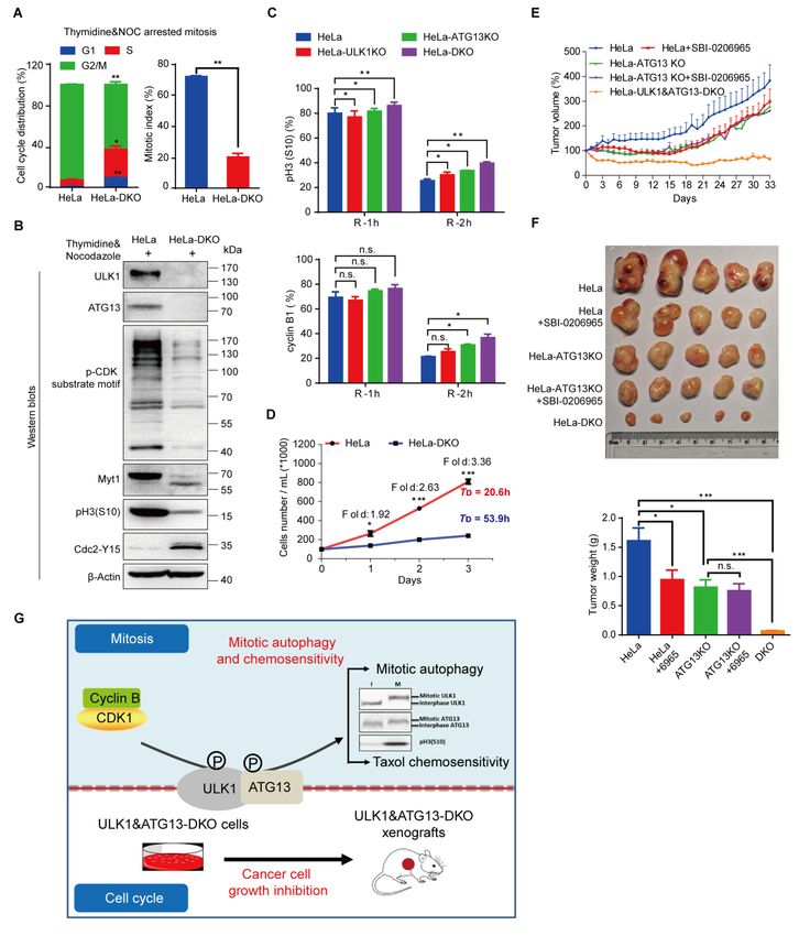
bioRxiv preprint first posted online May. 10, 2019; doi: http://dx.doi.org/10.1101/634733. The copyright holder for this preprint
 (which was not peer-reviewed) is the author/funder, who has granted bioRxiv a license to display the preprint in perpetuity.
                                  It is made available under a CC-BY 4.0 International license.

           Results

           ULK1-ATG13 is differentially regulated during cell cycle
           To dissect the underlying mechanism of ULK1-ATG13 regulation during cell cycle
           and mitosis, we synchronized HeLa cells using double-thymidine and nocodazole.
           Surprisingly, both ULK1 and ATG13 underwent a significant electrophoretic mobility
           shift in mitosis, while other ATGs such as ATG5, Beclin-1 or ATG101 did not
           (Figure 1A). In addition, thymidine was used in combination with RO-3306 [21], a
           specific CDK1 inhibitor , to synchronize cells to specific phases of mitosis. We found
           that the ULK1 and ATG13 electrophoretic mobility shift were closely correlated with
           mitotic progression (Figure 1B).
                Since phosphorylation-induced mobility shift is often indicative of
           phosphorylation on serine/threonine-proline residues [22], we seek to perform
           immunoprecipitation (IP) experiments and examined the ULK1 or ATG13 IP
           products using the motif antibodies for p-MAPK/CDK Substrate (PXS*P or
           S*PXR/K), p-CDK Substrate Motif [(K/H)pSP] and phospho-Threonine Proline
           [23-25]. Since mouse ULK1 has been validated in multiple studies [8, 9, 26-29], We
           constructed HEK-293T cell lines overexpressing FLAG-tagged mULK1,
           FLAG-tagged ATG13 or FLAG-tagged GFP control and performed
           immunoprecipitation in cells after thymidine block release with or without nocodazole
           (Figures 1C and 1D). Indeed, both the ULK1/FLAG antibodies and the CDK or
           MAPK/CDK substrate-specific motif antibodies could recognize a significant amount
           of ULK1 or ATG13 in both upshifted and non-shifted bands in mitotic cells compared
           with cells in other phases, which further indicates that ULK1-ATG13 was
           differentially regulated at both protein and phosphorylation levels (Figures 1C and
           1D).

                                                                       3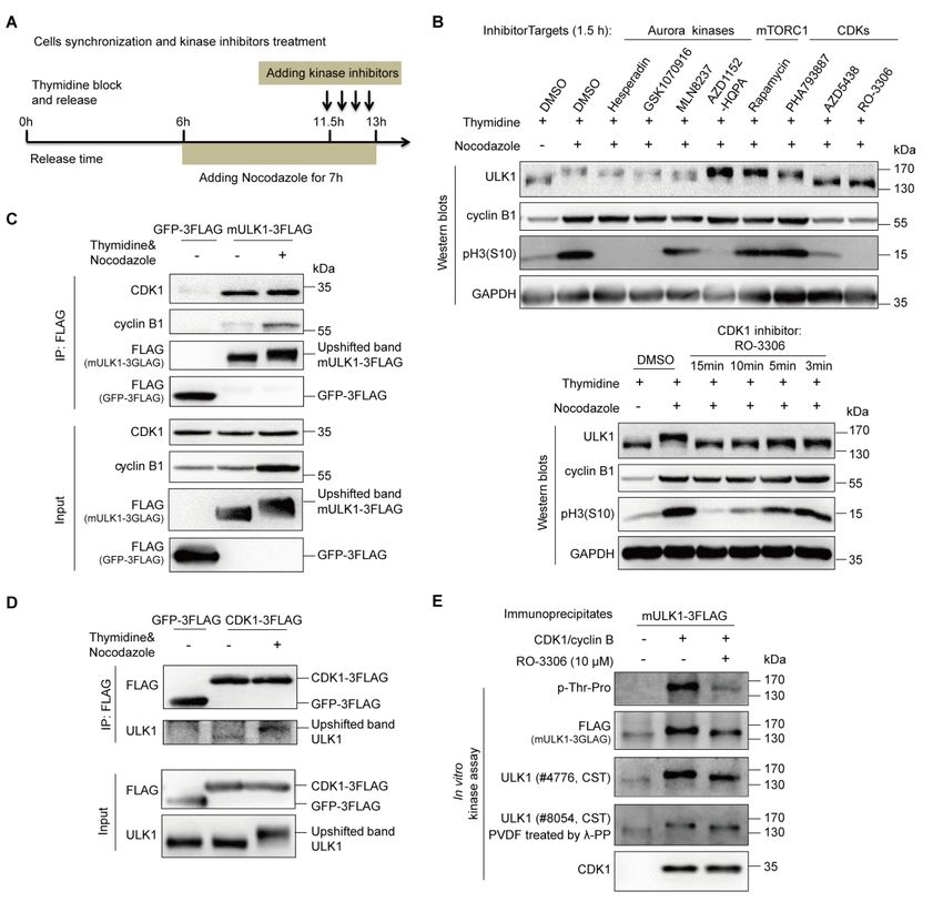
bioRxiv preprint first posted online May. 10, 2019; doi: http://dx.doi.org/10.1101/634733. The copyright holder for this preprint
 (which was not peer-reviewed) is the author/funder, who has granted bioRxiv a license to display the preprint in perpetuity.
                                  It is made available under a CC-BY 4.0 International license.

           Figure 1. ULK1-ATG13 is differentially regulated during the cell cycle.
           (A) ULK1-ATG13 shows a mobility shift in mitosis. HeLa cells synchronized by
           double-thymidine release in the presence or absence of nocodazole were subjected to SDS-PAGE
           and Western blots analysis.
                                                                       4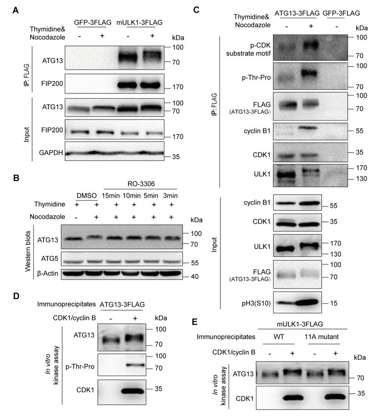
bioRxiv preprint first posted online May. 10, 2019; doi: http://dx.doi.org/10.1101/634733. The copyright holder for this preprint
 (which was not peer-reviewed) is the author/funder, who has granted bioRxiv a license to display the preprint in perpetuity.
                                  It is made available under a CC-BY 4.0 International license.

           (B) ULK1-ATG13 undergoes band shift during mitotic progression. HeLa cells synchronized by
           double-thymidine and RO-3306 were released into mitosis for Western blots analysis.
           (C-D) ULK1-ATG13 upshifted band in mitosis could be recognized by CDK substrate-specific
           antibodies. 293T cells stably expressing FLAG-tagged mULK1 or ATG13 were synchronized by
           single-thymidine and released in the presence or absence of nocodazole. The
           co-immunoprecipitates and input were immunoblotted with specific antibodies. Various cell cycle
           markers were detected to show the respective phases of cell cycle.

           ULK1 is highly phosphorylated in mitosis
           To better understand the detailed ULK1-ATG13 regulation mechanisms in mitosis,
           ULK1 and ATG13 were investigated in detail respectively. The immunoprecipitated
           ULK1 shows an obvious band shift in mitosis, both on the Coomassie brilliant blue
           stained gel as well as on Western blots (Figure 2A). Using a phospho-serine/threonine
           antibody, we confirmed that ULK1 is highly phosphorylated on serine/threonine in
           mitotic cells compared with asynchronous cells (Figure 2A).
                To confirm that the band shift of ULK1 in mitosis is due to phosphorylation, we
           treated the ULK1 immunoprecipitation products with lambda phosphatase in the
           presence or absence of its inhibitors (Figure 2B). The FLAG-tagged mULK1 band is
           downshifted by lambda phosphatase treatment, which can be reversed by phosphatase
           inhibitors. Moreover, the phospho-serine/threonine detected band was also diminished
           after lambda phosphatase treatment, which can also be reversed by phosphatase
           inhibitors (Figure 2B). This confirmed that the electrophoretic mobility upshift of
           ULK1 in mitosis is due to serine/threonine phosphorylation.
                To rule out the possibility that the band shift was caused by nocodazole-induced
           microtubule disruption, but not mitosis per se, we used STLC, a specific Eg5 inhibitor
           [30], to synchronize HeLa cells to mitosis. We found that STLC could induce ULK1
           band shift similar to nocodazole treatment, although to a less extent (Figure 2C). The
           dramatic band shift phenomena were also verified for endogenous ULK1 in HCT 116
           (human colorectal cancer cells) and RPE1 (human retinal pigmented epithelial cells)
           (Figure 2D), as well as endogenous ULK1 and exogenous mouse ULK1 in
           HEK-293T and HeLa cells with or without FLAG-tagged mULK1 overexpression
           (Figures S1A and S1B). Therefore, ULK1 is highly phosphorylated in mitosis of
           multiple cell types.
                It should be mentioned that the original antibody we used for ULK1 (Cell
           signaling technology, #8054) did not detect the upshifted band, but only showed
           decreased signal in mitosis (Figure S1C, the upper blot). However, it is interesting
           that when the PVDF membrane was treated with lambda phosphatase to remove the
           phosphorylation, the upshifted band appeared (Figure S1C, the lower blot), which
           indicates that the mitotic ULK1 phosphorylation might interfere with the recognition
           of this specific antibody. Therefore, for all work other than Figure S1C and Figure 3E,
                                                                       5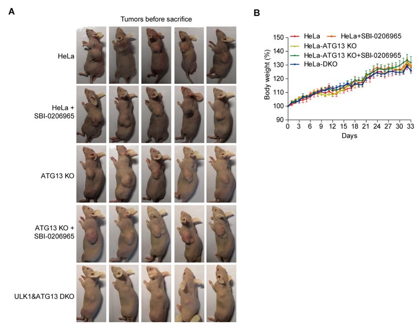
bioRxiv preprint first posted online May. 10, 2019; doi: http://dx.doi.org/10.1101/634733. The copyright holder for this preprint
 (which was not peer-reviewed) is the author/funder, who has granted bioRxiv a license to display the preprint in perpetuity.
                                  It is made available under a CC-BY 4.0 International license.

           we used ULK1 antibody (Cell signaling technology, #4776) instead because it is
           consistent with the FLAG antibody (detecting the FLAG-tagged mULK1) and
           Coomassie staining in the FLAG-tagged mULK1 expressing cells (Figure 2A).

           Figure 2. ULK1 is highly phosphorylated in mitosis.
           (A) ULK1 is phosphorylated in nocodazole-arrested mitosis. 293T cells overexpressing
           FLAG-tagged mULK1 or GFP were synchronized by single-thymidine and nocodazole. The
           immunoprecipitates using the FLAG antibody were subjected to Coomassie Brilliant Blue R-250
           staining and Western blots analysis. Statistical analysis for relative serine/threonine
           phosphorylated ULK1 was shown in Figure 2A, lower panel. n=4, **p < 0.01.
           (B) The immunoprecipitates from Figure 2A were treated with or without λ phosphatase in the
           presence or absence of phosphatase inhibitors and then subjected to Western blots analysis. λ PP,
           lambda phosphatase.
           (C) ULK1 undergoes similar mobility shift in both nocodazole- and STLC-arrested mitosis. HeLa
           cells synchronized with single-thymidine and nocodazole or STLC were analyzed by Western
           blots. The upper panel shows the immunoblotting and lower panel shows the ratio of upshifted and
           non-shifted ULK1. One-way ANOVA followed by Tukey's Multiple Comparison Test was used
           for the analysis. n=5, n.s., not significant, ***p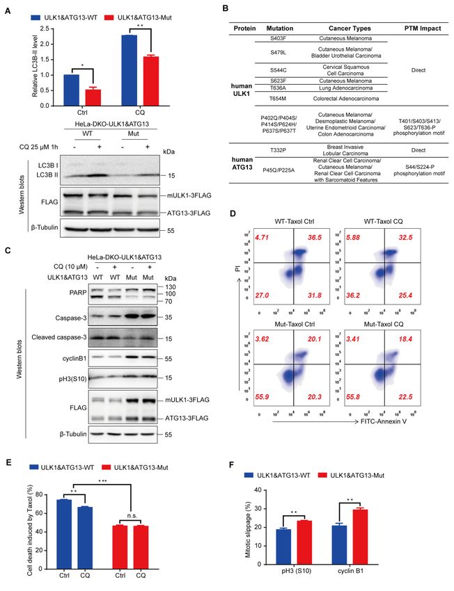
bioRxiv preprint first posted online May. 10, 2019; doi: http://dx.doi.org/10.1101/634733. The copyright holder for this preprint
 (which was not peer-reviewed) is the author/funder, who has granted bioRxiv a license to display the preprint in perpetuity.
                                  It is made available under a CC-BY 4.0 International license.

           and nocodazole synchronized mitotic HCT 116 and RPE1 cells analyzed by Western blots.

           ULK1 is a direct substrate of CDK1/cyclin B in mitosis
           In an attempt to elucidate the upstream kinase responsible for ULK1 phosphorylation
           in mitosis, we first used Scansite 3 (http://scansite3.mit.edu/) to predict the potential
           kinases with proline-directed serine/threonine motif preference, which indicated that
           CDK1, CDK5, MAPK1/3 are potential candidates. In the meantime, we used a
           combination of cell synchronization and various kinase inhibitors (Figure 3A),
           including Aurora kinase inhibitors Hesperadin, GSK1070916, MLN8237 and
           AZD1152-HQPA [31-34], mTORC1 inhibitor Rapamycin [35] and CDKs inhibitors
           PHA793887, AZD5438 and RO-3306 [21, 36, 37], to examine their effects on mitotic
           ULK1 mobility shift (Figure 3B). While none of the inhibitors of Aurora kinases,
           mTORC1, CDK2, CDK5 or CDK7 could significantly reduce the ULK1 mobility
           shift, it was interesting that both CDK1 inhibitors AZD5438 and RO-3306 completely
           abolished the ULK1 mobility shift. In contrast, the CDK2/5/7 inhibitor PHA793887
           did not (Figure 3B, the upper panel). This indicates that CDK1 is likely to be the
           ULK1 upstream kinase responsible for its mobility shift in mitosis.
                Considering that 1.5-hour treatment by CDK1 inhibitors could potentially affect
           the cell cycle themselves, which could in turn affect ULK1 phosphorylation. In fact,
           we did observe a significant decrease in both pH3(S10) and cyclin B1 levels (Figure
           3B, the upper panel), as well as dramatic cell morphological changes with pronounced
           blebbing, which is consistent with a previous report [21]. Therefore, it is possible that
           the reduction of mitotic ULK1 mobility shift was simply caused by CDK1
           inhibition-induced mitotic exit. Next, to avoid affecting the cell cycle, we treated the
           mitotic HeLa cells with RO-3306 for as short as 3 minutes, which did not reduce the
           pH3(S10) or cyclin B1 level (Figure 3B, the lower panel). However, we still observed
           the significant reduction of mitotic ULK1 mobility shift, which confirmed that CDK1
           is indeed the major upstream kinase that phosphorylates ULK1 and induces its
           mobility shift in mitosis (Figure 3B, the lower panel).
                Given that CDK1/cyclin B has multiple substrates [38], we further examined the
           in vivo association between ULK1 and the CDK1/cyclin B kinase complex. ULK1
           immunoprecipitation revealed that CDK1 and its mitotic partner cyclin B1 can be
           co-immunoprecipitated by ULK1 (Figure 3C). In contrast, the other predicted
           potential kinases by Scansite, such as MAPK1/3 and Aurora A, were not.
           Reciprocally, we also established a FLAG-tagged CDK1 overexpressing 293T cell
           line and found that ULK1 could also be co-immunoprecipitated by the FLAG-tagged
           CDK1 (Figure 3D), which confirmed that CDK1 interacts with ULK1. Importantly,
           we also performed in vitro kinase assay using purified CDK1/cyclin B kinase
           complex. We found that ULK1 could be highly phosphorylated and upshifted by

                                                                       7
bioRxiv preprint first posted online May. 10, 2019; doi: http://dx.doi.org/10.1101/634733. The copyright holder for this preprint
 (which was not peer-reviewed) is the author/funder, who has granted bioRxiv a license to display the preprint in perpetuity.
                                  It is made available under a CC-BY 4.0 International license.

           CDK1/cyclin B complex and the CDK1 inhibitor RO-3306 antagonized its
           phosphorylation and upshift (Figure 3E), which confirms that ULK1 is a direct
           substrate of CDK1.

           Figure 3. ULK1 is a substrate of CDK1/cyclin B in mitosis.
           (A) Illustration of cell synchronization and kinase inhibitors treatment. HeLa cells were
           synchronized with single-thymidine and nocodazole. Kinase inhibitors were added for different
           timepoints, ranging from 3 min to 1.5 h.
           (B) CDK1 inhibitors, but not Aurora kinase, mTORC1 and other CDKs inhibitors, abolished the
           ULK1 bandshift in mitosis. HeLa cells synchronized and treated as (A) were subjected to Western
           blots analysis.
           (C-D) ULK1 co-immunoprecipitates with CDK1 and vice versa. 293T cells stably overexpressing
           FLAG-tagged mULK1 or CDK1 were synchronized with single-thymidine and nocodazole and
           the co-immunoprecipitates were subjected to Western blots analysis.
           (E) ULK1 is upshifted and phosphorylated by purified CDK1/cyclin B complex in vitro. Purified
           CDK1/cyclin B complex as kinase and the ULK1 immunoprecipitates from asynchronous 293T
           cells overexpressing FLAG-tagged mULK1 as substrate with or without RO-3306 were subjected
           to in vitro kinase assay and Western blots analysis. λ-PP, lambda phosphatase.
                                                                       8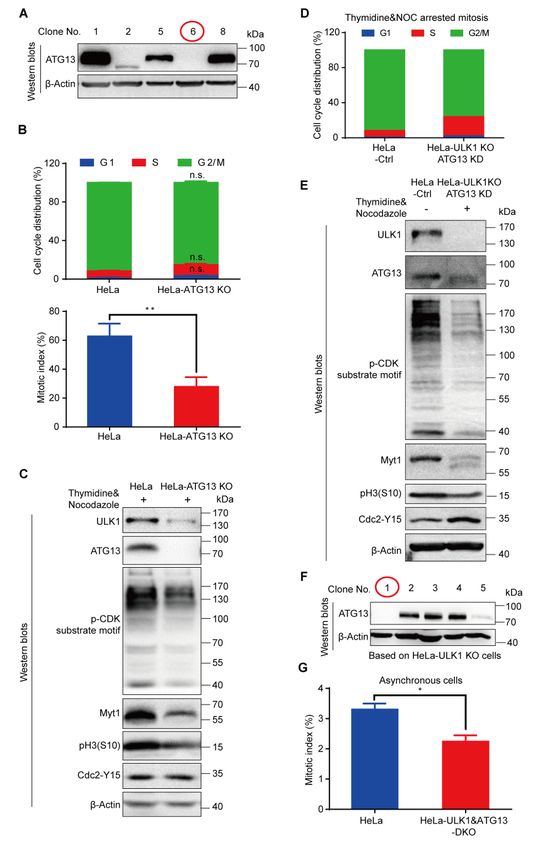
bioRxiv preprint first posted online May. 10, 2019; doi: http://dx.doi.org/10.1101/634733. The copyright holder for this preprint
 (which was not peer-reviewed) is the author/funder, who has granted bioRxiv a license to display the preprint in perpetuity.
                                  It is made available under a CC-BY 4.0 International license.

                Given that ULK1 could be autophosphorylated due to its kinase activity, the
           mobility shift and phosphorylation of kinase dead ULK1-K46I mutant [39] in mitosis
           were investigated. Our results show that ULK1-K46I could undergo mobility shift and
           mitotic phosphorylation as well (Figures S2A and S2B), which suggest that the kinase
           dead ULK1-K46I can also be phosphorylated in mitosis. Given that ATG13 is a
           substrate of ULK1, the ULK1-K46I could not phosphorylate ATG13, which reduced
           its electrophoresis bandshift and also had a significantly decreased interaction with
           ATG13 (Figure S2B, the input and IP for ATG13). Consistent with the cellular
           experiments, in vitro kinase assay also suggested that CDK1/cyclin B could
           phosphorylate ULK1-K46I and induce its upshift (Figure S2C). Additionally, ULK2,
           a member of ULK1 kinase family, was also found to be phosphorylated in mitosis
           with CDK1 substrate motif antibody and FLAG antibody (detecting FLAG-tagged
           mouse ULK2, mULK2-3FLAG) (Figure S3).

           ATG13 is also a direct substrate of CDK1/cyclin B in mitosis
           Co-immunoprecipitation was conducted using FLAG-tagged mULK1 and
           FLAG-tagged GFP as control. As components of the ULK1 complex, ATG13 but not
           FIP200 showed electrophoretic mobility shift in mitosis (Figure 4A). Similar to
           ULK1, ATG13 mobility shift in mitosis was also decreased by RO-3306 treatment
           (Figures 4B and S4A). To further confirm that the ATG13 electrophoretic mobility
           shift was caused by CDK1 as well, co-immunoprecipitation assay was performed in
           mitotic cells treated with RO-3306 for different timepoints. Consistent with ULK1,
           the electrophoresis upshift of ATG13 was also abolished by CDK1/cyclin B specific
           inhibitor RO-3306 (Figure S4B), which confirms that mitotic ATG13 is also
           phosphorylated by CDK1.
                Next, we performed co-immunoprecipitation to examine the interaction between
           ATG13 and ULK1, CDK1 in 293T cells overexpressing FLAG-tagged ATG13. Both
           Proline directed phospho-serine and threonine motif antibodies showed significant
           signal increase in mitosis (Figure 4C). The electrophoretic mobility shift of
           FLAG-tagged ATG13 was also apparent in mitosis. Moreover, both CDK1 and cyclin
           B1 could be co-immunoprecipitated by ATG13 (Figure 4C). These results confirmed
           the interactions among ATG13, ULK1 and CDK1/cyclin B1.
                It should be mentioned that in mitosis the ATG13 band shift are indistinguishable
           between ULK1-knockout and ULK1-overexpression cell lines, but are abrogated by
           RO-3306. This indicates that ATG13 phosphorylation-associated electrophoretic
           mobility shift is dependent on CDK1/cyclin B kinase activity but not its well-known
           kinase ULK1 (Figure S4C). Noteworthy, given that ATG13 could be phosphorylated
           by ULK1, despite in asynchronous condition, cells with ULK1 knockout showed
           downshifted band for ATG13. However, ATG13 band shift did not show any change
                                                                       9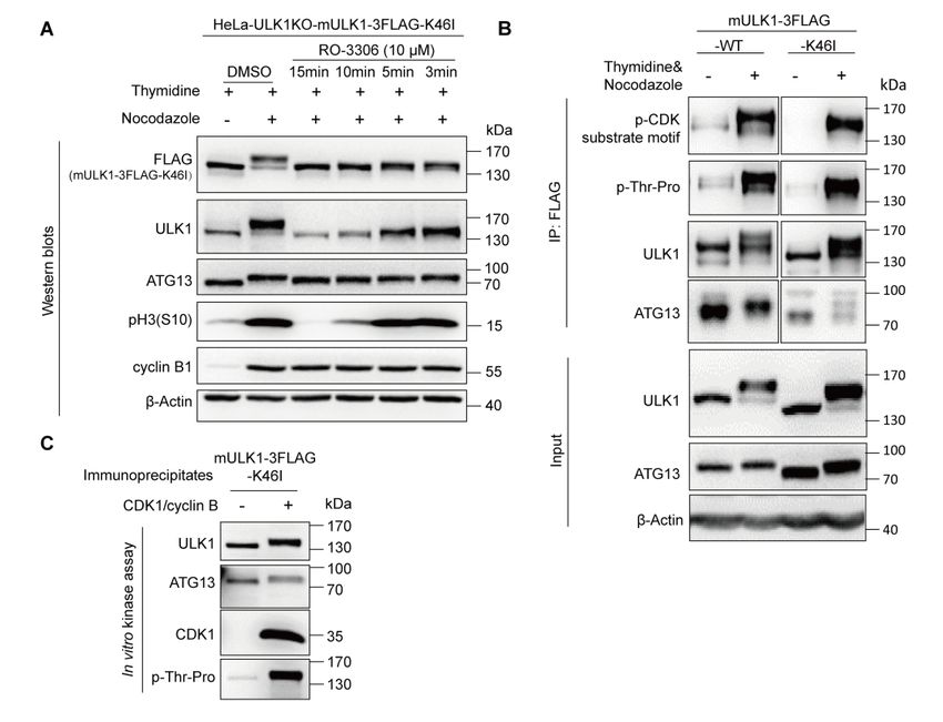
bioRxiv preprint first posted online May. 10, 2019; doi: http://dx.doi.org/10.1101/634733. The copyright holder for this preprint
(which was not peer-reviewed) is the author/funder, who has granted bioRxiv a license to display the preprint in perpetuity.
It is made available under a CC-BY 4.0 International license.
in asynchronous cells with or without ULK1 after RO-3306 treatment (Figure S4D).
In fact, in vitro kinase assay demonstrates that ATG13 could be directly
phosphorylated and upshifted by purified CDK1/cyclin B kinase complex, which are
shown by upshifted/phosphorylated ATG13 in both ATG13 immunoprecipitates and
ULK1 co-immunoprecipitates (Figures 4D and 4E).
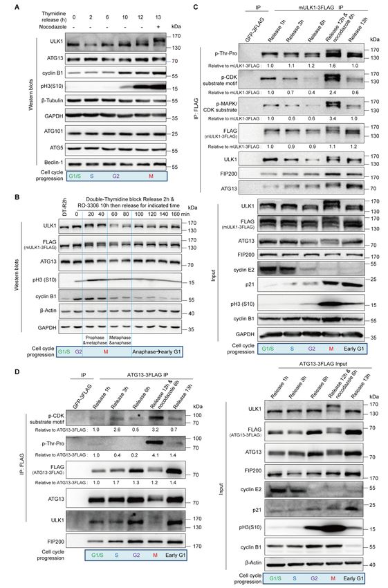
Figure 4. ATG13 is a substrate of CDK1/cyclin B in mitosis.
(A) Mitotic ATG13 undergoes mobility upshift in mitosis in electrophoresis. 293T cells
overexpressing FLAG-tagged mULK1 or GFP were synchronized by single-thymidine in the
presence or absence of nocodazole and co-immunoprecipitated by the FLAG antibody followed by
Western Blots. Size of endogenous ATG and expressed FLAG-tagged ATG13 were not
distinguishable in electrophoresis here.
(B) CDK1 inhibitor RO-3306 decreases the ATG13 bandshift in mitosis. HeLa cells synchronized
and treated as Figure 3A were subjected to Western blots analysis.
(C) ATG13 is phosphorylated and interacts with CDK1/cyclin B1 in mitosis. 293T cells
overexpressing FLAG-tagged ATG13 were synchronized by single-thymidine in the presence or
absence of nocodazole. The co-immunoprecipitates by FLAG antibody were subjected to Western
blots analysis.
10bioRxiv preprint first posted online May. 10, 2019; doi: http://dx.doi.org/10.1101/634733. The copyright holder for this preprint
(which was not peer-reviewed) is the author/funder, who has granted bioRxiv a license to display the preprint in perpetuity.
It is made available under a CC-BY 4.0 International license.
(D-E) ATG13 is directly phosphorylated by purified CDK1/cyclin B complex in vitro. The
ATG13 immunoprecipitates from asynchronous 293T cells overexpressing FLAG-tagged ATG13
or ULK1 and purified CDK1/cyclin B complex were subjected to in vitro kinase assay followed
by Western blots analysis. (D) shows representative Western blots of the ATG13
immunoprecipitates as substrate, and (E) shows the representative Western blots of the
ULK1-WT/11A-mutant (identified in Figure 5E and 5F) co-immunoprecipitates as substrate.
ULK1 and ATG13 phosphorylation sites in mitosis by CDK1/cyclin B
To identify the ULK1 phosphorylation sites in mitosis, we combined Scansite
prediction and mass spectrometry analysis. We first selected three potential sites of
mouse derived ULK1, serine 622, threonine 635 and threonine 653, because they had
high scores in Scansite prediction for Proline-dependent serine/threonine kinase group
(Pro_ST_kin), or their phosphorylation signals were specifically identified in mitotic
cells by mass spectrometry (Figure 5A). Site-directed mutagenesis was used to mutate
these sites from serine/threonine to unphosphorylatable alanine in FLAG-tagged
mULK1. Using 293T cells stably expressing FLAG-tagged GFP as control,
immunoprecipitation for FLAG-tagged mutant mULK1-S622A/T635A/T653A was
conducted in both asynchronous and mitotic cells. Although none of the single
mutations obviously disrupted ULK1 phosphorylation or band shift in mitosis (Figure
5B), S622A&T635A&T653A triple mutant significantly decreased the electrophoretic
mobility shift and the phospho-threonine-proline signals compared with wild-type
ULK1 (Figure 5C).
Since even the triple mutant did not completely abolish the band shift, we further
mutated more sites in the triple mutant (3A) background. It shows that the 11A
(S622&T635&T653&S479&S543&S413&T401&S403&S405&T282&T502A)
mutant collapsed the electrophoretic mobility shift and abolished ULK1
phosphorylation (as indicated by the p-Thr-Pro signal in Figure 5E) in mitosis, while
the other mutants, including the 5A, 7A, 9A and 10A, still have detectable
phosphorylation signals and upshift in mitosis (Figures 5D and 5E). It should be
mentioned that, as a substrate of ULK1, both ATG13 mobility shift and its interaction
with the ULK1-mutant were not much affected by ULK1 mutations, which indicates
that the kinase activity of ULK1 is not affected by 11A mutations (ATG13 Western
blot in Figure 5E) when compared to the decreased ATG13 interaction with the kinase
dead ULK1-K46I (Figure S2B). Importantly, in vitro kinase assay using purified
CDK1/cyclin B proteins also showed that the ULK1-11A mutant almost completely
abolished the phosphorylation signal and band shift compared with wild-type ULK1
(Figure 5F). Therefore, besides the three major sites (S622A/T635A/T653A),
CDK1/cyclin B phosphorylates multiple other sites of ULK1 in mitosis.
As for ATG13, we also combined Scansite prediction and mass spectrometry
analysis to identify its potential phosphorylation sites in mitosis. We selected four
11bioRxiv preprint first posted online May. 10, 2019; doi: http://dx.doi.org/10.1101/634733. The copyright holder for this preprint
(which was not peer-reviewed) is the author/funder, who has granted bioRxiv a license to display the preprint in perpetuity.
It is made available under a CC-BY 4.0 International license.
sites, ATG13-T342/T332/S44/S224, that were identified by both Scansite prediction
and mass spectrometry analysis (Figure 5G). Site-directed mutagenesis, Western blots
and immunoprecipitation were combined for site verification (Figures 5H and 5I), and
we found that the four-site mutant significantly decreased the electrophoretic mobility
shift and the phospho-serine/threonine-proline signals compared with wild-type
ATG13 (Figure 5I).
Figure 5. ULK1 and ATG13 phosphorylation sites in mitosis by CDK1/cyclin B.
(A) The specific phosphorylated sites identified by mass spectrometry in mitotic mULK1
compared with asynchronous mULK1.
(B-C) S622/T635/T653 phosphorylation contributes to ULK1 mobility shift in mitosis. 293T cells
overexpressing FLAG-tagged mULK1-S622/T635/T653 mutants were synchronized with
single-thymidine and nocodazole. Then immunoprecipitation for single mutant with FLAG
12bioRxiv preprint first posted online May. 10, 2019; doi: http://dx.doi.org/10.1101/634733. The copyright holder for this preprint
(which was not peer-reviewed) is the author/funder, who has granted bioRxiv a license to display the preprint in perpetuity.
It is made available under a CC-BY 4.0 International license.
antibody (B) or Western blots for double and triple mutant (C) was performed.
(D-E) More sites contribute to ULK1 band shift. Based on triple S622/T635/T653A mutant (3A),
the other 8 Ser/Thr sites were mutated into Ala. 293T cells expressing various mutants were
synchronized into mitosis with thymidine and nocodazole for immunoprecipitation by FLAG
antibody and Western blots. 5A, 3A-S479&S543A; 7A, 5A-S411&S413A; 9A,
5A-S413&T401&S403&S405A; 10A, 9A-T282A; 11A, 10A-T502A.
(F) ULK1-11A mutant was not upshifted or phosphorylated by CDK1/cyclin B kinase complex in
vitro. The ULK1-11A mutant immunoprecipitates from asynchronous 293T cells overexpressing
FLAG-tagged mULK1 and purified CDK1/cyclin B complex were tested in an in vitro kinase
assay and Western blots.
(G) The phosphorylated sites shared by mass spectrometry identification and Scansite prediction
in mitotic ATG13 compared with asynchronous ATG13.
(H-I) ATG13-T342/T332/S44/S224 phosphorylation contributes to ATG13 mobility shift in
mitosis. 293T cells overexpressing FLAG-tagged ATG13-T342/T332/S44/S224 mutants were
synchronized with single-thymidine and nocodazole. Then Western blots for four-site mutant (H)
or immunoprecipitation for mutants with FLAG antibody (I) was performed.
ULK1-ATG13 phosphorylation in mitosis positively regulates mitotic autophagy
and Taxol-induced cell death
To study the functional significance of these phosphorylation sites, we constructed
ULK1 and ATG13 wild-type or double mutant cell line based on HeLa-
ULK1&ATG13-DKO cell line (Figures S5A and S5B). Although ULK1-ATG13
complex is a key autophagy regulator, its function has only been investigated in
asynchronous cells. To unravel the function of these phosphorylation events that
occur specifically in mitosis, we first examined the autophagic flux in mitotic
HeLa-DKO cells expressing ULK1&ATG13 WT or mutant cell line. We used
autophagic flux, one of the most reliable methods to examine the amount of
autophagic degradation to indicate autophagy activity. Autophagic flux inhibitors
such as chloroquine (CQ) could cause autophagy marker LC3-II accumulation due to
blocked LC3-II degradation [3]. In fact, we found that the autophagic flux was
reduced by about 30% in mutant cell lines compared to the wild-type cell lines
(Figure 6A), which indicates that ULK1-ATG13 phosphorylation positively regulate
autophagy in mitosis.
It is very interesting that some ULK1/ATG13 mutations in patients were found to
be associated with the phosphorylation motif/sites identified in our study. For
example, in Desmoplastic Melanoma, Cutaneous Melanoma, Uterine Endometrioid
Carcinoma and Colon Adenocarcinoma patients, there are some proline mutations
such as P402Q/P404S/P414S/P624H/ P637S/P637T in the CDK1 substrate motifs in
ULK1 [40, 41], which is likely to indirectly affect CDK1 phosphorylation on ULK1.
There are also some direct serine/threonine mutations such as S403F, S479L, S544C,
S623F, T636A and T654M in Cutaneous Melanoma, Bladder Urothelial Carcinoma,
13bioRxiv preprint first posted online May. 10, 2019; doi: http://dx.doi.org/10.1101/634733. The copyright holder for this preprint
(which was not peer-reviewed) is the author/funder, who has granted bioRxiv a license to display the preprint in perpetuity.
It is made available under a CC-BY 4.0 International license.
Cervical Squamous Cell Carcinoma, Lung Adenocarcinoma and Colorectal
Adenocarcinoma patients [41-46], which could directly affect CDK1 phosphorylation
on ULK1. Similarly, in Renal Clear Cell Carcinoma, Cutaneous Melanoma and Renal
Clear Cell Carcinoma with Sarcomatoid Features patients, there are also mutations of
P45Q and P225A in the CDK1 substrate motifs in ATG13[41, 47, 48], which is likely
to indirectly affect CDK1 phosphorylation on ATG13. And the mutation for
ATG13-T332P in Breast Invasive Lobular Carcinoma [49] could directly affect
CDK1 phosphorylation on ATG13 (Figure 6B).
Besides the basic scientific question about autophagy regulation in mitosis, more
importantly, it has been shown that the combinatorial therapy by autophagy inhibitors
and chemotherapeutic drugs arresting cells in mitosis could have better anti-tumor
efficacy [50-52]. Next we combined CQ and chemotherapeutic drug Taxol, a reagent
stabilizing the microtubule polymer to arrest cells in mitosis [53]. Although neither
ULK1-11A nor ATG13-4A alone has significant effects on Taxol chemosensitivity
(Figures S6A and S6B), Taxol-induced cell death was substantially attenuated in
ULK1&ATG13 double mutant cell line compared with wild-type cell line (Figures
6C-6E). This decreased chemosensitivity in double mutant cell line may be due to its
low autophagy activity, which is consistent with previous report that autophagy
inhibition could inhibit cell death in mitotic arrest [18]. In addition, autophagy
inhibition by CQ could attenuate Taxol chemosensitivity in wild-type cell line (with
higher autophagy), but could not further decrease that of the double mutant cell line
(with lower autophagy), which indicates that ULK1-ATG13 is the major autophagy
pathway for Taxol-induced cell death in mitosis (Figures 6C-6E). Moreover, the
double mutant cell line displayed lower mitotic slippage induced by Taxol (Figure
6F), which may be an alternative mechanism for their differential chemosensitivity.
Considering the lower Taxol chemosensitivity in ULK1&ATG13 double mutant cell
line, our results indicate that patients with similar mutations in both ULK1 and
ATG13 might have increased chemotherapy resistance to Taxol.
14bioRxiv preprint first posted online May. 10, 2019; doi: http://dx.doi.org/10.1101/634733. The copyright holder for this preprint
(which was not peer-reviewed) is the author/funder, who has granted bioRxiv a license to display the preprint in perpetuity.
It is made available under a CC-BY 4.0 International license.
Figure 6. ULK1-ATG13 phosphorylation in mitosis regulates mitotic autophagy and Taxol
chemosensitivity.
(A) The autophagic flux of HeLa-DKO cells stably overexpressing double wild-type or mutant
FLAG-tagged mULK1 and ATG13. Cells synchronized to mitosis with thymidine and nocodazole
were shaken off and treated with or without autophagy inhibitor 25 μM CQ for 1h. Western blots
and statistical analysis for autophagy marker LC3B-II. The upper panel is the statistical result and
the lower panel is a representative Western blot. n=3, *p < 0.05, **p < 0.01.
(B) Human ULK1 and ATG13 mutations in cancer patients. The data were extracted from the
cBioPortal for Cancer Genomics and arranged according to phosphorylation sites identified in
15bioRxiv preprint first posted online May. 10, 2019; doi: http://dx.doi.org/10.1101/634733. The copyright holder for this preprint
(which was not peer-reviewed) is the author/funder, who has granted bioRxiv a license to display the preprint in perpetuity.
It is made available under a CC-BY 4.0 International license.
mitotic mouse ULK1 and human ATG13 (Figure 5). It should be pointed out that the human
ULK1 mutation sites of S544, S623, T636 and T654 in Figure 6B are equivalent to mouse ULK1
sites of S543, S622, T635 and T653. PTM, Post-translational modification.
(C) Analysis of cells treated with Taxol combined with CQ by Western blots. HeLa-DKO cells
stably overexpressing double wild-type or mutant FLAG-tagged mULK1 and ATG13 cells
released for 4h from thymidine block were treated with 20 nM Taxol with or without 10 μM CQ
for 24 h and analyzed by Western blots for apoptosis and mitotic/slippage markers.
(D) The representative flow cytometry result in cells as treated in Figure 6C. Cells treated as in
Figure 6C were subjected to co-staining with FITC-Annexin V and PI and analyzed by flow
cytometry.
(E) The statistical analysis for cell death induced by Taxol and/or CQ for Figure 6D. n=3, n.s., not
significant, **p < 0.01, ***p < 0.001.
(F) Statistical analysis for mitotic slippage markers cyclin B1 and pH3(S10) in cells as treated in
Figure 6C. Cells fixed with -20℃ 75% Ethanol overnight were subjected to either PI and
pH3(S10) co-staining or cyclin B1 staining for cell cycle, mitotic index and cyclin B1 level
analysis by flow cytometry. n=3, **p < 0.01.
ULK1-ATG13 is required for cell cycle progression
To examine the cell cycle regulation by ULK1-ATG13, we constructed
ULK1-knockout (KO) HeLa (human cervical cancer cells) and 293T (human
embryonic kidney cells) cells using CRISPR/Cas9 (Figures S7A and S7B).
ULK1-knockout cells had similar cell cycle distribution compared to wild-type (WT)
cells (Figure S7C). To further examine the effect of ULK1 on cell cycle progression,
we synchronized the cells by double-thymidine and found that the S/G2 transition in
ULK1-knockout cells was slightly delayed compared to wild-type cells (Figure S7D).
Given that the G2 and M phases are not distinguishable by propidium iodide
(PI) staining alone, we further used pH3(S10), one of the most commonly used
mitotic markers [54, 55], to examine whether ULK1 functions in G2/M transition.
Although no differences were detected for the G2/M percentage in wild-type and
ULK1-knockout cells, the mitotic progression is significantly decreased in
ULK1-knockout cells synchronized with thymidine and nocodazole, a microtubule
destabilizing reagent. It was shown by the percentage of pH3(S10) positive cells using
flow cytometry (Figure S8A) or Western blots analysis for cell cycle markers (Figure
S8B). The antibody for p-CDK Substrate Motif [23] recognizes the substrate of CDK,
whose phosphorylation level reflects the CDK activity and is used as a mitotic
marker. And either the upshifted band of Myt1 or the lower phosphorylation level of
Cdc2-Y15 in mitosis [56] could also be used as mitotic progression markers. Given
that ULK1 is a serine/threonine protein kinase, the contribution of ULK1 kinase
activity to mitotic progression is examined in cell lines expressing wild-type ULK1 or
kinase dead ULK1-K46I mutant [8], which indicates that the ULK1 kinase activity
has little effect on mitotic progression (Figure S8C).
16bioRxiv preprint first posted online May. 10, 2019; doi: http://dx.doi.org/10.1101/634733. The copyright holder for this preprint
(which was not peer-reviewed) is the author/funder, who has granted bioRxiv a license to display the preprint in perpetuity.
It is made available under a CC-BY 4.0 International license.
As the ULK1 partner in ULK1-ATG13 complex, ATG13 was reported to
function in mitotic catastrophe [57], ATG13-knockout cells were established using
CRISPR/Cas9 as well (Figure S9A). Cell cycle analysis indicated that ATG13
knockout inhibited G2/M transition and decreased the mitotic index (Figures S9B and
S9C), indicating that ATG13 is involved in cell cycle regulation. Noteworthy, it has
been reported that ATG13 is required for the kinase activity of ULK1 [26], the ULK1
kinase activity is not necessary for mitotic progression (Figure S8C). Therefore, the
phenotype of the ATG13-knockout cells is due to the lack of ATG13 rather than the
impairment in the kinase activity of ULK1.
To further investigate the role of ATG13 and ULK1 in cell cycle regulation, we
combined ULK1 knockout with ATG13 gRNA transient transfection and found that
the cell cycle progression was inhibited (Figures S9D and S9E). Then we constructed
a ULK1 and ATG13 double knockout (DKO) cell line (Figure S9F) and found that
both S/G2 and G2/M transitions were severely delayed (Figures 7A and S9G), while
ULK1 or ATG13 knockout alone (Figures S8A and S9B) did not simultaneously
delay both S/G2 and G2/M transitions. The immunoblotting of cell cycle markers
verified the mitotic index and cell cycle distribution data (Figure 7B). To rule out the
off-target effect, rescue assays were performed in knockout cells with exogenously
expressed ULK1-ATG13, which confirmed the specificity of ULK1 and ATG13
knockout (Figure S10). Furthermore, the effect of ULK1 and/or ATG13 on mitotic
exit markers was analyzed, which shows that DKO interfered with cyclin B1 and pH3
(S10) phosphorylation decrease more significantly than ULK1 or ATG13 single
knockout (Figure 7C). Accordingly, the growth rate of HeLa-DKO cells was
significantly slowed down compared with HeLa cells (Figure 7D).
To further test the effect of ULK1-ATG13 in vivo, the mouse models in nude
mice bearing ULK1 and ATG13 single or double knockout cells were established
(Figures S11A and S11B). The tumor weight and volume of DKO group were
significantly lower than all the other groups (Figures 7E and 7F, Figure S11A). In
contrast, ULK1 inhibitor SBI-0206965 [10] and/or ATG13-KO alone were not strong
enough to inhibit tumor growth as efficiently as DKO, which proved the synergistic
effects for ULK1 and ATG13 (Figures 7E and 7F), indicating that targeting ULK1
and ATG13 might be a potential anti-cancer strategy.
17bioRxiv preprint first posted online May. 10, 2019; doi: http://dx.doi.org/10.1101/634733. The copyright holder for this preprint
(which was not peer-reviewed) is the author/funder, who has granted bioRxiv a license to display the preprint in perpetuity.
It is made available under a CC-BY 4.0 International license.
Figure 7. ULK1-ATG13 is required for cell cycle progression.
(A) ULK1-ATG13 double knockout (DKO) inhibits S/G2 and G2/M transitions. HeLa wild-type
or ULK1&ATG13-DKO cells synchronized into mitosis were subjected to PI and pH3(S10)
co-staining for cell cycle and mitotic index analysis by flow cytometry. n=3, *p < 0.05, **p <
0.01.
(B) Representative Western blots suggest that ULK1&ATG13 DKO inhibits mitotic entry, which
is shown by mitotic markers and CDK1 substrate phosphorylation.
(C) Mitotic exit of ULK1, ATG13, or ULK1&ATG13 knockout cells. Cells were synchronized
into mitosis with thymidine and nocodazole and released into nocodazole-free complete DMEM
medium for different timepoints and then subjected to either PI and pH3(S10) co-staining or
cyclin B1 staining for cell cycle, mitotic index and cyclin B1 level analysis by flow cytometry.
n=3, n.s., not significant, *p < 0.05, **p < 0.01.
(D) ULK1&ATG13 DKO inhibits cell proliferation. HeLa wild-type or ULK1&ATG13 DKO
18bioRxiv preprint first posted online May. 10, 2019; doi: http://dx.doi.org/10.1101/634733. The copyright holder for this preprint
(which was not peer-reviewed) is the author/funder, who has granted bioRxiv a license to display the preprint in perpetuity.
It is made available under a CC-BY 4.0 International license.
cells were plated at 1×10⁵ cells/ml and cultured for 1, 2 or 3 days. The cells number was counted
by flow cytometry and the doubling time was calculated. TD indicates the average cell doubling
time and is calculated as: TD= t*[lg2/(lgNt-lgN0)], where t is the culture time, Nt is the cell
number after culturing, N0 is the original cell number plated. n=3, *p < 0.05, ***p < 0.001.
(E) The relative tumor volume growth curve of nude mice bearing different tumors with or
without SBI-0206965. The nude mice were injected with cells (1 × 10⁷) in 100 μL PBS/Matrigel
Matrix (1:1). Seven days post implantation, five mice in each group were injected with 0.5%
(M/V) methyl cellulose or SBI-0206965 in 0.5% methyl cellulose (20 mg/kg/d) every day for 33
days. Tumor growth was evaluated every day and tumor volume was calculated as: volume =
1/2(length × width2).
(F) Tumor growth in nude mice bearing wild-type or knockout cells with or without ULK1 kinase
inhibitor SBI-0206965. The protocols were indicated as Figure 7E, and the mice were sacrificed
and tumors were harvested and weighted up at the end of the experiment. n=5, n.s., not significant,
*p < 0.05, ***p < 0.001.
(G) Model illustrates functions of ULK1-ATG13 in cell cycle and its phospho-regulation in
mitosis. When cells are in mitosis, CDK1/cyclin B phosphorylates ULK1-ATG13 to induce
significant electrophoretic mobility shift. The phosphorylated ULK1-ATG13 regulates mitotic
autophagy and Taxol chemosensitivity. In asynchronous conditions, double knockout ULK1 and
ATG13 inhibits cancer cell proliferation in both cell and mouse models. I, interphase; M, mitosis.
Discussion
ULK1-ATG13 complex is mainly phosphorylated by AMPK and mTORC1 in
asynchronous conditions [8, 9, 19, 20], but little is known about its regulation in
mitosis. We found that the master cell cycle kinase CDK1 phosphorylates
ULK1-ATG13 complex to regulate its function in autophagy and Taxol
chemosensitivity. Besides the known function in autophagy, we also found that ULK1
and ATG13 coordinate to orchestrate cell cycle progression in cell line and mouse
models (Figure 7G).
Kinases involved in both autophagy and mitosis
Recent literature indicates that there are some kinases that are involved in both
autophagy and mitosis, which could bridge the autophagy and cell cycle regulation.
Cell cycle kinases such as CDK1, Aurora A, PLK-1 were found to regulate
autophagy, while kinases originally found in autophagy control were shown to
regulate mitosis as well, such as mTORC1 and AMPK [12, 14]. In addition, increased
evidence shows that some other autophagic proteins such as Beclin-1, SQSTM1/p62
and GABARAP are related to mitotic events [6, 58-60]. Although it has been reported
that ULK3, a member of ULK1 kinase family, could regulate cytokinesis and ATG13
could regulate Colchicinamide-induced mitotic catastrophe [57, 61], the roles of
autophagy kinase complex ULK1-ATG13 in mitotic regulation are still unclear.
While previous reports implicated possible links between ULK1 and CDK1 [62, 63],
19bioRxiv preprint first posted online May. 10, 2019; doi: http://dx.doi.org/10.1101/634733. The copyright holder for this preprint
(which was not peer-reviewed) is the author/funder, who has granted bioRxiv a license to display the preprint in perpetuity.
It is made available under a CC-BY 4.0 International license.
our study here is the first report that demonstrates CDK1 is the upstream kinase for
ULK1-ATG13 complex. ULK1-ATG13 phosphorylation could positively regulate
autophagy, which promoted Taxol-induced cell death and mitotic slippage. In our
opinion, the mitotic slippage may be an alternative mechanism for mitotic
ULK1-ATG13 phosphorylation mediated autophagy and chemosensitivity (Figure 6).
As to the role of ULK1-ATG13 in tumor proliferation, we propose that it is the
function of ULK1-ATG13 protein themselves rather than mitotic phosphorylation
(Figures 7E and 7F).
CDK1 interacts with ULK1/ATG13 in asynchronous conditions
It is interesting that CDK1 could be detected in ULK1/ATG13 Co-IP products in
asynchronous cells just as in mitotic cells, which indicates the constitutive interaction
between CDK1 and ULK1/ATG13. Since Cdc37 is a molecular chaperone linking
ULK1-ATG13 to Hsp90, similarly, it is possible that CDK1 may also function as a
molecular chaperone linking ULK1-ATG13 complex to Hsp90 or other unknown
proteins in interphase [64, 65], which may be important for ULK1-ATG13 stability.
CDK1 regulates the S/G2 and G2/M transitions via sequential coupling to cyclin A
and cyclin B [66], indicating that ULK1-ATG13 complex could be regulated by
CDK1 in both S/G2 and G2/M transitions, which is consistent with the role of
ULK1-ATG13 complex and their differential regulation in cell cycle progression
(Figures 1 and 7). Obviously, the functional significance for the CDK1 and
ULK1/ATG13 interaction in asynchronous conditions, whether other components of
ULK1 complex or other ATG proteins are subjected to similar regulation, need
further investigations, which would provide more insights for the molecular links
between autophagy and cell cycle progression.
VPS34- and ULK1-ATG13 complex-dependent mitotic autophagy regulation
Although increasing evidence indicates that autophagy in mitosis remains active in
mitosis but the regulation mechanism still unclear. Yuan’s group indicated that
activated CDK5 phosphorylates VPS34-Thr159 to inhibit VPS34-dependent
autophagy in mitosis [16]. Our finding here demonstrates that CDK1 phosphorylates
ULK1-ATG13 in mitosis to promote ULK1-ATG13-dependent autophagy, which at
least partially contributed to the active autophagy state in mitosis as reported in our
previous study [15]. However, it is likely that multiple autophagy regulators, not
limited to the VPS34 complex and ULK1-ATG13 complex, contribute to the mitotic
autophagy regulation, which certainly needs further investigations. In fact, we also
found that ULK2, the homolog of ULK1, is also phosphorylated in mitosis as ULK1
(Figure S3). And ULK1/ULK2-ATG13 complex in mitosis might orchestrate
respective functions for autophagy, chemosensitivity and beyond.
20bioRxiv preprint first posted online May. 10, 2019; doi: http://dx.doi.org/10.1101/634733. The copyright holder for this preprint
(which was not peer-reviewed) is the author/funder, who has granted bioRxiv a license to display the preprint in perpetuity.
It is made available under a CC-BY 4.0 International license.
Mitotic ULK1/ATG13 and beyond
The upshifted mitotic ULK1/ATG13 could be viewed as a hint for the other
autophagy related proteins potentially functioning in mitosis or cell cycle.
Furthermore, deciphering the mitotic phosphatase responsible for ULK1-ATG13
dephosphorylation should also be investigated in the future, which will be
complementary for CDK1-mediated phosphorylation. Importantly, controlling the
phosphorylation for ULK1-ATG13 by the candidate phosphatases and kinases might
be a promising therapeutic strategy for autophagy and mitosis related diseases.
In conclusion, we have uncovered the untraditional roles of ULK1-ATG13
complex in cell cycle progression and tumor growth, which provides ULK1-ATG13
as potential candidates in cancer therapy. We have also revealed its
phospho-regulation by CDK1/cyclin B in mitosis, which provides molecular
mechanisms not only for maintaining mitotic autophagy, but also for the potential
chemotherapy resistance in some cancer patients bearing ULK1-ATG13 mutations in
CDK1 motifs.
21bioRxiv preprint first posted online May. 10, 2019; doi: http://dx.doi.org/10.1101/634733. The copyright holder for this preprint
(which was not peer-reviewed) is the author/funder, who has granted bioRxiv a license to display the preprint in perpetuity.
It is made available under a CC-BY 4.0 International license.
Materials and Methods
Antibodies and reagents
The autophagy antibody sampler kit (#4445), ULK1 Antibody Sampler Kit (#8359),
Autophagy Induction (ULK1 Complex) Antibody Sampler Kit (#46486), the cell
cycle regulation antibody sampler kit II (#9870), Phospho-(Ser) Kinase Substrate
Antibody Sampler Kit (#9615), Phospho-Threonine-Proline Mouse mAb
(P-Thr-Pro-101) (#9391), anti-ULK1 (#4776) antibody, the HRP-linked anti-rabbit
and anti-mouse IgG antibodies were all from Cell signaling technology. The
anti-FLAG (F3165) antibody was acquired from Sigma and anti-β-Tubulin,
anti-GAPDH and anti-β-Actin antibodies from Beijing TransGen Biotech (Beijing,
China). The GlutaMAX supplement and puromycin dihydrochloride were from
Gibco. The secondary fluorescently conjugated antibodies, anti-fade prolong Gold
with DAPI were from Molecular Probes. Prestained Protein Ladder (26616) and
M-PER buffer were from Thermo Pierce. NH4Cl, RO-3306 and Thymidine were from
Sigma. Nocodazole and SBI-0206965 were from Selleckchem. FITC Annexin V
Apoptosis Detection Kit I (#556547) was from BD Biosciences. Methyl cellulose
(#69016260) was from Sinopharm Chemical Reagent Co., Ltd. Matrigel Matrix
(#354234) was from BD. Protease inhibitor and phosphatase inhibitor cocktails were
from Roche and the PVDF membrane from Millipore.
Cell culture and stable cell lines establishment
HeLa, HCT 116, RPE1 and HEK-293T cells were all cultured in DMEM medium
(without L-Glutamine) supplemented with 10% FBS, 2 mM GlutaMAX and 1%
penicillin/streptomycin (P/S). The plasmid for pBobi-FLAG-mULK1 contains one
FLAG tag and the affinity for FLAG antibody was lower than ULK1 antibody.
Therefore, in order to enhance its affinity to FLAG antibody, 3×FLAG was added to
mULK1 C-terminus [67]. Stable cell lines were constructed as described previously
[68]. HEK-293T or HeLa cells stably expressing mULK1-3×FLAG were maintained
in DMEM complete medium containing 1 μg/ml puromycin.
CRISPR/Cas9 technology
The gRNA targeted to human ULK1 was designed with CRISPR Design
(http://crispr.mit.edu). The sequence (human ULK1:
5’-GCCCTTGAAGACCACCGCGA-3’; human ATG13:
5’-CACATGGACCTCCCGACTGC-3’) was selected and subcloned into PX458
vector. HeLa and HEK-293T cells were transiently transfected with
PX458-ULK1/ATG13-gRNA with Fugene 6. The cells were diluted into 0.5 cell/100
μL at 96-well plate after 12 h transfection. Single cell in 96-well-plate was cultured in
DMEM complete medium to form single-cell-clone that was cultured in 24-well-plate
22bioRxiv preprint first posted online May. 10, 2019; doi: http://dx.doi.org/10.1101/634733. The copyright holder for this preprint
(which was not peer-reviewed) is the author/funder, who has granted bioRxiv a license to display the preprint in perpetuity.
It is made available under a CC-BY 4.0 International license.
and subjected to immunoblotting analysis using ULK1 or ATG13 specific antibody.
One of single-cell-clone could not be detected with ULK1/ATG13 antibody was
selected as ULK1/ATG13-knockout cell. ULK1 and ATG13 double knockout cells
were established based on ULK1-knockout cells using PX458- ATG13-gRNA.
Immunoprecipitation and Western blots
The procedure was instructed as previously [68]. Most immunoprecipitation
experiments were conducted in HEK-293T derived cell lines due to higher expression
level for the exogenous protein. Briefly, HEK-293T cells stably expressing
GFP-3FLAG or mULK1-3FLAG were lysed with M-PER supplemented with
protease inhibitors and phosphatase inhibitors and centrifuged at 4°C 14,000g for 10
min. The supernatant mixed with pre-incubated Protein G Dynabeads and FLAG
antibody at 4°C for 12 h and washed three times with lysis buffer. Then the
immunoprecipitate was denatured in 1×SDS-PAGE buffer at 95°C for 7 min and
subjected to SDS-PAGE and immunoblotting or Coomassie brilliant blue staining.
Apoptosis by flow cytometry
Cells treated with Taxol combine with or without chloroquine were subjected to
apoptosis assay according to the manufacturer instructions (BD Biosciences). Briefly,
cells were trypsinized and washed twice with ice cold PBS, and stained with PI and/or
FITC-Annexin V in apoptosis buffer for 30 min. The FITC and PE channel were used
for flow cytometry detection and the data were analyzed by FlowJo.
Cell cycle synchronization
Various cell synchronization methods are used in this paper, which have been used
previously. Briefly, a double-thymidine (2.5 μM) block arrested cells in G1/S border
and cells progress through S, G2 and M phase after release. A double-thymidine or
single-thymidine block in combination with nocodazole (100 ng/mL) or STLC (5 μM)
treatment arrested cells in prometaphase or prophase. A double-thymidine block in
combination with RO-3306 (10 μM) treatment arrested cells in late G2 phase and
progressed into mitosis after RO-3306 washout for three times with prewarmed PBS.
Cell cycle analysis by flow cytometry/FACS
Cells for cell cycle analysis were trypsinized with 0.25% Trypsin/EDTA. After
washing with ice-cold PBS twice, cells were fixed with -20°C 75% ethanol overnight
and then stained with PI/RNase staining buffer (BD pharmingen) for 15 minutes at
room temperature and analyzed with flow cytometry (Beckman Coulter, Cytoflex).
Alternatively, for mitotic index analysis, cells fixed were stained with
phospho-Histone H3 (S10) at 1:1600 for 2 hours at room temperature and washed
twice before Alexa-488 conjugated anti-rabbit IgG staining. After washing twice,
PI/RNase staining was conducted as described above before flow cytometry analysis.
23bioRxiv preprint first posted online May. 10, 2019; doi: http://dx.doi.org/10.1101/634733. The copyright holder for this preprint
(which was not peer-reviewed) is the author/funder, who has granted bioRxiv a license to display the preprint in perpetuity.
It is made available under a CC-BY 4.0 International license.
The data were analyzed by ModFit LT 4.1 and Flow Jo 7.6 software.
Lambda Phosphatase treatment
IP products from mULK1-3FLAG overexpressing HEK293T cells were aliquoted and
treated with reaction buffer, reaction buffer containing 1 μL (400 units) lambda
phosphatase (P0753S, NEB), reaction buffer containing 1 μL lambda phosphatase
plus 1×phosphatase inhibitors cocktail (Roche) in at 30°C for 30 min with gently
shaking. Then the reaction products were denatured at 95°C for 7 min and subjected
to immunoblotting with FLAG or Serine/Threonine specific antibody.
Mass spectrometry
For mass spectrometry assays, immunoprecipitates using FLAG antibody from
asynchronous or mitotic 293T cells expressing FLAG-tagged mULK1/ATG13 were
separated by SDS-PAGE, the gel was stained with Coomassie brilliant blue, and the
FLAG-tagged mULK1/ATG13 band in each lane was excised. Samples were
subjected to mass spectrometry analysis for mULK1/ATG13 phosphorylation by Core
Facility Center for Life Sciences, University of Science and Technology of China,
School of Life Science and Technology, ShanghaiTech University. The identified
phosphorylation sites specific in mitotic cells were selected as the candidates for
ULK1/ATG13 phosphorylation sites in mitosis.
In vitro kinase assay
Anti-FLAG immunoprecipitates from asynchronous 293T cells overexpressing
FLAG-tagged mULK1 (wild-type or K46I kinase dead) or ATG13 were washed three
times with M-PER and then resuspended in ice-cold kinase buffer (50 mM Tris-HCl
pH 7.5 @ 25°C, 10 mM MgCl2, 0.1 mM EDTA, 2 mM DTT, 0.01% Brij 35). The
immunoprecipitates were then incubated with or without 270 ng purified
CDK1/cyclin B (Life technologies, Part Number: PV3292, Lot Number: 1816161K)
in 20 μL reaction mix (kinase buffer and 20 μM ATP) pretreated with or without 10
μM CDK1 inhibitor RO-3306 at 30°C with constant shaking for 30 min. The reaction
was quenched by mixing with 5 μL 5×SDS-sample buffer and boiling at 95°C for 7
min.
Mouse Model
Four-week-old female BALB/c nude mice were purchased from Nanjing
Biomedical Research Institute of Nanjing University (Nanjing, China). All mice were
kept in an animal room under the specific-pathogen-free (SPF) condition. The mice
were fed with sterilized food and autoclaved tap water freely. The protocol involving
animals was approved by the ethical and humane committee of Hefei Institutes of
Physical Science, Chinese Academy of Sciences and carried out strictly in accordance
with the related regulations (Hefei, China). After one week, HeLa/HeLa-ATG13
24You can also read