PHGDH Is Upregulated at Translational Level and Implicated in Platin-Resistant in Ovarian Cancer Cells
←
→
Page content transcription
If your browser does not render page correctly, please read the page content below
ORIGINAL RESEARCH
                                                                                                                                              published: 10 June 2021
                                                                                                                                       doi: 10.3389/fonc.2021.643129

                                              PHGDH Is Upregulated at
                                              Translational Level and Implicated
                                              in Platin-Resistant in Ovarian
                                              Cancer Cells
                                              Fangfang Bi †, Yuanyuan An †, Tianshui Sun , Yue You and Qing Yang *

                                              Department of Obstetrics and Gynecology, Shengjing Hospital of China Medical University, Shenyang, China

                                              Background: Platinum-based chemotherapy is the first line option for ovarian cancer.
                                              The development of resistance to such chemotherapy results in treatment failure, while the
                          Edited by:          underlying mechanisms are poorly understood.
                         Yong Teng,
    Augusta University, United States         Methods: Clinical samples were collected from Shengjing Hospital of China Medical
                      Reviewed by:            University. MTT assay was used to see the proliferation and chemoresistance of ovarian
                Stephen John Ralph,           cancer cells. Transwell migration and Matrigel invasion assays was used to see the
          Griffith University, Australia
                              Ge Lou,
                                              invasion ability of ovarian cancer cells. In addition, polysome profiling and tissue
    Harbin Medical University Cancer          microarray and immunohistochemical staining were also used. The statistical
                      Hospital, China         significance of the difference was analyzed by ANOVA and post hoc Dunnett’s test.
                  *Correspondence:
                           Qing Yang          Results: PHGDH is the first enzyme responsible for serine biosynthesis pathway. The
                yangqing_sj@126.com           current study demonstrated that PHGDH is upregulated in platin-resistant ovarian cancer
     †
      These authors have contributed          cells and tissues at the protein level. Importantly, knockdown of PHGDH suppressed,
                 equally to this work
                                              while overexpression of PHGDH increased the survival upon cisplatin exposure,
                    Specialty section:
                                              invasiveness and spheroid formation of ovarian cancer cells. The current study
          This article was submitted to       demonstrated that PHGDH translation was upregulated in platin-resistant ovarian
                    Cancer Metabolism,
                                              cancer. In addition, our study provided evidence that LncRNA RMRP (RNA Component
                a section of the journal
                  Frontiers in Oncology       of Mitochondrial RNA Processing Endoribonuclease) was upregulated in platin-resistant
         Received: 17 December 2020           ovarian cancer, which promoted enrichment of RNA binding protein DDX3X (DEAD-Box
             Accepted: 12 May 2021            Helicase 3 X-Linked) on the PHGDH mRNA to promote its translation.
            Published: 10 June 2021

                              Citation:
                                              Conclusion: Collectively, the current study described that PHGDH was upregulated and
Bi F, An Y, Sun T, You Y and Yang Q           conferred resistance of ovarian cancer cells to cisplatin, suggesting that cisplatin
       (2021) PHGDH Is Upregulated            resistance could be overcome by targeting PHGDH. Our study also provided evidence
             at Translational Level and
         Implicated in Platin-Resistant       that differential PHGDH protein expression was defined by its translation, and RNA
              in Ovarian Cancer Cells.        binding protein DDX3X and LncRNA RMRP are regulators of its translation.
             Front. Oncol. 11:643129.
      doi: 10.3389/fonc.2021.643129           Keywords: PHGDH, DDX3X, platin-resistant, ovarian cancer cells, RMRP

Frontiers in Oncology | www.frontiersin.org                                         1                                         June 2021 | Volume 11 | Article 643129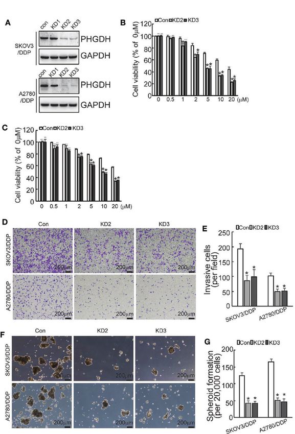
Bi et al.                                                                                                                 PHGDH in Ovarian Cancer

BACKGROUND                                                                   maintained in RPMI1640 containing 10% fetal bovine serum and
                                                                             100 IU/ml of penicillin, 100 µg/ml of streptomycin. The cells
Early-stage ovarian cancer is rarely detected until it progresses to         were incubated in a humidified atmosphere at 37°C with
late stage. Currently tumor debulking surgery combined with                  5% CO2.
platinum-based chemotherapy is commonly used for ovarian
cancer therapy. In many instances, however, some tumor cells                 Sphere Formation Assay
are intrinsically resistant to platinum, furthermore, some initially         The cells in the logarithmic growth phase were harvested and
responsive patients relapse due to acquired cisplatin resistance             resuspended in serum-free F12 medium supplemented with
(1). The mechanisms underlying intrinsic or acquired platinum                20 mg/ml Epidermal Growth Factor (EGF), 5 mg/ml Insulin
resistance are not completely clarified.                                      and 2% B27. Cells were plated in six-well Corning Spheroid
    Alike as many cytotoxic cancer chemotherapies increase reactive          Microplates. The media were changed every 3 days, images of
oxygen species (ROS) levels, platinum compounds increase                     cells were taken under an inverted microscope and the numbers
mitochondrial ROS by forming adducts on mitochondrial DNA,                   of spheroid were counted.
thereby compromising redox homeostasis (2). Not surprisingly the
capacity to maintain redox homeostasis plays an important role in            Western Blotting
cisplatin-resistance (3–7). Redox homeostasis is balanced by levels          In order to extract total protein, cells were lysed with ice-cold
between oxidants and antioxidants. Glutathione in a reduced form             RIPA lysis buffer for 30 min, then centrifugation at 12,000 rpm
is the principal cellular antioxidant, which is maintained by                for 30 min at 4°C. Proteins were quantified using BCA method
generation of Nicotinamide Adenine Dinucleotide Phosphate                    and separated with 10% SDS-PAGE. Proteins were transferred
(NADPH). Two shunt pathways branched from glycolysis,                        onto a polyvinylidene difluoride (PVDF) membrane, and then
pentose phosphate pathway and serine synthesis pathway are                   incubated in 1× TBST containing 5% milk for 2 h at room
responsible for intracellular NADPH generation via utilizing                 temperature. The PVDF membrane was incubated with anti-
glucose intermediate metabolites (8–12). The serine biosynthetic             human primary antibodies for overnight at 4°C, and then with
pathway is a branching pathway diverting from glycolysis by                  anti-rabbit secondary antibody at room temperature for 1.5 h.
conversion of 3-phosphoglycerate into serine. Serine is                      According to the manufacturer’s instructions, the gel
subsequently utilized as a substrate for one-carbon (folate cycle)           electrophoresis image analyzer GDS8000 (Thermo Fisher
metabolism, and biosynthesis of sphingolipids, nucleotides, and              Scientific) was then used to detect signals with ECL reagent.
glutathione. In addition, the intracellular methionine pool and,             The relative protein expression was analyzed by Image-J
thereby, methyl donor reactions are also supplemented by serine              software, represented as the density ratio versus GAPDH.
and glycine (13–15). Thus, serine synthesis pathway might play an
important role in regulating chemoresistance of ovarian cancer cells.        Construction of Lentiviral Vector and
    Phosphoglycerate dehydrogenase (PHGDH) is the rate-limiting              Preparation of Recombinant Lentivirus
enzyme responsible for the serine biosynthetic pathway. It has been          Lentiviral CRISPR/cas9 mediated PHGDH gene editing vector
reported that PHGDH is upregulated in some cancers derived from              was constructed by annealing gRNA oligonucleotide pairs and
distinct histology and functions as an oncogenic gene (11, 16–22).           subcloning them into pLenti-Cas9-sgRNA-puro lentivirus vector
In addition to its role in catalyzing de novo serine synthesis, it has       (Genechem Co., Ltd.). The gene encoding PHGDH labeled with
been reported that PHGDH promotes caner progression by                       Myc epitope (Myc-PHGDH) and RMRP were cloned into
production of D-2-hydroxyglutarate (D-2HG), an oncometabolite                pGCLV-GV166 lentivirus vector (Genechem Co., Ltd.). The
(23, 24). Genomic amplifications of PHGDH gene are observed in                shRNA targeting RMRP was designed and cloned into GV118
some breast cancers and melanomas (19, 25). PHGDH is also                    lentivirus vector (Genechem Co., Ltd.). The RNA sequence
transcriptionally regulated by various tumor suppressors and                 information was shown in the Supplementary Table 1.
oncogenes (26–28). Posttranslational regulation via proteasomal              Genechem Co., Ltd. performed DNA sequencing to verify the
degradation is also responsible for PHGDH expression in some                 sequence of the insert and identified it as 100%. After
cancers (29). The current study identifies a novel regulatory level at        construction, the recombinant lentiviral vector, plasmid
translational activation responsible upregulation of platinum-               pHelper 1.0 and plasmid pHelper 2.0 were co-transfected into
resistant ovarian cancer cells. In addition, PHGDH increases                 293T cells with liposome 3000 (Invitrogen). The recombinant
resistance of ovarian cancer cells to cisplatin and promotes                 lentivirus was harvested 72 h after transfection, centrifuged to
invasiveness and spheroid formation of ovarian cancer, making                remove cell debris, and then filtered through 0.22 mm cellulose
PHGDH a potential target for ovarian cancer therapy.                         acetate filter. The final titer of lentivirus was 1.0 × 10⁹ Tu/ml.

                                                                             Gene Expression Mediated by
MATERIALS AND METHODS                                                        Recombinant Lentivirus
                                                                             Cells were infected with recombinant virus targeting genes and
Cell Culture                                                                 corresponding empty vector without gene targeting as negative
Human ovarian cancer cell lines and their cisplatin resistant                control for 12 h. After 72 h, the infected cells were subjected to
cohorts SKOV3 and SKOV3/DDP, A2780 and A2780/DDP were                        puromycin selection. Western blot was used to measure the

Frontiers in Oncology | www.frontiersin.org                              2                                   June 2021 | Volume 11 | Article 643129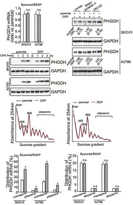
Bi et al.                                                                                                              PHGDH in Ovarian Cancer

infection efficiency of each gene and the negative control.                and 1% Triton X-100) and centrifuged at 13,000 rpm for 10 min
Specially, to purify the PHGDH in cells, cells were infected              at 4°C. The supernatants were then loaded on a sucrose density
recombinant virus expressing PHGDH tagged with Myc                        gradient system ranging from 7 to 47% (Teledyne Isco). The
epitope (Myc-PHGDH) for 12 h. After 72 h, the infected cells              gradients were centrifuged in an SW41 Beckman rotor at 35,000
were subjected to puromycin selection. Western blot was used to           rpm for 180 min at 4°C. Gradients were collected into 0.5 ml/tube
measure the infection efficiency of each gene and the negative             fractions by monitoring RNA absorbance at 254 nm using an ISCO
control, where the PHGDH expression level of Myc-PHGDH                    fractionator (Brandel, Inc.). RNA was isolated from monosome, and
and the control group was detected by incubating Myc-antibody             polysome fractions using the TRIzol reagent (Life Technologies)
as the primary antibody. Then cells were treated as indicated and         and quantified. Reverse transcription (RT) reactions were
subjected to further analysis.                                            performed using a SuperScript III first-strand synthesis system
                                                                          (Life Technologies) with a random primer. All polysomal analysis
3-(4,5-Dimethylthiazol-2-yl)-2,5-                                         was done a minimum of three times.
Diphenyltetrazolium Bromide (MTT) Assay
The MTT assay was performed according to the manufacturer’s               Clinical Samples
instruction. Briefly, cells were exposed to different concentrations       In the present study, 25 patients were recruited in this study, who
of cisplatin for 48 h, cells were incubated with MTT for 4 h,             underwent surgical resection at Shengjing Hospital of China
followed by addition of isopropyl alcohol to dissolve the                 Medical University from June 2014 to July 2017. The collected
formazan crystals. OD was measured at 570 nm using a                      tissues were immediately frozen in liquid nitrogen and stored in
Microplate Reader.                                                        −80°C freezer till further utilization. The patients were divided
                                                                          into two groups, platinum-sensitive (sixteen patients) and
Generation of Reporter Vectors and Dual-                                  platinum resistant (nine patients) groups with ages of 43 to 69
Luciferase Reporter Assay                                                 years (57 on the average). None of the patients had received
The 5′UTR (untranslational region), CDS (coding sequence),                chemotherapy or radiotherapy prior to the operation. The
and 3′UTR fragments of PHGDH mRNA was generated by PCR                    project was approved by Institutional Review Board of China
and inserted into the pMIR-REPORTTM Luciferase vector                     Medical University that informed consent was not needed to
(Promega, Madison, WI) just after the stop codon. The                     obtain from the patients or their family.
transfection was carried out with Lipofectamine 3000
(Invitrogen) according to the manufacturer’s instructions. Cells          Tissue Microarray and
were incubated for 48 h and harvested by adding 100 µl of                 Immunohistochemical Staining
reporter lysis buffer (Dual-Luciferase Assay System, Promega).            Tissue microarray sections were purchased from Shanghai
Luciferase activity was measured by Dual Luciferase Reporter              Outdo Biotech Co., LTD. Immunohistochemical staining was
Gene Assay Kit. Experiments were performed in triplicates and             performed on tissue sections using an antibody against PHGDH.
repeated for three times independently. The Renilla luciferase            A semi-quantitative H-score was assessed for each specimen by
activity values that reflect transfection efficiency were utilized to       multiplying the distribution areas (0–100%) at each staining
normalize the firefly luciferase activity values. Data are presented        intensity level by the intensities (0: negative; 1: weak staining;
as mean values (s.d.).                                                    2: moderate staining; 3: strong staining) as previously reported
                                                                          (30). The median value of the H-score was chosen as the cutoff
Transwell Migration and Matrigel                                          criterion to categorize into high and low expression subgroup.
Invasion Assays
Transwell invasion and migration assays were performed to                 Statistical Analysis
determine cell invasion and migration, respectively. Transwell            The statistical significance of the difference was analyzed by
inserts coated with Matrigel on the upper layers were used for            analysis of variance (ANOVA) and post hoc Dunnett’s test.
invasion assay. Uncoated inserts were used for migration assay.           Statistical significance was defined as P < 0.05. All experiments
Briefly, cells were seeded into the upper chamber with FBS-free            were repeated three times, and data were expressed as the mean ±
medium, and lower chamber was filled with full medium. The                 SD (standard deviation) from a representative experiment.
cells were incubated in a humidified 5% CO2 incubator at 37°C
for 24h. The invaded or migrated cells were fixed in 4%
paraformaldehyde for 5min and then stained with 0.3% crystal              RESULTS
violet. Invading cells or migrating cells were counted under a
light microscope.                                                         PHGDH Is Upregulated in Platin-Resistant
                                                                          Ovarian Cancer Cells and Predicts Poor
Polysome Profiling                                                         Prognosis
Some 1 × 10⁷ cells were incubated with 100 mg/ml cycloheximide            Western blot demonstrated that PHGDH was increased in
for 5 min to halt elongation. Cells were then harvested in 500 ml         platin-resistant (DDP) SKOV3 and A2780 cells, when
of polysome lysis buffer (15 mM Tris–HCl pH 7.4, 15 mM                    compared with their relative platin-sensitive parental partners
MgCl2, 0.3 M NaCl, 1 mg/ml heparin, 0.1 mg/ml cycloheximide,              (Figure 1A). PHGDH expression was also further investigated in

Frontiers in Oncology | www.frontiersin.org                           3                                   June 2021 | Volume 11 | Article 643129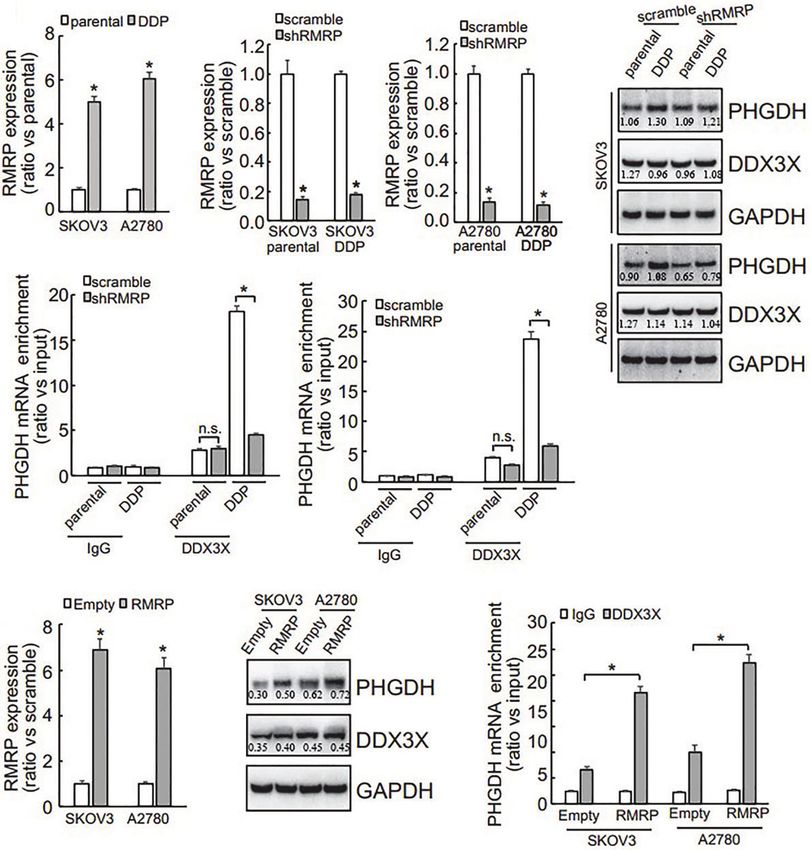
Bi et al. PHGDH in Ovarian Cancer
16 platin-sensitive and eight platin-resistant ovarian cancer predicted significantly poor overall survival of patients with
tissues. PHGDH expression was higher in most of platin- ovarian cancer (Figure 1D).
resistant ovarian cancer tissues than in platin-sensitive tissues
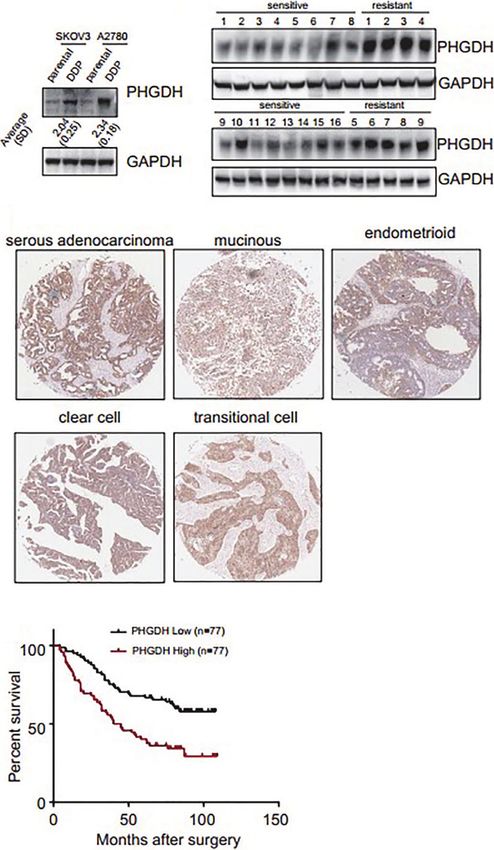
(Figure 1B). PHGDH expression was then investigated in a Knockdown of PHGDH Increases
panel of ovarian cancer tissues using immunohistochemical Responsiveness to Cisplatin and
staining. PHGDH was expressed in 118 of 154 epithelial Suppresses Capacities of Invasion and
ovarian cancer specimens (Figure 1C). Based on the Spheroid Formation in Platin-Resistant
expression of PHGDH expression, patients with ovarian cancer Ovarian Cancer
were grouped into high and low expression groups. Kaplan– To investigate the potential role of PHGDH in ovarian cancer,
Meier survival analysis demonstrated that high PHGDH PHGDH was knocked down using CASPR-Cas9 system. Tow of
B
A
C
D
FIGURE 1 | PHGDH is increased in platin-resistant ovarian cancer and predicts poor prognosis of patients with ovarian cancer. (A) PHGDH expression was
assessed using Western blot in platin-sensitive (parental) and their platin-resistant (DDP) SKOV3 and A2780 cells. The relative expression was noted under the blots.
(B) PHGDH expression was investigated in 16 cisplatin-sensitive and nine cisplatin-resistant ovarian cancer tissues using Western blot. (C) Ovarian cancer tissue
microarray was subjected to immunohistochemical staining, and representative images of immunohistochemistry staining with PHGDH were presented. (D) Kalpan–
Meier plot shows the overall survival of patients with ovarian cancer grouped by PHGDH expression.
Frontiers in Oncology | www.frontiersin.org 4 June 2021 | Volume 11 | Article 643129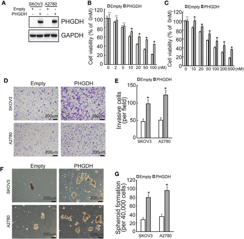
Bi et al.                                                                                                                                    PHGDH in Ovarian Cancer

gRNAs against PHGDH significantly decreased PHGDH                                       Ectopic Expression of PHGDH Decreases
expression in SKOV3/DDP and A2780/DDP cells compared to                                Responsiveness to Cisplatin and
the cells infected with the pLenti-Cas9-sgRNA-puro lentivirus                          Promotes Invasiveness and Spheroid
vector without targeting PHGDH (Figure 2A). CCK8 assays                                Formation of Ovarian Cancer Cells
demonstrated the knockdown of PHGDH significantly decreased                             To further confirmed the potential role of PHGDH in ovarian
cell viability of SKVO3/DDP (Figure 2B) and A2780/DDP                                  cancer, PHGDH was ectopically overexpressed in SKOV3 and
(Figure 2C) cells when exposed to cisplatin. Matrigel-coated                           A2780 cells infected with lentivirus containing PHGDH labeled
Transwell assay demonstrated that knockdown of PHGDH                                   with Myc epitope (Myc-PHGDH) compared to SKOV3 and A2780
significantly decreased invasiveness of SKOV3/DDP and                                   cells infected with the empty pGCLV-GV166 lentivirus vector
A2780/DDP cells (Figures 2D, E). In addition, PHGDH                                    (empty) (Figure 3A). In addition, SKOV3 or A2780 cells were
knockdown also suppressed spheroid formation capacity of                               infected with lentivirus containing PHGDH (PHGDH) and the
SKOV3/DDP and A2780/DDP cells (Figures 2F, G).                                         empty pGCLV-GV166 lentivirus vector (empty) as control.

  FIGURE 2 | PHGDH knockdown increases responsiveness to cisplatin and decreases capacities of invasion and spheroid formation in platin-resistant ovarian
  cancer. (A) SKOV3/DDP or A2780/DDP cells were infected with CASPR-Cas9 lentivirus containing specific gRNA against PHGDH and the pLenti-Cas9-sgRNA-puro
  lentivirus vector without PHGDH targeting as control, knockdown of PHGDH was confirmed by Western blot. (B, C) The indicated cells were treated with the solvent
  control and different doses of cisplatin for 48 h, and cell viability was analyzed using CCK8 assays. (D, E) The indicated cells were plated on the Matrigel-coated
  transwell, invaded cells were stained with crystal violet and photographed (D), cell numbers were counted and plotted (E). (F, G) The indicated cells were floating
  cultured with serum-free media for 14 days, spheroid was photographed (F), and spheroid numbers were counted and plotted (G). *P < 0.01; N.S., not significant.

Frontiers in Oncology | www.frontiersin.org                                        5                                          June 2021 | Volume 11 | Article 643129
Bi et al.                                                                                                                                      PHGDH in Ovarian Cancer

Overexpression of PHGDH significantly increased cell viability of                        proteasomal degradation of PHGDH, respectively. Neither
SKOV3 (Figure 3B) and A2780 (Figure 3C) cells upon cisplatin                            E64D plus pepstatin A nor MG132 altered the different
treatment. Matrigel-coated transwell assays demonstrated that                           expression of PHGDH in platin-sensitive ovarian cancer cells
overexpression increased invasion capacity of SKOV3 and A2780                           and their platin-resistant partners compared to the cells treated
cells (Figures 3D, E). In addition, PHGDH overexpression also                           with the solvent vehicle (Figure 4B), indicating that synthesis,
increased spheroid formation of SKOV3 and A2780 cells                                   but not degradation of PHGDH might be responsible for
(Figures 3F, G).                                                                        upregulation of PHGDH in platin-resistant ovarian cancer
                                                                                        cells. Cells were then treated with Cycloheximide (CHX) for
PHGDH Is Increased in Cisplatin-Resistant                                               16 h to completely block protein synthesis, then CHX was
Ovarian Cancer Cells at the Translational                                               washed out to resume protein synthesis for different period.
Initiation Level                                                                        PHGDH expression was apparently recovered after 1 h free of
The promotive role of PHGDH in cisplatin-resistant, invasion                            CHX in SKOV3/DDP and A2780/DDP cells, while its expression
and spheroid formation of ovarian cancer cells promoted us to                           was delayed in their parental partners (Figure 4C), confirming
further investigate the underlying mechanisms of its                                    that PHGDH translation is activated in platin-resistant ovarian
upregulation in platin-resistant ovarian cancer. qRT-PCR                                cancer cells. PHGDH mRNA distribution was also investigated
found that no obvious alteration of PHGDH mRNA was                                      using ribosome profiling. Distribution of total RNAs was similar
observed in platin-resistant SKOV3 and A2780 cells when                                 between platin-resistant and platin-sensitive SKOV3
compared with their platin-sensitive partners (Figure 4A),                              (Figure 4D) and A2780 (Figure 4E) cells. Occupation of
indicating that PHGDH was upregulated in platin-resistant                               PHGDH mRNA on both monosomes and polysomes was
ovarian cancer cells at the protein level. E64D plus pepstatin A                        significantly augmented in SKOV3/DDP and A2780/DDP cells
and MG132 were then utilized to suppress lysosomal and                                  when compared with their parental control partners (Figure 4F).

  FIGURE 3 | PHGDH overexpression suppresses responsiveness to cisplatin and promotes invasion and spheroid formation of platin-sensitive ovarian cancer cells.
  (A) SKOV3 or A2780 cells were infected with lentivirus containing PHGDH labeled with Myc epitope (Myc-PHGDH, labeled as PHGDH) and the empty pGCLV-
  GV166 lentivirus vector (empty) as control, expression of PHGDH was confirmed by Western blot. (B, C) The indicated cells (SKOV3 or A2780 cells were infected
  with lentivirus containing PHGDH (PHGDH) and the empty pGCLV-GV166 lentivirus vector (empty) as control) were treated with the solvent control and were treated
  with different doses of cisplatin for 48 h, and cell viability was analyzed using CCK8 assays. (D, E) The indicated cells were plated on the Matrigel-coated tranwell,
  invaded cells were stained with crystal violet and photographed (D), cell numbers were counted and plotted (E). (F, G) The indicated cells were floating cultured with
  serum-free media for 14 days, spheroid was photographed (F), and spheroid numbers were counted and plotted (G). *P < 0.01; N.S., not significant.

Frontiers in Oncology | www.frontiersin.org                                         6                                           June 2021 | Volume 11 | Article 643129
Bi et al. PHGDH in Ovarian Cancer
A B
C
D E
F G
FIGURE 4 | Translational activation is responsible for upregulation of PHGDH in platin-resistant ovarian cancer cells. (A) PHGDH mRNA expression was analyzed
using real-time RT-PCR in ovarian cancer cell lines. (B) PHGDH expression was analyzed using Western blot after cells were incubated with the solvent vehicle,
MG132, or E64D and pepstatin A for 24 h. (C) Ovarian cancer cells were treated with cycloheximide (CHX) for 16 h, then CHX were completely washed out. PHGDH
expression was analyzed using Western blot analysis after Cells were cultured under complete media for the additional indicated time. (D, E) SKOV3 (D) or A2780
(E) Cells were exposed to CHX for 10 h, cell homogenate was subjected for sucrose gradient fractionation. Absorbance at 254 nm was measured in each fraction.
(F, G) total RNA was isolated from monosome and polysome fractions, PHGDH mRNA (F) and GAPDH mRNA (G) occupation on monosome and polysome
fractions was analyzed using real-time RT-PCR. *P < 0.05, n.s., not significant.
On the other hand, distribution of GAPDH was not different containing CDS alone or 5’UTR and CDS significantly increased
between platin-resistant ovarian cancer cells and their parental PHGDH expression, while constructs containing 3’UTR fragment
control partners (Figure 4G). was not overexpressed efficiently in SKOV3 and A2780 cells
(Figure 5B), indicating that 3’UTR of PHGDH might suppress its
DDX3X Recruitment on PHGDH mRNA Is translation. miRNAs containing RISC are well-known to regulate
Increased and Implicated in Translation of translation via 3’UTR of target mRNAs, RNA immunoprecipitation
PHGDH in Platin-Resistant Ovarian (RIP) was then performed using pan-Ago antibody, a key
Cancer Cells component of RISC. Enrichment of PHGDH mRNA by pan-Ago
To investigate the potential responsive cis-acting element on was not significantly different between platin-resistant and platin-
PHGDH transcript, eukaryotic expression constructs containing sensitive SKOV3 (Figure 5C) and A2780 (Figure 5D) cells,
various fragment of PHGDH transcript were generated as illustrated indicating that miRISCs might not be implicated in differential
(Figure 5A). Transfection of eukaryotic expression construct translation of PHGDH between platin-resistant and platin-sensitive
Frontiers in Oncology | www.frontiersin.org 7 June 2021 | Volume 11 | Article 643129Bi et al. PHGDH in Ovarian Cancer
ovarian cancer cells. In addition to miRNAs, RNA-binding proteins indicating that other factor(s) might guide DDX3X to the
are also involved in translation of target mRNAs. Using biotin- PHGDH mRNA in platin-resistant ovarian cancer. Since
labeled 3’UTR of PHGDH transcript, quantitative analysis of except for RNA binding proteins, non-coding RNAs are also
PHGDH RNA binding proteins were evaluated by biotin pull implicated in translation of target mRNAs via interaction, non-
down followed by quantitative mass spectrometry. Among coding RNAs that can potentially interact with both PHGDH
differentially potential PHGDH mRNA-binding proteins, mRNA and DDX3X were screened via online database
recruitment of Dead-box helicase 3 X-linked (DDX3X) was (starbase.sysu.edu.cn) and RNA component of mitochondrial
significantly increased. Western blot showed that no apparent RNA processing endoribonuclease (RMRP) attracted our
difference of DDX3X expression was observed in platin-resistant attention. qRT-PCR demonstrated that RMRP was significantly
and platin-sensitive SKOV3 and A2780 cells (Figure 5E), while increased in SKOV3/DDP and A2780/DDP cells (Figure 6A),
DDX3X recruitment on PHGDH mRNA was significantly while LINC00662 was undetectable in any samples. RMRP was
increased in platin-resistant SKOV3 (Figure 5F) and A2780 then knocked down in SKOV3 (Figure 6B) and A2780
(Figure 5G) cells, when compared with their parental control (Figure 6C) cells. Western blot demonstrated that knockdown
partners. In addition, knockdown of DDX3X significantly of RMRP significantly decreased PHGDH expression in SKOV3/
decreased PHGDH expression in SKOV3/DDP and A2780/DDP DDP and A2780/DDP cells (Figure 6). RIP was also performed
cells (Figure 5H). and demonstrated that recruitment of DDX3X on the PHGDH
mRNA was significantly compromised by RMRP knockdown in
SKOV3/DDP (Figure 6D) and A2780/DDP (Figure 6E) cells.
LncRNA RMRP Is Increased and Promotes RMRP was also ectopically expressed in platin-sensitive SKOV3
Recruitment of DDX3X on the PHGDH and A2780 cells (Figure 6F). Overexpression of RMRP
mRNA in Platin-Resistant Ovarian Cancer significantly increased PHGDH expression in SKOV3 and
Cells A2780 cells (Figure 6G). In addition, overexpression of RMRP
DDX3X expression was not different (Figure 5E), while its also promoted recruitment of DDX3X on the PHGDH mRNA in
enrichment on the PHGDH mRNA was increased (Figures 5F, G), SKOV3 and A2780 cells (Figure 6H).
A B
C D
E
F G
H
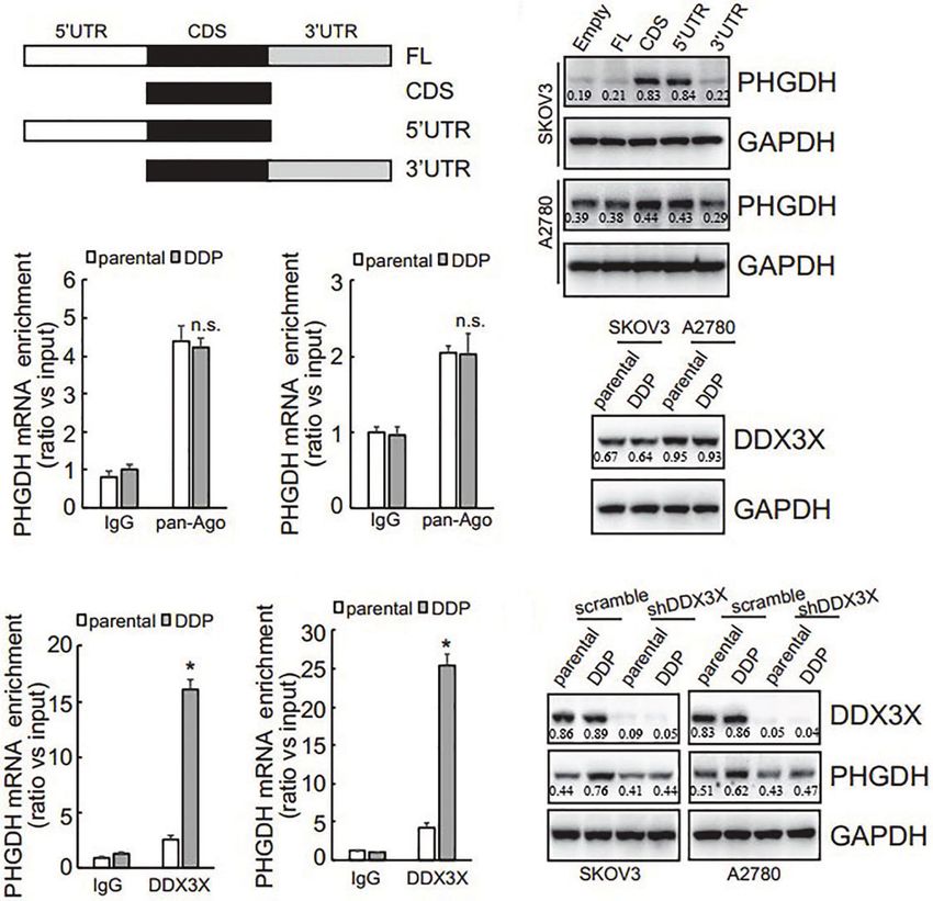
FIGURE 5 | PHGDH expression is upregulated via 3’UTR of its transcript in a DDX3X-dependent pattern. (A) The PHGDH expression pMIR-REPORTTM Luciferase
vectors were cloned as illustrated. (B) cells were transfected with empty pMIR-REPORTTM Luciferase vector (empty) and PHGDH vector (FL, CDS, 5’UTR, 3’UTR),
and ectopic PHGDH expression was investigated using Western blot analysis. (C, D) RIP was performed using IgG or pan-Ago antibody from control and DDP
paired A2780 (C) or SKOV3 (D) cells, and enrichment of PHGDH mRNA was evaluated using qRT-PCR. (E) DDX3X expression was analyzed using Western blot
analysis. (F, G) RIP was performed using IgG or DDX3X antibody from control and DDP paired A2780 (F) or SKOV3 (G) cells, and enrichment of PHGDH mRNA
was evaluated using qRT-PCR. (H) Cells were infected with pGCLV-GV166 lentivirus vector (scramble) or shRNAs against DDX3X (shDDX3X), PHGDH expression
was analyzed using Western blot. *P < 0.05, n.s., not significant.
Frontiers in Oncology | www.frontiersin.org 8 June 2021 | Volume 11 | Article 643129Bi et al. PHGDH in Ovarian Cancer
DISCUSSION occurred in cisplatin resistant cells. Serine is essential for cell
synthesis of nucleotides, proteins and lipids. Many types of cancer
Ovarian cancer is rarely diagnosed during its early stages since lack need to synthesize serine to maintain a rapid and stable growth
of obvious symptoms, which ranks the fifth leading cause of pattern. PHGDH is a key enzyme in serine biosynthesis pathway,
cancer-related death in female. Standard treatment for ovarian which plays a key role in serine biosynthesis and mitochondrial
cancer includes staging and optimal debulking surgery followed by redox homeostasis in cancer cells. Furthermore, we found that
adjuvant platinum-based (e.g. cisplatin-based) chemotherapy as PHGDH knockout inhibited the proliferation of ovarian cancer
the first line treatment. Despite the apparent benefits for patients cells, while PHGDH overexpression increased the survival rate,
with ovarian cancer utilizing platinum-based chemotherapy, invasiveness and spheroid formation of ovarian cancer cells after
initial or acquired resistance is a clinical barrier leading to cisplatin exposure. The current study demonstrated that PHGDH,
therapeutic failure. Therefore, it is of clinical significance to the key enzyme implicated in serine biosynthesis pathway, was
better understand the mechanisms underlying cisplatin upregulated in cisplatin-resistant ovarian cancer cells and tissues.
resistance. Metabolic reprogramming is one of the Knockdown of PHGDH decreased cell viability upon cisplatin
characteristics of tumor cells, which is closely related to exposure, suppressed invasion and spheroid formation of
tumorigenesis, progression and drug resistance. As for cisplatin-resistant ovarian cancer cells, while overexpression of
intracellular glucose metabolism pathway, in addition to PHGDH increased cell viability upon cisplatin exposure,
glycolysis and mitochondrial oxidative phosphorylation, there promoted invasion and spheroid formation of cisplatin-sensitive
are also some branch pathways, such as serine synthesis ovarian cancer cells. Importantly, high PHGDH expression
pathway. The expression of PHGDH, the rate limiting enzyme predicted a poor prognosis of patients with ovarian cancers.
of serine metabolism pathway, increased significantly in cisplatin These data indicated that upregulation of PHGDH might play a
resistant cells, suggesting that serine metabolism rearrangement role on cisplatin resistance, and PHGDH might function as an
A B C
D
E
F G H
FIGURE 6 | RMRP is increased and promotes recruitment of DDX3X to PHGDH transcript in cisplatin-resistant ovarian cancer cells. (A) qRT-PCR was performed to
analyze the RMRP expression. (B, C) cells were infected with scramble GV118 lentivirus vector (scramble) or shRNAs against RMRP (shRMRP), knockdown
efficiency was confirmed by qRT-PCR (B), PHGDH expression was analyzed using Western blot (C). (D, E) RIP was performed using IgG or DDX3X antibody from
parental and DDP paired SKOV3 (D) and A2780 (E) infected with scramble or shRMRP. (F, H) cisplatin-sensitive SKOV3 and A2780 cells were infected with empty
pGCLV-GV166 lentivirus vector (empty) or lentivirus containing RMRP (RMRP), or RMRP, expression of RMRP was analyzed using qRT-PCR (F), PHGDH expression
was investigated using Western blot (G), recruitment of DDX3X on PHGDH transcript was studied using RIP (H). *P < 0.05, n.s., not significant.
Frontiers in Oncology | www.frontiersin.org 9 June 2021 | Volume 11 | Article 643129Bi et al. PHGDH in Ovarian Cancer
oncogene in ovarian cancer. Therefore, targeting PHGDH might DDX3X on the PHGDH transcript. In addition, the current study
render resistant cells sensitive to cisplatin, providing a potential also demonstrates that LncRNA RPRM is upregulated, which plays
strategy for treatment of cisplatin resistant ovarian cancer. an important role in promotion of DDX3X recruitment. Thus, this
PHGDH has been shown to be regulated at multiple levels, study suggests that targeting PHGDH may provide a potential
including gene amplification (25, 31), transcriptional activation (27), opportunity to overcome cisplatin resistance in ovarian cancer.
as well proteasomal degradation (29). The current study provides an
additional regulatory mechanism by which PHGDH is regulated at
the translational level. Our data suggest that PHGDH translational
CONCLUSIONS
efficiency is different in platin-resistant and platin-sensitive ovarian
cancer cells, which further adds diversity to the regulation of Collectively, the current study described that PHGDH was
PHGDH expression in the distinct cancers. PHGDH is highly upregulated and conferred resistance of ovarian cancer cells to
expressed in many cancer cells and plays a critical role to support cisplatin, suggesting that cisplatin resistance could be overcome
a variety biosynthetic processes important for cell proliferation) (14, by targeting PHGDH. In addition, our study also provided
32, 33), thereby our findings might provide a novel potential evidence that differential PHGDH protein expression was
therapeutic targets for cancer therapy. defined by its translation, and RNA binding protein DDX3X
DDX3X is a member of the Asp-Glu-Ala-Asp (DEAD) box and LncRNA RMRP are regulators of its translation.
protein family. DEAD motif containing proteins have ATP-
dependent RNA helicase activity, which are involved in alteration
of RNA secondary structure, thereby implicated in a variety of
cellular processes such as splicing and translation initiation. Nuclear
DATA AVAILABILITY STATEMENT
DDX3X is involved in transcriptional regulation, mRNP assembly, The original contributions presented in the study are included in
pre-mRNA splicing, and mRNA export, while cytoplasmic DDX3X the article/Supplementary Material. Further inquiries can be
is thought to be implicated in translation, cellular signaling, and directed to the corresponding author.
viral replication (34–36). Dysregulation of DDX3X has been
implicated in tumorigenesis and development (37–44). The
current study found that although total DDX3X expression was
not distinct between cisplatin-sensitive and cisplatin-resistant AUTHOR CONTRIBUTIONS
ovarian cancer cells, while its recruitment on PHGDH mRNA
QY designed the article. FB and TS did the experiments. YA and
was augmented in cisplatin-resistant ovarian cancer cells.
YY wrote the manuscript. All the figures were prepared by FB
Importantly, knockdown of DDX3X significantly decreased
and revised by QY. All authors contributed to the article and
PHGDH expression in cisplatin-resistant ovarian cancer cells.
approved the submitted version.
These data indicated that DDX3X enrichment might play a
critical role in promoting PHGDH translation in cisplatin-
resistant ovarian cancer cells, while additional factor(s) are
necessary for its recruitment to the PHGDH mRNA. The current FUNDING
study found that long non-coding RNA (Lnc RNA) RPRM was
upregulated in cisplatin-resistant ovarian cancer cells and promoted Supported by grants from National Natural Science Foundation
enrichment of DDX3X on the PHGDH mRNA. Thereby, our of China (No.81872125), 345 Talent Project of Shengjing
findings identified cooperation of DDX3X and RPRM to promote Hospital of China Medical University (No.M0695), and
translation of PHGDH transcript in cisplatin-resistant ovarian Outstanding Scientific Fund of Shengjing Hospital (No. 201704).
cancer cells.
In conclusion, the current study demonstrates that PHGDH is
upregulated and plays a critical role in cisplatin resistance in ovarian SUPPLEMENTARY MATERIAL
cancer cells. We show that high PHGDH expression is correlated
with lower overall survival of patients with ovarian cancer. We show The Supplementary Material for this article can be found online
that PHGDH is upregulated at the translational level in cisplatin at: https://www.frontiersin.org/articles/10.3389/fonc.2021.
resistant ovarian cancer cells, which is regulated by recruitment of 643129/full#supplementary-material
REFERENCES 3. Salatino A, Aversa I, Battaglia AM, Sacco A, Di Vito A, Santamaria G, et al. H-
Ferritin Affects Cisplatin-Induced Cytotoxicity in Ovarian Cancer Cells
1. Chen J, Solomides C, Parekh H, Simpkins F, Simpkins H. Cisplatin Resistance Through the Modulation of ROS. Oxid Med Cell Longevity (2019)
in Human Cervical, Ovarian and Lung Cancer Cells. Cancer Chemother 2019:3461251. doi: 10.1155/2019/3461251
Pharmacol (2015) 75:1217–27. doi: 10.1007/s00280-015-2739-2 4. Sun X, Wang S, Gai J, Guan J, Li J, Li Y, et al. Sirt5 Promotes Cisplatin
2. Kleih M, Bopple K, Dong M, Gaissler A, Heine S, Olayioye MA, et al. Direct Impact Resistance in Ovarian Cancer by Suppressing Dna Damage in a ROS-
of Cisplatin on Mitochondria Induces ROS Production That Dictates Cell Fate of Dependent Manner Via Regulation of the Nrf2/HO-1 Pathway. Front
Ovarian Cancer Cells. Cell Death Dis (2019) 10:851. doi: 10.1038/s41419-019-2081-4 Oncol (2019) 9:754. doi: 10.3389/fonc.2019.00754
Frontiers in Oncology | www.frontiersin.org 10 June 2021 | Volume 11 | Article 643129Bi et al. PHGDH in Ovarian Cancer
5. Sun C, Guo E, Zhou B, Shan W, Huang J, Weng D, et al. A Reactive Oxygen 26. Svoboda LK, Teh SSK, Sud S, Kerk S, Zebolsky A, Treichel S, et al. Menin
Species Scoring System Predicts Cisplatin Sensitivity and Prognosis in Ovarian Regulates the Serine Biosynthetic Pathway in Ewing Sarcoma. J Pathol (2018)
Cancer Patients. BMC Cancer (2019) 19:1061. doi: 10.1186/s12885-019-6288-7 245:324–36. doi: 10.1002/path.5085
6. Lee H, Lee D, Kang KS, Song JH, Choi YK. Inhibition of Intracellular Ros 27. DeNicola GM, Chen PH, Mullarky E, Sudderth JA, Hu Z, Wu D, et al. NRF2
Accumulation by Formononetin Attenuates Cisplatin-Mediated Apoptosis in Regulates Serine Biosynthesis in non-Small Cell Lung Cancer. Nat Genet
LLC-PK1 Cells. Int J Mol Sci (2018) 19:813. doi: 10.3390/ijms19030813 (2015) 47:1475–81. doi: 10.1038/ng.3421
7. Lv X, Song DM, Niu YH, Wang BS. Inhibition of Heme Oxygenase-1 Enhances 28. Amelio I, Markert EK, Rufini A, Antonov AV, Sayan BS, Tucci P, et al. p73
the Chemosensitivity of Laryngeal Squamous Cell Cancer Hep-2 Cells to Cisplatin. Regulates Serine Biosynthesis in Cancer. Oncogene (2014) 33:5039–46. doi:
Apoptosis (2016) 21:489–501. doi: 10.1007/s10495-016-1216-7 10.1038/onc.2013.456
8. Ghanbari Movahed Z, Rastegari-Pouyani M, Mohammadi MH, Mansouri K. 29. Zhang B, Zheng A, Hydbring P, Ambroise G, Ouchida AT, Goiny M, et al.
Cancer Cells Change Their Glucose Metabolism to Overcome Increased ROS: Phgdh Defines a Metabolic Subtype in Lung Adenocarcinomas With Poor
One Step From Cancer Cell to Cancer Stem Cell? Biomed Pharmacother = Prognosis. Cell Rep (2017) 19:2289–303. doi: 10.1016/j.celrep.2017.05.067
Biomed Pharmacother (2019) 112:108690. doi: 10.1016/j.biopha.2019.108690 30. Detre S, Saclani Jotti G, Dowsett M. A “Quickscore” Method for
9. Cho ES, Cha YH, Kim HS, Kim NH, Yook JI. The Pentose Phosphate Pathway Immunohistochemical Semiquantitation: Validation for Oestrogen Receptor
as a Potential Target for Cancer Therapy. Biomol Ther (2018) 26:29–38. doi: in Breast Carcinomas. J Clin Pathol (1995) 48:876–8. doi: 10.1136/jcp.48.9.876
10.4062/biomolther.2017.179 31. Locasale JW, Grassian AR, Melman T, Lyssiotis CA, Mattaini KR, Bass AJ,
10. Chen X, Xu Z, Zhu Z, Chen A, Fu G, Wang Y, et al. Modulation of G6PD et al. Phosphoglycerate Dehydrogenase Diverts Glycolytic Flux and
Affects Bladder Cancer Via ROS Accumulation and the AKT Pathway In Contributes to Oncogenesis. Nat Genet (2011) 43:869–74. doi: 10.1038/ng.890
Vitro. Int J Oncol (2018) 53:1703–12. doi: 10.3892/ijo.2018.4501 32. Pacold ME, Brimacombe KR, Chan SH, Rohde JM, Lewis CA, Swier LJ, et al. A
11. Wu X, Xia J, Zhang J, Zhu Y, Wu Y, Guo J, et al. Phosphoglycerate PHGDH Inhibitor Reveals Coordination of Serine Synthesis and One-Carbon
Dehydrogenase Promotes Proliferation and Bortezomib Resistance Through Unit Fate. Nat Chem Biol (2016) 12:452–8. doi: 10.1038/nchembio.2070
Increasing Reduced Glutathione Synthesis in Multiple Myeloma. Br J 33. Mullarky E, Lairson LL, Cantley LC, Lyssiotis CA. A Novel Small-Molecule
Haematol (2020) 190(1):52–66. doi: 10.1111/bjh.16503 Inhibitor of 3-Phosphoglycerate Dehydrogenase. Mol Cell Oncol (2016) 3:
12. Saber MM, Al-Mahallawi AM, Nassar NN, Stork B, Shouman SA. Targeting Colorectal e1164280. doi: 10.1080/23723556.2016.1164280
Cancer Cell Metabolism Through Development of Cisplatin and Metformin Nano- 34. Song H, Ji X. The Mechanism of RNA Duplex Recognition and Unwinding by DEAD-
Cubosomes. BMC Cancer (2018) 18:822. doi: 10.1186/s12885-018-4727-5 box Helicase DDX3X. Nat Commun (2019) 10:3085. doi: 10.1038/s41467-019-11083-2
13. Li AM, Ye J. The PHGDH Enigma: Do Cancer Cells Only Need Serine or Also a 35. Ku YC, Lai MH, Lo CC, Cheng YC, Qiu JT, Tarn WY, et al. Ddx3 Participates
Redox Modulator? Cancer Lett (2020) 476:97–105. doi: 10.1016/j.canlet.2020.01.036 in Translational Control of Inflammation Induced by Infections and Injuries.
14. Samanta D, Semenza GL. Serine Synthesis Helps Hypoxic Cancer Stem Cells Regulate Mol Cell Biol (2019) 39:3595–607. doi: 10.1128/MCB.00285-18
Redox. Cancer Res (2016) 76:6458–62. doi: 10.1158/0008-5472.CAN-16-1730 36. Fullam A, Gu L, Hohn Y, Schroder M. DDX3 Directly Facilitates IKKalpha
15. Reid MA, Allen AE, Liu S, Liberti MV, Liu P, Liu X, et al. Serine Synthesis Through Activation and Regulates Downstream Signalling Pathways. Biochem J (2018)
PHGDH Coordinates Nucleotide Levels by Maintaining Central Carbon 475:3595–607. doi: 10.1042/BCJ20180163
Metabolism. Nat Commun (2018) 9:5442. doi: 10.1038/s41467-018-07868-6 37. Yang F, Fang E, Mei H, Chen Y, Li H, Li D, et al. Cis-Acting Circ-CTNNB1
16. Wei L, Lee D, Law CT, Zhang MS, Shen J, Chin DW, et al. Genome-Wide CRISPR/ Promotes Beta-Catenin Signaling and Cancer Progression Via DDX3-
Cas9 Library Screening Identified PHGDH as a Critical Driver for Sorafenib Mediated Transactivation of YY1. Cancer Res (2019) 79:557–71. doi:
Resistance in HCC. Nat Commun (2019) 10:4681. doi: 10.1038/s41467-019-12606-7 10.1158/0008-5472.CAN-18-1559
17. Mullarky E, Xu J, Robin AD, Huggins DJ, Jennings A, Noguchi N, et al. 38. Lin TC. Ddx3x Multifunctionally Modulates Tumor Progression and Serves as
Inhibition of 3-Phosphoglycerate Dehydrogenase (PHGDH) by Indole a Prognostic Indicator to Predict Cancer Outcomes. Int J Mol Sci (2019) 21:
Amides Abrogates De Novo Serine Synthesis in Cancer Cells. Bioorg Med e00285–18. doi: 10.3390/ijms21010281
Chem Lett (2019) 29:2503–10. doi: 10.1016/j.bmcl.2019.07.011 39. Wang Z, Shen GH, Xie JM, Li B, Gao QG. Rottlerin Upregulates DDX3
18. Ma X, Li B, Liu J, Fu Y, Luo Y. Phosphoglycerate Dehydrogenase Promotes Expression in Hepatocellular Carcinoma. Biochem Biophys Res Commun
Pancreatic Cancer Development by Interacting With eIF4A1 and Eif4e. J Exp (2018) 495:1503–9. doi: 10.1016/j.bbrc.2017.11.198
Clin Cancer Res CR (2019) 38:66. doi: 10.1186/s13046-019-1053-y 40. He Y, Zhang D, Yang Y, Wang X, Zhao X, Zhang P, et al. A Double-Edged
19. Unterlass JE, Basle A, Blackburn TJ, Tucker J, Cano C, Noble MEM, et al. Function of DDX3, As an Oncogene or Tumor Suppressor, in Cancer
Validating and Enabling Phosphoglycerate Dehydrogenase (PHGDH) as a Progression (Review). Oncol Rep (2018) 39:883–92. doi: 10.3892/or.2018.6203
Target for Fragment-Based Drug Discovery in PHGDH-amplified Breast 41. Chen HH, Yu HI, Yang MH, Tarn WY. Ddx3 Activates CBC-Eif3-Mediated
Cancer. Oncotarget (2018) 9:13139–53. doi: 10.18632/oncotarget.11487 Translation of Uorf-Containing Oncogenic mRNAs to Promote Metastasis in
20. Yoshino H, Nohata N, Miyamoto K, Yonemori M, Sakaguchi T, Sugita S, et al. HNSCC. Cancer Res (2018) 78:4512–23. doi: 10.1158/0008-5472.CAN-18-0282
PHGDH as a Key Enzyme for Serine Biosynthesis in HIF2alpha-Targeting 42. Cannizzaro E, Bannister AJ, Han N, Alendar A, Kouzarides T. Ddx3x RNA
Therapy for Renal Cell Carcinoma. Cancer Res (2017) 77:6321–9. doi: Helicase Affects Breast Cancer Cell Cycle Progression by Regulating Expression of
10.1158/0008-5472.CAN-17-1589 KLF4. FEBS Lett (2018) 592:2308–22. doi: 10.1002/1873-3468.13106
21. Zhu J, Ma J, Wang X, Ma T, Zhang S, Wang W, et al. High Expression of 43. Adjibade P, Grenier St-Sauveur V, Bergeman J, Huot ME, Khandjian EW,
PHGDH Predicts Poor Prognosis in Non-Small Cell Lung Cancer. Trans Mazroui R. DDX3 Regulates Endoplasmic Reticulum Stress-Induced ATF4
Oncol (2016) 9:592–9. doi: 10.1016/j.tranon.2016.08.003 Expression. Sci Rep (2017) 7:13832. doi: 10.1038/s41598-017-14262-7
22. Samanta D, Park Y, Andrabi SA, Shelton LM, Gilkes DM, Semenza GL. Phgdh 44. Zhao L, Mao Y, Zhao Y, He Y. DDX3X Promotes the Biogenesis of a Subset of
Expression is Required for Mitochondrial Redox Homeostasis, Breast Cancer miRNAs and the Potential Roles They Played in Cancer Development. Sci Rep
Stem Cell Maintenance, and Lung Metastasis. Cancer Res (2016) 76:4430–42. (2016) 6:32739. doi: 10.1038/srep32739
doi: 10.1158/0008-5472.CAN-16-0530
23. Mattaini KR, Brignole EJ, Kini M, Davidson SM, Fiske BP, Drennan CL, et al. Conflict of Interest: The authors declare that the research was conducted in the
An Epitope Tag Alters Phosphoglycerate Dehydrogenase Structure and absence of any commercial or financial relationships that could be construed as a
Impairs Ability to Support Cell Proliferation. Cancer Metab (2015) 3:5. doi: potential conflict of interest.
10.1186/s40170-015-0131-7
24. Fan J, Teng X, Liu L, Mattaini KR, Looper RE, Vander Heiden MG, et al. Copyright © 2021 Bi, An, Sun, You and Yang. This is an open-access article
Human Phosphoglycerate Dehydrogenase Produces the Oncometabolite D-2- distributed under the terms of the Creative Commons Attribution License (CC BY).
Hydroxyglutarate. ACS Chem Biol (2015) 10:510–6. doi: 10.1021/cb500683c The use, distribution or reproduction in other forums is permitted, provided the
25. Mullarky E, Mattaini KR, Vander Heiden MG, Cantley LC, Locasale JW. PHGDH original author(s) and the copyright owner(s) are credited and that the original
Amplification and Altered Glucose Metabolism in Human Melanoma. Pigment publication in this journal is cited, in accordance with accepted academic practice. No
Cell Melanoma Res (2011) 24:1112–5. doi: 10.1111/j.1755-148X.2011.00919.x use, distribution or reproduction is permitted which does not comply with these terms.
Frontiers in Oncology | www.frontiersin.org 11 June 2021 | Volume 11 | Article 643129You can also read