Activating transcription factor 4 mediates adaptation of human glioblastoma cells to hypoxia and temozolomide
←
→
Page content transcription
If your browser does not render page correctly, please read the page content below
www.nature.com/scientificreports
OPEN Activating transcription factor 4
mediates adaptation of human
glioblastoma cells to hypoxia
and temozolomide
Nadja I. Lorenz1,2,3,4,7, Alina C. M. Sittig1,2,3,4,7, Hans Urban1,2,3,4, Anna‑Luisa Luger1,2,3,4,
Anna L. Engel1,2,3,4, Christian Münch3,5,6, Joachim P. Steinbach1,2,3,4 &
Michael W. Ronellenfitsch1,2,3,4*
The integrated stress response (ISR) is a central cellular adaptive program that is activated by
diverse stressors including ER stress, hypoxia and nutrient deprivation to orchestrate responses via
activating transcription factor 4 (ATF4). We hypothesized that ATF4 is essential for the adaptation
of human glioblastoma (GB) cells to the conditions of the tumor microenvironment and is
contributing to therapy resistance against chemotherapy. ATF4 induction in GB cells was modulated
pharmacologically and genetically and investigated in the context of temozolomide treatment as well
as glucose and oxygen deprivation. The relevance of the ISR was analyzed by cell death and metabolic
measurements under conditions to approximate aspects of the GB microenvironment. ATF4 protein
levels were induced by temozolomide treatment. In line, ATF4 gene suppressed GB cells (ATF4sh)
displayed increased cell death and decreased survival after temozolomide treatment. Similar results
were observed after treatment with the ISR inhibitor ISRIB. ATF4sh and ISRIB treated GB cells were
sensitized to hypoxia-induced cell death. Our experimental study provides evidence for an important
role of ATF4 for the adaptation of human GB cells to conditions of the tumor microenvironment
characterized by low oxygen and nutrient availability and for the development of temozolomide
resistance. Inhibiting the ISR in GB cells could therefore be a promising therapeutic approach.
With a median survival of less than one year in unselected cohorts, glioblastoma (GB) represents the most fre-
quent, malignant primary brain tumor in adults1. So far, standard therapy is palliative and consists of surgical
resection followed by alkylating chemotherapy with temozolomide and radiotherapy and optional alternating
electrical fields for patients stable after radiochemotherapy2,3. For the improvement of existing therapy schemes,
new approaches are urgently needed.
One reason for the poor prognosis of GB is therapy resistance—either tumor cells do not even initially
respond to therapy (primary resistance) or develop resistance during the course of treatment (secondary resist-
ance). In addition to therapeutic interventions, tumor cells are also challenged by a local decline of oxygen and
nutrient supply in the GB microenvironment. Thus, the selective pressure favors tumor cells capable of mounting
an effective stress response to enable adaptation and ensure cell survival. The integrated stress response (ISR) is
a conserved adaptive program that can be activated by distinct stress signals including ER stress caused by the
accumulation of unfolded proteins in the endoplasmic reticulum (unfolded protein response, UPR), hypoxia as
well as glucose and amino acid deprivation4–7. While the ISR is primarily a pro-survival cell response, in some
cellular models chronic or severe stress has also been reported to induce cell death5. Activation of the ISR is
accomplished by one of four eIF2α kinases (PERK, GCN2, PKR and HRI) with distinct regulatory domains and
activating triggers7,8. Activation of these kinases results in phosphorylation of the eukaryotic translation initiation
1
Dr. Senckenberg Institute of Neurooncology, University Hospital Frankfurt, Goethe University, Schleusenweg
2‑16, 60528 Frankfurt am Main, Germany. 2German Cancer Consortium (DKTK), Partner Site Frankfurt/Mainz,
Frankfurt am Main, Germany. 3Frankfurt Cancer Institute (FCI), University Hospital Frankfurt, Goethe University,
Frankfurt am Main, Germany. 4University Cancer Center Frankfurt (UCT), University Hospital Frankfurt, Goethe
University, Frankfurt am Main, Germany. 5Institute of Biochemistry II, Goethe University, Frankfurt am Main,
Germany. 6Cardio-Pulmonary Institute, Frankfurt am Main, Germany. 7These authors contributed equally: Nadja
I. Lorenz and Alina C. M. Sittig. *email: M.Ronellenfitsch@gmx.net
Scientific Reports | (2021) 11:14161 | https://doi.org/10.1038/s41598-021-93663-1 1
Vol.:(0123456789)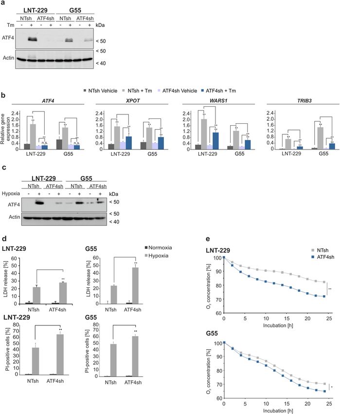
www.nature.com/scientificreports/
factor 2α (eIF2α) which in turn leads to a reduction in global translation initiation and facilitates preferential
translation of selected mRNAs, including the core effector of the ISR activating transcription factor 4 (ATF4)5.
Thus, ATF4 is frequently used as a marker for ISR a ctivation5,9. ATF4 induction has been demonstrated in conse-
quence of hypoxic conditions, ER stress as well as glucose and amino acid deprivation6,8–12. In GB ATF4 has not
yet been studied in the context of hypoxia and combined glucose deprivation which characterize the conditions
of the tumor microenvironment.
Via its leucine zipper region, ATF4 interacts with other transcription factors and activates an array of tar-
get genes including genes involved in amino acid synthesis, angiogenesis, metastasis, differentiation and drug
resistance9. The termination of the stress response is regulated by a negative feedback loop that leads to dephos-
phorylation of eIF2α mainly by GADD349.
ATF4 expression has been reported to be upregulated in different types of human cancers and overexpres-
sion correlated with earlier tumor progression and therapy r esistance13. Pharmacological inhibition of the ISR
could be an opportunity to sensitize tumor cells to conditions of the tumor microenvironment and increase
their sensitivity towards existing therapy strategies. Previous studies demonstrated that PERK inhibition itself
can reduce tumor cell survival but is also associated with severe side effects in vivo especially on pancreatic
function14. The small molecule inhibitor ISRIB blocks the ISR downstream of P-eIF2α by targeting eIF2B, which
is required as guanidine nucleotide exchange factor in the translation initiation process and thereby sustains
global protein t ranslation15–17.
Few studies on ATF4 in GB have been conducted and indicate an elevated expression in comparison to normal
brain tissue18. Also in the context of chemotherapy, ATF4 has been suggested to interfere with a utophagy19,20.
While induction of ATF4 by typical conditions of the GB microenvironment with severe hypoxia and low
nutrient supply21,22 is plausible, the relevance of ATF4 in this context has not been investigated in detail y et23.
Similarly, effects on clonal survival, the main readout to evaluate temozolomide effects in vitro have not been
performed18,20,24.
In this experimental study we investigated the regulation of ATF4, the main orchestrator of the ISR, under
conditions of the glioma microenvironment. Hypoxia and temozolomide chemotherapy induced ATF4 mRNA
expression and protein levels. Using pharmacological and genetic approaches we found that ATF4 directs tumor
cell protective responses to mediate therapy resistance. This included protection from combined hypoxia-/
nutrient restriction-induced cell death as well as from temozolomide chemotherapy. Therefore, ATF4 could be
a promising candidate for therapeutic inhibition.
Results
ATF4 is induced by pharmacological ER stress activators, glutamine depletion and temozo‑
lomide treatment in human GB cell lines. To investigate the importance of the ISR in human GB cell
lines basal ATF4 mRNA expression levels were analyzed (Fig. S1a). Based on these results one GB cell line with
low and one with high ATF4 mRNA expression, LNT-229 and G55 respectively, were chosen for further experi-
ments. ATF4 mRNA levels of primary GB cells (NCH690, NCH644 and NCH421K) were comparable to the
expression levels of GB cell lines. For comparison with non-tumor cells human astrocytes were investigated.
Here, under basal conditions ATF4 mRNA levels were also similar to the expression levels of primary GB cells
(Fig. S1a). ER stress was pharmacologically induced in LNT-229 and G55 cells with tunicamycin or thapsigar-
gin. Treatment led to reduced cell growth after 24 h and even more pronounced after 96 h incubation (Fig. S1b).
Pharmacological activation resulted in an upregulated ATF4 expression on protein level in LNT-229 and G55
cells in all tested concentrations (Fig. 1a). Additionally, both ER stress inducers led to an increased mRNA level
of ATF4 itself as well as its target genes XPOT, WARS1 and TRIB38,25–28 (Fig. 1b).
ATF4 is known to be a major regulator of the ISR under amino acid deprivation conditions10. Glutamine
withdrawal alone was sufficient to induce ATF4 expression in LNT-229 and G55 cells; concomitant glucose
restriction did not result in an additional induction of the transcription factor (Fig. S1c, S1d). Serum withdrawal
in the presence of glutamine had no effect on ATF4 expression (Fig. S1c). ATF4 itself as well as the ATF4 target
genes XPOT, WARS1 and TRIB3 were transcriptionally upregulated during glutamine starvation in LNT-229
and G55 cells (Fig. S1e).
The anti-glioma chemotherapeutic temozolomide has been reported to increase ATF4 expression as a con-
sequence of UPR induction18,20. In both, LNT-229 and G55 cells treatment with temozolomide resulted in an
increased ATF4 expression compared to vehicle treated cells and a transcriptional upregulation of ATF4 and its
target genes XPOT and WARS1 (Fig. 1c,d).
Gene suppression of ATF4 inhibits activation of the integrated stress response under phar‑
macological ER stress induction and glutamine deprivation. To investigate the effects of ATF4 in
human GB cell lines LNT-229 and G55 ATF4 gene suppressed cells (ATF4sh) were generated for further analy-
ses. Stable gene suppression was confirmed via qPCR analysis and revealed a reduction of ATF4 mRNA in both
LNT-229 and G55 ATF4sh cells compared to control cells (Fig. S2a). Tunicamycin dramatically induced ATF4
expression in LNT-229 and G55 control cells (NTsh) whereas no or only a slight effect after pharmacological ER
stress induction was observed in ATF4sh cells on protein (Fig. 2a) and mRNA level (Fig. 2b). Glutamine with-
drawal resulted in an upregulation of ATF4 protein in LNT-229 control cells but not in the corresponding ATF4
gene-suppressed cells (Fig. S2b). This effect was also observed to a lesser extent in G55 cells most likely due to a
lesser gene suppression efficiency of G55 ATF4sh cells (Fig. S2b). Glucose restriction alone did not result in an
ATF4 induction under all tested conditions in both control and ATF4 gene-suppressed cells.
Scientific Reports | (2021) 11:14161 | https://doi.org/10.1038/s41598-021-93663-1 2
Vol:.(1234567890)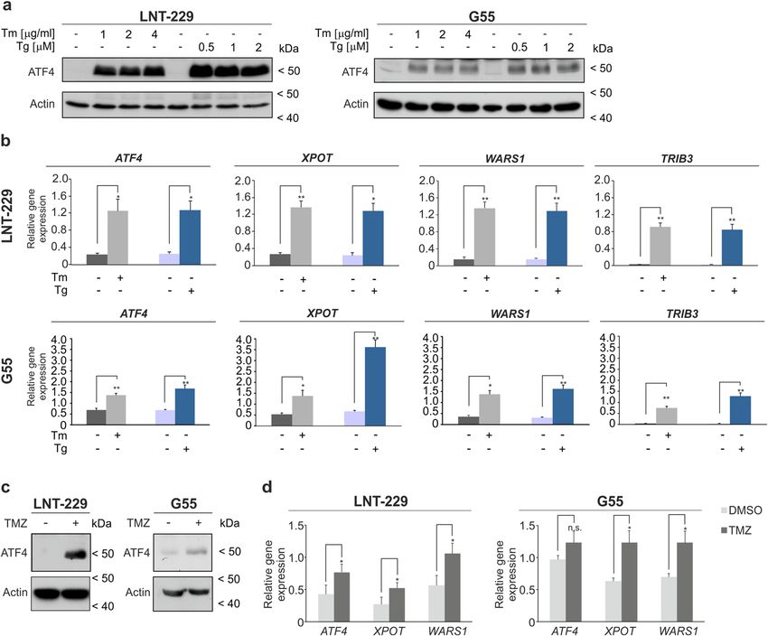
www.nature.com/scientificreports/
Figure 1. ATF4 is regulated by pharmacological ER stress activators and is induced by TMZ treatment
in human glioblastoma cells. (a) LNT-229 and G55 cells were incubated with increasing concentration
of thapsigargin (Tg) and tunicamycin (Tm) in serum-free DMEM for 8 h. Cellular lysates were analyzed
by immunoblot with antibodies for ATF4 and actin. (b) LNT-229 and G55 cells were treated with 2 µg/
ml tunicamycin (Tm) or 1 µM thapsigargin (Tg) for 8 h in serum-free DMEM. cDNA was analyzed for the
expression of ATF4 and its target genes XPOT, WARS1 and TRIB3 by qPCR. 18S and SDHA were used as
housekeeping genes for normalization (n = 3, mean ± SD, *p < 0.05, **p < 0.01, Student’s t test). (c) LNT-229 and
G55 cells were treated with 400 μM temozolomide (TMZ) in serum-free DMEM for 24 h. Cellular lysates were
analyzed by immunoblot with antibodies for ATF4 and actin. (d) cDNA was analyzed for induction of ATF4 and
target genes XPOT and WARS1 by qPCR (n = 3, mean ± SD, n.s. not significant, *p < 0.05, Student’s t test). SDHA
and 18S were used as housekeeping genes for normalization.
ATF4 gene suppression sensitizes human GB cells to hypoxia‑induced cell death and increases
oxygen consumption. The GB tumor microenvironment is characterized by limited oxygen and glucose
supply that drive activation of the cellular stress response machinery which enables cellular adaptation to sustain
survival29. To analyze the effects of the ER stress response in the context of hypoxia-induced cell death in GB
cells we used experimental conditions mimicking the in vivo situation of reduced glucose availability and severe
hypoxia30. The relevance of ATF4 for adaptation under such conditions was investigated by pharmacological as
well as genetic ATF4 modulation. Treatment with tunicamycin which induces ATF4 as shown in Fig. 1a, con-
ferred protection towards combined hypoxia- and nutrient depletion-induced cell death in LNT-229 and G55
cells (Fig. S2c). Immunoblot analysis showed a strong induction of ATF4 only in LNT-229 control (NTsh) and
only a reduced induction of ATF4 in LNT-229 ATF4sh cells after treatment under hypoxic, glucose restricted
conditions (Fig. 2c). Both, LNT-229 and G55 ATF4sh cells were sensitized to hypoxia-/nutrient depletion-
induced cell death compared to control cells (Fig. 2d). In line with these findings, LNT-229 and G55 ATF4sh
cells displayed an increased oxygen consumption compared to NTsh cells (Fig. 2e). Conversely, treatment with
the ISR inducer tunicamycin led to a reduced oxygen consumption in LNT-229 and G55 cells (Fig. S2d).
ATF4 gene suppression sensitizes GB cells to temozolomide. To investigate a potential effect of
temozolomide on ATF4 expression, experiments with LNT-229 and G55 ATF4sh and corresponding NTsh cells
were performed. In both cell lines, ATF4 expression was increased to a smaller extent in ATF4sh cells on pro-
tein level (Fig. 3a). In line with these results, clonogenicity assays revealed that LNT-229 and G55 ATF4sh cells
displayed a reduced clonal survival confirming a higher sensitivity towards temozolomide compared to the cor-
Scientific Reports | (2021) 11:14161 | https://doi.org/10.1038/s41598-021-93663-1 3
Vol.:(0123456789)www.nature.com/scientificreports/
Figure 2. ATF4sh cells are insensitive for ATF4 induction via pharmacological ER stress activation and
sensitized to hypoxia-induced cell death. (a) LNT-229 and G55 NTsh and ATF4sh cells were incubated with
2 µg/ml tunicamycin (Tm) in serum-free DMEM for 8 h. Cellular lysates were analyzed by immunoblot with
antibodies for ATF4 and actin. (b) cDNA was analyzed by qPCR with primers for ATF4, XPOT, WARS1
and TRIB3 (n = 3, mean ± SD, n.s. not significant, **p < 0.01, Student’s t-test). SDHA and 18S were used as
housekeeping genes for normalization. (c) LNT-229 and G55 NTsh or ATF4sh cells were incubated in serum-
free DMEM containing 2 mM glucose and 4 mM glutamine in normoxia or hypoxia (0.1% O 2) for 8 h. Cellular
lysates were analyzed for the expression of ATF4 or actin by immunoblot. (d) Cells were incubated in serum-
free DMEM supplemented with 2 mM glucose and 4 mM glutamine in normoxia or hypoxia (0.1% O 2). Cell
death was analyzed by propidium iodide (PI) uptake and quantified by flow cytometry using BD FACS Diva
software (version 6.1.3) (n = 3, mean ± SD, **p < 0.01, Student’s t-test) or by LDH release assay (n = 4, mean ± SD,
**p < 0.01, Student’s t-test). (e) Oxygen consumption of LNT-229 and G55 NTsh and ATF4sh cells was measured
in serum-free DMEM supplemented with 2 mM glucose and 4 mM glutamine overlaid with paraffin oil with
a fluorescence-based assay. Oxygen consumption is shown relative to the start of the experiment (n = 3, mean,
*p < 0.05, **p < 0.01, Student’s t-test).
Scientific Reports | (2021) 11:14161 | https://doi.org/10.1038/s41598-021-93663-1 4
Vol:.(1234567890)www.nature.com/scientificreports/
Figure 3. Gene suppression of ATF4 sensitizes human GB cells to temozolomide induced stress. (a) LNT-229
and G55 control (NTsh) or ATF4 knockdown (ATF4sh) cells were treated with 400 µM temozolomide (TMZ) in
serum-free DMEM for 8 h. Cellular lysates were analyzed by immunoblot with antibodies for ATF4 and actin.
(b) LNT-229 and G55 NTsh and ATF4sh cells were treated with 20 µM TMZ or 200 µM TMZ respectively for
24 h and were further allowed to grow in DMEM for 5 days. Clonogenicity is shown relative to vehicle (DMSO)
(n = 3, mean ± SD, *p < 0.05, Student’s t-test). (c) LNT-229 and G55 NTsh and ATF4sh cells were treated with
800 µM TMZ for 72 h in serum-free DMEM. Cell death was analyzed by propidium iodide (PI) staining and
quantified by flow cytometry (n = 3, mean ± SD, *p < 0.05, **p < 0.01, Student’s t-test). Data were analyzed with
BD FACS Diva software (version 6.1.3).
responding NTsh control (Fig. 3b). Furthermore LNT-229 ATF4sh cells showed an enhanced susceptibility to
temozolomide treatment (Fig. 3c).
Pharmacological inhibition of ATF4 activation sensitizes GB cells to hypoxia induced cell death
and increases temozolomide susceptibility. To further investigate the effect of ISR inhibition in the
context of hypoxia induced cell death and to evaluate whether ISR inhibition could improve temozolomide treat-
ment, we employed the pharmacological ISR inhibitor ISRIB.
While ISRIB treatment did not influence ATF4 expression in LNT-229 and G55 cells, co-treatment with
tunicamycin and ISRIB resulted in decreased ATF4 expression on protein level compared to cells treated with
tunicamycin alone (Fig. 4a). Similar results were observed on mRNA level of target genes where combined
treatment with both substances resulted in reduced translation of ATF4 itself and ATF4 target genes XPOT
and WARS1 (Fig. 4b). Under glucose restricted, hypoxic conditions, where ATF4 gene suppression resulted in
increased cell death (Fig. 2d), ISR inhibition with ISRIB similarly sensitized LNT-229 and G55 cells to hypoxia
induced cell death (Fig. 4c).
Temozolomide induced ATF4 activation was reduced in LNT-229 and G55 cells when ISRIB was present
(Fig. 4d). ISRIB treatment did not influence cell growth in LNT-229 and G55 cells after 72 h, but in combination
with temozolomide it further decreased cell density compared to treatment with temozolomide alone (Fig. 4e).
Furthermore, ISR inhibition with ISRIB significantly increased temozolomide susceptibility in LNT-229 and
G55 cells after 72 h (Fig. 4f) in line with our results that ATF4 is important for resistance against temozolomide
(Fig. 3b,c).
Discussion
Adaptation to the conditions of the tumor microenvironment is a central challenge for GB cells in order to enable
survival and proliferation. ER stress and amino acid deprivation, frequently present in the GB microenvironment,
trigger activation of the transcription factor ATF4 which induces a cellular adaptive stress response. Therefore,
ATF4 is a logical candidate for therapeutic inhibition to enhance the susceptibility of GB cells towards existing
therapy approaches.
Employing our paradigm of hypoxia-induced cell death with severe hypoxia and glucose r estriction30, we
here provide evidence that supports an important role of ATF4 and the ISR to cope with conditions found in the
GB microenvironment (Fig. S3). ATF4 was induced under hypoxic conditions and pharmacological ER stress
induction in human GB cells (Figs. 1a, 2c). Gene suppression of ATF4 sensitized GB cells to hypoxia induced
cell death (Figs. 2c,d, S2c) indicating a protective role of ATF4 expression and induction of the ISR. In line,
we found an increased oxygen consumption in ATF4sh cells indicating that these cells are more dependent on
oxygen (Fig. 2e). This concept that a higher oxygen consumption correlates with enhanced sensitivity to hypoxia-
induced cell death has previously been reported for PPARGC1A (PGC-1α) overexpressing GB cells31. In contrast,
pharmacological ATF4 induction with tunicamycin resulted in a reduced oxygen consumption compared to
Scientific Reports | (2021) 11:14161 | https://doi.org/10.1038/s41598-021-93663-1 5
Vol.:(0123456789)www.nature.com/scientificreports/
Figure 4. ISRIB treatment sensitizes human GB cells to hypoxia induced cell death and increases temozolomide toxicity.
(a) LNT-229 and G55 cells were treated with 2 µg/ml tunicamycin (Tm), 0.2 µM or 1 µM ISRIB or a combination of both
substances in serum-free DMEM for 8 h as indicated. Cellular lysates were analyzed by immunoblot with antibodies for ATF4
and actin. (b) Isolated cDNA of G55 cells was analyzed by qPCR for the expression of ATF4, XPOT and WARS1. SDHA and
18S were used for normalization (n = 3, mean ± SD, n.s. not significant, **p < 0.01, Student’s t-test). (c) LNT-229 and G55
cells were incubated in serum-free DMEM without glutamine in normoxia or hypoxia (0.1% O 2). Cell death was analyzed
by LDH release assay (n = 4, mean ± SD, n.s. not significant, **p < 0.01, Student’s t-test). (d) Cellular lysates of LNT-229 and
G55 cells treated with temozolomide and ISRIB for 24 h in serum-free DMEM as indicated were analyzed by immunoblot
with antibodies for ATF4 and actin. (e) LNT-229 and G55 cells were treated for 72 h in serum-free DMEM as indicated. Cell
densities were measured by CV staining (n = 3, mean ± SD, **p < 0.01, Student’s t-test). (f) Cell death of LNT-229 and G55 cells
was analyzed by propidium iodide (PI) uptake and quantified by flow cytometry (n = 3, mean ± SD, **p < 0.01, Student’s t-test)
after treatment with temozolomide and ISRIB in serum-free DMEM for 72 h as indicated. Data analysis was performed with
BD FACS Diva software (version 6.1.3).
Scientific Reports | (2021) 11:14161 | https://doi.org/10.1038/s41598-021-93663-1 6
Vol:.(1234567890)www.nature.com/scientificreports/
vehicle treated LNT-229 and G55 cells (Fig. S2d) indicating that GB cells with an intact ISR reduce their oxygen
consumption during ER stress conditions. Interestingly, it has recently been reported that the stress responses
mediated by the ISR and via mTOR display a significant overlap in their translational p rograms32. In line with
this finding, we have previously shown that a defective mTOR inhibition sensitizes GB cells to hypoxia-induced
cell death33 in a similar manner as a defective ISR (Fig. 2d). In parallel, dysfunction of both stress responses was
associated with an increased oxygen consumption (Fig. 2e)33.
Pharmacological ER stress induction with tunicamycin and thapsigargin is known to induce ATF4 and a
number of ATF4 target g enes8,25–28. In our experiments, treatment with tunicamycin as well as thapsigargin led
to a significant increase of ATF4 and its target genes in human GB cell lines (Fig. 1a,b). This effect was strongly
reduced in ATF4sh cells compared to control cells in LNT-229 and G55 (Fig. 2a,b). Furthermore, we could
demonstrate that ATF4 was induced by glutamine depletion in GB cells whereas glucose starvation alone did
not modulate ATF4 expression (Fig. S1c,d). This finding is consistent with data showing an upregulation of
ATF4 via the GCN2/P-eIF2α/ATF4 axis during glutamine starvation conditions in colorectal adenocarcinoma
and fibrosarcoma cell lines10. Glutamine is an important non-essential amino acid used by cancer cells not only
for the synthesis of nucleotides and proteins but also as an energy source for ATP production and to support
glutathione biosynthesis in redox stress conditions34. It has been reported that glutamine depletion led to a 50%
reduction in survival in ATF4sh cells compared to a 25% reduction in control c ells10. Our results emphasize the
role of glutamine depletion in the context of the ISR in human GB cells where an ATF4 upregulation possibly
helps the tumor cells to adjust to altered nutrient availability.
Previous studies in human GB cells have shown that ATF4 expression is induced by the alkylating chemo-
therapeutic agent temozolomide18,20. In our experiments we found an ATF4 dependent ISR activation after
temozolomide treatment in human GB cell lines (Fig. 1c,d) which was strongly reduced in ATF4sh cells (Fig. 3a).
Furthermore, gene suppression of ATF4 led to a reduction of clonogenicity after temozolomide treatment in
LNT-229 and G55 cells compared to the corresponding control cells (Fig. 3b). The fact that the impact of temo-
zolomide treatment on the clonogenicity is more pronounced in G55 cells could be due to the higher basal level
of ATF4 (Fig. S1a) and therefore a higher dependency of those cells on ATF4 activity. ATF4sh cells were also
more susceptible to temozolomide induced acute cell death compared to control cells (Fig. 3c). Thus, our results
demonstrate that initiation of the ISR and induction of ATF4 are essential for the activation of cellular resistance
mechanisms which allow GB cells to overcome temozolomide toxicity (Fig. S3). In previous studies it has been
demonstrated that PERK inhibition can be beneficial for cancer treatment14. While many ISR inhibitors target
PERK itself, ISRIB specifically inhibits the stress response downstream of PERK by reversing the attenuation
of eIF2B by phosphorylated eIF2α15–17. PERK inhibition was reported to lead to side effects in in vivo models,
especially on pancreatic function, whereas targeting phosphorylated eIF2α with ISRIB does not show pancre-
atic toxicity14,35. ISRIB treatment sensitized GB cells to hypoxia induced cell death (Fig. 4c), indicating that ISR
inhibition leads to an increased vulnerability to conditions of the tumor microenvironment (hypoxia, nutrient
deprivation) and impaired tumor cell survival. Furthermore, our experiments using combined temozolomide and
ISRIB treatment highlight the importance of the ISR and especially ATF4 induction for temozolomide resistance
in GB cells. ISR inhibition enhanced temozolomide susceptibility in GB cell lines (Fig. 4e,f).
While the ISR inducing agents thapsigargin and tunicamycin are frequently used to study ATF4-mediated
cellular effects, these substances are certainly not specific for ATF4. Also with regard to shRNA approaches
off-target effects cannot be ruled out. Nevertheless, our work provides consistent evidence that supports an
important role of eIF2α-ATF4 axis and the integrated stress response of GB cells to conditions found in the
tumor microenvironment (low glucose and hypoxia) and in response to temozolomide.
Analyses of databases suggested elevated ATF4 expression in gliomas in comparison to normal brain tissue
and an association of increased ATF4 transcription with poor overall survival in glioma cohorts18,36. Several
in vitro investigations have implicated ATF4 in tumor cell survival with sometimes opposing cytoprotective and
cytotoxic roles depending on context: ATF4 has been shown to attenuate ferroptosis under dihydroartemisinin-
induced ER stress or sorafenib treatment to sustain glioma cell survival and inhibition of this axis was therefore
proposed as a therapeutic s trategy37. On the contrary, another study in GB cells demonstrated that ATF4 played
a key role to induce autophagic cell death via reticulophagy (i.e. the selective degradation of the endoplasmic
reticulum) in response to loperamide treatment and in this context activation of ATF4 was suggested to be ben-
eficial for tumor therapy38. These studies highlight the ambiguous character of the ATF4 mediated stress response
depending on the experimental context. A more in depth understanding of ATF4 effects is therefore urgently
needed before advancing ATF4 targeting strategies to clinical trials. In our study we approximated in vivo con-
ditions both with regard to metabolic (oxygen and glucose deprivation) and treatment (with temozolomide as
the standard chemotherapeutic for gliomas) scenarios. In both scenarios ATF4 mediated protective adaptation
in GB cells. A next step is to validate the advantages of a combined temozolomide and anti-ATF4 (e.g. ISRIB
treatment) approach in in vivo GB mouse models.
Our preclinical results have potentially high translational value. Primary or secondary resistance to temozo-
lomide is a major problem in GB treatment. Several scenarios of combining temozolomide with an ISR inhibi-
tor (e.g. ISRIB) could be of interest: including ISR inhibition directly in the first line temozolomide treatment
algorithm could enhance temozolomide efficacy particularly in MGMT-promoter methylated tumors and maybe
also help achieve some efficacy in MGMT unmethylated tumors. Another scenario could be to include ISR
inhibition as a treatment concept to (re-)sensitize GB to temozolomide in the recurrent disease setting. In this
context, it would be interesting to analyze histology specimens of recurrent gliomas after temozolomide treat-
ment for activation of the ISR which would be a rationale for such an approach in the recurrent disease setting.
The ISR has gained interest across the cancer field and it is likely that candidates for clinical testing will
become available. Therapeutic inhibition could be a promising clinical strategy to improve treatment of GBs.
Scientific Reports | (2021) 11:14161 | https://doi.org/10.1038/s41598-021-93663-1 7
Vol.:(0123456789)www.nature.com/scientificreports/
MGMT IDH p53 PTEN
LNT-229 Methylated Wildtype Wildtype Wildtype
G55 Unmethylated Wildtype Mutant n.a
Table 1. Genetic background of LNT-229 and G55 cells. MGMT, IDH, p53 and PTEN status of G55 and LNT-
229 cells33.
Material and methods
Reagents, cell lines and culture conditions. Tunicamycin and thapsigargin were purchased from Toc-
ris (Bristol, UK). All reagents not specified elsewhere were purchased from Sigma (Taufkirchen, Germany).
LNT-229 cells were a kind gift of N. de Tribolet (Lausanne, Switzerland). LNT-229 cells were authenticated
using STR analysis by Multiplexion (Heidelberg, Germany). The STR profile of the tested cells matched with the
known profile for LN-229. LNT-229 and LN-229 cells only differ in their p53 status39. G55 T2 cells (named G55
for simplicity throughout the manuscript) were a kind gift of Manfred Westphal and Kathrin Lamszus (Ham-
burg, Germany)40, a STR profile for this cell line has not been deposited in databases yet. MGMT, IDH, p53 and
PTEN status of LNT-229 and G55 cells have been described previously and are listed (Table 1)33,41.
All cell lines were regularly checked for mycoplasma contamination throughout the study and only contam-
ination-free cells were used for experiments.
Cell lines were cultured in Dulbecco´s modified Eagle´s medium (DMEM) containing 10% fetal calf serum
(Biochrom KG, Berlin, Germany), 100 IU/ml penicillin and 100 µg/ml streptomycin (Life Technologies,
Karlsruhe, Germany) in cell culture incubators at 37 °C and 5% C O242. For experiments comparing sub-cell
lines, equal cell densities at the start of the experiment were confirmed by crystal violet (CV) staining in parallel
assays42.
Human primary GB cells NCH690, NCH421K and NCH644 were purchased from CLS (Eppelheim, Ger-
many) and were cultured in Neurobasal A medium (Thermo Fisher Scientific, Dreieich, Germany) supplemented
with 1 × B27 supplement (Thermo Fisher Scientific, Dreieich, Germany), 2 mM glutamine (Thermo Fisher Sci-
entific, Dreieich, Germany), 1U/ml heparin, 100 IU/ml penicillin and 100 µg/ml streptomycin (Life Technolo-
gies, Karlsruhe, Germany) and 20 ng/ml EGF and FGF-2 (ReliaTech GmbH, Wolfenbüttel, Germany). Human
astrocytes were purchased from Innoprot (Derio, Spain) and were cultured in Astrocyte medium (Innoprot,
Derio, Spain) according to the manufacturer’s protocol.
Temozolomide treatment. The different temozolomide concentrations were chosen based on the experi-
mental setup and cell line characteristics (i.e. MGMT promoter methylation status). The effect of low doses of
temozolomide on clonal survival are a known phenomenon and are usually by several factors lower than doses
required to induce acute t oxicity43,44. For clonal survival analyses of MGMT promoter methylated LNT-229 cells
20 µM temozolomide and for MGMT promoter unmethylated G55 cells 200 µM temozolomide were chosen.
For acute toxicity cells were treated with higher doses of temozolomide for 72 h. To investigate acute effects of
temozolomide treatment on mRNA expression and protein levels, cells were incubated with 400 µM temozolo-
mide for 8 h.
Generation of ATF4 gene suppressed cells. pLKO.1 plasmids targeting ATF4 (ATF4sh,
TRCN0000329695) and non-targeting control (NTsh, #1864) were purchased from Sigma and Addgene respec-
tively. HEK293 cells were used for lentiviral production with pCMV-dR8.2 dvpr (Addgene, #8455) and pCMV-
VSVG (Addgene, #8454) as packaging and envelope plasmids according to the manufacturer’s protocol. Lenti-
viral transduction was performed using polybrene (Merck Millipore, Darmstadt, Germany). ATF4sh and NTsh
cells were cultured in standard medium supplemented with 2 µg/ml puromycin (AppliChem, Darmstadt, Ger-
many). Experiments were performed with early passage pooled clones.
Induction of hypoxia. Cells were seeded and allow to attach overnight under normoxia. Experiments were
performed in serum-free DMEM containing 2 mM glucose and 4 mM glutamine under normoxia or 0.1% O2
hypoxia in BD GasPak pouches (Becton–Dickinson, Heidelberg, Germany) for anaerobic culture for the indi-
cated intervals33,45.
Immunoblot. Cell lysate preparation and immunoblot analysis was performed as described previously33.
Primary antibodies for ATF4 (Proteintech, Rosemont, IL, USA, 10835-1-AP and Cell Signaling Technology, Dan-
vers, MA, USA, #11815) or actin (Santa Cruz Biotechnology, Santa Cruz, CA, USA, #sc-1616) were employed as
indicated. Secondary anti-rabbit and anti-goat antibodies were purchased from Jackson ImmunoResearch (West
Grove, PA, USA, #111-036-144) and Santa Cruz (#sc-2020). Chemiluminescence was used for detection. All full
length immunoblots are shown in Fig. S4.
RNA extraction and quantitative PCR analysis. Total RNA was extracted using TRIzol and
EXTRACTME RNA isolation kit (Blirt, Gdansk, Poland). cDNA was synthesized using the Vilo cDNA synthesis
kit (Invitogen, Carlsbad, CA, USA)33. Quantitative RT-PCR (qPCR) was performed using the Absolute Blue
SYBR Green Fluorescein q-PCR Mastermix (Thermo Fisher Scientific, Dreieich, Germany) with the correspond-
Scientific Reports | (2021) 11:14161 | https://doi.org/10.1038/s41598-021-93663-1 8
Vol:.(1234567890)www.nature.com/scientificreports/
ing primer pairs (Table S1). 18S and SDHA were used as housekeeping genes for normalization. Data was ana-
lyzed using the Vandesompele method46.
Cell density and viability assays. For cell growth analyses cells were seeded in 96-well plates and allowed
to attach overnight. After treatment, cell density was analyzed by CV staining after the indicated intervals as
previously described42,47. Cell viability was analyzed by lactate dehydrogenase release assays with the Cytotox-
icity Detection Kit (LDH) (Roche, Mannheim, Germany) or by propidium iodide (PI) uptake assays via flow
cytometry as previously d escribed30,33. Data were acquired with a BD Canto II flow cytometer and analyzed with
BD FACS Diva software (version 6.1.3).
Clonal survival assays. For analysis of clonal survival 500 cells were seeded per well of a 6 well plate and
allowed to attach overnight. Cells were treated with temozolomide in serum-free DMEM. Medium was replaced
with fresh DMEM containing 10% FCS after 24 h. CV staining was performed 5 days later. Clones were counted
manually48.
Oxygen consumption. Cells were seeded in 24-well plates with oxygen sensors (PreSens, Regensburg,
Germany) and allowed to attach overnight. Medium was replaced and cells were overlaid with sterile paraffin
oil. Oxygen consumption was measured in optical sensor plates and concentrations of remaining oxygen were
recorded over time33. For statistical analysis values of the end-points of the experiments were analyzed by a two-
tailed student’s t-tests.
Statistics. Quantitative data are expressed as mean and standard deviation (SD). A two-tailed student’s t-test
was used for the calculation of p-values. Values of p < 0.01 were considered as highly significant, p < 0.05 as sig-
nificant and p > 0.05 as not significant (n.s.).
Data availability
The datasets used or analyzed during the current study are available from the corresponding author on reason-
able request.
Received: 18 February 2021; Accepted: 18 June 2021
References
1. Ohgaki, H. et al. Genetic pathways to glioblastoma: a population-based study. Can. Res. 64, 6892–6899. https://doi.org/10.1158/
0008-5472.CAN-04-1337 (2004).
2. Stupp, R. et al. Radiotherapy plus concomitant and adjuvant temozolomide for glioblastoma. N. Engl. J. Med. 352, 987–996. https://
doi.org/10.1056/NEJMoa043330 (2005).
3. Stupp, R. et al. Effect of tumor-treating fields plus maintenance temozolomide vs maintenance temozolomide alone on survival in
patients with glioblastoma: A randomized clinical trial. JAMA 318, 2306–2316. https://doi.org/10.1001/jama.2017.18718 (2017).
4. PeñarandaFajardo, N. M., Meijer, C. & Kruyt, F. A. E. The endoplasmic reticulum stress/unfolded protein response in gliomagenesis,
tumor progression and as a therapeutic target in glioblastoma. Biochem. Pharmacol. 118, 1–8. https://doi.org/10.1016/j.bcp.2016.
04.008 (2016).
5. Pakos-Zebrucka, K. et al. The integrated stress response. EMBO Rep. 17, 1374–1395. https://doi.org/10.15252/embr.201642195
(2016).
6. Rzymski, T. et al. Regulation of autophagy by ATF4 in response to severe hypoxia. Oncogene 29, 4424–4435. https://doi.org/10.
1038/onc.2010.191 (2010).
7. Wek, R. C., Jiang, H.-Y. & Anthony, T. G. Coping with stress: eIF2 kinases and translational control. Biochem. Soc. Trans. 34, 7–11.
https://doi.org/10.1042/BST20060007 (2006).
8. Harding, H. P. et al. Regulated translation initiation controls stress-induced gene expression in mammalian cells. Mol. Cell 6,
1099–1108. https://doi.org/10.1016/S1097-2765(00)00108-8 (2000).
9. Ameri, K. & Harris, A. L. Activating transcription factor 4. Int. J. Biochem. Cell Biol. 40, 14–21. https://doi.org/10.1016/j.biocel.
2007.01.020 (2008).
10. Ye, J. et al. The GCN2-ATF4 pathway is critical for tumour cell survival and proliferation in response to nutrient deprivation.
EMBO J. 29, 2082–2096. https://doi.org/10.1038/emboj.2010.81 (2010).
11. Blais, J. D. et al. Activating transcription factor 4 is translationally regulated by hypoxic stress. Mol. Cell. Biol. 24, 7469–7482.
https://doi.org/10.1128/MCB.24.17.7469-7482.2004 (2004).
12. Iurlaro, R. et al. Glucose deprivation induces ATF4-mediated apoptosis through TRAIL death receptors. Mol. Cell. Biol. https://
doi.org/10.1128/MCB.00479-16 (2017).
13. Singleton, D. C. & Harris, A. L. Targeting the ATF4 pathway in cancer therapy. Expert Opin. Ther. Targets 16, 1189–1202. https://
doi.org/10.1517/14728222.2012.728207 (2012).
14. Atkins, C. et al. Characterization of a novel PERK kinase inhibitor with antitumor and antiangiogenic activity. Can. Res. 73,
1993–2002. https://doi.org/10.1158/0008-5472.CAN-12-3109 (2013).
15. Rabouw, H. H. et al. Small molecule ISRIB suppresses the integrated stress response within a defined window of activation. Proc.
Natl. Acad. Sci. USA. 116, 2097–2102. https://doi.org/10.1073/pnas.1815767116 (2019).
16. Sidrauski, C. et al. Pharmacological brake-release of mRNA translation enhances cognitive memory. Elife https://doi.org/10.7554/
eLife.00498 (2013).
17. Sidrauski, C., McGeachy, A. M., Ingolia, N. T. & Walter, P. The small molecule ISRIB reverses the effects of eIF2α phosphorylation
on translation and stress granule assembly. Elife https://doi.org/10.7554/eLife.05033 (2015).
18. Chen, D. et al. ATF4 promotes angiogenesis and neuronal cell death and confers ferroptosis in a xCT-dependent manner. Oncogene
36, 5593–5608. https://doi.org/10.1038/onc.2017.146 (2017).
19. He, Y. et al. Regulation of integrated stress response sensitizes U87MG glioblastoma cells to temozolomide through the mitochon-
drial apoptosis pathway. Anat. Rec. 301, 1390–1397. https://doi.org/10.1002/ar.23839 (2018).
20. Chen, D., Rauh, M., Buchfelder, M., Eyupoglu, I. Y. & Savaskan, N. The oxido-metabolic driver ATF4 enhances temozolamide
chemo-resistance in human gliomas. Oncotarget 8, 51164–51176. https://doi.org/10.18632/oncotarget.17737 (2017).
Scientific Reports | (2021) 11:14161 | https://doi.org/10.1038/s41598-021-93663-1 9
Vol.:(0123456789)www.nature.com/scientificreports/
21. Evans, S. M. et al. Hypoxia is important in the biology and aggression of human glial brain tumors. Clin. Cancer Res. 10, 8177–8184.
https://doi.org/10.1158/1078-0432.CCR-04-1081 (2004).
22. Monteiro, A. R., Hill, R., Pilkington, G. J. & Madureira, P. A. The role of hypoxia in glioblastoma invasion. Cells https://doi.org/
10.3390/cells6040045 (2017).
23. Moeckel, S. et al. ATF4 contributes to autophagy and survival in sunitinib treated brain tumor initiating cells (BTICs). Oncotarget
10, 368–382. https://doi.org/10.18632/oncotarget.26569 (2019).
24. Weatherbee, J. L., Kraus, J.-L. & Ross, A. H. ER stress in temozolomide-treated glioblastomas interferes with DNA repair and
induces apoptosis. Oncotarget 7, 43820–43834. https://doi.org/10.18632/oncotarget.9907 (2016).
25. Han, J. et al. ER-stress-induced transcriptional regulation increases protein synthesis leading to cell death. Nat. Cell Biol. 15,
481–490. https://doi.org/10.1038/ncb2738 (2013).
26. Ohoka, N., Yoshii, S., Hattori, T., Onozaki, K. & Hayashi, H. TRB3, a novel ER stress-inducible gene, is induced via ATF4-CHOP
pathway and is involved in cell death. EMBO J. 24, 1243–1255. https://doi.org/10.1038/sj.emboj.7600596 (2005).
27. Armstrong, J. L., Flockhart, R., Veal, G. J., Lovat, P. E. & Redfern, C. P. F. Regulation of endoplasmic reticulum stress-induced cell
death by ATF4 in neuroectodermal tumor cells. J. Biol. Chem. 285, 6091–6100. https://doi.org/10.1074/jbc.M109.014092 (2010).
28. Fusakio, M. E. et al. Transcription factor ATF4 directs basal and stress-induced gene expression in the unfolded protein response
and cholesterol metabolism in the liver. Mol. Biol. Cell 27, 1536–1551. https://doi.org/10.1091/mbc.E16-01-0039 (2016).
29. Jawhari, S., Ratinaud, M.-H. & Verdier, M. Glioblastoma, hypoxia and autophagy: A survival-prone “ménage-à-trois”. Cell Death
Dis. 7, e2434. https://doi.org/10.1038/cddis.2016.318 (2016).
30. Steinbach, J. P., Wolburg, H., Klumpp, A., Probst, H. & Weller, M. Hypoxia-induced cell death in human malignant glioma cells:
energy deprivation promotes decoupling of mitochondrial cytochrome c release from caspase processing and necrotic cell death.
Cell Death Differ. 10, 823–832. https://doi.org/10.1038/sj.cdd.4401252 (2003).
31. Bruns, I. et al. Disruption of peroxisome proliferator-activated receptor γ coactivator (PGC)-1α reverts key features of the neoplastic
phenotype of glioma cells. J. Biol. Chem. 294, 3037–3050. https://doi.org/10.1074/jbc.RA118.006993 (2019).
32. Klann, K., Tascher, G. & Münch, C. Functional translatome proteomics reveal converging and dose-dependent regulation by
mTORC1 and eIF2α. Mol. Cell 77, 913-925.e4. https://doi.org/10.1016/j.molcel.2019.11.010 (2020).
33. Thiepold, A.-L. et al. Mammalian target of rapamycin complex 1 activation sensitizes human glioma cells to hypoxia-induced cell
death. Brain 140, 2623–2638. https://doi.org/10.1093/brain/awx196 (2017).
34. Jiang, J., Srivastava, S. & Zhang, J. Starve cancer cells of glutamine: Break the spell or make a hungry monster?. Cancers https://
doi.org/10.3390/cancers11060804 (2019).
35. Halliday, M. et al. Partial restoration of protein synthesis rates by the small molecule ISRIB prevents neurodegeneration without
pancreatic toxicity. Cell Death Dis. 6, e1672. https://doi.org/10.1038/cddis.2015.49 (2015).
36. Peñaranda-Fajardo, N. M. et al. ER stress and UPR activation in glioblastoma: identification of a noncanonical PERK mechanism
regulating GBM stem cells through SOX2 modulation. Cell Death Dis. 10, 690. https://doi.org/10.1038/s41419-019-1934-1 (2019).
37. Chen, Y. et al. Dihydroartemisinin-induced unfolded protein response feedback attenuates ferroptosis via PERK/ATF4/HSPA5
pathway in glioma cells. J. Exp. Clin. Cancer Res. 38, 402. https://doi.org/10.1186/s13046-019-1413-7 (2019).
38. Zielke, S. et al. ATF4 links ER stress with reticulophagy in glioblastoma cells. Autophagy 1, 1–17. https://doi.org/10.1080/15548
627.2020.1827780 (2020).
39. Wischhusen, J., Naumann, U., Ohgaki, H., Rastinejad, F. & Weller, M. CP-31398, a novel p53-stabilizing agent, induces p53-depend-
ent and p53-independent glioma cell death. Oncogene 22, 8233–8245. https://doi.org/10.1038/sj.onc.1207198 (2003).
40. Eckerich, C. et al. RON receptor tyrosine kinase in human gliomas: Expression, function, and identification of a novel soluble
splice variant. J. Neurochem. 109, 969–980. https://doi.org/10.1111/j.1471-4159.2009.06027.x (2009).
41. Ishii, N. et al. Frequent co-alterations of TP53, p16/CDKN2A, p14ARF, PTEN tumor suppressor genes in human glioma cell lines.
Brain Pathol. 9, 469–479. https://doi.org/10.1111/j.1750-3639.1999.tb00536.x (1999).
42. Ronellenfitsch, M. W. et al. Antagonism of the mammalian target of rapamycin selectively mediates metabolic effects of epider-
mal growth factor receptor inhibition and protects human malignant glioma cells from hypoxia-induced cell death. Brain 132,
1509–1522. https://doi.org/10.1093/brain/awp093 (2009).
43. Hermisson, M. et al. O6-methylguanine DNA methyltransferase and p53 status predict temozolomide sensitivity in human malig-
nant glioma cells. J. Neurochem. 96, 766–776. https://doi.org/10.1111/j.1471-4159.2005.03583.x (2006).
44. Gramatzki, D. et al. Glioma cell death induced by irradiation or alkylating agent chemotherapy is independent of the intrinsic
ceramide pathway. PLoS ONE 8, e63527. https://doi.org/10.1371/journal.pone.0063527 (2013).
45. Wanka, C. et al. Synthesis of cytochrome C oxidase 2: A p53-dependent metabolic regulator that promotes respiratory function
and protects glioma and colon cancer cells from hypoxia-induced cell death. Oncogene 31, 3764–3776. https://doi.org/10.1038/
onc.2011.530 (2012).
46. Vandesompele, J. et al. Accurate normalization of real-time quantitative RT-PCR data by geometric averaging of multiple internal
control genes. Genome Biol. 3, 0034. https://doi.org/10.1186/gb-2002-3-7-research0034 (2002).
47. Grady, J. E., Lummis, W. L. & Smith, C. G. An improved tissue culture assay. III. Alternate methods for measuring cell growth.
Cancer Res. 20, 1114–1117 (1960).
48. Fischer, S. et al. Hypoxia enhances the antiglioma cytotoxicity of B10, a glycosylated derivative of betulinic acid. PLoS ONE 9,
e94921. https://doi.org/10.1371/journal.pone.0094921 (2014).
Author contributions
M.W.R. and J.P.S. conceived the study and designed the experiments. A.C.M.S., N.I.L., H.U., A.-L.L., A.L.E.
and M.W.R. performed the experiments. A.C.M.S., N.I.L., H.U., A.-L.L., A.L.E., C.M. and M.W.R. analyzed the
data. A.C.M.S., N.I.L. and M.W.R. wrote the manuscript. All authors helped to draft the manuscript and read
and approved the final version.
Funding
Open Access funding enabled and organized by Projekt DEAL. The Senckenberg Institute of Neurooncology is
supported by the Senckenberg Foundation. A.-L.L. has received funding from the Frankfurt Research Funding
(FFF) of the Medical Faculty of the Goethe University Frankfurt (program “Patenschaftsmodell”) and a “Cli-
nician Scientist” fellowship from the Else Kröner-Forschungskolleg (EKF). C.M. acknowledges funding from
the German Research Foundation (DFG) Emmy Noether Program (MU 4216/1-1). J.P.S. and M.W.R. received
funding by the State of Hessen within the LOEWE program. M.W.R also received funding from the Frankfurt
Research Funding (FFF) (programs “Nachwuchsforscher” & “Clinician Scientists”) as well as a fellowship by the
University Cancer Center Frankfurt (UCT).
Scientific Reports | (2021) 11:14161 | https://doi.org/10.1038/s41598-021-93663-1 10
Vol:.(1234567890)www.nature.com/scientificreports/
Competing interests
JPS has a consulting or advisory board membership with, or has received honoraria or travel or accommodation
expenses from Abbvie, Medac, Novocure, Roche, and UCB. MWR has received research funding from UCB. All
other authors declare that they have no competing interests.
Additional information
Supplementary Information The online version contains supplementary material available at https://doi.org/
10.1038/s41598-021-93663-1.
Correspondence and requests for materials should be addressed to M.W.R.
Reprints and permissions information is available at www.nature.com/reprints.
Publisher’s note Springer Nature remains neutral with regard to jurisdictional claims in published maps and
institutional affiliations.
Open Access This article is licensed under a Creative Commons Attribution 4.0 International
License, which permits use, sharing, adaptation, distribution and reproduction in any medium or
format, as long as you give appropriate credit to the original author(s) and the source, provide a link to the
Creative Commons licence, and indicate if changes were made. The images or other third party material in this
article are included in the article’s Creative Commons licence, unless indicated otherwise in a credit line to the
material. If material is not included in the article’s Creative Commons licence and your intended use is not
permitted by statutory regulation or exceeds the permitted use, you will need to obtain permission directly from
the copyright holder. To view a copy of this licence, visit http://creativecommons.org/licenses/by/4.0/.
© The Author(s) 2021
Scientific Reports | (2021) 11:14161 | https://doi.org/10.1038/s41598-021-93663-1 11
Vol.:(0123456789)You can also read