Experimental And Theoretical Studies of Green Synthesized Cu2O Nanoparticles Using Datura Metel L
←
→
Page content transcription
If your browser does not render page correctly, please read the page content below
Experimental And Theoretical Studies of Green
Synthesized Cu2O Nanoparticles Using Datura
Metel L
Karuppaiah Chinnaiah
Kalasalingam University: Kalasalingam Academy of Research and Education
Vivek Maik
SRM-RI: SRM Research Institute Kattankulathur
Karthik Kannan
Kumoh National University of Technology: Kumoh National Institute of Technology
V. Potemkin
South Ural State University (National Research University): Uzno-Ural'skij gosudarstvennyj universitet
M. Grishina
South Ural State University (National Research University): Uzno-Ural'skij gosudarstvennyj universitet
M. Gohulkumar
Vivekanandha College of Arts and Sciences for Women
Ratnesh Tiwari
Dr CV Raman University
K. Gurushankar ( gurushankar01051987@gmail.com )
Kalasalingam University: Kalasalingam Academy of Research and Education https://orcid.org/0000-
0003-1173-2859
Research Article
Keywords: Datura metel L, Cu2O nanoparticles, UV, XRD, FT-IR, DFT
Posted Date: September 20th, 2021
DOI: https://doi.org/10.21203/rs.3.rs-804953/v1
License: This work is licensed under a Creative Commons Attribution 4.0 International License.
Read Full License
Page 1/20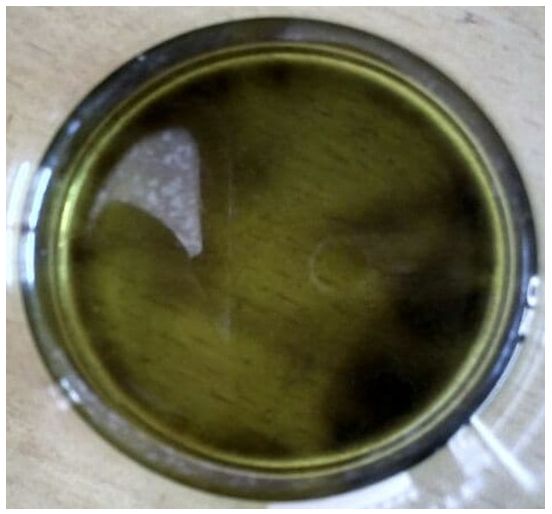
Abstract
In biomedical applications, Cu2O nanoparticles are of great interest. The bioengineered route is eco-
friendly for the synthesis of nanoparticles. Therefore, in the present study, there is an attempt to synthesis
of Cu2O nanoparticles using Datura metel L. The synthesized nanoparticles were characterized by UV-Vis,
XRD, and FT-IR. UV-Vis results suggest the presence of hyoscyamine, atropine in Datura metel L, and also,
nanoparticles formation has been confirmed by the presence of absorption peak at 790 nm. The average
crystallite size (19.56 nm) obtained by XRD. Further, the various functional groups have been confirmed
through FT-IR. To highlight the peak of the dominant frequencies, Fourier Power Spectrum was also used
to analyze the synthesized nanomaterials spectrum results. Density functional theory (DFT) further also
used over a period of time to measure the energy of the substance, which seems to suggest a stable
compound. Furthermore, the calculated energies, thermodynamic characteristics (such as enthalpies,
entropies, Gibbs-free energies), modeled structures of complexes, crystals, and clusters, and predicted
yields, rates, and regio- and stereospecificity of reactions were in good agreement with the experimental
ones. Overall, the findings indicate the successful synthesis of Cu2O nanoparticles using Datura metel L.
correlates with theoretical study.

Introduction
Now a day’s engineered nanoparticles and their technological impacts are creating new revolutionary
thoughts in the industrial process through the utilization of optical, electronic, and magnetic properties
[1]. Cu2O is an important promising candidate among the transition-metal oxides due to high abounded,
nontoxic and cheap availability. Generally, it consists of + 1, +2 oxidation states known as cuprous and
cupric ions [2]. The nanoparticles of Cu2O is naturally P-type semiconductor material with a narrow
bandgap of ̴ 1.2 and ̴ 2 eV, which could be favor on light absorption in several biomedical applications
such as MRI-ultra sound dual imaging, microbial activity [3, 4] and bandgap of 2.98 eV used as a
photoelectrode for solar energy conversion [5]. The synthesis of copper oxide nanoparticles via the green
route is an easy production strategy and eco-friendly method [2]. Hence, green synthesis is the best
adopted route for nanoparticles production when compared with the chemical method due to without
requirement of any reducing and stabilizing agent. The plant metabolism like flavanoid, alkaloids,
terpenoids, polyphenols, and proteins are a reservoir of plant tissue, it could be used as a reducing agent
on various nanoparticles synthesis [6]. Datura metel L. is a Solanacae family agriculture plant that is
geographically spread over on gibbous world either as an origin or foreign plant in Asia, America, and
Europe. The species of Datura metel L. have been used as classical Chinese medicine for the treatment of
asthma, coughs and antitumor activities, etc. Particularly, the major constituent of this plant namely
withanolides having plant steroids build on by ergostane skeleton in the side chain of δ- lactone and their
predominant alkaloid content of hyoscyamine, atropine has given the medicinal properties on
nanoparticles formulation [7].

                                                  Page 2/20
DFT has been used for understanding and studying the stability of new compounds in previous
documents [8, 9]. Generally, a new compound is considerably longer than that in a stable orbital at a
constant energy level. The electron exchange and correlation of geometry optimization, electronic
structure, and optical properties were determined by using a general approximation gradient (GGA) with
the interpolation formula [10]. An ultra-soft potential provided by the Vanderbilt optimization method
defines the interaction between the electron and ion. In a plane wave basis, the electronic valence wave
characteristics are extended to a 16 eV power reduction. In addition to DFT, Fourier power values can also
be used for the indigenous signature of any compound [11, 12]. For different compounds and the same
for the same compounds, the four spectral picks are unique. Studying the pureness, stability and other
intrinsic characteristics of the compound, which can be directly seen at the peaks of Fourier, can benefit
us. Therefore, there is an attempt to synthesize Cu2O nanoparticles using Datura metel L. in the current
research. UV-Vis, XRD, and FT-IR used to characterize the synthesized nanoparticles and further the
compound stabilization has calculated by DFT method. In addition, the calculated energies,
thermodynamic characteristics (such as enthalpies, entropies, Gibbs-free energies), modeled structures of
complexes, crystals, and clusters, and predicted yields, rates, and regio- and stereospecificity of reactions
are also studied.

Experimental
Materials & Methods
The homogeneous Datura metel L. leaves collected from farmland on Krishnankoil, Tamil Nadu, India and
Copper nitrate (Cu(NO3)2) purchased from SRL chemicals Mumbai, India, used without any further
purification for the synthesis of Cu2O nanoparticles. Datura metel L. leaves dried at room temperature up
to brown after then grained by mortar and pestle. The prepared powder consumed for all the
characterization and double distilled (DD) water was used throughout the experiment.

Dust and organic moieties contain in leaves of Datura metel L. cleaned through the process of washing in
tap water followed by DD water after than leaves cut into small pieces. 10 mg of leaves poured into the
100 ml of DD water boiled at 60℃ for 20 min in a magnetic stirrer with the speed of 70 rpm. After
cooling, the prepared extract solution has filtered through the Whatman No.1 filter paper. This extract was
used as a reducing agent in copper oxide nanoparticles (Cu2O NPs) synthesis.

In this experiment, 20 ml of extract added drop by drop with 100 ml of 3 mM copper nitrate aqueous
solution. The reaction mixture of copper nitrate and leaf extract stirrer for 20 min in this mean time the
colour of the solution changed from blue to sea green, which indicates the formation Cu2O NPs. The
formed NPs are maintaining the room temperature up to 15 days after that start to obtained black
precipitation. The prepared NPs are filtered and dried at 60˚C for 3 hrs.

Characterization
                                                  Page 3/20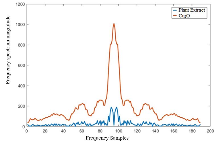
The UV-Vis absorption spectra are recorded on Shimadzu UV–1800 suited with 10 mm quartz cuvette.
XRD measurements carried out on Brucker Eco D8 Advance diffractometer with Cu-Kα radiation (λ=
1.54060 Å). FTIR analysis was performed at room temperature on IRTracer-100 Shimadzu.

Results And Discussion
Spectral Characterization

The inhomogeneous electron gas (the Born–Oppenheimer approximation) is a collection of interacting
point electrons traveling quantum–mechanically in the potential region of a set of atomic nuclei that are
considered to be static. The independent electron approximation, Hartree theory, and Hartree-Fock theory
are the most common approximation schemes used to solve such models. However, over the last thirty
years or so, another approach-Density Functional Theory (DFT)-has become increasingly popular for the
solution of such problems. This approach has the advantage of being able to solve a wide variety of
problems with high precision while still being computationally simple. Density functional theory can be
used to measure the electronic structure of atoms, molecules, and solids (DFT). Its aim is to use quantum
mechanics' fundamental laws to obtain a quantitative understanding of material properties.

Using traditional electronic structure approaches, the Schrödinger equation of N interacting electrons
moving in an external, electrostatic potential is solved (typically the Coulomb potential generated by the
atomic nuclei). However, there are major disadvantages to this approach: (1) the problem is nontrivial
even for small numbers N, and the resulting wave functions are complicated objects; and (2) the
computational effort increases exponentially with increasing N, rendering the description of larger
systems prohibitive. DFT takes a different approach, using the one-body density as the fundamental
variable instead of the many-body wave function. Density-functional theory is computationally feasible
also for large systems since the density n(r) is a function of only three spatial coordinates (rather than the
3N coordinates of the wave function). A significant development influenced spectral analysis
methodology, as the numerical approach was improved by Cooley and Tukey in 1965, known as the Fast
Fourier Transformation. The continuum of power density or power range indicates the value of the Fourier
process in spectral analysis. The function of autocorrelation of a continuous signal plays an important
role as well. The Fourier transform of a finite continuous and their relation to infinite, continuous signals
will provide the Dirichlet condition and zero means for the continuous operation. The experimental
measures of the compound in our paper are limited to a limited amount of time. By multiplying the
infinite continuous recording by a data window as defined, the record duration can be limited to a certain
period T.

                                       (1)

The window in fourier integral leads to the identity as

                                                   Page 4/20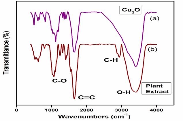
w(t) = 1 for |t| ≤ T/2 (2)
The Fourier transform of this product is the transformation of the infinite record which is paired with
transformations of the window when multiplying the infinite record. The Convolution Theorem
establishes this relationship, which affirms that:
F[f(t).w(t)] = F(α) * W(α) (3)
Discrete-time Fourier transform (DTFT) is helpful in calculating the diffracted wave information and the
obtained peaks tell us all about the molecular design's properties without having to measure the
molecular structure. The DTFT is in the direction of x, y, z for a given compound.
(4)
The p(x,y,x) gives the density distribution of the crystalline state of the compound and (a,b,c) represent
the edge length in the (x,y,z) directions. The DTFT biosynthesis analysis of Cu2O as shown (Fig. 1)
reveals a distinct difference in the spectral density peak for the three experiments. DTFT peaks shown
above indicate that the peak value of 1100 is unique to the synthesized compound and that of the
previous compound, the lower peak of 200 is. The sharp rise in maximum value also indicates that the
compound is more active and reactive than the other compound.
UV-Vis Analysis
The optical properties of biomolecules loaded Cu2O NPs were analyzed through UV-Vis absorbance
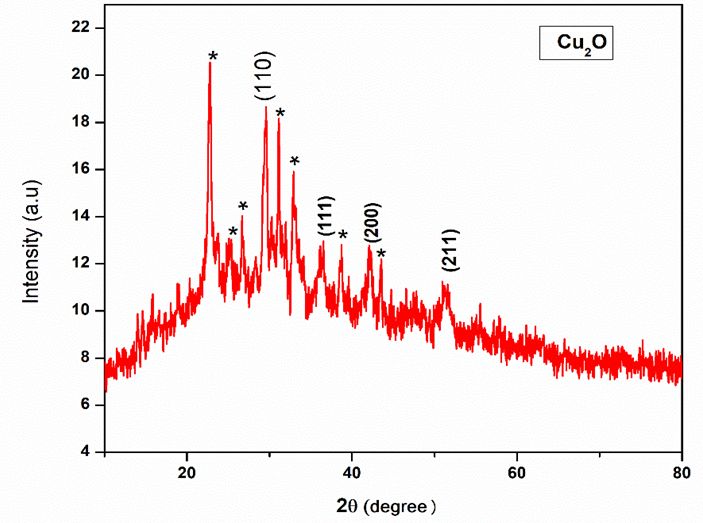
spectra. Fig. 2 shows the optical absorbance spectra of the Cu2O NPs. The formation of Cu2O NPs
confirmed by the colour change of copper nitrate aqueous solution from bluish green to sea green when
adding of Datura metel L. leaf extract shown (Fig. 3). The SPR absorbance peak was found at 790 nm by
the oscillation of electrons on the surface of the Cu2O NPs, which is clearly revealing the reduction of
Cu2O NPs. The formed Cu2O NPs absorbance peak at 790 nm shows the electronic d-d transitions
making by the Cu2+ ions in d orbital, in this kind of absorption favored for the extended lifetime of
photogenerated carriers [13]. The serious of alkaloids present on the Datura metel L. extract such as
hyoscyamine, atropine is exhibited the characteristic absorption band at 200-350 nm in UV-Vis spectra
corresponds to π-π* transition [14]. The bandgap energy of prepared Cu2O nanoparticles is 2.98 eV from
Tauc’s plot analysis. This bandgap energy well suited for solar cell and optical device applications
according to the report of earlier researcher. Awed et al., 2019 has achieved 2.98 eV for nonlinear optical
susceptibility through annealing process and Singh et al., 2004 has attained the same bandgap energy
for nanocrystalline CdTe film for electroluminescent display devices [15, 16].
XRD Analysis
Page 5/20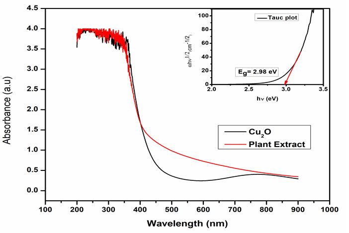
Crystallinity, size, and phase of the biosynthesized Cu2O NPs were determined through XRD analysis and
their diffraction pattern shown (Fig. 4). Biosynthesized Cu2O NPs have characteristic diffraction peaks at
2θ angle 22.77°, 25.08°, 26.54°, 29.48°, 31.28°, 32.95°, 36.47°, 38.67°, 42.19°, 47.66°, 51.62°. Here, the
observed diffraction reflections peaks at 29.48°, 36.47°, 42.19°, 51.62° indicates the presence of Cu2O
NPs and indexed by Bragg’s reflections (110), (111), (200), and (211). According to the JCPDS Card No:
77-0199 the mentioned lattice planes such as (110), (111), (200) are exhibit primitive lattice structures of
Cu2O NPs. Similar kinds of Cu2O NPS XRD diffractogram are reported by some other researchers for
various plant extract [2, 6, 17]. The remaining unassigned peaks and background noises in the XRD
pattern represented by the star symbol, which reveals the Datura metel L. biomolecules, encapsulated
around the Cu2O NPs [18]. The prepared Cu2O NPs average crystallite size is 19.56 nm calculated from
the result of XRD analysis using Debye – Scherer’s equation.

                   (5)

In this case, k is the dimensionless shape factor taken as 0.9, λ known as X-ray wavelength, β is line
broadening at half the maximum intensity (FWHM) and θ is the Bragg angle. This result illustrated
biomolecules are well bound with Cu2O nanoparticles during synthesis and Datura metel L. extract is one
of the promising candidates for reduction and stabilization of Cu2O NPs.

Fourier Transform Infrared Spectroscopy Analysis

Phytochemicals present on the plant extract and the formation of Cu2O were identified through the FTIR
spectra analysis. The IR spectra of Cu2O NPs compared with Datura metel L. is shown (Fig. 5). The FTIR
spectrum (Fig. 5b) Datura metel L. leaf extract shows a broad absorption band at 3406 cm-1 which is due
to the O-H stretching mode of phenol and alcohols. The peak at 2937 cm-1 indicates C-H stretching of
alkyl groups and strong peaks 1651 cm-1 show the C=C stretching vibration of carboxylic groups. The
peaks at 1546 cm-1 reveal that the C-N stretch of aliphatic amines and peaks at 1406, 1359, and 1317 cm-
1
    are representing C-C stretch (in-ring) of aromatics, N=O bending vibration of nitro compounds, C-N
stretch of aromatic amines, respectively. The peaks that appeared at 1105, and 1068 cm-1 are belong to
the C-N stretch of aliphatic amines. The peaks at 752, 621, and 526 cm-1 are show the existence of C-Cl
stretch alkyl halides, C-H bends alkanes, and C-I stretches aliphatic iodo compounds. The FTIR spectrum
of Cu2O NPs showed again the presence of O-H stretching mode of phenol and alcohols and C-H
stretching of alkyl groups at 3404, and 1620 cm-1, which supports the idea of phenol, alcohols, and alkyl
group, are free from Cu2O NPs formation. The peaks at 1409, 1359, and 1317 cm-1 on Cu2O NPs
represent a diminishment of C-C stretch (in-ring) of aromatics, N=O bending vibration of nitro compounds,
C-N stretch of aromatic amines, respectively. After bioreduction, peak shifts have occurred at 752 to 709
cm-1 on C-Cl alkyl halides, 621 to 650 cm-1 on C-H bend of alkanes, and 526 to 609 cm-1 on C-I stretch in
aliphatic iodo compounds. Above mentioned biocompounds are acting as a capping agent as well as
                                                   Page 6/20
bound along with the Cu2O nanoparticles. The disappearance of peaks at 2937, 1546, 879 cm-1 and
newly formed peaks at 1043, 999 cm-1 confirms the C-H stretching of alkyl, C-N stretch of aliphatic
amines, N-H bending vibration of nitro compounds, and C-OH of carboxylic acid are responsible for
structural changes on NPs formation. The strong peaks obtained at 819 cm-1 and the existence of new
peaks at 499 cm-1 are correspond to the characteristic formation of Cu2O NPs (Fig. 5a) [19]. Particularly
aliphatic amines in Datura metel L. leaf extract are mainly responsible for the reduction of copper ions
into Cu2O NPs.
Density Functional Theory Analysis
The optical properties of a molecule or crystal are among the most useful classes of properties that can
predict distinctive characteristics. These can be used to locate wavelengths of optical radiation based on
its electronic structure either in the absorption or emission spectrum. DFT lets us calculate these
properties, related to electron motion evolution under electric field control. DFT is the theory of differential
and functional functions. The spectrum data are shown (Fig. 6). NUM is first standardized with [01] which
Gaussian has reoriented to speed up the two calculations of electron energy models. The internal nuclear
energy has been measured using the spectral data measurement. The next step is to measure every
single electron transaction using the Hamiltonian Fock matrix. Once we know the propagation of the
electron, we measure the angular momentum of the electrons that are then used to detect an electron's
energy gap.
The energy of the synthesized compound stabilized over some time to a constant value of 16.6378 eV
and remained the same indicating compound stability. As shown in Fig. 6. The initial fluctuation is
caused by the excitation of the electron that tends to return the electron to its normal condition in time.
The oscillation reaction shows that the compound is erratic, but as the proposed compound can be
shown, its behavior is very stable.
Computational modeling
Calculation of the thermodynamic and surface characteristics of Cu2O thin films at all temperatures was
carried out using MERA software with periodic boundary conditions along with a, b and c axes of Cu2O
unit cell like it were described in [20, 21] and applied in studying organic, inorganic and combined
systems in [20-37].
The MOPS algorithm has been used to model oxyhydrate gel formation [20, 25, 29], crystal structures of
triosmium clusters [21, 23, 24, 28, 30, 33, 35], organic molecule complexation during chemical reactions
[31], protein affinity [34], and crystal structures and interaction energies of gas hydrates [26, 37].
Calculated energies, thermodynamic properties (such as enthalpies, entropies, and Gibbs-free energies),
modeled structures of complexes, crystals, and clusters, and predicted yields, rates, and regio- and
stereospecificity of reactions were all in good agreement with experimental results.
Page 7/20The structures hyoscyamine and atropine were optimized at the DFT B3LYP 6-311G (d,p) level of theory.
Then, the UV-Vis spectrum was calculated using TD DFT B3LYP 6-311G (d,p) which shows the absorption
band is 253.3 nm that in good agreement with the experimental data.
The structure of cuprite [38] (Crystallography Open Database ID 1000063) was taken as the initial
structure for the computer simulation of Cu2O nanoparticles (cubic syngony, space group Pn m, a = b =c
= 4.252(2), α = β = γ = 90°).
1000 multiplications of the crystal cell in random directions were performed to the composition
Cu2474O1237 (this composition corresponds to the experimental size of the particles) and the structure
with the minimum energy is chosen. Calculation using the Bragg’s equation showed that the resulting
modelled nanoparticles should have diffraction reflections peaks at 2θ angle 29.69°, 36.57°, 42.49°,
52.69°, 61.65°, 69.90°, 73.86°, 77.74°, 85.35°. The first four reflections are in good agreement with the
experimental diffractogram and correspond to reflections (110), (111), (200), and (211) that show a good
quality of the simulation.
The initial structure of atropine and hyoscyamine in an aqueous solution (they are similar, since atropine
is a racemate and hyoscyamine is an L-isomer of the same compound) modeled within the MOPS
software [20, 25, 29] with the continual account of the solvent influence shown (Fig. 7a). The structure
contains the intramolecular hydrogen bond =O…H-O with a length of 2.22 Å.
Subsequently, the modeling of the complex of this nanoparticle with hyoscyamine and with atropine was
carried out. The calculated Gibbs free energy of the complex formation is -179.4 kJ/mol. The structure
and conformation of atropine (hyoscyamine) remain almost unchanged during the formation of the
complex. The complex is formed by three short contacts (Fig. 7b). Two of them are carried out by
carbonyl oxygen with two copper atoms of nanoparticles (2.16 and 2.19 Å). These bifurcate interactions
become possible due to a defect in the surface of the nanoparticles, when two copper atoms at once turn
out to be with a lack of valence. The third contact is the hydrogen bond of the hydroxyl hydrogen of
atropine with the oxygen of the nanoparticle surface (2.09 Å). The intramolecular hydrogen bond =O…H-O
is retained, but slightly extended to 2.37 Å.
Conclusions
A simple and cost-effective method has been proposed to the synthesis of Cu2O nanoparticles at room
temperature within the reaction time of 30 minutes and abundant, the unappreciated plant of Indian
surroundings (Datura metel L.) extract utilized as a reducing and stabilizing agent. The UV-Vis analysis
shows Cu2O NPs formation at 790 nm through the making of cu2+ ions transition in d-orbital. The result
of the XRD pattern reveals that biomolecules of Datura metel L. encapsulation on synthesized Cu2O NPs
and found in crystalline nature with an average crystallite size of 19.56 nm. This biomolecules
encapsulation is validated by FTIR characterization, the aliphatic amines in Datura metel L. responsible
for the reduction of Cu2O NPs, and their Phytochemicals are effectively utilized as a bio-capping agent
Page 8/20and it has been bound along around the Cu2O NPs. The synthesized compound was tested using DFT
and Fourier power spectrum for energy band stability as well as for spectral signatures. These two
algorithmic evaluations have enabled us to calculate the stability and unique spectral signature of the
proposed Cu2O compounds. In the first time, we successfully applied spectral characterization with
Fourier transform for the biosynthesized Cu2O NPs and highlighted their spectrum. High transformation
of the high frequency of spectral characterization suggests that synthesized Cu2O NPs to be in more
active and reactive than other compound but oscillation reactions are erratic as well as their stability is
very stable in behavior even after 10 iterations of density functional theory analysis. Moreover, the
present report is used to study the stabilization of nanoparticles in various solar cell, wastewater
treatment, and biomedical applications.
Declarations
Author’s Declarations
The authors declare no conflict of interest.
Acknowledgments
The project was supported by Ministry of Science and Higher Education of Russia (Grant no. FENU-2020-
0019). Authors acknowledged Kalasalingam Academy of Research and Education, Krishnankoil,
Tamilnadu, India.
Authors’ Contributions
Experimental and characterization of green synthesized Cu2O nanoparticles using Datura Metel L done by
KC and supervised by KG, KK, MGR and RT. Theoretical studies performed by KG, VM, VP and MG and
supervised by VP and VM. Manuscript written by KC and checked by all authors.
Data Availability All data generated during this study are included in this published article
Funding
The project was supported by Ministry of Science and Higher Education of Russia (Grant no. FENU-2020-
0019).
Ethical Approval Not applicable.
Consent to Participate Not applicable.
Consent for Publication Not applicable.
Conflict of interest The authors declare that they have no known competing financial interests.
Page 9/20References
1. Azhar W, Khan AR, Muhammad N, Liu B, Song G, Hussain A, Yasin MU, Khan S, Munir R, Gan Y
(2020) Ethylene mediates CuO np-induced ultrastructural changes and oxidative stress in
arabidopsis thaliana leaves. Environ. Sci. Nano 7:938-953. https://doi.org/10.1039/C9EN01302D
2. Jadhav MS, Kulkarni S, Raikar P, Barretto DA, Vootlac SK, Raikar US (2018) Green biosynthesis of
CuO & Ag–CuO nanoparticles from malus domestica leaf extract and evaluation of antibacterial,
antioxidant and DNA cleavage activities. New J. Chem 42: 204-213.
https://doi.org/10.1039/C7NJ02977B
3. Kargar A, Jing Y, Kim SJ, Riley CT, Pan X, Wang D (2013) ZnO/CuO Heterojunction branched
nanowires for photoelectrochemical hydrogen generation. ACS Nano 7(12):11112-11120.
https://doi.org/10.1021/nn404838n
4. Wongrakpanich A, Mudunkotuwa IA, Geary SM, Morris AS, Mapuskar KA, Spitz DR, Grassian VH,
Salem AK (2016) Size-dependent cytotoxicity of copper oxide nanoparticles in lung epithelial cells.
Environ. Sci. Nano 3:365-374. https://doi.org/10.1039/C5EN00271K
5. Awed AS, El-Ghamaz NA, El-Nahass MM, Zeyada HM (2019) Linear and nonlinear optical properties
of alizarin red S thin films. Indian J Phys 93:861-868. https://doi.org/10.1007/s12648-018-01359-6
6. Siddiqui VU, Ansari A, Chauhan R, Siddiqi WA (2021) Green synthesis of copper oxide (CuO)
nanoparticles by punica granatum peel extract. Materials Today Proceedings 36: 751-755.
https://doi.org/10.1016/j.matpr.2020.05.504
7. Nasir B, Baig MW, Majid M, Ali SM, Khan MJI, Kazmi STB, Haq I (2020) Preclinical anticancer studies
on the ethyl acetate leaf extracts of Datura Stramonium and Datura Inoxia. BMC Complement Med
Ther 20:188. https://doi.org/10.1186/s12906-020-02975-8
8. Haunschild R, Barth A, French BA (2019) Comprehensive analysis of the history of DFT based on the
bibliometric method RPYS. J Cheminform 11:72. https://doi.org/10.1186/s13321-019-0395-y
9. Fermi E (1928) Eine Statistische Methode Zur Bestimmung Einiger Eigenschaften Des Atoms und
ihre Anwendung auf die Theorie des periodischen Systems der Elemente. Z. Physik 48:73-
79. https://doi.org/10.1007/BF01351576
10. Haunschild R, Barth A, Marx W (2016) Evolution of DFT studies in view of a scientometric
perspective. J Cheminform 8:52. https://doi.org/10.1186/s13321-016-0166-y
11. Ciaccio EJ, Biviano AB, Whang W, Coromilas J, Garan H (2011) A new transform for the analysis of
complex fractionated atrial electrograms. BioMed Eng OnLine 10:35. https://doi.org/10.1186/1475-
925X-10-35
12. Mesgaran SD, Eggert A, Höckels P, Derno M, Kuhla B (2020) The use of milk Fourier transform mid-
infrared spectra and milk yield to estimate heat production as a measure of efficiency of dairy cows.
J Animal Sci Biotechnol 11:43. https://doi.org/10.1186/s40104-020-00455-0
13. Luo Z, Jiang H, Li D, Hu L, Geng W, Wei P, Ouyang P (2014) Improved photocatalytic activity and
mechanism of Cu2O/N–TiO2 prepared by a two-step method. RSC Adv 4:17797-17804.
Page 10/20https://doi.org/10.1039/C3RA47973K
14. Ramalechume C, Shamili P, Krishnaveni R, Swamidoss CMA (2020) Synthesis of copper oxide
nanoparticles using tree gum extract, its spectral characterization, and a study of its anti- bactericidal
properties. Materials Today Proceedings 33:4151-4155. https://doi.org/10.1016/j.matpr.2020.06.587
15. Grad L, Novotny Z, Hengsberger M, Osterwalder J (2020) Influence of surface defect density on the
ultrafast hot carrier relaxation and transport in Cu2O photoelectrodes. Sci Rep 10:10686.
https://doi.org/10.1038/s41598-020-67589-z
16. Singh RS, Rangari VK, Sanagapalli S, Jayaraman V, Mahendra S, Singh VP (2004) Nano-structured
CdTe, CdS and TiO2 for thin film solar cell applications. Sol. Energy Mater. Sol. Cells 82:315-330.
https://doi.org/10.1016/j.solmat.2004.02.006
17. Sasidharan D, Namitha TR, Johnson SP, Jose V, Mathew P (2020) Synthesis of silver and copper
oxide nanoparticles using myristica fragrans fruit extract: antimicrobial and catalytic applications.
Sustain. Chem. Pharm 16:100255. https://doi.org/10.1016/j.scp.2020.100255
18. Sarkar J, Chakraborty N, Chatterjee A, Bhattacharjee A, Dasgupta D, Acharya K (2020) Green
synthesized copper oxide nanoparticles ameliorate defence and antioxidant enzymes in lens
culinaris. Nanomaterials 10:312. https://doi.org/10.3390/nano10020312
19. Anand GT, Sundaram SJ, Kanimozhi K, Nithiyavathi R, Kaviyarasu K (2021) Microwave assisted
green synthesis of CuO nanoparticles for environmental applications. Materials Today Proceedings
36:427-434. https://doi.org/10.1016/j.matpr.2020.04.881
20. Sukharev YI, Potemkin VA, Markov BA (2001) Autowave processes of forming gels as a cause of the
coloring of oxyhydrate gels (the chromatic effect) of some rare earth metals (yttrium, gadolinium).
Colloids Surf. A 194:75-84. https://doi.org/10.1016/S0927-7757(01)00757-9
21. Potemkin VA, Maksakov VA, Kirin VP (2003) Conformational states of triosmium clusters with
aminoacid ligands: a theoretical study. J. Struct. Chem 44:741-747.
https://doi.org/10.1023/B:JORY.0000029809.88411.8b
22. Potemkin VA, Krasnov VP, Levit GL, Bartashevich EV, Andreeva IN, Kuzminsky MB, Anikin NA,
Charushin VN, Chupakhin ON (2004) Kinetic resolution of (±)-2,3-dihydro-3-methyl-4H-1,4-
benzoxazine in the reaction with (S)-naproxen chloride: a theoretical study. Mendeleev Commun
14:69-70. https://doi.org/10.1070/MC2004v014n02ABEH001887
23. Potemkin VA, Maksakov VA, Kirin VP (2004) Theoretical study of the conformations of triosmium
clusters with a chiral carane ligand. J. Struct. Chem 45:405-409. https://doi.org/10.1007/s10947-
005-0006-9
24. Potemkin VA, Maksakov VA, Korenev VS (2005) Theoretical study of the conformational states of
triosmium clusters with a chiral pinane ligand. J Struct. Chem 46:43-
48. https://doi.org/10.1007/s10947-006-0007-3
25. Sukharev YI, Avdin VV, Lymar AA, Belkanova MY, Potemkin VA (2006) Directions in structure
formation of oxyhydrate gels of zirconium and rare earth elements. J. Struct. Chem. 47:151-155.
https://doi.org/10.1007/s10947-006-0280-1
Page 11/2026. Aladko EY, Ancharov AI, Goryainov SV, Kurnosov AV, Larionov EG, Likhacheva AY, Manakov AY,
Potemkin VA, Sheromov MA, Teplykh AE, Voronin VI, Zhurko FV (2006) New type of phase
transformation in gas hydrate forming system at high pressures. Some experimental and
computational investigations of clathrate hydrates formed in the SF6−H2O system. J. Phys. Chem. B
110:21371-21376. https://doi.org/10.1021/jp061698r
27. Grishina MA, Potemkin VA, Bartashevich EV, Sinyaev AN, Rusinov GL, Latosh NI, Ganebnykh IN,
Koryakova OV, Ishmetova RI (2006) Modeling of 1,2,4,5-tetrazine complexes with organic amines. J.
Struct. Chem 47:1155-1160. https://doi.org/10.1007/s10947-006-0438-x
28. Potemkin VA, Maksakov VA, Korenev VS (2007) Theoretical study of the conformational states of
triosmium clusters with a chiral μ-1-NH pinane ligand. J. Struct. Chem 48:225-230.
https://doi.org/10.1007/s10947-007-0036-6
29. Avdin VV, Lymar AA, Batist AV, Nikitin EA, Belkanova MY, Potemkin VA (2007) Structure formation in
heavy metal oxyhydrates at low rates of gel formation, J. Struct. Chem 48:747-752.
https://doi.org/10.1007/s10947-007-0114-9
30. Korenev VS, Kirin VP, Maksakov VA, Virovets AV, Tkachev SV, Potemkin VA, Agafontsev AM, Tkachev
AV (2007) Triosmium cluster with the bridging aminooxime derivative of pinane: synthesis, crystal
structure and conformational analysis. Russ. J. Coord. Chem 33:594-
600. https://doi.org/10.1134/S1070328407080088
31. Shchur IV, Khudina OG, Burgart YV, Saloutin VI, Grishina MA, Potemkin VA (2007) Synthesis,
structure, and complexing ability of fluoroalkyl-containing 2,2′-(biphenyl-4,4′-diyldihydrazono)-bis(1,3-
dicarbonyl) compounds. Russ. J. Org. Chem 43:1781-1787.
https://doi.org/10.1134/S107042800712007X
32. Grishina MA, Potemkin VA, Matern AI (2008) Theoretical study of acridane oxidation reactions. J.
Struct. Chem 49:7-12. https://doi.org/10.1007/s10947-008-0002-y
33. Maksakov VA, Pervukhina NV, Podberezskaya NV, Afonin MY, Potemkin VA, Kirin VP (2008) X-ray and
conformation analysis of the new trinuclear cluster of osmium Os3(μ,η2-OCC6H5)(CO)9. J. Struct.
Chem 49:894-900. https://doi.org/10.1007/s10947-008-0154-9
34. Kuzmicheva GA, Jayanna PK, Eroshkin AM, Grishina MA, Pereyaslavskaya ES, Potemkin VA, Petrenko
VA (2009) Mutations in Fd phage major coat protein modulate affinity of the displayed peptide.
Protein Eng., Des. Sel 22:631-639. https://doi.org/10.1093/protein/gzp043
35. Potemkin VA, Ivshina NN, Maksakov VA (2009) Theoretical Study of the conformational features of
triosmium clusters. J. Struct. Chem 50:143-151. https://doi.org/10.1007/s10947-009-0202-0
36. Ivshina NN, Bartashevich EV, Potemkin VA, Grishina MA, Ishmetova RI, Rusinov GL, Latosh NI,
Slepukhin PA, Charushin VN (2010) Changes in the vibrational characteristics of substituted 1,2,4,5-
tetrazines after complexation with 1,2,3-benzotriazole: A theoretical study. J. Struct. Chem 50:1053-
1058. https://doi.org/10.1007/s10947-009-0155-3
37. Manakov AY, Likhacheva AY, Potemkin VA, Ogienko AG, Kurnosov AV, Ancharov AI (2011)
Compressibility of gas hydrates. ChemPhysChem 12:2476-
Page 12/202484. https://doi.org/10.1002/cphc.201100126
38. Neuburger M. C (1931) Präzisionsmessung der Gitterkonstante von Cuprooxyd Cu2O. Zeitschrift für
Physik 67:845-850. https://doi.org/10.1007/BF01390765
Tables
Table 1 FTIR wavenumbers and their corresponding functional group analysis of Cu2O synthesized by
Datura metel L
Wavenumbers (cm-1) Vibrational assignments Functional groups
Datura metel L Cu2O NPs
3406 3404 O-H stretching phenol, alcohols
1651 1620 C=C stretching carboxylic groups
1406 1409 C-C stretch (in-ring) Aromatics
1359 1359 N=O bending nitro compounds
1317 1317 C-N stretch aromatic amines
1105 1124 C-N stretch aliphatic amines
1068 943 C-N stretch aliphatic amines
752 709 C-Cl stretch alkyl halides
621 650 C-H bend Alkanes
526 609 C-I stretch aliphatic iodo
2937 - C-H stretching Alkyl
1546 - C-N stretch aliphatic amines
879 - N-H bending vibration nitro compounds
- 819 Characteristic formation of Cu2O NPs
- 499
Figures
Page 13/20Figure 1
Displays the spectral characterization with a Fourier Transform of the proposed Cu2O compound results.
The higher transformation of the high frequency suggests the biosynthesis of Cu2O
Page 14/20Figure 2
UV-Vis absorption spectra of Cu2O NPs
Page 15/20Figure 3
Formation of Cu2O NPs
Page 16/20Figure 4
XRD analysis of Cu2O NPs
Page 17/20Figure 5
FTIR spectra (a) Cu2O NPs and (b) Plant extract
Page 18/20Figure 6
Energy (eV) versus DFT iterations show the compound's stability after 10 iterations, since the energy
levels obtain a constant value and remain the same
Page 19/20Figure 7
The structure of a) atropine and b) its complex with the fragment of Cu2O nanoparticles
Page 20/20You can also read