Brain reactivity to emotional stimuli in women with premenstrual dysphoric disorder and related personality characteristics
←
→
Page content transcription
If your browser does not render page correctly, please read the page content below
www.aging-us.com                                                              AGING 2021, Vol. 13, No. 15
                                                                                                     Research Paper
Brain reactivity to emotional stimuli in women with premenstrual
dysphoric disorder and related personality characteristics
Mingzhou Gao1,2,3,*, Mingqi Qiao1,2,3,*, Li An4, Guangbin Wang5, Jieqiong Wang2,3,
Chunhong Song6, Fengqin Wei1,3, Yanhong Yu1,2,3, Tao Gong5, Dongmei Gao1,2,3
1
  College of Traditional Chinese Medicine, Shandong University of Traditional Chinese Medicine, Jinan, Shandong
Province, China
2
  Research and Innovation Team of Emotional Diseases and Syndromes in Shandong University of Traditional
Chinese Medicine, Jinan, Shandong Province, China
3
  Key Laboratory of Traditional Chinese Medicine Classical Theory, Ministry of Education, Shandong University of
Traditional Chinese Medicine, Jinan, Shandong Province, China
4
  Jinan Central Hospital, Jinan, Shandong Province, China
5
  Department of Radiology, Shandong Medical Imaging Research Institute, Jinan, Shandong Province, China
6
  Department of Laboratory Animal Center, Central Hospital Affiliated to Shandong First Medical University, Jinan,
Shandong Province, China
*Equal contribution and Co-first author

Correspondence to: Dongmei Gao; email: 2017101002@sdutcm.edu.cn
Keywords: premenstrual dysphoric disorder, anger, depression, eysenck personality questionnaire, emotion regulation
Received: April 5, 2021        Accepted: July 8, 2021               Published: August 4, 2021

Copyright: © 2021 Gao et al. This is an open access article distributed under the terms of the Creative Commons Attribution
License (CC BY 3.0), which permits unrestricted use, distribution, and reproduction in any medium, provided the original
author and source are credited.

ABSTRACT

Aims: Premenstrual dysphoric disorder (PMDD) is a psychiatric condition that is associated with the menstrual
cycle. Elucidation of the neural regulation mechanisms of brain reactivity to emotional stimuli among women
with PMDD may inform PMDD treatment.
Methods: Eighty-six women (42 PMDD, 44 healthy controls) were allocated into two groups (anger-induced
group: 23 PMDD vs. 23 controls; depression-induced group: 19 PMDD vs. 21 controls). During the luteal phases
of the menstrual cycle, all the women were subjected to functional magnetic resonance imaging (fMRI). fMRI
resting-state scans were performed before and after the study participants had performed an emotional stimuli
task. After the emotional stimuli task, emotional status of the participants were evaluated by Self-Rating
Depression Scales (SDS) and Trait Anger Expression Inventory–II (STAXI-II). In addition, all the participants were
requested to complete the Eysenck Personality Questionnaire (EPQ) and the Twenty-Item Toronto Alexithymia
Scale (TAS-20).
Results: Compared to healthy controls, all women with PMDD exhibited significantly high scores in Tas-20
(p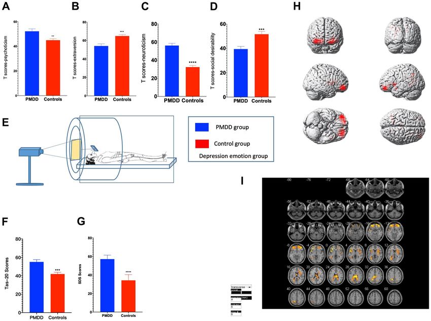
Conclusions: Women with more neuroticism and psychoticism, less extraversion and social desirability tend to
report PMDD symptoms. Women with this condition experience difficulties in regulating emotions during the
luteal phase of the menstrual cycle. Abnormal ReHo levels in the precuneus, superior frontal gyrus, lobulus
paracentralis, and right cerebellum may contribute to anger dysregulation. Hypoactivation in the middle frontal
gyrus, the middle gyrus and the cingulate gyrus may be generally associated with depression dysregulation in
PMDD.

INTRODUCTION                                                   suppressed ventral striatum responses to negative
                                                               stimuli during the luteal phase [16]. Moreover, during
Premenstrual dysphoric disorder (PMDD), a severe               the menstrual cycle, women with PMDD exhibit
form of premenstrual syndrome (PMS), is characterized          lower activations of the perennial anterior cingulate
by significant premenstrual mood disturbances, often           and the ventromedial prefrontal cortex [17]. A recent
with ―a cluster of affective, behavioral and somatic           study reported that, compared to healthy controls,
symptoms‖ [1]. Based on the latest diagnostic criteria,        women with PMDD have different intrinsic network
PMDD was classified as a subclass of depressive                dynamics in the brain [18].
disorders in the Diagnostic and Statistical Manual of
Mental Disorders (DSM–5) in 2013 [2]. According to             Based on the above findings, this study aimed at
DSM-5, 3-8% of women in the reproductive age have              investigating differences in brain reactions when
PMDD [3], which causes a severe decrease in the                women with PMDD and healthy controls (HCs) are
quality of life and psychological problems [4, 5].             subjected to emotional stimuli.

Depression, anxiety, and irritability are the three most       MATERIALS AND METHODS
evaluated PMDD symptoms [6]. Emotional problems
constitute most of the PMDD symptoms, therefore, it            Ethical statement
has been postulated that women with PMDD experience
greater difficulties with emotion regulation. Emotional        After being informed of the procedures in the study as
regulation is the ability to identify the emotions a           well as the significance of the study, participants were
person feels, and how those emotions are experienced,          asked to sign an informed consent form before inclusion
expressed and regulated [7]. The mechanisms through            in the study. Ethical approval was obtained from the
which PMDD leads to emotional dysregulation have not           Medicine Ethics Committee of the First Affiliated
been elucidated. However, it has been postulated that          Hospital of Shandong University of Traditional Chinese
central nervous system (CNS) sensitivity to                    Medicine, Shandong, China. All research procedures
reproductive hormones, such as progesterone and                were performed in accordance with the Declaration of
allopregnanolone, genetic factors, as well as                  Helsinki.
psychosocial factors, such as trauma history or
emotional and physical abuse may be contribute to              Study participants
emotional dysregulation [8]. Imaging studies have
reported differences in brain structure and function           We performed an epidemiological survey whereby a
between women with and without PMS/PMDD [9–11].                total of 868 questionnaires were distributed among
These findings regarding abnormal activities of the            women in universities in Jinan, Shandong, China. A
brain in PMDD may be potential key factors for the             total of 786 questionnaires were recovered for
occurrence of it [12].                                         collecting demographic data. Based on the DSM-5
                                                               criteria, 46 women were diagnosed with PMDD [19]. A
Functional magnetic resonance imaging (fMRI) based             total of 46 healthy volunteers with no history of mental
on blood-oxygen-level dependent (BOLD) techniques              illnesses and who were in good physical health were
has widely been used to study functional activities and        recruited from universities through newspaper, online
cognitive behaviours of the brain in response to               and leaflet advertising. All participants with PMDD, in
induced stimuli with tasks, that is, task fMRI (tfMRI)         accordance with the random number table, were
or without tasks, that is, resting state fMRI (rsfMRI)         randomly allocated into the anger-induced group (23
[13, 14]. Structurally, women with PMDD have been              PMDD vs. 23 controls) and the depression-induced
shown to exhibit greater grey matter density in the            group (19 PMDD vs. 21 controls). Data for four PMDD
hippocampal cortex and lower grey matter density in            participants and two controls in the depression-induced
the parahippocampal cortex [15]. Functionally,                 group were excluded from the final analysis because
women with PMDD have elevated amygdala and                     two participants failed to experience anger while four

www.aging-us.com                                       19530                                                  AGING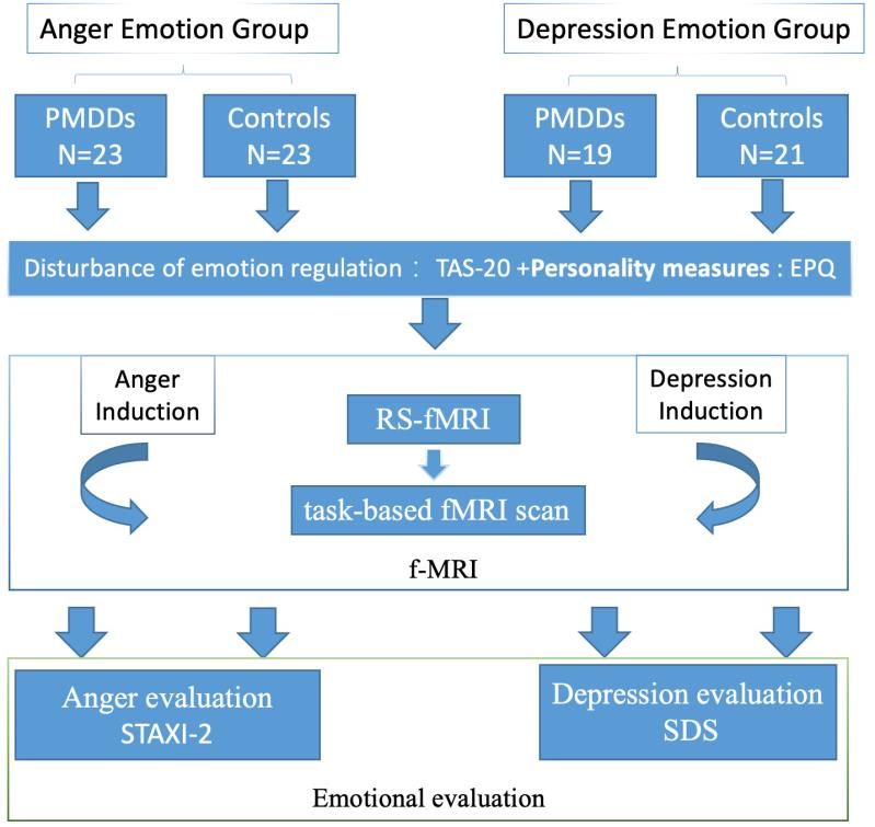
participants had correctly guessed the purpose of the               88-item questionnaire that measures the four personality
experiment before it had started.                                   dimensions (21 items for extraversion, 23 items for
                                                                    psychoticism, 24 items for neuroticism, and 20 items for
Inclusion criteria for PMDD                                         lying/social desirability) [20]. Scores were summed and
                                                                    converted into T scores using the equation:
The inclusion criteria for the PMDD group were: i.                  (T=50+10*(X-M)/SD).
Women who met the DSM-5 PMDD diagnostic criteria;
ii. Female, 18–45 years old, ethnic origin was not a                Emotional evaluation
consideration; iii. Women with a normal menstrual
cycle (differences in the range of duration of menstrual            Emotional regulation was measured using the Twenty-
flow ≤ 3 d), cycle 21–35 d; iv. Women who had                       Item Toronto Alexithymia Scale (TAS-20), which was
demonstrated an understanding of the purpose of this                used to test participants’ self-inability to identify and
study and were willing to volunteer; v. Women without               describe emotions before f-MRI. Emotional evaluation
major diseases including cardiac, liver, and kidney                 after induction was performed using the Self-Rating
diseases as well as brain tumors or other brain diseases;           Depression Scale (SDS) while anger was measured
vi. Women without a history of drug abuse.                          using the State-Trait Anger Expression Inventory-2
                                                                    (STAXI-2) created by Spielberger. The State–Trait
Inclusion criteria for HC                                           Anger Expression Inventory–II (STAXI-II) is a
                                                                    psychometric assessment tool that is used to measure
The inclusion criteria for HC were: i. Female, 20–25                anger experience, expression, and control in research
years old, right-handed, volunteer college students; ii.            and in clinical settings.
Regarding consciousness and independent judgment, we
included women who demonstrated an understanding of                 Experimental paradigm
the purpose of this study and who were willing to
volunteer; iii. Women with normal visual acuity with or             Participants in the two PMDD groups and their related
without correction; iv. Lack of metal objects in the body           HC groups were subjected to fMRI examination during
(including pacemakers, metal dental materials, and                  the late luteal phase (ranging from 1 to 5 d before
braces among others); v. Healthy individuals without                menstruation). To confirm the relatively stable and low
frequent headaches, dizziness, seizures, or other                   levels of endogenous cortisol and oestradiol, the scan
neurological diseases; vi. Women in good mental states,             tests were performed between 19:00 and 22:00 pm [21].
with good sleep quality and appetite.                               To verify menstrual cycle stages, we obtained self-
                                                                    reports regarding when menstruation started and
Exclusion criteria for all study participants                       combined this information with primary gynaecological
                                                                    examinations.
Exclusion criteria for the study were: i. mental illness or
women with previous histories of mental illness; ii.                In the entire experimental procedure, anger and
Serious physical illness; iii. Women with a history of              depression were induced in different groups (Figure 1).
drug abuse (including three months of treatment with                Each study participant was subjected to an fMRI scan
PMDD drugs); iv. Those with hematological diseases;                 composed of 6 min of 3D structure image scanning and
v. Pregnant or lactating women; vi. Aphasia,                        8 min of resting-state (RS-fMRI) scanning followed by
disturbance of consciousness, dementia, and other                   a task-based fMRI scan during which emotion images
circumstances such that participants could not cooperate            were presented. During the RS-fMRI scan, each study
with the examiner; vii. Months of unilateral                        participant was instructed to keep her eyes closed, not to
ovariectomy or abortion, taking contraceptives; viii.               think about anything and to stay awake.
Head movement more than 3 mm, in any direction, and
more than 1° during motion correction.                              During task-based fMRI scans, participants had an
                                                                    option of keeping their eyes open and view negative
Participants were terminated from the study if they: i.             (anger for anger-induced group and depression for
Exhibited symptoms requiring emergency treatment,                   depression-induced group). Neutral (NEU) emotion
thereby interfering with clinical study of the case; ii. Were       images were selected from the International Affective
unable to adhere to the study; and iii. Became pregnant.            Picture System [22] based on our previous studies. The
                                                                    task-based fMRI scan was performed in two runs, with
Personality measures                                                one set of neutral and negative images (6 images each)
                                                                    presented in each run. The first run consisted of a
Personality characteristics were determined using the               30-sec presentation of anger images (each image was
Eysenck Personality Questionnaire (EPQ), which is an                presented for 5 s, 6 images in a block) followed by

www.aging-us.com                                            19531                                                    AGING
a 30-sec presentation of neutral images (each image was Data acquisition
presented for 5 s, 6 images in a block). The second run
was performed in the opposite order. The fMRI imaging device is a magnetic device
manufactured by the Philips Company, Netherlands. It
Stimulation images were presented using a brain consists of a 3.0t TX superconducting MR instrument,
function audio-visual stimulation system (SAMRTEC and an eight-channel phase-control front ring. FMRI
SA-9900; Shenzhen Meide Medical Electronics images were obtained using a 3.0-T MR scanner
Technology Co., Ltd). This system composed of a equipped with a prototype fast gradient system for echo-
general console, cabinets, mirrors, vinyl screens, and planar imaging (EPI) at the Institute of Medical Imaging
liquid crystal displays among others. Based on of Shandong. Functional images were obtained using an
experimental requirements, using a projector, study echo planar imaging sequence with the following
participants viewed clear visual images as selected by parameters: TE = 35 ms, TR = 2000 ms, slice thickness
experimenters. The experimental visual stimulation = 4 mm, gap = 1 mm, flip angle = 90°, FOV = 24 cm,
system was programmed with Eprime. Both groups and in-plane resolution = 64 × 64. The resting-state
were shown negative emotional pictures and neutral session lasted 6 min, during which participants were
emotional pictures as stimuli for the task. Negative instructed not to; move, think systematically or to fall
emotions were induced by viewing the pictures. asleep. In addition, a T1-weighted sagittal three-
dimensional magnetization-prepared rapid gradient echo
After the scan, subjective reports of picture-evoked (MP-RAGE) sequence was acquired with the following
emotional effects were evaluated using the self-rating parameters: 144 slices, TR = 2300 ms, TE = 3.39 ms,
depression scale (SDS) and the emotional statement and slice thickness = 1 mm, flip angle = 7°, inversion time =
guidance language implementation checklist. 1100 ms, FOV = 200 × 256 mm2, and in-plane
Participants’ subjective feelings were evaluated using resolution = 200 × 256.
the visual scale test, which ranged from 0 (no feeling) to
8 (very strong feeling) points. Higher scores implied fMRI data analysis
that participants experienced higher emotional
strengths. Participants were asked to carefully assess Functional MRI data were pre-processed using
their emotional intensities. Statistical Parametric Mapping (SPM8) [23]. The first 3
Figure 1. Schematic presentation of the experimental procedure through which participants watched various images in the
anger- and depression-induction stages.
www.aging-us.com 19532 AGINGvolumes of functional images were discarded because between-group differences met the criteria of
of signal equilibrium and to allow participants to adapt uncorrected p < 0.01 at the voxel level and cluster size >
to the scanning noise. All images were time-shifted so 40 voxels, corresponding to a corrected p < 0.05. Then,
that the slices were temporally aligned. Then, images to examine the altered activation difference, one-sample
were realigned, after which we verified that all t-tests were performed on the individual activation maps
participants had moved no more than 3 mm in the of between-group peak voxels in the two groups, with a
translational dimension or 3° in the rotational significance criterion of p < 0.05 in the SPSS 25.0
dimension. Anatomical images were spatially software package. Finally, we evaluated the dissociable
normalized to the Montreal Neurological Institute anomaly of activation patterns between two groups in
(MNI) template. Normalization parameters were applied the whole brain using the criterion of corrected p < 0.05
to functional images. Images were smoothed using a for voxel level and cluster size > 389 voxels. The alpha
Gaussian filter with a full width of 8 mm at half for all significant results was two-tailed, except where
maximum. indicated.
All images were time-shifted so that slices were Data
temporally aligned. Then, images were realigned, after
which we verified that all participants had moved no The data analyzed in this study are available from the
more than 3 mm in the translational dimension or 3° in authors upon reasonable request.
the rotational dimension. Then, images were co-
registered with anatomical images, which were Ethical standards
segmented into grey matter and white matter.
Anatomical images were spatially normalized to the This study was ethically approved by the Medicine Ethics
Montreal Neurological Institute (MNI) template, and Committee of the First Affiliated Hospital of Shandong
normalization parameters were applied to the functional University of Traditional Chinese Medicine, Shandong,
images. Images were smoothed using a Gaussian filter China. All research procedures were conducted in
with a full width of 8 mm at half maximum. After accordance with the Declaration of Helsinki.
further pre-processing, which included the removal of
linear trend and temporal bandpass filtering (0.01-0.08 RESULTS
Hz), regional homogeneity (regional homogeneity,
ReHo) was determined using the Resting-State fMRI Participant characteristics
Data Analysis Toolkit (REST, by Song et al.,
http://www.restfmri.net). There were no significant differences in age,
menstruation (days), menophania (years), or length of
Statistical analysis menstrual cycle (days) between the anger- or
depression-induced groups.
To examine the personality and effects of the two emotion
induction procedures on participants’ subjective feelings Personality characteristics and emotion regulation
of anger, depression as well as their positive and negative
emotions, we performed unpaired T tests to compare In the anger-induced group, women with PMDD
emotional scores for PMDD vs. controls in the two exhibited higher Tas-20 scores (p=0.0001), higher
emotion groups. All statistical tests were performed using neuroticism and psychoticism T-scores (prespectively) when evaluated using STAXI-2. These anger induction, the PMDD group mainly exhibited findings imply a higher level of anger. For depression elevated activation in the middle frontal gyrus induction, both groups showed a strong degree of (BA10), temporal lobe (BA42), and left cerebellum depression and anxiety. However, compared to the (BA37) as well as decreased activation in the controls, women with PMDD exhibited higher SDS precuneus (BA7), superior frontal gyrus (BA8), scores (p
DISCUSSION verbal aggression and neuroticism and lower with
regards to socialization [25]. Impaired cognitive
Clinically, difficulties in regulating emotions are linked functions are key in defining PMDD. In this study, we
to core PMDD symptoms [18]. In this study, tas-20 adopted EPQ to investigate susceptible traits. Compared
scales indicated that all PMDD women had to HCs, neuroticism and psychoticism scores were
dysregulated feelings. Personality traits reflect people’s higher while extraversion and lying scores were lower
characteristic patterns of thoughts, feelings, and in PMDD patients, implying that there are certain
behaviours. The EPQ is a questionnaire used in personality tendencies in PMDD.
psychology to assess personality traits of an individual.
The questionnaire was initially devised by psychologists Eysenck’s biological model of personality suggests that
Hans Jürgen Eysenck and Sybil B. G. Eysenck [24]. a quitting behaviour is strongly correlated with
According to the EPQ, compared to controls, PMS extraversion scores. In other words, extroverted
patients score significantly higher with regard to individuals tend not to persevere when solving boring,
somatic anxiety, muscular tension, indirect aggression, frustrating problems [26]. YA Zhang found that
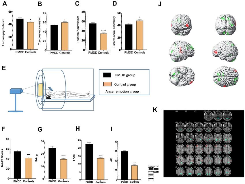
Figure 3. Comparisons of emotional changes and brain reactivity to emotional stimuli among PMDD and control study
participants (PMDD=19, Controls=21) of the depression-induced group. (A–D) Extraversion, psychoticism, neuroticism, and social
desirability T-scores. (E) Schematic presentation of participants in the experiment. (F) TAS-20 scores for both groups. (G) Depression
emotions of participants in the depression-induced group. (H) Illustration of activations in various brain areas: the middle frontal gyrus,
middle gyrus and cingulate gyrus for PMDD and HC groups. (I) PMDD and HC groups Depression mood Subtract Neutral Frontal Mid-Back
Picture Condition T-test activation Differential area; p < 0.05, cluster size > 389 warm (T value positive) represents PMDD group higher than
the HC group, cool (T negative) on behalf of the normal group than the patient group.
www.aging-us.com 19535 AGINGdifferent personalities lead to different PMS symptoms patients was significantly enhanced when compared to
and coping styles [27]. A recent study evaluating that of healthy controls [21, 39].
recurrent depression in recurrent brief depression
(RBD) revealed that there were significant differences The cerebellum processes a wide range of behavioral
in the distribution of neuroticism (N) and extraversion effects, including pain, emotional, and administrative
(E) scores between patients and controls. Anxiety functions [40, 41]. In the luteal phase, PMS patients
groups exhibited high N scores while RBD patients show increased cerebellar activation, especially in the
were found to have low E scores [28]. Most women cerebellar vermis. Enhanced cerebellar activity is
with menstrual disorders are characterized by associated with emotional deterioration [42, 43]. These
neuroticism, which can affect their quality of life [29]. findings imply that the cerebellar nucleus, which is
Neurotic women are less vulnerable to negative associated with other mood disorders, may also be
emotions during their mid-late luteal phase than during involved in PMDD patients. The involved primary
other phases. Sensitive responses of neurotic women to regions are the top of the cerebellum midline and
fluctuations in progesterone levels during menstrual cerebellar vermis [44, 45]. We found that brain regions
cycles may be among the reasons accounting for their presented in the experiment were more specific than in
mood changes [30]. previous studies, further demonstrating that the
occurrence of PMDD is associated with the cerebellum.
According to Hans Eysenck’s theory, personality traits
have a close relationship with brain activity [31]. In The frontal lobe is located in the front of the brain and it
this study, we found that compared to healthy controls, includes four main gyri. The frontal lobe is the most
after depression induction, brain activities were developmentally advanced brain structure, possessing
enhanced in the middle frontal gyrus, middle gyrus and different emotional processing functions. The frontal
in the cingulate gyrus of PMDD women. Moreover, cortex edge affects individual decision making and
after anger induction, PMDD women exhibited emotional regulation [33]. It also affects almost all
increased activations mainly in the middle frontal gyrus psychological functions to which emotional control of an
(BA10), temporal lobe (BA42), left cerebellum individual are inextricably linked. As an important part
(BA37), as well as decreased activations in the of brain emotional control, the cingulate gyrus may also
precuneus (BA7), superior frontal gyrus (BA8), lobulus play a key regulatory role in individual cognitive
paracentralis (BA6), and right cerebellum (BA48). functions, emotional regulation and so on. The anterior
Furthermore, our findings indicate that brain function cingulate gyrus also plays an important role in
abnormalities occur in patients before menstruation, measuring both external and self-expected matches [46].
which affects their emotional capacities as well as When the cingulate gyrus is damaged, human
cognitive abilities. Our findings are consistent with implementation of cognitive, emotional and other brain
those of previous studies, the only difference is that functions is dysregulated, which triggers individual
previous studies did not perform in-depth research on indifference, attention disorders, autonomic dysfunction,
specific PMDD emotions [10, 32]. emotional instability and other clinical symptoms [47].
Abnormal changes in the cingulate gyrus are associated
Clinical and neurological studies have not with individual mental anxiety [48, 49]. Comasco et al.
conclusively determined whether the frontal lobe plays [50] found out anterior cingulate cortex activation by
a key role in emotional processing. Some studies have emotional stimulation in PMDD patients.
reported that frontal lobe lesions can lead to changes in
patient moods [33, 34]. In particular, associative fibers Strengths and limitations
in the frontal part of the frontal lobe are closely
associated with mental activities [35, 36]. This series This study has some strengths, for instance, the sample
of direct or indirect neural connections are the size was prospectively determined and was bigger than
anatomical bases for the regulation of physiological that of existing published studies, which evaluated brain
and psychological functions of the prefrontal cortex activity in women with PMDD [18, 51, 52]. Our
[37]. For PMDD patients during the luteal phase, findings inform on treatment avenues for PMDD. A
emotional responses to negative emotion pictures were major limitation of this study was that study participants
significantly reduced. This indicates that positive were obtained from universities and lacked women of
emotional adjustments for patients were weakened other ages, which limits the application of our findings.
while negative emotional regulations were increased
during the premenstrual period [38]. Our results are CONCLUSIONS
consistent with those of Gingnell and Baller who used
PET or fMRI to investigate CNS activity and Neuroticism and psychoticism are susceptible traits of
confirmed that prefrontal cortex reactivity in PMDD PMDD patients and are associated with brain reactivity
www.aging-us.com 19536 AGINGto emotional stimuli. Upon exposure to depressive PMID:24345853
stimuli, we found increased functions of the middle
4. Yamada K, Kamagata E. Reduction of quality-adjusted
frontal gyrus, the middle gyrus and the cingulate gyrus life years (QALYs) in patients with premenstrual
in PMDD women. Upon exposure to anger stimuli, the dysphoric disorder (PMDD). Qual Life Res. 2017;
frontal lobe (especially the upper, middle, and central
26:3069–73.
lobule), parietal lobes (mainly the precuneus), temporal
https://doi.org/10.1007/s11136-017-1642-1
lobe, and cerebellum (mainly the left and right
PMID:28674766
cerebellum) were activated in PMDD women. More
studies using larger samples are needed to confirm our 5. Sabourin-Guardo E, Gamache D, Dubois-Comtois K.
findings and to identify neural circuit mechanisms to Premenstrual dysphoric disorder: Mental disorder or
emotion regulation. adjustment difficulty? Annales Medico-Psychologiques.
2020; 178:283–89.
AUTHOR CONTRIBUTIONS https://doi.org/10.1016/j.amp.2018.12.015
6. Ko CH, Long CY, Chen SY, Chen IJ, Huang TH, Yen JY.
Methodology (performing the experiments), Mingzhou Depression, irritability, and anxiety in women with
Gao, Li An, Guangbin Wang, Jieqiong Wang, premenstrual dysphoric disorder. Int J Psychiatry Med.
Chunhong Song, Fengqin Wei, Tao Gong and Yanhong 2013; 46:39–55.
Yu. data curation, Mingzhou Gao and Tao Gong; https://doi.org/10.2190/PM.46.1.d PMID:24547609
writing-original draft preparation, Mingzhou Gao;
writing—review and editing, Mingzhou Gao and 7. Gross JJ. The Emerging Field of Emotion Regulation: An
Dongmei Gao; supervision, Mingqi Qiao and Dongmei Integrative Review. Review of General Psychology.
Gao; funding acquisition, Dongmei Gao. All authors 1998; 2:271–99.
have read and agreed to the published version of the https://doi.org/10.1037/1089-2680.2.3.271
manuscript. 8. Hantsoo L, Epperson CN. Allopregnanolone in
premenstrual dysphoric disorder (PMDD): Evidence for
ACKNOWLEDGMENTS dysregulated sensitivity to GABA-A receptor
modulating neuroactive steroids across the menstrual
Our gratitude to all public health agencies that cycle. Neurobiol Stress. 2020; 12:100213.
collaborated in this study. https://doi.org/10.1016/j.ynstr.2020.100213
PMID:32435664
CONFLICTS OF INTEREST
9. Duan G, Liu H, Pang Y, Liu P, Liu Y, Wang G, Liao H,
The authors declare that they have no conflicts of Tang L, Chen W, Mo X, Wen D, Lin H, Deng D.
interest. Hippocampal fractional amplitude of low-frequency
fluctuation and functional connectivity changes in
FUNDING premenstrual syndrome. J Magn Reson Imaging. 2018;
47:545–53.
This study was financially supported by the National https://doi.org/10.1002/jmri.25775
Science and Technology Major Project "Key New Drug PMID:28577332
Creation and Manufacturing Program" of China 10. Liao H, Duan G, Liu P, Liu Y, Pang Y, Liu H, Tang L, Tao J,
[SQ2017ZX091064] and the National Natural Science Wen D, Li S, Liang L, Deng D. Altered fractional
Foundation of China [81001484; 81473558]. amplitude of low frequency fluctuation in
premenstrual syndrome: A resting state fMRI study. J
REFERENCES Affect Disord. 2017; 218:41–48.
https://doi.org/10.1016/j.jad.2017.04.045
1. Leminen H. Paavonen J. Duodecim. 2013; 129: PMID:28458114
1756–63.
11. Liu Q, Li R, Zhou R, Li J, Gu Q. Abnormal Resting-State
2. American Psychiatric Association. DSM-5: Diagnostic Connectivity at Functional MRI in Women with
and Statistical Manual of Mental Disorders, 5th Edition. Premenstrual Syndrome. PLoS One. 2015;
American Psychiatric Publishing. Arlington. 2013. 10:e0136029.
3. Hartlage SA, Breaux CA, Yonkers KA. Addressing https://doi.org/10.1371/journal.pone.0136029
concerns about the inclusion of premenstrual PMID:26325510
dysphoric disorder in DSM-5. J Clin Psychiatry. 2014; 12. Epperson CN. Premenstrual dysphoric disorder and the
75:70–76. brain. Am J Psychiatry. 2013; 170:248–52.
https://doi.org/10.4088/JCP.13cs08368
www.aging-us.com 19537 AGINGhttps://doi.org/10.1176/appi.ajp.2012.12121555 22. Lang PJ, Bradley MM, Cuthbert BN. International
PMID:23450284 Affective Picture System (IAPS): Technical Manual
13. Fox MD, Raichle ME. Spontaneous fluctuations in brain and Affective Ratings. Center for Research in
activity observed with functional magnetic resonance Psychophysiology University of Florida. 1997.
imaging. Nat Rev Neurosci. 2007; 8:700–11. 23. Friston KJ. Statistical parametric maps in functional
https://doi.org/10.1038/nrn2201 PMID:17704812 imaging: A general linear approach. Human Brain
14. Heeger DJ, Ress D. What does fMRI tell us about Mapping. 1994; 2:189–210.
neuronal activity? Nat Rev Neurosci. 2002; 3:142–51. https://doi.org/10.1002/hbm.460020402
https://doi.org/10.1038/nrn730 24. Eysenck H, Eysenck S. Manual of the Eysenck
PMID:11836522 personality questionnaire. San Diego, Calif.:
15. Jeong HG, Ham BJ, Yeo HB, Jung IK, Joe SH. Gray EdITS/Educational and Industrial Testing Service, 1994.
matter abnormalities in patients with premenstrual 25. Hallman J, Oreland L, Edman G, Schalling D.
dysphoric disorder: an optimized voxel-based Thrombocyte monoamine oxidase activity and
morphometry. J Affect Disord. 2012; 140:260–67. personality traits in women with severe
https://doi.org/10.1016/j.jad.2012.02.010 premenstrual syndrome. Acta Psychiatr Scand.
PMID:22381950 1987; 76:225–34.
16. Protopopescu X, Tuescher O, Pan H, Epstein J, Root J, https://doi.org/10.1111/j.1600-0447.1987.tb02890.x
Chang L, Altemus M, Polanecsky M, McEwen B, Stern PMID:3673649
E, Silbersweig D. Toward a functional neuroanatomy of 26. Cooper C, Taylor R. Personality and performance on a
premenstrual dysphoric disorder. J Affect Disord. 2008; frustrating cognitive task. Percept Mot Skills. 1999;
108:87–94. 88:1384.
https://doi.org/10.1016/j.jad.2007.09.015 https://doi.org/10.2466/pms.1999.88.3c.1384
PMID:18031826 PMID:10485127
17. Toffoletto S, Lanzenberger R, Gingnell M, Sundström- 27. Zhang Y, Ruifang A, Tang Z, Jing L, Haimiao Z.
Poromaa I, Comasco E. Emotional and cognitive Relationships between premenstrual syndrome
functional imaging of estrogen and progesterone symptoms and coping styles in female college students
effects in the female human brain: a systematic with different personalities. Zhongguo Shiyong Fuke Yu
review. Psychoneuroendocrinology. 2014; 50:28–52. Chanke Zazhi. 2013; 029:62–65.
https://doi.org/10.1016/j.psyneuen.2014.07.025
PMID:25222701 28. Williams WR, Richards JP, Ameen JR, Davies J.
Recurrent brief depression and personality traits in
18. Petersen N, Ghahremani DG, Rapkin AJ, Berman SM, allergy, anxiety and premenstrual syndrome patients: a
Liang L, London ED. Brain activation during emotion general practice survey. Med Sci Monit. 2007;
regulation in women with premenstrual dysphoric 13:CR118–24.
disorder. Psychol Med. 2018; 48:1795–802. PMID:17325634
https://doi.org/10.1017/S0033291717003270
PMID:29145910 29. Liu G, Yao Q, Zhan D, Sheng H, Qingxiong Y, Shaohong L.
Correlation between the life quality and personality for
19. Ghanizadeh A. Agreement between Diagnostic and patients with menstrual disorder. Hainan Yixueyuan
Statistical Manual of Mental Disorders, Fourth Edition,
Xuebao. 2011; 17:1254–56.
and the proposed DSM-V attention deficit
hyperactivity disorder diagnostic criteria: an 30. Renlai Z. Effects of Menstrual Cycle and Neuroticism on
exploratory study. Compr Psychiatry. 2013; 54:7–10. Emotional Responses of Healthy Women. Acta
https://doi.org/10.1016/j.comppsych.2012.06.001 Psychologica Sinica. 2014; 45:1–11.
PMID:22809622 31. Tran Y, Craig A, Boord P, Connell K, Cooper N, Gordon
20. Yaoxiao G. Eysenck Personality Questionnaire Revised E. Personality traits and its association with resting
in China. Psychological Science. 1984. regional brain activity. Int J Psychophysiol. 2006;
https://doi.org/10.1037/t12641-000 60:215–24.
https://doi.org/10.1016/j.ijpsycho.2005.05.008
21. Bao AM, Ji YF, Van Someren EJ, Hofman MA, Liu RY,
PMID:16019096
Zhou JN. Diurnal rhythms of free estradiol and cortisol
during the normal menstrual cycle in women with 32. Liao H, Pang Y, Liu P, Liu H, Duan G, Liu Y, Tang L, Tao J,
major depression. Horm Behav. 2004; 45:93–102. Wen D, Li S, Liang L, Deng D. Abnormal Spontaneous
https://doi.org/10.1016/j.yhbeh.2003.09.004 Brain Activity in Women with Premenstrual Syndrome
PMID:15019795 Revealed by Regional Homogeneity. Front Hum
www.aging-us.com 19538 AGINGNeurosci. 2017; 11:62. PMID:17786822
https://doi.org/10.3389/fnhum.2017.00062 41. Strick PL, Dum RP, Fiez JA. Cerebellum and nonmotor
PMID:28243196 function. Annu Rev Neurosci. 2009; 32:413–34.
33. Berridge KC, Kringelbach ML. Neuroscience of affect: https://doi.org/10.1146/annurev.neuro.31.060407.125
brain mechanisms of pleasure and displeasure. Curr 606 PMID:19555291
Opin Neurobiol. 2013; 23:294–303. 42. Rapkin AJ, Berman SM, Mandelkern MA, Silverman DH,
https://doi.org/10.1016/j.conb.2013.01.017 Morgan M, London ED. Neuroimaging evidence of
PMID:23375169 cerebellar involvement in premenstrual dysphoric
34. Li W, Qin W, Liu H, Fan L, Wang J, Jiang T, Yu C. disorder. Biol Psychiatry. 2011; 69:374–80.
Subregions of the human superior frontal gyrus and https://doi.org/10.1016/j.biopsych.2010.09.029
their connections. Neuroimage. 2013; 78:46–58. PMID:21092938
https://doi.org/10.1016/j.neuroimage.2013.04.011 43. Stoodley CJ, Valera EM, Schmahmann JD. Functional
PMID:23587692 topography of the cerebellum for motor and cognitive
35. De Bondt T, De Belder F, Vanhevel F, Jacquemyn Y, tasks: an fMRI study. Neuroimage. 2012; 59:1560–70.
Parizel PM. Prefrontal GABA concentration changes in https://doi.org/10.1016/j.neuroimage.2011.08.065
women-Influence of menstrual cycle phase, hormonal PMID:21907811
contraceptive use, and correlation with premenstrual
44. Kimbrell TA, Ketter TA, George MS, Little JT, Benson BE,
symptoms. Brain Res. 2015; 1597:129–38.
Willis MW, Herscovitch P, Post RM. Regional cerebral
https://doi.org/10.1016/j.brainres.2014.11.051
glucose utilization in patients with a range of severities
PMID:25481417
of unipolar depression. Biol Psychiatry. 2002;
36. Gusnard DA, Akbudak E, Shulman GL, Raichle ME. 51:237–52.
Medial prefrontal cortex and self-referential mental https://doi.org/10.1016/s0006-3223(01)01216-1
activity: relation to a default mode of brain function. PMID:11839367
Proc Natl Acad Sci USA. 2001; 98:4259–64.
https://doi.org/10.1073/pnas.071043098 45. Ketter TA, Kimbrell TA, George MS, Dunn RT, Speer
PMID:11259662 AM, Benson BE, Willis MW, Danielson A, Frye MA,
Herscovitch P, Post RM. Effects of mood
37. Qing W, Keyong W, Zhihua Z, Liangjun P, Yongmei W, and subtype on cerebral glucose metabolism
Bao H, Wangfa L. Study on prospective memory of in treatment-resistant bipolar disorder. Biol
male alcohol dependent patients with withdrawal. Psychiatry. 2001; 49:97–109.
Chinese Journal of Behavioral Medicine and Brain https://doi.org/10.1016/s0006-3223(00)00975-6
Science. 2013; 22:134–36. PMID:11164756
https://doi.org/10.3760/cma.j.issn.1674-
6554.2013.02.012 46. Yan Li YZ. Correlation between resting brain activity
and anxiety symptoms in patients with depression.
38. Batra NA, Seres-Mailo J, Hanstock C, Seres P, Khudabux Chinese Journal of Behavioral Medicine and Brain
J, Bellavance F, Baker G, Allen P, Tibbo P, Hui E, Le Science. 2012; 21:988–90.
Melledo JM. Proton magnetic resonance spectroscopy
measurement of brain glutamate levels in 47. Bush G, Luu P, Posner MI. Cognitive and emotional
premenstrual dysphoric disorder. Biol Psychiatry. 2008; influences in anterior cingulate cortex. Trends Cogn Sci.
63:1178–84. 2000; 4:215–22.
https://doi.org/10.1016/j.biopsych.2007.10.007 https://doi.org/10.1016/S1364-6613(00)01483-2
PMID:18061146 PMID:10827444
39. Gingnell M, Bannbers E, Wikström J, Fredrikson M, 48. Jiayu G, Liwen T, Deqing Z, Jie W, Wei Z. Research on
Sundström-Poromaa I. Premenstrual dysphoric metabolic characteristics of brain cuff in patients with
disorder and prefrontal reactivity during anticipation of generalized anxiety disorder. Chinese Journal of Clinical
emotional stimuli. Eur Neuropsychopharmacol. 2013; Psychology. 2015; 23:60–62.
23:1474–83. https://doi.org/10.16128/j.cnki.1005-
https://doi.org/10.1016/j.euroneuro.2013.08.002 3611.2015.01.013
PMID:24001875 49. Ying X, Yang Y. Chinese attachment type in outpatient
40. Schmahmann JD, Weilburg JB, Sherman JC. The anxiety disorder. Chin J Clin Psychol. 2013; 21:81–84.
neuropsychiatry of the cerebellum - insights from the
50. Comasco E, Hahn A, Ganger S, Gingnell M, Bannbers E,
clinic. Cerebellum. 2007; 6:254–67.
Oreland L, Wikström J, Epperson CN, Lanzenberger R,
https://doi.org/10.1080/14734220701490995
Sundström-Poromaa I. Emotional fronto-cingulate
www.aging-us.com 19539 AGINGcortex activation and brain derived neurotrophic factor https://doi.org/10.1016/j.jad.2018.08.033
polymorphism in premenstrual dysphoric disorder. PMID:30195172
Hum Brain Mapp. 2014; 35:4450–58.
52. Petersen N, Ghahremani DG, Rapkin AJ, Berman SM,
https://doi.org/10.1002/hbm.22486 PMID:24615932
Wijker N, Liang L, London ED. Resting-state functional
51. Flores-Ramos M, Alcauter S, López-Titla M, Bernal- connectivity in women with PMDD. Transl Psychiatry.
Santamaría N, Calva-Coraza E, Edden RA. Testosterone 2019; 9:339.
is related to GABA+ levels in the posterior-cingulate in https://doi.org/10.1038/s41398-019-0670-8
unmedicated depressed women during reproductive PMID:31827073
life. J Affect Disord. 2019; 242:143–49.
www.aging-us.com 19540 AGINGSUPPLEMENTARY MATERIALS
Supplementary Tables
Supplementary Table 1. PMDD group and HC group depression
mood subtract neutral frontal mid-back picture condition t-test
activation differential area.
MNI coordinate
Central position Voxel t BA
X Y Z
the middle frontal gyrus -30 48 -12 588 3.321 11
middle gyrus 39 42 -15 957 3.963 47
cingulate gyrus -6 -24 30 1072 4.154 23
Note: PMDD, premenstrual dysphoric disorder; HC, healthy control;
MNI, Montreal Neurological Institute; BA, Brodmann area.
Supplementary Table 2. PMDD group and HC group anger
mood subtract neutral frontal mid-back picture condition t-test
activation differential area.
MNI coordinates
Brain region Voxel t BA
X Y Z
Superior frontal gyrus 24 42 51 225 −3.4197 8
Middle frontal gyrus −42 45 0 188 2.8693 10
paracentral lobule −6 −33 60 142 −3.1293 6
Precuneus −9 −57 45 730 −3.6001 7
Temporal lobe 72 −30 6 138 2.4814 42
Right cerebellum 24 −39 −21 185 −2.8195 48
Left cerebellum −21 −57 −18 200 2.9857 37
Note: PMDD, premenstrual dysphoric disorder; HC, healthy control;
MNI, Montreal Neurological Institute; BA, Brodmann area.
www.aging-us.com 19541 AGINGYou can also read