Comparative analysis of morphological and molecular approaches integrated into the study of the dinoflagellate biodiversity within the recently ...
←
→
Page content transcription
If your browser does not render page correctly, please read the page content below
Biodiversity Data Journal 8: e55172
              doi: 10.3897/BDJ.8.e55172

                                                     Research Article

Comparative analysis of morphological and
molecular approaches integrated into the study of
the dinoflagellate biodiversity within the recently
deposited Black Sea sediments – benefits and
drawbacks
Nina Dzhembekova‡, Fernando Rubino§, Satoshi Nagai|, Ivelina Zlateva‡, Nataliya Slabakova‡, Petya
Ivanova‡, Violeta Slabakova¶, Snejana Moncheva‡
    ‡ Institute of Oceanology “Fridtjof Nansen”, Marine Biology and Ecology Department, Bulgarian Academy of Sciences,
    Varna, Bulgaria
    § Water Research Institute, Unit Talassografico “A. Cerruti”, National Research Council CNR-IRSA, Taranto, Italy
    | National Research Institute of Fisheries Science, Research Center for Aquatic Genomics, Fisheries Research and Education
    Agency, Yokohama Kanagawa, Japan
    ¶ Institute of Oceanology “Fridtjof Nansen”, Ocean Technologies Department, Bulgarian Academy of Sciences,
    Varna, Bulgaria

   Corresponding author: Snejana Moncheva (snejanam@abv.bg)

   Academic editor: Anne Thessen

   Received: 05 Jun 2020 | Accepted: 27 Jul 2020 | Published: 18 Aug 2020

   Citation: Dzhembekova N, Rubino F, Nagai S, Zlateva I, Slabakova N, Ivanova P, Slabakova V, Moncheva S
   (2020) Comparative analysis of morphological and molecular approaches integrated into the study of the
   dinoflagellate biodiversity within the recently deposited Black Sea sediments – benefits and drawbacks.
   Biodiversity Data Journal 8: e55172. https://doi.org/10.3897/BDJ.8.e55172

Abstract
One of the assets, assigned to the phytoplankton resting stages, is that of serving as the
“memory” of the aquatic ecosystems and preserved biodiversity in the course of time.
However, an accurate cyst identification proves to be a more difficult and extremely
challenging process, even today. In order to gain a better taxonomic coverage of cyst
assemblages in the Black Sea, an integrated approach of the classical morphological
identification with metabarcoding methods (MySeq sequencing of V7-V9 regions of the 18S
rDNA) was applied on thirteen surface sediment samples collected from different sites. A

© Dzhembekova N et al. This is an open access article distributed under the terms of the Creative Commons Attribution License
(CC BY 4.0), which permits unrestricted use, distribution, and reproduction in any medium, provided the original author and source
are credited.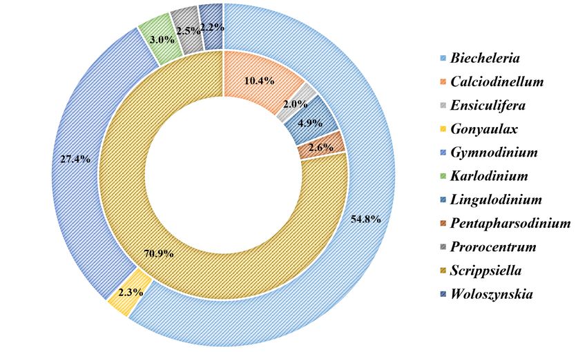
2                                   Dzhembekova N et al

total number of 112 dinoflagellate taxa was detected at the species level and ascribed to
51 genera. In general, it is the molecular analysis that yields a higher number of taxa as
compared to those obtained through the morphological taxonomy (66 taxa based on the
DNA sequences versus 56 morphologically-identified taxa). Besides, it should be pointed
out that the integrated dataset includes 14 potentially toxic dinoflagellate species.
Discerned, subsequently, was a good dataset consistency for ten species, followed by
some discrepancies as to a number of taxa, identified with one of the methods only, due to
specific methodological biases. On the whole, it could be concluded that the combination of
morphological and molecular methods is likely to increase the potential for a more reliable
taxonomic assessment of phytoplankton diversity in marine sediments which, in turn,
proves conclusively the utmost importance of the integrated approach.

Keywords
Black Sea, phytoplankton, cyst, morphology, metabarcoding

Introduction
Biodiversity of phytoplankton as key primary producers is of utmost importance for the
state and activity of the marine ecosystems (Ptacnik et al. 2008) and the precise and
accurate information about the species diversity is fundamental for the proper
understanding of their functioning (McCann 2000, Strong et al. 2015). The production of
dormant resting stages is a life-cycle trait common to many planktonic taxa (see Belmonte
and Rubino 2019 for a review on this topic). Resting cysts accumulated in sediments will
remain viable for years up to a century (Lundholm et al. 2011, Ribeiro et al. 2011), serving
as potential seed banks and maintaining biodiversity over time (Kremp et al. 2015). At the
same time, the occurrence of phytoplankton species in the water column is often
discontinuous, with a vast number of taxa, some of them represented in a very small
concentration, others growing in high abundance (sometimes forming blooms) for a very
short period, followed by a marked decrease or even complete disappearance from the
plankton community (Rubino et al. 1998, Moscatello et al. 2004). As a result, some species
cannot be detected in routine monitoring programmes as viable cells, but are much more
readily identified in the sediment at dormant stage only (Bravo et al. 2006, Rubino et al.
2010b, Salgado et al. 2011). Moreover, some of the resting cysts are produced by species
causing harmful algal bloom (HAB) and play an important role in bloom initiation,
representing a “dormant threat” for the ecosystem (Anderson et al. 2003, Garces 2004).
Thus, the survey of cyst assemblages in recent sediments is a suitable tool, not only for
studying phytoplankton biodiversity, but also for detecting harmful microalgae and species
not previously reported in a given area (Rubino et al. 2010a).

Dinoflagellate cysts play a decisive role in the very functioning of marine coastal plankton,
both from a biological and ecological point of view (Dale 1983) and information about their
density and distribution add value to our knowledge of phytoplankton biodiversity (Bravo et
al. 2006, Rubino et al. 2010a), improving our understanding of dinoflagellate population
Comparative analysis of morphological and molecular approaches integrated ...    3

dynamics (Natsuike et al. 2013, Anderson et al. 2014) and predicting possible HAB events
(Tang and Gobler 2012, Satta et al. 2013). Furthermore, having reliable data on the resting
stage diversity is an essential prerequisite for accurate species identification. The existing
species inventories of delineated dinoflagellate cysts are mostly based on morphological
characteristics (Matsuoka and Fukuyo 2000) and it is precisely this classical approach that
has been successfully applied in the Black Sea species inventory (Rubino et al. 2010b,
Aydin et al. 2015, Mudie et al. 2017), regardless of the fact that there are some cases in
which such an approach could lead to an ambiguous identification. In reality, however,
many cyst morphotypes produced by Gonyaulacales, Gymnodiniales or Peridiniales
species share the same “simple” subspherical shape with no evidence of any processes
and are characteried by the following common distinctive features - the size, the presence
of mucous or the outline of the archeopyle, when visible. This is precisely the reason why
the recent molecular methods have appeared to be gaining in popularity when studying
microbial diversity in sediment samples, unlocking their potential for more sensitive
detection and exact taxonomical identification (Penna et al. 2010, Forster et al. 2016).
Besides, the morphological analysis applied together with the next-generation sequencing
(NGS) tool is likely to generate greater discrimination power giving a better insight into the
resting stage communities in a given environment (Piredda et al. 2017, Jung et al. 2018).
The classical morphological method proves to be particularly effective in estimating the
abundance of different morphotypes, whereas the obtained environmental high-throughput
sequence data are considered appropriate for a better assessment and re-evaluation of the
taxon composition of resting propagules in the benthos.

The present study, taking a more integrated approach - i.e. combining next-generation
sequencing of V7-9 hypervariable regions of the 18S rDNA with morphological observation,
focuses exclusively on the dinoflagellate diversity and distribution in the Black Sea recent
sediments as modern cyst assemblages. It is worth noting that the same NGS dataset has
been previously used for assessment of overall phytoplankton composition in the Black
Sea floor and which revealed a rich diversity of species not reported earlier in Black Sea
sediments (Dzhembekova et al. 2018). Advanced in the study is a detailed comparative
analysis of the dinoflagellate cyst diversity giving, thus, due prominence to the strengths
and weaknesses of the two methods employed.

Material and methods

Sampling

Surface sediment samples were collected using a multicorer (the top 0–5 cm of the core)
or Van-Veen Grab sampler, by a 10 x 10 cm frame at 13 stations located in different areas
across the Black Sea during the period from May to June 2016 (Fig. 1, Table 1). All the
samples were stored in a refrigerator at 8°C and kept in dark conditions in order to ensure
only the survival of the resting stages in the sediments. The samples were well
homogenised and split into two equal subsamples, each of which to be used for
subsequent molecular and morphological analyses, respectively.
4                                        Dzhembekova N et al

    Table 1.
    Overview of samplings in the Black Sea

Date of sampling      Sampling station   Latitude N /Longitude E   Station depth   Sampling device

17.5.2016             1                  46°12,098'/30°49,649′     28.6            multicorer

18.5.2016             2                  45°30,969′/29°51,728'     20.4            multicorer

19.5.2016             3                  45°11,999′/29°48,616'     20.5            multicorer

19.5.2016             4                  45°18,676'/29°51,200′     22.0            multicorer

21.5.2016             5                  46°30,535′/30°49,432′     13.1            Van-Veen grab

21.5.2016             6                  46°19,474'/31°27,999′     16.0            multicorer

24.5.2016             7                  43°22,049'/31°49,994′     1933.0          multicorer

26.5.2016             8                  42°14,070′/39°53,161'     1904.0          multicorer

29.5.2016             9                  42°07,359′/41°36,987'     37.0            Van-Veen grab

30.5.2016             10                 41°54,259′/41°44,948′     42.0            Van-Veen grab

30.5.2016             11                 41°45,763′/41°42,883′     63.0            Van-Veen grab

2.6.2016              12                 43°31,558′/36°04,183'     2100.0          multicorer

3.6.2016              13                 44°38,163′/31°23,298′     391.0           multicorer

    Figure 1.
    Map of sampling stations in the Black Sea (●coastal CO, ■shelf SH, ▲open sea OS)

Morphological identification

An aliquot of homogenied sediment (from 2.00 to 2.20 cm3) was selected from each
subsample for the cyst analyses while another one (≈ 10 cm3) was concurrently oven-dried
for one night at 70°C to calculate the water content. The wet aliquots were weighed and
screened through a 10 µm mesh (Endecotts Limited steel sieves, ISO3310-1, London,
England) using filtered natural (0.45 µm) seawater (Montresor et al. 2010). The material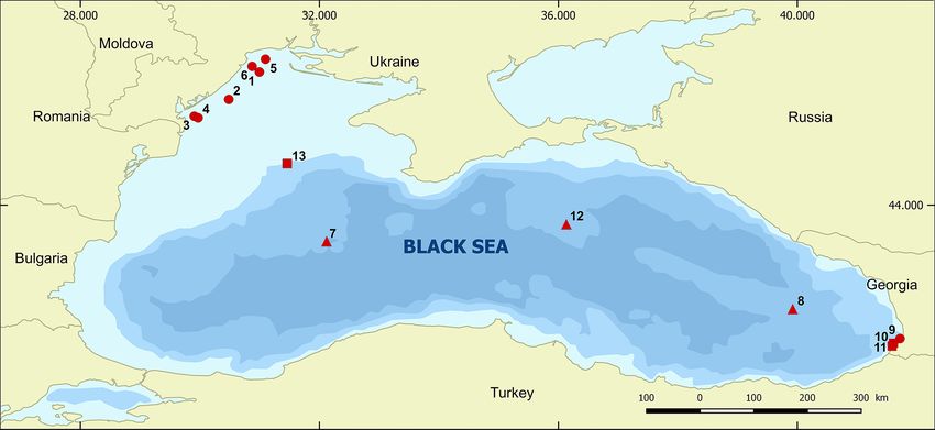
Comparative analysis of morphological and molecular approaches integrated ...   5

retained on to the sieve was then subjected to ultrasound for 1 min at low frequency and
screened again through a sieve battery (200, 75, 20 and 10 µm mesh sizes). Two fine-
grained fractions (10-20 µm and 20-75 µm), containing mainly protistan cysts, were
obtained along with a > 75 µm fraction containing larger dinoflagellate cysts (e.g.
Lingulodinium and Polykrikos) interspersed with zooplankton resting eggs. The material
retained on to the 200 µm mesh was discarded. No chemicals were used to dissolve the
sediment particles in order to preserve the calcareous cyst walls.

A quali-quantitative analysis for cysts taxonomic identification was conducted under an
inverted microscope (Zeiss Axiovert 200M), equipped with a Leica MC17o HD digital
camera at 320-400x magnification. Next determined was the number of both the full (i.e.
with cytoplasmic content) and the empty (i.e. already germinated) cysts. As for the 10-20
µm and 20-75 µm fractions, at least 200 full cysts were counted in an attempt to obtain
such values of abundance that are as homogeneous as possible and as a means to
evaluate the rare species as well. From this perspective, a detailed and thorough analysis
was performed on the > 75 µm fractions, while all the resting stage morphotypes were
identified on the basis of published descriptions and the Modern Dinocyst Key Website
(https://www.marum.de/en/Modern_Dinocyst_Key.html). The identification was performed
at the species level whenever possible and, as a rule, the modern taxonomic appellation
was applied. For those taxa whose active stages are not determined, the paleontological
nomenclature was adopted. Quantitative data are reported as cysts x g-1 of dry sediment
(cysts g-1).

DNA extraction, PCR amplification, sequencing and bioinformatics

Total DNA was extracted from 0.5 g of surface sediment samples (in triplicate for each
sampling location) using the ISOIL DNA extraction kit (NIPPON GENE, Tokyo, Japan). All
DNA samples were used as templates for PCR amplification of the V7–9 hypervariable
regions of the 18S-rRNA gene (amplicon length ~484 bp) using universal primers SSR-
F1289-sn and F: TGGAGYGATHTGTCTGGTTDATTCCG; SSR-R1772-sn, R:
TCACCTACGGAWACCTTGTTACG (modified from Tanabe et al. 2015). The massively
parallel paired-end sequencing workflow was derived from the document 16S
metagenomics sequencing library preparation: preparing 16S ribosomal gene amplicons
for the Illumina MiSeq system distributed by Illumina (part no. 15044223 Rev. B). Two-step
PCR for the construction of paired-end libraries and HTS on Illumina Miseq 300 PE
platform (Illumina, USA) followed the protocols described in Dzhembekova et al. (2017). All
the procedures and techniques, applicable to the treatment of the obtained sequences,
selection and taxonomic identification of operational taxonomic units (OTUs), were
administered according to the workflow described by Dzhembekova et al. (2017). When
considering the taxonomic identification, a reference similarity threshold ≥ 98% was found
appropriate.

DNA sequence dataset for this study can be found in the DDBJ Sequence Read Archive
under access no. DRA009586.
6                                    Dzhembekova N et al

Statistical analyses

As similarity between morphological and molecular datasets is only possible for species
identified by both methods, the two datasets were analysed under certain initial conditions:
first, the datasets were aggregated and further referred to as coastal, shelf and open sea
sampling sites (habitats) and, next, only species that were present at all sites were
selected and viewed as “shared”. The shared species datasets comprised between 17 and
57% of the species numbers per station identified by the morphological method and
between 12 and 32% of those specified by the molecular approach. The percentage
fraction of each “shared” species in the total cyst abundance was calculated and
expressed in numerical value per habitat.

Euclidean distances analysis was used for assessment of similarity between shared
species datasets, by abundance. The data were normalised in the interval [0,1] and the
resultant euclidean distances were further converted to similarity scores. The threshold
similarity score value was set to 0.75 (SimScoreTreshold = 0.75) (Malone 2017).

Results

Morphological identification and enumeration of dinoflagellate cysts

From the total amount of 56 taxa, 51 were morphologically differentiated as full cysts and
33 as empty cysts, out of which 39 (almost 70%) were identified at the species level.
Considerable spatial variability in cysts abundance has been observed, ranging between
269 (st. 9) and 6963 (st. 1) cysts g-1 for the full dinoflagellate cysts and between 66 (st. 9)
and 5296 (st. 12) cysts g-1 for the empty dinocysts. On the whole, most species were
unevenly spread. Scrippsiella acuminata (Ehrenberg) Kretschmann, Elbrächter,
Zinssmeister, S.Soehner, Kirsch, Kusber & Gottschling, 2015 and Scrippsiella sp.
dominated by frequency (found at all stations) and abundance (accounting for more than
70% of the total cyst abundance). Amongst the identified resting stages, five were assigned
to potentially toxic dinoflagellates (Alexandrium minutum Halim, 1960, Gonyaulax spinifera
(Claparède & Lachmann) Diesing, 1866, Lingulodinium polyedra (F.Stein) J.D.Dodge,
1989, Polykrikos hartmannii W.M.Zimmermann, 1930 and Protoceratium reticulatum
(Claparède & Lachmann) Bütschli, 1885). Nonetheless, the cysts of these harmful
microalgae were too sporadic and occasional to be found in great abundance with the
exception of L. polyedra, identified at 11 out of 13 stations and measured in high densities
(up to 1722 full cysts g-1).

The majority of cysts belong to the predominant Black Sea-specific phytoplankton species,
except for those originally known as fossils only (Calciodinellum operosum
Deflandre, 1947 †, Calciperidinium asymmetricum Versteegh, 1993 †, Follisdinellum
splendidum Versteegh, 1993 †, Melodomuncula berlinensis Versteegh, 1993 †, Posoniella
tricarinelloides (G.Versteegh) Streng, Banasová, D.Reháková & H.Willems).
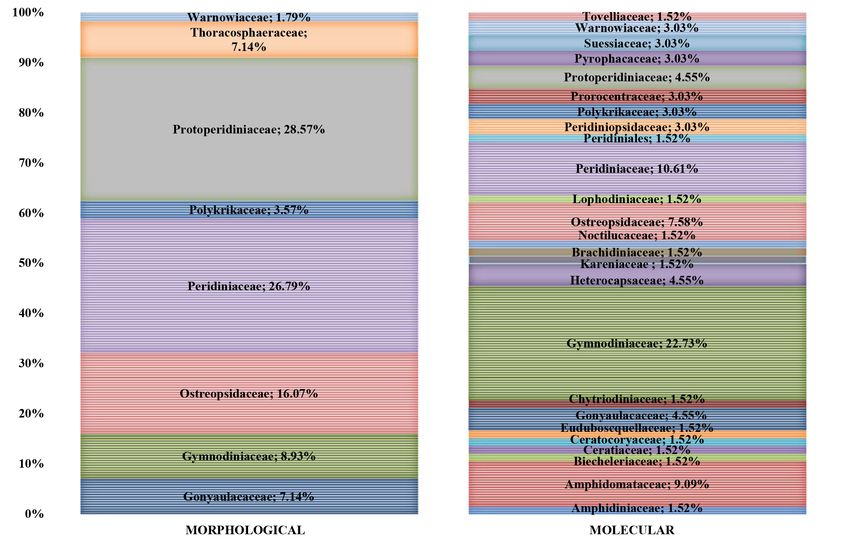
Comparative analysis of morphological and molecular approaches integrated ...     7

Dinoflagellate sediment diversity estimated on the basis of next-generation
sequencing

The total number of dinoflagellate sequences in the dataset was 904,816 representing
36.4% of all sequences obtained by massively parallel sequencing (MPS). The sequences
were clustered into 348 18S operational taxonomic units (OTUs) accounting for 15.5% of
all the obtained OTUs. More than half of the dinoflagellate sequences (60.4%) could be
assigned to references with high value of similarity varying from 100 to 98%. The number
of OTUs that satisfied the taxonomic assignment criteria (> 0.980 BLAST top hit similarity)
was 99 ranging by samples between 29 (st. 3) and 64 (st. 11). The number of sequences
also fluctuated between samples with a minimum value of 18,871 (st. 8) and maximum
approaching 87,693 (st. 7). More than 71% of OTUs (66 taxa) were determined at the
genus level and 58% were identified at the species level (56 species) (Suppl. material 1).
Only 7% of the OTUs, identified at the genus level, were assigned to reference sequences
deposited in GenBank as sp. aside from the 6%, due to a similarity with two different
species from the same genus. The remaining 29% of the OTUs in compliance with the
assignment criteria could not be identified at a lower taxonomic rank due to a similarity with
sequences deposited at a higher taxonomic level (6%) or through the same similarity with
species from different genera (23%). The vast majority of OTUs were unevenly distributed
amongst the stations, with a smaller number of species dominated by both sequence and
sample numbers (e.g. Biecheleria sp., Gymnodinium aureolum (E.M.Hulburt) Gert Hansen,
2000). It is worth noting that, amongst the most abundant OTUs found at all the stations,
some HAB species (Gymnodinium catenatum H.W.Graham, 1943 and Karlodinium
veneficum (D.Ballantine) J.Larsen, 2000) were also preserved in the records. The selected
OTUs, closely affiliated with potentially toxic dinoflagellates (Alexandrium andersonii
Balech, 1990 and Alexandrium pacificum R.W.Litaker, 2014), were represented with just a
few sequences and a single record, whereas the others, such as Gonyaulax spinifera,
Prorocentrum cordatum (Ostenfeld) J.D.Dodge, 1975 and Protoceratium reticulatum,
appeared more regularly with higher sequence numbers.

Integrated morphological and molecular data

The integrated morphological and molecular approach allowed for the detection of a total
number of 26 dinoflagellate taxa at the family level, despite the inconsistent taxonomic
composition between the morphologically- and metagenetically-derived datasets (Fig. 2).
Some families were found only in the NGS dataset represented by a few species (OTUs),
while the morphologically-identified fossil species in the family Thoracosphaeraceae
escaped the molecular analysis probably due to the lack of a reference database. The
highest taxonomic diversity was preserved in Peridiniaceae, followed by
Protoperidiniaceae for morphologically-identified cysts while easily traced in
Gymnodiniaceae and Peridiniaceae for molecular OTUs.
8 Dzhembekova N et al
Figure 2.
Family-level taxonomic compositions of dinoflagellates identified with the morphological and
molecular approach.
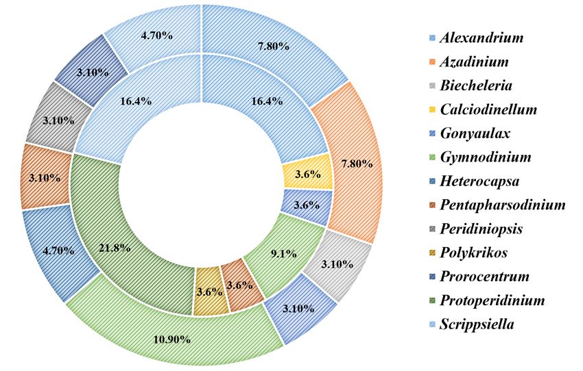
At the lower taxonomic level, 51 different genera were detected (21 retrieved by
morphological/LM analyses and 41 by the molecular/NGS approach). Most of them were
represented by a single species, each accounting for less than 3% of the total species
number and only 13 yielding a higher proportion. Alexandrium, Gonyaulax, Gymnodinium,
Pentapharsodinium and Scrippsiella were amongst the most diverse genera in both
datasets (Fig. 3). By abundance, Scrippsiella dominated in the morphological dataset while
Biecheleria and Gymnodinium were the taxa with the highest percentage in the molecular
dataset (Fig. 4). Some of the dominant (most abundant and common) morphologically-
identified genera, such as Scrippsiella, Gymnodinium, Gonyaulax and Pentapharsodinium,
were clearly reflected in the NGS dataset. For the other taxa, there was not a good match
between the two approaches. For example, Alexandrium, Diplopsalis, Lingulodinium and
Protoperidinium were more frequent in the LM analyses, whereas Polykrikos and
Protoceratium were detected in more samples via molecular analyses. Some dominant in
NGS dataset genera (e.g. Biecheleria, Biecheleriopsis, Heterocapsa, Karlodinium,
Lepidodinium, Pelagodinium and Prorocentrum) were overlooked by the classical method,
just as others (e.g. Ensiculifera, Kryptoperidinium, Nematodinium and Oblea) that were
subject to morphological identification only (Figs 3, 4).
Species-level taxonomic compositions revealed a total number of 112 dinoflagellates (56 -
by the morphological approach versus 66 by the DNA sequences) with 85 species that
could be clearly distinguished and the remaining ones identified as sp. (Suppl. material 1).
The number of species detected by stations ranged between 11 (st. 10) and 32 (st. 4) forComparative analysis of morphological and molecular approaches integrated ... 9 the morphologically-derived dataset and from 21 (st. 3) to 48 (st. 11) for the molecular dataset. In general, the total molecular classified OTU number was higher than the number of the morphologically-identified species in the samples. At one distinct station, there was a high discrepancy between the number of species identified through the morphological and molecular methods - the greatest difference was found at station 11, where 18 species were morphologically identified vs. 50 taxa detected via the molecular approach. From amongst the species readily distinguished, only 10 were shared between the two datasets (Table 2). Originally, the NGS method demonstrated higher sensitivity, detecting the species in more samples relative to LM, but the opposite was also noticed. Yet, in the final analysis, no correlation was found between the cysts abundance and the sequence reads for the shared species. Figure 3. Species composition (number of taxa) percentage share by genera for the morphological dataset (inner ring) and the molecular dataset (outer ring). Only genera with percentage share ≥ 3% are presented on the chart (non-assigned taxa – below 3% represent 23.2% of morpohological and 48.8% of molecular datasets). In total, 14 HAB dinoflagellate species were identified in the combined dataset, out of which five were accurately identified by the two methods. Apparently, the dominant species recorded with NGS (Gymnodinium catenatum and Karlodinium veneficum) were not present in the morphological results (K. veneficum is not known as a cyst producer). The potentially-toxic species detected by LM (Alexandrium minutum, Gonyaulax spinifera, Lingulodinium polyedra, Polykrikos hartmannii and Protoceratium reticulatum) were reflected in NGS data, but with a low degree of congruence.
10 Dzhembekova N et al
Figure 4.
Abundance of taxa (cyst concentration and DNA sequence number) percentage share by
genera for the morphological dataset (inner ring) and the molecular dataset (outer ring). Only
genera with percentage share ≥ 3% are presented on the chart (non-assigned taxa – below
3% represent 11.11% of morphological and 17.83% of molecular datasets).
Table 2.
Number of samples with shared species identified by the morphological and NGS approach.
SHARED SPECIES Number of samples (n)
Full cysts Empty cysts Molecular
Alexandrium minutum 4 0 5
Diplopsalis lenticula 7 8 1
Gonyaulax spinifera 1 0 10
Gymnodinium litoralis 4 1 12
Gymnodinium nolleri 12 8 13
Lingulodinium polyedra 9 9 4
Pentapharsodinium dalei 8 3 4
Pentapharsodinium tyrrhenicum 10 9 13
Polykrikos hartmannii 3 0 9
Protoceratium reticulatum 2 0 10
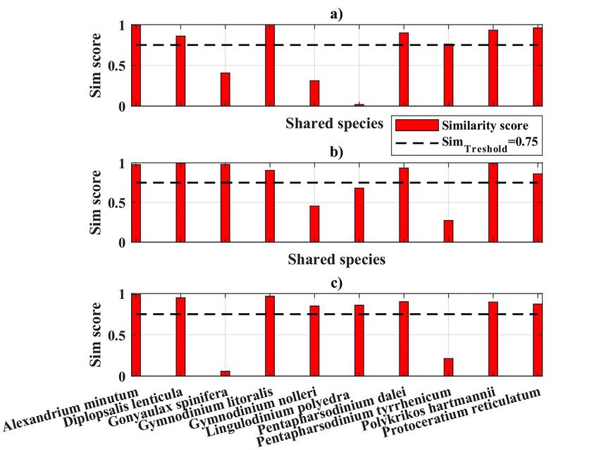
With regard to the shared species by habitats and in accordance with the initial conditions
set above, the statistical analysis indicated that 7 out of 10 shared species (Alexandrium
minutum, Diplopsalis lenticula Bergh, 1881, Gymnodinium litoralis A.Reñé, 2011,Comparative analysis of morphological and molecular approaches integrated ... 11 Pentapharsodinium dalei Indelicato & Loeblich III, 1986, Pentapharsodinium tyrrhenicum (Balech) Montresor, Zingone & Marino, 1993, Polykrikos hartmannii and Protoceratium reticulatum) have a similarity score above the threshold ( SimScoreTreshold = 0.75); for example, 70% overall similarity between datasets for the coastal habitat (Fig. 5). The same result was produced for the shelf habitat (e.g. 70% overall similarity between datasets), but the species with a similarity score above the threshold (SimScoreTreshold = 0.75), were slightly different (Alexandrium minutum, Diplopsalis lenticula, Gonyaulax spinifera, Gymnodinium litoralis, Pentapharsodinium dalei, Polykrikos hartmannii and Protoceratium reticulatum) (Fig. 5). In the open sea habitat, the comparative analysis showed that 8 out of 10 shared species, Alexandrium minutum, Diplopsalis lenticula, Gymnodinium litoralis, Gymnodinium nolleri M.Ellegaard & Ø.Moestrup, 1999, Lingulodinium polyedra, Pentapharsodinium dalei, Polykrikos hartmannii and Protoceratium reticulatum, have a similarity score above the threshold; for example, 80% overall similarity between datasets (Fig. 5). Evidently, the three habitats seemed to have 6 out of the 10 shared species in common: Alexandrium minutum, Diplopsalis lenticula, Gymnodinium litoralis, Pentapharsodinium dalei, Polykrikos hartmannii and Protoceratium reticulatum. Figure 5. Similarity scores of shared species concentrations (identified by both morphological and molecular approaches) per habitat (resp.: a) coastal, b) shelf and c) open sea).
12                                  Dzhembekova N et al

Discussion
Principally, even nowadays, detailed and extensive research studies into the marine
phytoplankton dormant stages in the Black Sea sediments remain scarce and limited
compared to the intensive planktonic studies. The proposed study, therefore, was designed
to give a sharper focus on species-level taxonomic composition, which yielded a total
number of 56 morphologically-differentiated dinocyst taxa. Within them, 39 species were
immediately identified, which is relatively high when compared with other morphology-
based studies of recent Black Sea sediments (Rubino et al. 2010b, Aydin et al. 2015) and
also represents quite a substantial proportion against those reported by Mudie et al.
(2017), exceeding the number of 71, but at 185 datapoints. In addition, four different cyst
types (data not shown) were identified for Scrippsiella acuminata, revealing much higher
intraspecific morphological variability of these species over those reported previously
(Rubino et al. 2017). The cyst concentration of S. acuminata, a typical bloom causing
species in the Black Sea (Oguz 2008), as the most abundant cysts in the region, most
likely is a good example of the existing benthic-pelagic coupling in the ecosystem
processes. Nevertheless, there are some morphotypes of cysts associated with the genera
Gymnodinium, Alexandrium, Protoperidinium and Scrippsiella for which it was not possible
to be successfully identified to species level due to certain methodological constraints, for
example, lack of clearly defined morphological features (Bolch et al. 1999, Bravo et al.
2006) or extremely low germination success. In case of difficult morphological
identification, the general perception is that molecular methods could be the preferred
option for dinocyst identification as it shows greater efficiency and higher resolution at
species-level (Penna et al. 2010). In this study, metabarcoding data identified 66
dinoflagellate taxa with a high level of similarity with the reference database. Although the
taxonomic coverage was fairly satisfactory, the identification power was contingent upon
key issues of DNA methodology itself, such as insufficient resolution capacity of the
barcode and the quality and completeness of the reference database.

Taking into consideration the advantages and drawbacks of the morphological and
molecular methods and their appropriateness for biodiversity assessment, suggests that
the two methods should be cross-checked if they are to ensure the accuracy of the
dinocyst identification. To that effect, the integration and comparison of data derived by
different approaches seem imperative, regardless of the subtle interpretation of the
combined dataset. A critical issue in the comparative analyses is to use the same
taxonomic concept in all attempted approaches as biodiversity can be measured at
different taxonomic levels (Colwell 2009). Thus the detailed assessment of the
comparability of the two methodologies, a comparison between the taxa lists derived from
both NGS and morphological approaches revealed that basically, the DNA metabarcoding
yielded higher taxonomic diversity than that derived by morphology. While all the families
that were found by LM were recovered by NGS, metabarcoding data recovered the
presence of 18 families that were not detected morphologically. Thus, it could be reasoned
that the molecular approach was highly beneficial for detecting a higher number of taxa at
the genus and species level, while both morphology and NGS seemed more efficient in
revealing the degree of similarity between some of the dominant genera. Other taxa were
Comparative analysis of morphological and molecular approaches integrated ...      13

further detected by either morphology or NGS methods exclusively. Our findings were in
line with those indicated in other studies integrating molecular and morphological
approaches, not only for resting cysts, but also for planktonic stages (e.g. Penna et al.
2010, Eiler et al. 2013, Zimmermann et al. 2015, Hirai et al. 2017, Jung et al. 2018, Perini
et al. 2019). It should also be acknowledged that the discrepancy between LM and NGS
could be attributed to a number of different methodological biases – from sampling to
database biases (Matsuoka and Fukuyo 2000, Zimmermann et al. 2015, Danovaro et al.
2016, Dzhembekova et al. 2017, Harvey et al. 2017, Smith et al. 2017) (Table 3). The
sampling biases can be primarily triggered by the limited amount of sediment subsample
used for initial sample processing, both for DNA isolation and morphological analyses and
most notably for rare species, i.e. present in very low densities. In addition, the natural
heterogeneity of sediments should be considered as a possible cause of the wide disparity
in the taxonomic composition and abundance. Amongst the limitations of the NGS method
that may affect the reliability of the resting cysts detection, are the controversial issues
concerning the DNA from the sedimented vegetative cells and those related to the
extracellular DNA preserved in the sediment (Corinaldesi et al. 2011). Another factor that
might explain the observed incongruities is the difficult morphological identification of some
cysts; for example, a broad range of morphotypes assigned to Alexandrium,
Protoperidinium and Scrippsiella could not be identified at the species level, as the lack of
specific features has an adverse effect on the successful accuracy of the subsequent
identification (Bravo et al. 2006, Matsuoka et al. 2006). The resolution of the primers
applied was insufficient for some taxa with high intragenus and intergenus similarity. Thus,
for instance, within Biecheleria (Biecheleria brevisulcata K.Takahashi & Iwataki, 2014 and
Biecheleria cincta (Siano, Montresor & Zingone) Siano, 2012)), Prorocentrum (
Prorocentrum texanum Henrichs, Steidinger, P.S.Scott & L.Campbell, 2013, Prorocentrum
micans Ehrenberg, 1834 and Prorocentrum mexicanum Osorio-Tafall, 1942), Scrippsiella/
Peridinium (Scrippsiella hangoei (J.Schiller) J.Larsen, 1995 and Peridinium aciculiferum
Lemmermann, 1900) and Scrippsiella/Theleodinium ( Scrippsiella acuminata and
Theleodinium calcisporum Craveiro, Pandeirada, Daugbjerg, Moestrup & Calado, 2013)
species, it could not be established in view of the inadequate set of recognisable identifiers
in the target region. The 18S rRNA region is not useful for differentiating some species
because of a low variability (Henrichs et al. 2013). Irrefutably, incorporating different target
regions leads to improved detection efficiency (Smith et al. 2017, Sildever et al. 2019,
Atherton and Jondelius 2020). Indeed, the reference database deficiency may also hinder
the correct interpretation of metabarcoding data (some sequences were assigned to
references deposited as sp.: for example Fragilidium sp., Gymnodinium sp.,
Proterythropsis sp., Scrippsiella sp. and Warnowia sp.). A practical solution to such an
issue might be achieved through filling the taxonomic gaps with integrated registration for
both morphological and genetic information. The development of reliable databases with
data from different regions will further facilitate the precise identification of OTUs. In the
current study, the morphological dataset covered 13 Protoperidinium species (nine
identified at the species level and four at the genus level), whereas the filtered NGS
dataset, including only sequence with similarity ≥ 98% with the reference was related to
only one species. Actually, there were more Protoperidinium species determined in the
14                                          Dzhembekova N et al

molecular data, but were excluded from any further analyses due to their low similarity
(below the set threshold).

 Table 3.
 Possible biases explaining discrepancies between morphological and NGS data in sediment resting
 cyst analyses (modified from Harvey et al., 2017).

Bias        Name              Description                           Possible effect
type

Sampling Fractional           Morphological and NGS methods were Numbers of individuals and possibly
         sampling             applied to a subset of each sample taxonomic composition may not be
                                                                 identical between subsamples

            DNA extraction    DNA extraction from sediment samples Taxa and/or abundances may be under-
            effectiveness     is difficult/ challenging            represented in the resulting data

Organism Life stage           Sequencing technologies cannot        DNA from sedimented vegetative cells and/
         identifications      differentiate between different       or the extracellular DNA preserved in the
                              phytoplankton life stages (cysts/     sediment may affect the reliability of the
                              vegetative cells)                     results

PCR         Taxonomic         Target gene copy number vary          Taxa with more target gene copies per cell
            variation         amongst taxa                          may be favoured over those with fewer
                                                                    copies

            Primer bias       Primer design supports some taxa      Favoured taxa may be preferentially
                              against others                        amplified, whereas other taxa may be
                                                                    missed

            Amplification     DNA from some taxa is more easily     Privileged taxa may be preferentially
            bias              amplified than from other taxa        amplified and over-represented

Database Database             Morphological knowledge and/or NGS    Inability to assign taxonomy to organisms
         composition          databases may not                     and/or incorrect assignments are made
                              include all sampled taxa

            Data resolution   Available morphological and DNA       Morphology and/or NGS data may not
                              sequence data resolution              contain sufficient information to assign
                              may vary amongst taxa                 taxonomy at finer scales (e.g. genus,
                                                                    species)

            Database errors   Possible mismatch of taxonomic        Incorrect taxonomic assignments
                              information to morphological or DNA
                              sequence data

Given the fact that the application of different methods increases the potential for
substantial growth in biodiversity studies, both approaches can benefit from the integration
of the information contained in the datasets generated by metabarcoding and morphology
methods (Zimmermann et al. 2015). Our results highlight the significance of the combined
data for a better interpretation of the results and advanced understanding of the current
species diversity. On one hand, environmental DNA metabarcoding, shedding new light on
the species richness and infinite variety, could speed up the proper identification of
dinoflagellate resting stages, refine their cryptic and pseudo-cryptic alignment, providing a
vivid picture of the immense benthic microalgal diversity. For example, five different
species were genetically identified within genus Alexandrium, though, discerned under a
Comparative analysis of morphological and molecular approaches integrated ... 15
light microscopy, there were two species and seven noted as sp. On the other hand, the
traditional identification technique via LM proved to be even more indispensable for the
correct validation of the data obtained by metabarcoding. Yet, some identified
morphospecies exist that could not be detected using the NGS analyses (eight
Protoperidinium species were precisely morphologically identified as full cysts vs. only one
species determined genetically).
The combined results broaden our knowledge on the dinoflagellate diversity in Black Sea
sediments and highlight the importance of carrying out integrated investigations. Moreover,
the presence of species, both typical and exceptional for the present-day plankton
community, defines the role of sediments as seed banks where resting stages accumulate
over time. Some cysts are ubiquitously distributed in Black Sea sediments, whereas others
are irregularly scattered in patchy dispersion. Most notably, however, is that, unlike other
studies (Gong et al. 2015), a separate depth-dependent gradient of dinoflagellate cyst
diversity was not found. In a nutshell, the patterns of cysts richness, quantitative and
spatial distribution should be more thoroughly explored within the context of environmental
conditions, which might additionally support the overall analysis of the results. Further
complex multidisciplinary studies are needed to unravel the factors underlying the structure
of plankton cyst banks in the sediments.
Acknowledgements
This study was supported by the National Science Fund, Ministry of Education and Science
(MES), Bulgaria under project “Phytoplankton cysts – an intricacy between a “memory” or a
“potential” for Black Sea biodiversity and algal blooms” (Grant number 01/8, 16.12.2016)
and MASRI – Infrastructure for Sustainable Development of Marine Research including the
Participation of Bulgaria in the European Infrastructure Euro-Argo, National Roadmap for
Scientific Infrastructure (2017-2023) of Republic of Bulgaria (Contract number
D01-158/28.08.2018).
Manuela Belmonte (CNR-IRSA of Taranto) performed the morphological quali-quantitative
analysis of cysts.
References
• Anderson D, Keafer B, Kleindinst J, McGillicuddy D, Martin J, Norton K, Pilskaln C,
Smith J, Sherwood C, Butman B (2014) Alexandrium fundyense cysts in the Gulf of
Maine: Long-term time series of abundance and distribution, and linkages to past and
future blooms. Deep Sea Research Part II: Topical Studies in Oceanography 103: 6‑26.
https://doi.org/10.1016/j.dsr2.2013.10.002
• Anderson DM, Fukuyo Y, Matsuoka K (2003) Cyst methodologies. In: Hallegraeff GM,
Anderson DM, Cembella AD (Eds) Manual on harmful marine microalgae. 11.
UNESCO, Paris. [ISBN 92-3-103871-0].16 Dzhembekova N et al
• Atherton S, Jondelius U (2020) Biodiversity between sand grains: Meiofauna
composition across southern and western Sweden assessed by metabarcoding.
Biodiversity Data Journal 8 https://doi.org/10.3897/bdj.8.e51813
• Aydin H, Balci M, Uzar S, Balkis N (2015) Dinoflagellate cyst assemblages in surface
sediments of southwestern Black Sea and Çanakkale Strait (Dardanelles). Fresenius
Environmental Bulletin 24: 4789‑4798.
• Belmonte G, Rubino F (2019) Resting cysts from coastal marine plankton.
Oceanography and Marine Biology 1‑88. https://doi.org/10.1201/9780429026379-1
• Bolch CS, Negri A, Hallegraeff G (1999) Gymnodinium microreticulatum sp. nov.
(Dinophyceae): a naked, microreticulate cyst-producing dinoflagellate, distinct from
Gymnodinium catenatum and Gymnodinium nolleri. Phycologia 38 (4): 301‑313.
https://doi.org/10.2216/i0031-8884-38-4-301.1
• Bravo I, Garcés E, Diogène J, Fraga S, Sampedro N, Figueroa R (2006) Resting cysts
of the toxigenic dinoflagellate genus Alexandriumin recent sediments from the Western
Mediterranean coast, including the first description of cysts of A. kutnerae and A.
peruvianum. European Journal of Phycology 41 (3): 293‑302.
https://doi.org/10.1080/09670260600810360
• Colwell RK (2009) Biodiversity: concepts, patterns, and measurement. In: Colwell RK,
Levin SA, Carpenter SR, Godfray HC, Kinzig AP, Loreau M, Losos JB, Walker B,
Wilcove DS (Eds) The Princeton Guide to Ecology. Princeton University Press, 832 pp.
[ISBN 978-1-4008-3302-3]. https://doi.org/10.1515/9781400833023
• Corinaldesi C, Barucca M, Luna GM, Dell’Anno A (2011) Preservation, origin and
genetic imprint of extracellular DNA in permanently anoxic deep-sea sediments.
Molecular Ecology 20 (3): 642‑654. https://doi.org/10.1111/j.1365-294x.2010.04958.x
• Dale B (1983) Dinoflagellate resting cysts: “benthic plankton”. In: Fryxell GA (Ed.)
Survival strategies of the algae. Cambridge University Press Cambridge, 144 pp.
• Danovaro R, Carugati L, Berzano M, Cahill A, Carvalho S, Chenuil A, Corinaldesi C,
Cristina S, David R, Dell'Anno A, Dzhembekova N, Garcés E, Gasol J, Goela P, Féral J,
Ferrera I, Forster R, Kurekin A, Rastelli E, Marinova V, Miller P, Moncheva S, Newton A,
Pearman J, Pitois S, Reñé A, Rodríguez-Ezpeleta N, Saggiomo V, Simis SH, Stefanova
K, Wilson C, Lo Martire M, Greco S, Cochrane SJ, Mangoni O, Borja A (2016)
Implementing and innovating marine monitoring approaches for assessing marine
environmental status. Frontiers in Marine Science 3 https://doi.org/10.3389/fmars.
2016.00213
• Dzhembekova N, Urusizaki S, Moncheva S, Ivanova P, Nagai S (2017) Applicability of
massively parallel sequencing on monitoring harmful algae at Varna Bay in the Black
Sea. Harmful Algae 68: 40‑51. https://doi.org/10.1016/j.hal.2017.07.004
• Dzhembekova N, Moncheva S, Ivanova P, Slabakova N, Nagai S (2018) Biodiversity of
phytoplankton cyst assemblages in surface sediments of the Black Sea based on
metabarcoding. Biotechnology & Biotechnological Equipment 32 (6): 1507‑1513.
https://doi.org/10.1080/13102818.2018.1532816
• Eiler A, Drakare S, Bertilsson S, Pernthaler J, Peura S, Rofner C, Simek K, Yang Y,
Znachor P, Lindström E (2013) Unveiling distribution patterns of freshwater
phytoplankton by a next generation sSequencing based approach. PLOS One 8 (1).
https://doi.org/10.1371/journal.pone.0053516
• Forster D, Dunthorn M, Mahé F, Dolan J, Audic S, Bass D, Bittner L, Boutte C, Christen
R, Claverie J, Decelle J, Edvardsen B, Egge E, Eikrem W, Gobet A, Kooistra WC,Comparative analysis of morphological and molecular approaches integrated ... 17
Logares R, Massana R, Montresor M, Not F, Ogata H, Pawlowski J, Pernice M, Romac
S, Shalchian-Tabrizi K, Simon N, Richards T, Santini S, Sarno D, Siano R, Vaulot D,
Wincker P, Zingone A, de Vargas C, Stoeck T (2016) Benthic protists: the under-charted
majority. FEMS Microbiology Ecology 92 (8). https://doi.org/10.1093/femsec/fiw120
• Garces E (2004) Relationship between vegetative cells and cyst production during
Alexandrium minutum bloom in Arenys de Mar harbour (NW Mediterranean). Journal of
Plankton Research 26 (6): 637‑645. https://doi.org/10.1093/plankt/fbh065
• Gong J, Shi F, Ma B, Dong J, Pachiadaki M, Zhang X, Edgcomb V (2015) Depth shapes
α- and β-diversities of microbial eukaryotes in surficial sediments of coastal
ecosystems. Environmental Microbiology 17 (10): 3722‑3737.
https://doi.org/10.1111/1462-2920.12763
• Harvey JJ, Johnson S, Fisher J, Peterson W, Vrijenhoek R (2017) Comparison of
morphological and next generation DNA sequencing methods for assessing
zooplankton assemblages. Journal of Experimental Marine Biology and Ecology 487:
113‑126. https://doi.org/10.1016/j.jembe.2016.12.002
• Henrichs D, Scott P, Steidinger K, Errera R, Abraham A, Campbell L (2013) Morphology
and phylogeny of Prorocentrum texanum sp. nov. (Dinophyceae): A new toxic
dinoflagellate from the Gulf of Mexico coastal waters exhibiting two distinct
morphologies. Journal of Phycology 49 (1): 143‑155. https://doi.org/10.1111/jpy.12030
• Hirai J, Nagai S, Hidaka K (2017) Evaluation of metagenetic community analysis of
planktonic copepods using Illumina MiSeq: Comparisons with morphological
classification and metagenetic analysis using Roche 454. PLOS One 12 (7).
https://doi.org/10.1371/journal.pone.0181452
• Jung SW, Kang D, Kim H, Shin HH, Park JS, Park SY, Lee T (2018) Mapping
distribution of cysts of recent dinoflagellate and Cochlodinium polykrikoides using next-
generation sequencing and morphological approaches in South Sea, Korea. Scientific
Reports 8 (1). https://doi.org/10.1038/s41598-018-25345-4
• Kremp A, Oja J, LeTortorec A, Hakanen P, Tahvanainen P, Tuimala J, Suikkanen S
(2015) Diverse seed banks favour adaptation of microalgal populations to future climate
conditions. Environmental Microbiology 18 (2): 679‑691.
https://doi.org/10.1111/1462-2920.13070
• Lundholm N, Ribeiro S, Andersen T, Koch T, Godhe A, Ekelund F, Ellegaard M (2011)
Buried alive – germination of up to a century-old marine protist resting stages.
Phycologia 50 (6): 629‑640. https://doi.org/10.2216/11-16.1
• Malone M (2017) The microbiome of diabetic foot ulcers and the role of biofilms. The
Microbiology of Skin, Soft Tissue, Bone and Joint Infections 41‑56.
https://doi.org/10.1016/b978-0-12-811079-9.00003-3
• Matsuoka K, Fukuyo Y (2000) Technical guide for modern dinoflagellate cyst study.
WESTPAC-HAB/WESTPAC/IOC, 29 pp.
• Matsuoka K, Kawami H, Fujii R, Iwataki M (2006) Further examination of the cyst-theca
relationship of Protoperidinium thulesense (Peridiniales, Dinophyceae) and the
phylogenetic significance of round brown cysts. Phycologia 45 (6): 632‑641. https://
doi.org/10.2216/05-42.1
• McCann KS (2000) The diversity–stability debate. Nature 405 (6783): 228‑233.
https://doi.org/10.1038/35012234
• Montresor M, Bastianini M, Cucchiari E, Giacobbe MG, Penna A, Rubino F, Satta CT
(2010) STADI DI RESISTENZA DEL FITOPLANCTON. In: Socal G, Buttino I, Cabrini M,18 Dzhembekova N et al
Mangoni O, Penna A, Totti C (Eds) Metodologie di studio del plancton marino. ISPRA—
Istituto Superiore per la Protezione e la Ricerca Ambientale, Roma, 623 pp.
• Moscatello S, Rubino F, Saracino OD, Fanelli G, Belmonte G, Boero F (2004) Plankton
biodiversity around the Salento Peninsula (South East Italy): an integrated water/
sediment approach. Scientia Marina 68: 85‑102. https://doi.org/10.3989/scimar.
2004.68s185
• Mudie P, Marret F, Mertens K, Shumilovskikh L, Leroy SG (2017) Atlas of modern
dinoflagellate cyst distributions in the Black Sea Corridor: from Aegean to Aral Seas,
including Marmara, Black, Azov and Caspian Seas. Marine Micropaleontology 134:
1‑152. https://doi.org/10.1016/j.marmicro.2017.05.004
• Natsuike M, Nagai S, Matsuno K, Saito R, Tsukazaki C, Yamaguchi A, Imai I (2013)
Abundance and distribution of toxic Alexandrium tamarense resting cysts in the
sediments of the Chukchi Sea and the eastern Bering Sea. Harmful Algae 27: 52‑59.
https://doi.org/10.1016/j.hal.2013.04.006
• Oguz T (Ed.) (2008) BSC: State of the Environment of the Black Sea (2001-2006/7).
Commission on the Protection of the Black Sea Against Pollution, 421 pp. [ISBN
978-9944-245-33-3]
• Penna A, Battocchi C, Garcés E, Anglès S, Cucchiari E, Totti C, Kremp A, Satta C,
Grazia Giacobbe M, Bravo I, Bastianini M (2010) Detection of microalgal resting cysts in
European coastal sediments using a PCR-based assay. Deep Sea Research Part II:
Topical Studies in Oceanography 57: 288‑300. https://doi.org/10.1016/
j.dsr2.2009.09.010
• Perini F, Bastianini M, Capellacci S, Pugliese L, DiPoi E, Cabrini M, Buratti S, Marini M,
Penna A (2019) Molecular methods for cost-efficient monitoring of HAB (harmful algal
bloom) dinoflagellate resting cysts. Marine Pollution Bulletin 147: 209‑218.
https://doi.org/10.1016/j.marpolbul.2018.06.013
• Piredda R, Sarno D, Lange C, Tomasino MP, Zingone A, Montresor M (2017) Diatom
resting stages in surface sediments: A pilot study comparing next generation
sequencing and serial dilution cultures. Cryptogamie, Algologie 38 (1): 31‑46.
https://doi.org/10.7872/crya/v38.iss1.2017.31
• Ptacnik R, Solimini A, Andersen T, Tamminen T, Brettum P, Lepistö L, Willén E,
Rekolainen S (2008) Diversity predicts stability and resource use efficiency in natural
phytoplankton communities. Proceedings of the National Academy of Sciences 105
(13): 5134‑5138. https://doi.org/10.1073/pnas.0708328105
• Ribeiro S, Berge T, Lundholm N, Andersen T, Abrantes F, Ellegaard M (2011)
Phytoplankton growth after a century of dormancy illuminates past resilience to
catastrophic darkness. Nature Communications 2 (1). https://doi.org/10.1038/
ncomms1314
• Rubino F, Saracino OD, Fanelli G, Belmonte G, Miglietta A, Boero F (1998) Life cycles
and pelago-benthos interactions. Biologia Marina Mediterranea 5: 253‑259.
• Rubino F, Belmonte M, Caroppo C, Giacobbe M (2010a) Dinoflagellate cysts from
surface sediments of Syracuse Bay (Western Ionian Sea, Mediterranean). Deep Sea
Research Part II: Topical Studies in Oceanography 57: 243‑247. https://doi.org/10.1016/
j.dsr2.2009.09.011
• Rubino F, Moncheva S, Belmonte M, Slabakova N, Kamburska L (2010b) Resting
stages produced by plankton in the Black Sea—biodiversity and ecological perspective.Comparative analysis of morphological and molecular approaches integrated ... 19
Rapport Commission International pour l’Exploration Scientifique de la Mer
Méditerranée 39: 399.
• Rubino F, Belmonte M, Galil B (2017) Plankton resting stages in recent sediments of
Haifa port, Israel (Eastern Mediterranean) - Distribution, viability and potential
environmental consequences. Marine Pollution Bulletin 116: 258‑269.
https://doi.org/10.1016/j.marpolbul.2016.12.078
• Salgado P, Troncoso VA, Montresor M, Salamanca M, Lange CB (2011) First record of
Lingulodinium polyedrum (Dinophyceae) resting cysts in coastal sediments from the
Inner Sea of Chiloé, Los Lagos Region, southern Chile (~41°-43°S). Gayana. Botánica
68 (1): 106‑109. https://doi.org/10.4067/s0717-66432011000100012
• Satta C, Anglès S, Lugliè A, Guillén J, Sechi N, Camp J, Garcés E (2013) Studies on
dinoflagellate cyst assemblages in two estuarine Mediterranean bays: A useful tool for
the discovery and mapping of harmful algal species. Harmful Algae 24: 65‑79.
https://doi.org/10.1016/j.hal.2013.01.007
• Sildever S, Kawakami Y, Kanno N, Kasai H, Shiomoto A, Katakura S, Nagai S (2019)
Toxic HAB species from the Sea of Okhotsk detected by a metagenetic approach,
seasonality and environmental drivers. Harmful Algae 87 https://doi.org/10.1016/j.hal.
2019.101631
• Smith K, Kohli G, Murray S, Rhodes L (2017) Assessment of the metabarcoding
approach for community analysis of benthic-epiphytic dinoflagellates using mock
communities. New Zealand Journal of Marine and Freshwater Research 51 (4):
555‑576. https://doi.org/10.1080/00288330.2017.1298632
• Strong JA, Andonegi E, Bizsel KC, Danovaro R, Elliott M, Franco A, Garces E, Little S,
Mazik K, Moncheva S, Papadopoulou N, Patrício J, Queirós A, Smith C, Stefanova K,
Solaun O (2015) Marine biodiversity and ecosystem function relationships: The
potential for practical monitoring applications. Estuarine, Coastal and Shelf Science
161: 46‑64. https://doi.org/10.1016/j.ecss.2015.04.008
• Tanabe A, Nagai S, Hida K, Yasuike M, Fujiwara A, Nakamura Y, Takano Y, Katakura S
(2015) Comparative study of the validity of three regions of the 18S-rRNA gene for
massively parallel sequencing-based monitoring of the planktonic eukaryote community.
Molecular Ecology Resources 16 (2): 402‑414. https://doi.org/10.1111/1755-0998.12459
• Tang YZ, Gobler C (2012) The toxic dinoflagellate Cochlodinium polykrikoides
(Dinophyceae) produces resting cysts. Harmful Algae 20: 71‑80. https://doi.org/10.1016/
j.hal.2012.08.001
• Zimmermann J, Glöckner G, Jahn R, Enke N, Gemeinholzer B (2015) Metabarcoding
vs. morphological identification to assess diatom diversity in environmental studies.
Molecular Ecology Resources 15 (3): 526‑542. https://doi.org/10.1111/1755-0998.1233620 Dzhembekova N et al
Supplementary material
Suppl. material 1: Minimum and maximum abundance of the dinoflagellate species
identified in the Black Sea sediment samples by morphological and molecular
approach
Authors: Dzhembekova, N., Rubino, F., Nagai, S., Zlateva, I., Slabakova, N., Ivanova, P.,
Slabakova, V., Moncheva, S.
Data type: Table
Download file (61.20 kb)You can also read