Utilization of patterned bioprinting for heterogeneous and physiologically representative reconstructed epidermal skin models
←
→
Page content transcription
If your browser does not render page correctly, please read the page content below
www.nature.com/scientificreports
OPEN Utilization of patterned
bioprinting for heterogeneous
and physiologically representative
reconstructed epidermal skin
models
Sabrina Madiedo‑Podvrsan1,2, Jean‑Philippe Belaïdi1,2, Stephanie Desbouis1,
Lucie Simonetti1, Youcef Ben‑Khalifa1, Christine Collin‑Djangone1, Jérémie Soeur1,2* &
Maïté Rielland1,2*
Organotypic skin tissue models have decades of use for basic research applications, the treatment
of burns, and for efficacy/safety evaluation studies. The complex and heterogeneous nature of
native human skin however creates difficulties for the construction of physiologically comparable
organotypic models. Within the present study, we utilized bioprinting technology for the controlled
deposition of separate keratinocyte subpopulations to create a reconstructed epidermis with two
distinct halves in a single insert, each comprised of a different keratinocyte sub-population, in
order to better model heterogonous skin and reduce inter-sample variability. As an initial proof-of-
concept, we created a patterned epidermal skin model using GPF positive and negative keratinocyte
subpopulations, both printed into 2 halves of a reconstructed skin insert, demonstrating the feasibility
of this approach. We then demonstrated the physiological relevance of this bioprinting technique by
generating a heterogeneous model comprised of dual keratinocyte population with either normal or
low filaggrin expression. The resultant model exhibited a well-organized epidermal structure with
each half possessing the phenotypic characteristics of its constituent cells, indicative of a successful
and stable tissue reconstruction. This patterned skin model aims to mimic the edge of lesions as seen
in atopic dermatitis or ichthyosis vulgaris, while the use of two populations within a single insert
allows for paired statistics in evaluation studies, likely increasing study statistical power and reducing
the number of models required per study. This is the first report of human patterned epidermal model
using a predefined bioprinted designs, and demonstrates the relevance of bioprinting to faithfully
reproduce human skin microanatomy.
Human skin is highly heterogeneous with variability across individuals, anatomical sites and during skin pathol-
ogy. A skin region can be of thickness ranging from 0.1 on the eye-lid up to more than 1 mm on the palm and
sole, glabrous or hairy, and of different cell content and extra-cellular matrix (ECM) organization across different
anatomical sites1–3. There are also dramatic interpersonal variations in pH, moisture, thickness, organization and
microbiota. Furthermore, skin status is continually changing due to aging, UV exposure, pollution, environment,
lifestyle or disease states1. Accurately modelling the physiology and the physiopathology of human skin requires
in vitro organotypic models capable of reproducing key areas of diversity.
Skin was the first organ to be reconstructed in vitro by tissue engineering in 1 9814, and has since been utilized
for knowledge studies, the treatment of burns, scars and for plastic surgery5, and forms the backbone of efficacy
and toxicity screening strategies used in the pharmaceutical and cosmetic industries6–8. Despite considerable
advances in tissue engineering for skin, the organotypic model state-of-the-art remains far from reproducing
the complexity of native human skin, resulting in limited predictability in research and development applica-
tions and evaluation tests9,10. While the need for more physiological skin models is clear, creating such models
1
L’Oréal Research and Innovation, Aulnay‑sous‑Bois, France. 2These authors contributed equally: Sabrina Madiedo-
Podvrsan, Jean-Philippe Belaïdi, Jérémie Soeur and Maïté Rielland. *email: jeremie.soeur@rd.loreal.com; maite.
rielland@rd.loreal.com
Scientific Reports | (2021) 11:6217 | https://doi.org/10.1038/s41598-021-85553-3 1
Vol.:(0123456789)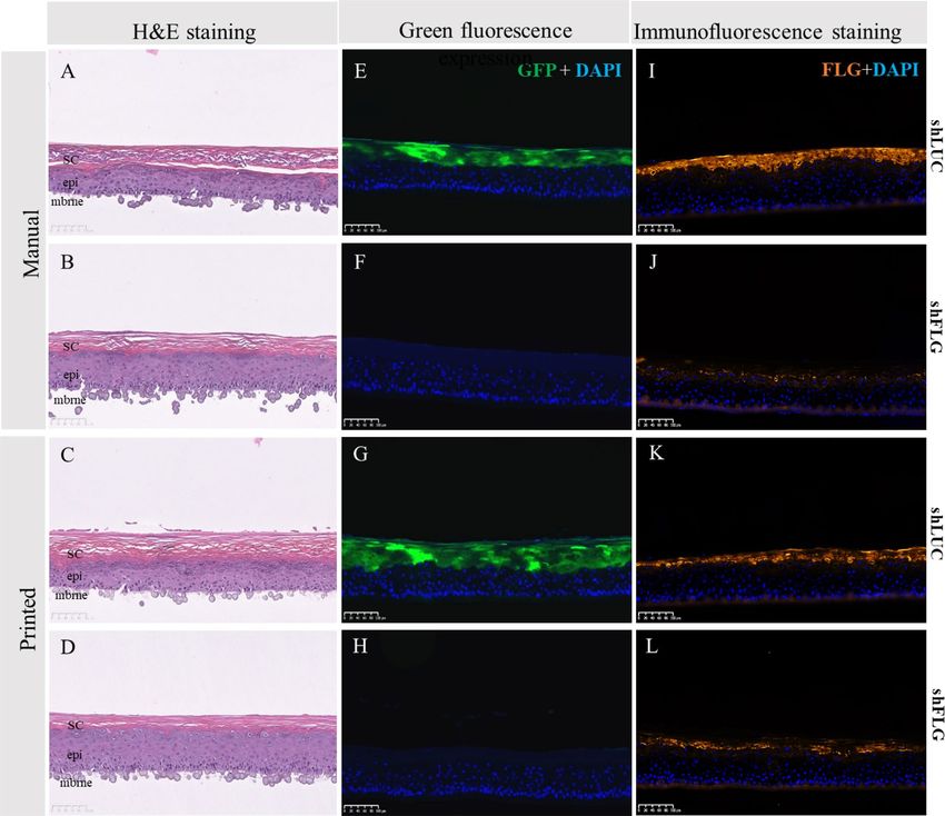
www.nature.com/scientificreports/
using conventional culture methods is highly challenging due to insufficient cell deposition precision to accurate
recreate native human skin conditions.
Three-dimensional bioprinting, more commonly simply known as bioprinting, is an additive manufacturing
technique that utilizes a printing system able to precisely deposit biological material such as cells and biocompat-
ible matrices into complex and functional 3D architectures, resulting in organ reconstructions more reflective
of human tissue than otherwise p ossible11–13. The accurate cell deposition available in bioprinting offers the
possibility to reproduce skin heterogeneity, and therefore create more physiologically relevant skin models. This
potential is exemplified by the work of Ng and colleagues, who developed a bioprinted pigmented full thickness
skin model in which the melanocytes were spatially positioned to mimic pigmented b lemishes14. Leveraging the
spatial positioning accuracy of bioprinting to reproduce keratinocyte heterogeneity within in vitro skin models
would allow for improved understanding skin physiology and accurate modelling of skin pathology such as that
seen within photoaging, atopic dermatitis (AD) or ichthyosis vulgaris (IV)15–17. Furthermore, the ability to create
a patterned reconstructed model of two separate skin phenotypes, for example one healthy and one pathologi-
cal, within a single skin model insert may accurately reflect the heterogeneity of pathological skin, while also
reducing inter-sample variability and improving the robustness of evaluation studies.
To validate the concept of patterned epidermal model, we first generated a proof-of-concept heterogeneous
epidermal model using a normal human keratinocyte (NHK) population divided into GFP positive and negative
subpopulations. We then extended this concept to a published physiologically relevant filaggrin down-regulated
epidermal model, which mimics numerous defects observed in the epidermis of AD and IV patients, especially
the drastic reduction of keratohyalin g ranules18–20. One-half of the reconstructed skin insert was reconstructed
with shLuciferase-GFP transduced NHKs to function as a control section, with the other half of the insert
reconstructed with fillagrin knockdown (shFLG) transduced NHKs.
This is the first report of a patterned bioprinting approach being used to reproduce the heterogeneity of
the epidermis within an organotypic epidermal model, and represents a significant advancement in skin tissue
engineering for both knowledge and evaluation studies.
Results
Lentiviral transduction and bioprinting of primary human keratinocytes does not impair epi‑
dermis reconstruction. As an initial proof of concept we utilized two populations of NHK; one of native
cells (hereafter referred to as wild type (WT) cells), the second transfected with a lentiviral particle expressing
Green Fluorescent Protein (WT-GFP) (Supplementary Fig. 1C,D). Both cell types demonstrated normal polygo-
nal cell morphology in 2D monoculture (Supplementary Fig. 1A,B). The two groups were then either bioprinted
or manually seeded to determine if inkjet bioprinting was influencing GFP expression or compromise the abil-
ity of the cells to form a reconstructed epidermis. Models using both manual and printed WT and WT-GFP
keratinocytes demonstrated a well-organized basal layer, spinous layer, granular layer and stratum corneum
(Fig. 1A,B,E,F).
GFP expression was particularly prominent in the granular layer and in the stratum corneum of the WT-GFP
epidermis (Fig. 1D,H), but was still present in the basal and spinous layers, albeit at a lower intensity. As expected
GFP expression was not detected in WT epidermis (Fig. 1C,G).
The expression and localization of key epidermal markers keratin 14 (K14, basal layer marker), keratin 10
(K10, spinous layer marker), and transglutaminase 1 (TGM1), loricrin (LOR) and filaggrin (FLG), three key
markers of final differentiation, were all consistent with published reports21,22, with no difference between manual
or printed epidermal models or WT and WT-GFP NHKs reconstructed models (Supplementary Fig. 2). These
data demonstrate neither GFP transduction nor inkjet bioprinting hamper the ability of NEK to form a recon-
structed epidermis consistent with native skin.
Patterned human keratinocytes are stable in culture and form a fully differentiated patterned
epidermis. We next adapted the bioprinting technique to obtain controlled and reproducible reconstructed
epidermis patterns. We utilized two designs; semi-circles, which easily allow for half-and-half tissue collection,
or concentric rings, which resemble a dermatological spot (Fig. 2A,B). Identifying the correct NHK concentra-
tions with the appropriate media deposition is the most critical part of the process to obtain the optimal space
between both compartments. If the cells are not sufficiently concentrated, the 3D models will not reconstruct
correctly; if the cell concentration is too high clots may occur during printing and NHK mortality will increase
(data not shown). After fixing the printing protocol, each compartment was separated by an empty space of
1.5 mm by using a hydrophobic membrane. We optimized of the quantity of liquid, number of cells and patterns
of drop depositions to obtain an acceptable separation distance between compartments (Fig. 2A,B). The printing
process took ~ 20 s per sample and was highly reproducible (data not shown).
To examine if the printed NHK subpopulations are spatially stable within the sample we printed both WT
and WT-GFP groups in semi-circle and the concentric circle depositions and examined the tissue histology
and GFP distribution pattern (Figs. 3A,B, 4A,B). For both designs, the epidermis of printed models was fully
differentiated with the presence of all epidermal layers (Figs. 3B-Z1, Z2 and Z3, 4B-Z1, Z2 and Z3). Histological
analysis shows that the original gap between the patterned compartments to have been populated by cells and
closed (Figs. 3A,B-Z2, 4A,B-Z2, Supplementary Fig. 3). Moreover, the clear demarcation of GFP expression
shows that both compartments kept their original design with no merging of cell populations (Figs. 3A,B-Z2,
4A,B-Z3). We also found the expression of epidermal markers K14, K10, TGM1, LOR and FLG to be homoge-
neously expressed all along the samples, with no visible difference between WT and WT-GFP printed sections
(Supplementary Fig. 4). These data demonstrate that NHK bioprinted patterning results in separate and stable
populations without compromising reconstructed epidermis structure or morphology.
Scientific Reports | (2021) 11:6217 | https://doi.org/10.1038/s41598-021-85553-3 2
Vol:.(1234567890)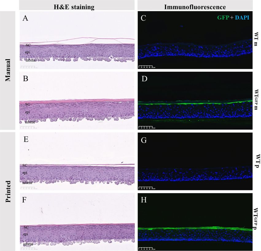
www.nature.com/scientificreports/
Figure 1. Effect of GFP transfection and bioprinting on epidermal reconstruction morphology. H&E
morphology between skin models reconstructed from WT or WT-GFP cells was consistent for both manually
(A,B) or bioprinted (E,F) samples. As anticipated, samples printed with WT-GFP (D,H) but not WT cells (C,G)
express GPF, with no difference between those using manual (C,D) or (G,H) reconstruction techniques. SC
stratum corneum, epi epidermis, mbrne polycarbonate membrane support, WTm manual reconstructed skin
substitute from WT NHKs, WTp printed reconstructed skin substitute from WT NHKs, WT-GFPm manual
reconstructed skin substitute from WT-GFP NHK, WT-GFPm printed reconstructed skin substitute from
WT-GFP NHK (n = 6 for each condition).
Bioprinting of shLUC and shFLG human keratinocytes does not affect epidermis reconstruc‑
tion. We next applied the pattern bioprinting technique to a more physio-pathologically relevant condition in
order to create healthy “control” and “pathological” skin sections within the same sample, by utilizing a filaggrin
(FLG)-deficient keratinocyte model of Atopic D ermatitis19. We created a FLG-deficient cell line via lentiviral
particles targeting FLG (named thereafter shFLG cells), and a control line transfected with a lentivirus inserting
GFP in order to further distinguish between FLG positive and negative populations within the model (shLUC
cells) (Supplementary Fig. 5A). Both shFLG and shLUC NHKs demonstrated the correct polygonal morphology
within 2D monoculture (Supplementary Fig. 5A, a and b), while only shLUC NHKs displayed green fluores-
cence as anticipated (Supplementary Fig. 5A, c and d).
To evaluate the impact of the bioprinting process and shFLG and shLUC cell phenotypes on epidermal dif-
ferentiation, we created 3D reconstructed epidermal models using either conventional manual cell seeding or
bioprinting of both NHK populations on a single polycarbonate membrane. As shLUC and shFLG NHK have
different doubling times of 17.7 h and 22 h, respectively, we adjusted their initial seeding concentrations in
order to obtain homogenous epidermal proliferation and differentiation (Supplementary Fig. 5B). After 14 days
of culture we correlated tissue histology with GPF and filaggrin expression, finding both manually seeded or
printed shLUC epidermis were fully differentiated (Fig. 5A,C), positive for GFP (Fig. 5E,G) and expressed FLG
Scientific Reports | (2021) 11:6217 | https://doi.org/10.1038/s41598-021-85553-3 3
Vol.:(0123456789)
www.nature.com/scientificreports/
Figure 2. Macroscopic visualization of the cellular deposition design. Cells were printed in patterned cellular
suspension in either semi-circles (A) or concentric rings (B) schematically illustrated as 2 separate NHK
populations (left) or after seeding on a polycarbonate membrane after the bioprinting process (right). Pattern
designs were drawn internally using BioCaD software (RegenHU).
in spinous, granular and stratum corneum layers as anticipated (Fig. 5I,K). Conversely, shFLG samples displayed
a characteristic reduction of the granular layer while basal and spinous layers were unaffected (Fig. 5B,D), con-
sistent with literature r eports19. Similarly, shFLG samples lacked GFP expression (Fig. 5F,H) while exhibiting a
pronounced reduction of FLG expression as expected of lentivirus-transfected FLG knockdown cells (Fig. 5J,L).
Immunofluorescence staining demonstrated the expression and localization of main epidermal markers K14,
K10, TGM1 and LOR to be equivalent between printed and manually seeded shLUC and shFLG samples, with
homogenous expression and similarly no difference between shLUC and shFLG reconstructions (Supplemen-
tary Fig. 6A). We verified the expression of FLG via western blot in our printed shLUC and shFLG epidermis,
confirming the downregulation of FLG in shFLG but not shLUC samples (Supplementary Fig. 6B). These results
demonstrate that both cell populations formed well-stratified epidermal reconstructions phenotypically consist-
ent with literature reports, and that bioprinting has no deleterious effect on the reconstructed model.
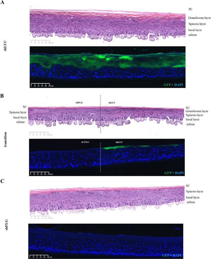
Patterned shLUC and shFLG human keratinocytes keep their designs over the culture period
and form fully differentiated patterned epidermises. As shFLG and shLUC epidermal reconstruc-
tions were possible and kept their associated phenotypes, we next bioprinted each population within the same
insert to form 2 semi-circles, each printed with either shFLG or shLUC cells. The shLUC half of the model dem-
onstrated strong co-localization of GFP and FLG expression as anticipated, while the half printed with shFLG
cells lacked GFP expression with no discernible FLG expression, demonstrating the model to be accurately
printed and stable after 14 days in culture (Fig. 6A,B, Supplementary Fig. 7A).
The ShLUC half of the reconstructed epidermis presents a normal differentiation with a clear granular layer
and presence high FLG expression (Fig. 7A,B), whereas shFLG printed half demonstrate a lack of keratohyalin
granules and down-regulation of FLG as previously observed (Fig. 7C, Supplementary Fig. 7A). GFP and FLG
expression patterns demonstrate a clear demarcation between shFLG and shLUC printed sections, indicating the
cell populations do not stray from their original printed region. Furthermore, the gap between the populations
creating during the printing was organically closed by the keratinocyte proliferation over the culture period,
resulting in a seamless epidermis composed of the two subpopulations (Fig. 7B). Immunostaining of major
epidermal markers was consistent across both shLUC and shFLG printed halves of the reconstruction (Supple-
mentary Fig. 7B). Collectively, these data demonstrate that each printed compartment retains their anticipated
phenotype with a clear separation between the printed subpopulations.
Scientific Reports | (2021) 11:6217 | https://doi.org/10.1038/s41598-021-85553-3 4
Vol:.(1234567890)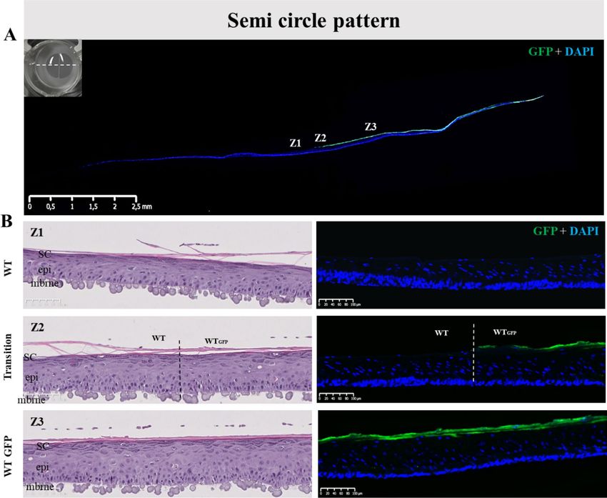
www.nature.com/scientificreports/
Figure 3. Histology of pattern bioprinted epidermis comprised of two distinct cell populations. (A) Lateral view
of the reconstructed skin stained for GFP (green) and cell nuclei (DAPI, blue) at day 14 post-reconstruction.
Indicated positions Z1–3 correspond to (B) H&E and immunofluorescence stained sections Z1–3, whereby Z1
is from the WT printed cell half, Z2 is the WT/WT-GFP cell interface, and Z3 is representative of the WT-GFP
printed half. The H&E images (left) show no difference in gross morphology, while the immunofluorescence
images (right) reveal no GFP staining on the WT cell side of the model, a clear demarcation where the WT
and WT-GFP halves meet, and a consistent GFP signal in the WT-GFP printed half, demonstrating the model
patterning to be stable across 14 days of culture. Images representative of 10 separate models.
Discussion
Within the present study, we demonstrate how separate populations of NHK can be bioprinted in a precise and
controlled way to create accurate patterns, retain their localization over the culture period and form a stable,
compartmentalized single sample. This approach allows for complex modelling of skin, and likely increases
evaluation study statistical power by enabling the use of pairwise statistics and resulting in a reduced number
of models required per study.
Skin is a dynamic living tissue with a high degree of cellular and structural h eterogeneity1,2. Accurate repro-
duction of this heterogeneity within in vitro reconstructed skin models is an absolute requirement for transla-
tion to both knowledge studies and in vitro evaluation predictability. Traditional manual seeding reconstruc-
tion techniques are hampered by cell placement insufficient of the precision required to create more complex
and therefore physiologically relevant models of skin heterogeneity. Cellular patterning has already previously
examined in 2D and 3D models, but the techniques used within the present study facilitate a higher degree of
viability and epidermal stability than previous r eports14,23. In a pioneering study, Min and colleagues created a
full thickness skin model with a controlled deposition of melanocytes to mimic skin b lemishes24. This approach
was not without difficultly however, as after air–liquid interface culture the melanocytes spread across the epi-
dermis rather than restricted to the basal layer as seen within native human skin6. A strength of our study is that
native skin architecture is well maintained, with a well-stratified differentiation of keratinocytes, from the basal
layer up to the stratum corneum25, which also result in a model that is highly stable during the culture period.
To better approximate complex in vivo skin, we have validated proof-of-concept-patterning approaches with
two different populations of NHK, following two different designs. Utilizing a semi-circle design allows for
Scientific Reports | (2021) 11:6217 | https://doi.org/10.1038/s41598-021-85553-3 5
Vol.:(0123456789)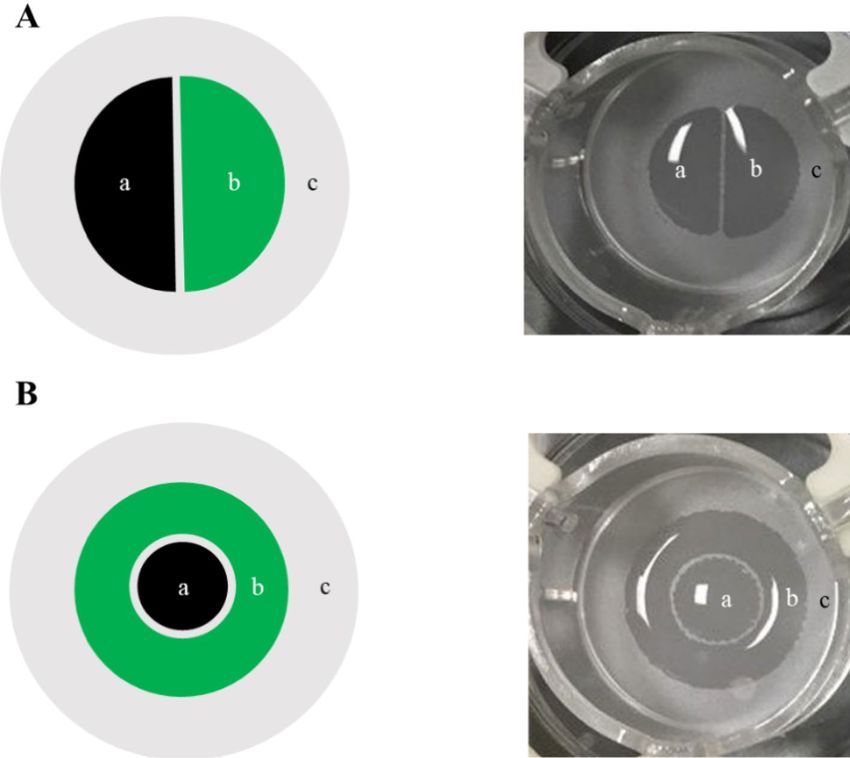
www.nature.com/scientificreports/
Figure 4. Bioprinted RHE constructs were achieved using distinct populations of NHKs on a same sample
in order to pattern the concentric design. Morphological analysis of the printed epidermal substitutes and
validation of the pattern using H&E staining and fluorescence analysis were done after 14 days of culture at
air–liquid interface. (A) GFP staining (Green) on the entire skin substitute with nuclei counterstain with DAPI
(blue) where we can observe both conserved parts of the epidermal sample, Z1, Z2 and Z3 are for zone 1, zone
2 and zone 3 represent the areas illustrated in (B); a macroscopic picture of the semi-circle sample has been
represented with the orientation of sample slices for the analysis; (B) H&E staining and Green fluorescence
analysis on the WT part of the sample (Z1), the transition between the WT and the WTGFP parts (Z2) and the
WTGFP part (Z3) proving that pattern is conserved after 14 days of culture and that both NHK populations
form at the end of the culture a unique sample. Sc stratum corneum, epi epidermis, mbrne polycarbonate
membrane support, WT reconstructed epidermal part from native phenotypic keratinocytes, WTGFP
reconstructed skin substitute from GFP-transduced native phenotypic keratinocytes, Transition specific region
of the patterned reconstructed skin substitute where both WT and WTGFP are in contact (n = 11).
evaluation of two phenotypically distinct populations in a single insert, which can easily be separated into two
halves for analysis. The bioprinted concentric rings approach resembles a dermatological spot, resulting in a
more accurate in vivo approximation of spots on skin. Cell printed with either technique showed the correct
differentiation of all epidermal layers for their phenotype and express key epidermal markers consistent with
published data21,22, demonstrating the stability of the pattern printed approach.
The FLG down-regulated NHK epidermal model is a key model for the study of AD and IV, which are wide-
spread in industrialized countries and among the most common human epidermal d isorders18,19,26–28. We chose
to examine this model based on clinical importance, applicability to reconstructed skin approaches, and that
the patterned approach is highly suited to examining the edges of AD and IV lesions. Consistent with published
data, we note a characteristic dramatic decrease of keratohyalin granules, demonstrating our reconstruction to
be consistent with those published p reviously29. Of note, we did not observe difference for intermediate and final
Scientific Reports | (2021) 11:6217 | https://doi.org/10.1038/s41598-021-85553-3 6
Vol:.(1234567890)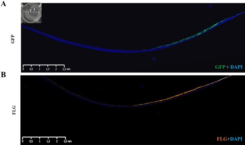
www.nature.com/scientificreports/
Figure 5. Histological examination of skin models reconstructed via manual seeding or bioprinting with
shLUC or shFLG cells. (A) H&E sections of shLUC reconstructed samples manually seeded show a well-
organized epidermal architecture, while (B) shFLG reconstructed samples demonstrate a pronounced
hypogranulosis, consistent with FLG down-regulated skin. These data are consistent with those obtained via
bioprinting (C,D). Manually seeded shLUC models expressed high levels of (E) GFP, which is not presented in
shFLG samples (F). Manually seeded samples are indistinguishable from their bioprinted counterparts (G,H).
Lastly, manually printed shLUC models highly express FLG (I), in contrast to samples reconstructed with
shFLG NHK (J). There is no discernible difference in expression in models reconstructed using bioprinting
(K,J). Images representative of 8 separate samples. SC stratum corneum, epi epidermis, mbrne polycarbonate
membrane support.
differentiation markers as K10 or LOR in our shFLG models compared to controls, consistent with the results of
the FLG down-regulated epidermal model of Mildner and co-workers, but contradictory to the model of Pendar-
ies and c olleagues18,19. It is interesting that the presence of shFLG cells within our patterned AD model did not
influence the differentiation of the control shLUC cells, with all layers of the epidermis and epidermal marker
expression being normal, while shFLG printed sections showed down-regulation of the FLG protein associated
with attended hypogranulosis exclusively in the shFLG section. Our results demonstrate that these cells can both
exist in separate sides of a single insert, a prerequisite for practical use of patterned skin bioprinting.
The compartmentalization of several cellular types on a same reconstruction, including functionalized cells
modified by genome editing, allows for greater understanding of physiological and pathophysiological mecha-
nisms of human skin. The principles described within this paper are likely to be applicable to any model using
keratinocytes as the constituent cells that requires a pattern design. For instance, patterned epidermal models
may have applications for knowledge studies of skin diseases, particularly skin cancers of keratinocyte origin.
Such a model would be valuable to examine preventive treatment for photoaging associated with actinic kera-
tosis, whereby the epidermis presents patches of p53 mutated NHKs that may progress to invasive squamous
cell carcinoma15. The patterning approach is similarly applicable for raw material evaluation safety and efficacy
studies. Skin from different parts of the body could be pattern onto a single model (for example to model skin
Scientific Reports | (2021) 11:6217 | https://doi.org/10.1038/s41598-021-85553-3 7
Vol.:(0123456789)www.nature.com/scientificreports/
Figure 6. Expression of FLG and GFP in the reconstructed epidermis created via patterned bioprinting with
shLUC and shFLG cell populations. (A) GFP (green) and (B) FLG expression is only present on the shLUC half
of the reconstructed skin. Images representative of 8 samples.
of the face or arm), with cells derived from donors of different ethnicities or ages in order to study their pheno-
typic differences. Such heterogenous models may be used to examine contrasting responses to environmental or
chemical stresses. The shFLG model could be of interest to study such as microbiota interactions, impact of UV
associated with humidity and temperature, or pollutants to better understand some mechanisms associated to
AD and I V19,29–31. The precision of bioprinting also offers the potential to increase the complexity of epidermal
models by integrating langerhans cells or melanocytes, or by printing a fibroblast-populated dermal compart-
ment with immune cells as T helper cells to study AD p athology30,32,33. Patterning melanocytes, langerhans cells
or creating complex dermal compartment reconstructions will need adaptations of protocols and the materials
however, as they all present increased cellular mobility, facilitating inter- and intra-compartmental migration.
A particular strength of the patterning approach is that the dual population model can be used to test a raw
material or drug of interest on both sides simultaneously. This technique creates the opportunity to use paired
statistical approaches, reducing the inter-section variability and thus increasing the statistical power the evalua-
tion studies. Similarly, the pairwise approach of having two conditions on a single sample facilitates a dramatic
reduction in the total amount of samples required to achieve meaningful results within a study, leading to reduce
study costs and higher result reliability. To get high reproducibility in the cell deposition and to be able to process
our samples always the same way after culture, we have patented an adaptor for insert. This adaptor blocks inserts
during printing and culture with a precise orientation, which is essential for sample pooling and/or comparison
regarding in vitro evaluations and knowledge.
In summary, by integrating different cellular populations in a same model using a high precision patterned
bioprinting approach we open the possibility of creating heterogeneous yet stable reconstructive skin models
more reflective of native human skin. The patterned human epidermis model described within this study is the
first proof-of-concept description that such a bioprinting based-strategy can create a viable and stable NHK
patterned reconstructed epidermis. This innovation in tissue engineering offers potential for basic research into
skin pathology using transfected keratinocytes to model skin conditions, and provides a stable and robust basis
of evaluation studies with potential for improved statistical power and reduced tissue use.
Materials and methods
Keratinocyte culture and preparation. Primary NHK were collected from healthy foreskin originating
from surgical waste of consenting subjects. The cells were cultured on a feeder layer of Mitomycin C-treated
3T3 fibroblasts and in presence of ROCK Inhibitor (Y-27632, Sigma-Aldrich, USA) in an incubator at 37 °C
and in a humidified atmosphere containing 5% of CO2 in NHK growth media (Episkin, France). Cells were
detached using 0.05% Trypsin–EDTA (1×) (Gibco, USA) after phosphate buffered saline (PBS, ThermoFisher,
USA) washing, and then suspended in a SGM medium (Episkin, France) at concentrations ready to use for
manual seeding or printing.
Mitomycin C treatment for feeder production. Murine 3T3 fibroblasts were cultured in DMEM
(Gibco, USA) enriched with 10% of Bovine calf serum defined/supplemented (BCS, HyClone, USA) medium for
Scientific Reports | (2021) 11:6217 | https://doi.org/10.1038/s41598-021-85553-3 8
Vol:.(1234567890)www.nature.com/scientificreports/
Figure 7. Histological analysis and GFP expression in shLUC and shFLG halves of pattern bioprinted
reconstructed skin. (A) The suLUC half of the patterned model is histologically normal and shows high levels of
GFP staining as anticipated, which abruptly ceases at the border with the shFLG printed half (B). (C) The shFLG
printed half of the model demonstrates a lack of keratohyalin granules in H&E staining and no GFP signal,
demonstrating the model to be accurately printed and stable with no migration of cell populations after 14 days
in culture. Images representative of 8 individual samples.
5 days, at an initial density of 1.5 × 10⁶ cells, in an incubator at 37 °C and in a humidified atmosphere containing
5% of CO2, with a medium change every two days. When 3T3 covered 70–80% of the flask area, they were treated
with a solution of 0.5 mg/ml of Mitomycin C (Sigma, USA) and Dulbecco’s phosphate-buffered saline enriched
with calcium and magnesium (PBS+, ThermoFisher, USA) solution for two hours in a 37 °C/5% CO2 incubator.
Mitomycin C-treated 3T3 were washed with phosphate buffered saline (PBS, ThermoFisher, USA) and harvested
at passage 13 using 0.05% Trypsin EDTA (1×) (Gibco, USA), resuspended in DMSO (Sigma, USA) 7.5% diluted
Scientific Reports | (2021) 11:6217 | https://doi.org/10.1038/s41598-021-85553-3 9
Vol.:(0123456789)www.nature.com/scientificreports/
in DMEM + 15% of BCS and homogenously distributed in cryopreserved vial at 1 × 10⁷ cells per vial and stored
in liquid nitrogen for future use.
Normal human keratinocytes GFP transduction. Primary NHK were collected from foreskin of
young healthy subjects under plastic surgery were purchased from Alphenyx (France), or Biopredic (France),
both of which are accredited by the French Minister of Research. Informed consent was obtained from all sub-
jects or from a parent and/or legal guardian for donors under 18 years of age. All samples of skin cells collected
NHK were transduced at multiplicity of infection (MOI) of 5 with highly purified lentiviral particles (Flash
Therapeutics, Toulouse, France) with either a Green Fluorescent Protein (GFP), a control luciferase targeting
shRNA (shLuc) or a filaggrin targeting shRNA (shFLG: 5′-TAGTTTGGTGGTAGCTTATTT-3′), with 4 μg/ml
of polybrene (SIGMA, USA). After 18 h of incubation, plates were rinsed in PBS (ThermoFisher, USA) and
cultured in NHK Growth medium (Episkin, Lyon, France). shFLG and GFP cells were then selected with puro-
mycin (InvivoGen, USA) at 5 µg/ml in NHK Growth medium (Episkin, France) for 48 h.
Name Vector description
shLUC rLV.H1.shLuc.EF1.GFP
shFLG rLV.U6.sh4FLG.hPGK.PURO
GFP rLV.EF1.EGFP.hPGK.PURO
Doubling time. Transduced cells were trypsinized and counted 5 days after plating to evaluate the popula-
tion doubling time compared to non-transduced keratinocytes using the following equation:
Doubling time = (log(2) × Duration of culture/(log(final concentration) − log(initial concentration)).
Inkjet bioprinting. Keratinocyte suspensions were transferred into sterile UV shielding bioprinting car-
tridges (Nordson EFD, USA) and loaded onto the bioprinter along with an additional mechanical cell mixer
device (RegenHU, Switzerland). Normal and transduced NHK were seeded seperately at an initial cellular den-
sity of 200 × 103 up to 300 × 103 NHK/0.5 cm2, directly onto air-lifted polycarbonate culture inserts (Episkin,
Lyon, France) blocked on 6 well-plates by an internal adaptor (French patent application No FR3067040, filed
on 2nd of June 2017). Cellular suspensions were extruded through a CF300 ID (inner diameter) = 0.15 mm/S
(stroke) = 0.1 mm jetting piezoelectric microvalve inkjet printhead nozzle (RegenHU, Switzerland) at a pneu-
matic pressure of 0.010 MPa into semi-circles and concentric rings patterns. Single layers were printed for both
designs at 800 μm printing resolution (distances between the droplet dispensing) and using a valve opening and
closing duration of 2000 µs. After bioprinting, the reconstructed epidermis models were cultured in an incubator
(37 °C/5% CO2) at air-lift interphase for the entirety of the reconstruction period. For WT/WTGFP, experiments
were carried out two times and for shFLG/shLUC, experiments were carried out 3 times.
Manual cell deposition. Normal and transduced NHK were manually seeded at an initial cellular den-
sity of between 200 × 103 to 300 × 105 NHK/0.5 cm2, then directly onto air-lifted polycarbonate culture inserts
(Episkin, France) and blocked on 6 well-plates by an internal adaptor. The models were then cultured in an
incubator (37 °C/5% CO2) at air-lift interphase for the entirety of the reconstruction period. For WT/WTGFP
experiments were carried out 2 times, while shFLG/shLUC, experiments were done 3 times.
Patterning designs. BioCAD (RegenHU, Switzerland) bioprinting software was used to draw designs in all
bioprinting studies. Two patterns were generated; semi-circles and concentric rings. The concentric rings design
is composed of two compartments; the inner circle and the outer circle. The inner circle includes an area of 0.38
cm2 and the outer circle an area of 0.98 cm2. Both are composed of consecutive circles separated by 500 µm each.
Inner and outer circles are separated by a 1.5 mm space. The semi-circles design is also composed of two mir-
rored semi-circle compartments separated by a 1.5 mm space. Each semi-circle has an area of 0.56 cm2 and are
composed of vertical lines each separated by 800 µm.
Human epidermis reconstruction. Reconstructed human epidermis (RHE) were cultured for 14 days
with SGM medium at air–liquid interface and media was replaced every other day.
Sample preparation and immunostaining on frozen sections. Mature epidermis constructs were
collected and cut in half, perpendicularly to the compartmentalization imposed by the pattern. Half was embed-
ded in optimal cutting temperature (OCT) compound (Tissue Tek, Sakura, Japan) in cryomold biopsy molds
(Fisher USA) and frozen in a dry ice/100% ethanol bath. The samples were then sent to a subcontractor, (ABS,
Saint-Herblain, France) in order to perform immunostaining. The samples were cut with a cryostat at − 20 °C
(block and chamber support temperature) to create 7 µm thick sections. The frozen sections were soaked 10 min
in acetone at − 20 °C, then washed in 1 × PBS for 5 min at room temperature. The samples were blocked in
1 × PBS/ Bovine Serum Albumin (BSA, Sigma, USA) 3%/Normal Goat Serum (NGS, Diagomics, France) 10%
solution for 30 min at room temperature. Primary antibodies for Keratin 10 (DAKO, Agilent, USA, 1/100 dilu-
tion), Keratin 14 (Abcam, GB, 1/1000 dilution), Loricrin (Ozyme, France, 1/500 dilution), Transglumatinase 1
(Santa Cruz, USA, 1/400 dilution and Novus Biological, USA, 1/500 dilution) were incubated over night at 4 °C.
Filaggrin (Santa Cruz, USA, 1/200 dilution) was stained for 30 min at room temperature in an Antibody diluent
(Diagomics, France). Then, sections were rinsed 3 times with 1 × PBS for 5 min at room temperature. Second-
Scientific Reports | (2021) 11:6217 | https://doi.org/10.1038/s41598-021-85553-3 10
Vol:.(1234567890)www.nature.com/scientificreports/
ary antibodies (Goat anti-mouse AF555, Life Technologies, 1/500 or Goat anti-rabbit AF555 Life technologies
1/500) were incubated for 1 h at room temperature in a dark chamber. Then, sections were rinsed 3 times with
1 × PBS for 5 min at room temperature. Slides were then mounted in Prolong Gold antifade reagent with DAPI
(Life Technologies, USA). Immunofluorescence and fluorescence microscopy scans were achieved with a Nano-
zoomer S60 (Hamamatsu, Japan) and analyzed with NDP view software.
Sample preparation and histological staining on paraffin sections. Unfixed mature printed epi-
dermis constructs were collected and cut in half, perpendicularly to the compartmentalization imposed by the
pattern. Half is placed in a histological cassette between 2 foam pads (both Dutscher, Belgium) prewet in 4% par-
aformaldehyde (w/v) buffered at pH 6.9 (Carlo Erba Reagents, France) and incubated at 4 °C. The samples were
then sent to a subcontractor (Atlantic Bone Screen, France) in order to perform H&E staining. The paraformal-
dehyde fixed samples are dehydrated by a succession of ethanol 70%, ethanol 100% and xylene baths and embed-
ded into a paraffin block. Samples are cut with a microtome at room temperature (block and chamber support
temperature) to 5 µm thickness. Paraffin embedded samples underwent deparaffinization (ethanol, xylene and
distilled water baths) before being placed onto SuperFrost Plus (ThermoFisher, USA) microscope. After being
deparaffinized and rehydrated, sections are immersed in Hematoxylin solution (ThermoFisher, USA) for 2 min.
Then, they were washed and immersed 2 min in bluing solution (ThermoFisher, USA) and washed again. The
samples were then stained 6 min with 1% Eosin Y solution (ThermoFisher, USA). After the washing step, sec-
tions are dehydrated with ethanol and xylene, and mounted with Coverquick 2000 Q path mounting medium
(VWR). Sample scans were achieved with a Nanozoomer S60 (Hamamatsu, Japan) and analyzed with the NDP
view software.
FLG quantification by Western Blot. RHE model samples were homogenized, in a lysis buffer with 300 µl
of RIPA containing a cocktail of protease inhibitors (Roche, Switzerland), pepstatine (Roche, Switzerland) and
steel balls, 2 × 3 min at 30 Hz. Lysates were clarified by centrifugation at 10,000g at 4 °C. Protein determination
was performed on subsequent soluble protein supernatants using a BCA assay (Thermo Scientific, USA) accord-
ing to the manufacturer’s instructions. Eight µg aliquots of soluble protein extracts from samples were separated
on TGX stain free 8–16% gradient SDS-PAGE gels (Bio-Rad, USA). Proteins were then transferred to nitrocellu-
lose membranes (Bio-Rad) under standard conditions 7 min at 25 V. Membranes were incubated 2 × 15 min with
TBS Tween/1% Blotting Grade Blocker (Bio-Rad, USA). Then primary antibodies were incubated overnight at
4 °C, followed by anti-HRP conjugated secondary antibody (Bio-Rad, USA) in TBS Tween/1% Blotting Grade
Blocker (Bio-Rad, USA) 1.5 h at room temperature. Protein bands were revealed by ECL Prime (GE Healthcare,
USA) and visualized on ChemiDoc MP (Chemi Hi Resolution).
Protein Reference Dilution Anti-species MW
Actin sc47778 Santa Cruz 1/1000 Mouse 43 kDa
Filaggrin sc66192 Santa Cruz 1/200 Mouse 38 kDa
Received: 8 September 2020; Accepted: 1 March 2021
References
1. Wong, R., Geyer, S., Weninger, W., Guimberteau, J. C. & Wong, J. K. The dynamic anatomy and patterning of skin. Exp. Dermatol.
25, 92–98. https://doi.org/10.1111/exd.12832 (2016).
2. Oh, J. et al. Biogeography and individuality shape function in the human skin metagenome. Nature 514, 59–64. https://doi.org/
10.1038/nature13786 (2014).
3. Fernandez-Flores, A. Regional variations in the histology of the skin. Am. J. Dermatopathol. 37, 737–754. https://doi.org/10.1097/
dad.0000000000000353 (2015).
4. Bell, E., Ehrlich, H. P., Buttle, D. J. & Nakatsuji, T. Living tissue formed in vitro and accepted as skin-equivalent tissue of full thick-
ness. Science (New York, N.Y.) 211, 1052–1054. https://doi.org/10.1126/science.7008197 (1981).
5. Albanna, M. et al. In situ bioprinting of autologous skin cells accelerates wound healing of extensive excisional full-thickness
wounds. Sci. Rep. 9, 1856. https://doi.org/10.1038/s41598-018-38366-w (2019).
6. Zhang, Z. & Michniak-Kohn, B. B. Tissue engineered human skin equivalents. Pharmaceutics 4, 26–41. https://doi.org/10.3390/
pharmaceutics4010026 (2012).
7. Yan, W. C. et al. 3D bioprinting of skin tissue: From pre-processing to final product evaluation. Adv. Drug Deliv. Rev. 132, 270–295.
https://doi.org/10.1016/j.addr.2018.07.016 (2018).
8. Lelievre, D. et al. The EpiSkin phototoxicity assay (EPA): Development of an in vitro tiered strategy using 17 reference chemicals
to predict phototoxic potency. Toxicol. In Vitro 21, 977–995. https://doi.org/10.1016/j.tiv.2007.04.012 (2007).
9. De Wever, B., Kurdykowski, S. & Descargues, P. Human skin models for research applications in pharmacology and toxicology:
Introducing NativeSkin®, the “missing link” bridging cell culture and/or reconstructed skin models and human clinical testing.
Appl. In Vitro Toxicol. 1, 26–32. https://doi.org/10.1089/aivt.2014.0010 (2015).
10. Abdallah, M. A., Pawar, G. & Harrad, S. Evaluation of in vitro vs in vivo methods for assessment of dermal absorption of organic
flame retardants: A review. Environ. Int. 74, 13–22. https://doi.org/10.1016/j.envint.2014.09.012 (2015).
11. Murphy, S. V. & Atala, A. 3D bioprinting of tissues and organs. Nat. Biotechnol. 32, 773–785. https://doi.org/10.1038/nbt.2958
(2014).
12. Heinrich, M. A. et al. 3D Bioprinting: From benches to translational applications. Small 15, e1805510. https://doi.org/10.1002/
smll.201805510 (2019).
Scientific Reports | (2021) 11:6217 | https://doi.org/10.1038/s41598-021-85553-3 11
Vol.:(0123456789)www.nature.com/scientificreports/
13. Levato, R. et al. From shape to function: The next step in bioprinting. Adv. Mater. 32, e1906423. https://doi.org/10.1002/adma.
201906423 (2020).
14. Ng, W. L., Qi, J. T. Z., Yeong, W. Y. & Naing, M. W. Proof-of-concept: 3D bioprinting of pigmented human skin constructs. Bio-
fabrication 10, 025005. https://doi.org/10.1088/1758-5090/aa9e1e (2018).
15. Sim, C. S., Slater, S. & McKee, P. H. Mutant p53 expression in solar keratosis: An immunohistochemical study. J. Cutan. Pathol.
19, 302–308. https://doi.org/10.1111/j.1600-0560.1992.tb01366.x (1992).
16. Weidinger, S. & Novak, N. Atopic dermatitis. Lancet 387, 1109–1122. https://doi.org/10.1016/s0140-6736(15)00149-x (2016).
17. Thyssen, J. P., Godoy-Gijon, E. & Elias, P. M. Ichthyosis vulgaris: The filaggrin mutation disease. Br. J. Dermatol. 168, 1155–1166.
https://doi.org/10.1111/bjd.12219 (2013).
18. Mildner, M. et al. Knockdown of filaggrin impairs diffusion barrier function and increases UV sensitivity in a human skin model.
J. Investig. Dermatol. 130, 2286–2294. https://doi.org/10.1038/jid.2010.115 (2010).
19. Pendaries, V. et al. Knockdown of filaggrin in a three-dimensional reconstructed human epidermis impairs keratinocyte differ-
entiation. J. Investig. Dermatol. 134, 2938–2946. https://doi.org/10.1038/jid.2014.259 (2014).
20. Huet, F. et al. Reconstructed human epidermis for in vitro studies on atopic dermatitis: A review. J. Dermatol. Sci. 89, 213–218.
https://doi.org/10.1016/j.jdermsci.2017.11.015 (2018).
21. Candi, E., Schmidt, R. & Melino, G. The cornified envelope: A model of cell death in the skin. Nat. Rev. Mol. Cell Biol. 6, 328–340.
https://doi.org/10.1038/nrm1619 (2005).
22. Edqvist, P.-H.D. et al. Expression of human skin-specific genes defined by transcriptomics and antibody-based profiling. J. Histo-
chem. Cytochem. 63, 129–141. https://doi.org/10.1369/0022155414562646 (2014).
23. Guillotin, B. et al. Laser assisted bioprinting of engineered tissue with high cell density and microscale organization. Biomaterials
31, 7250–7256. https://doi.org/10.1016/j.biomaterials.2010.05.055 (2010).
24. Min, D. et al. Bioprinting of biomimetic skin containing melanocytes. Exp. Dermatol. 27, 453–459. https://doi.org/10.1111/exd.
13376 (2018).
25. Wikramanayake, T. C., Stojadinovic, O. & Tomic-Canic, M. Epidermal differentiation in barrier maintenance and wound healing.
Adv. Wound Care (New Rochelle) 3, 272–280. https://doi.org/10.1089/wound.2013.0503 (2014).
26. McLean, W. H. Filaggrin failure—from ichthyosis vulgaris to atopic eczema and beyond. Br. J. Dermatol. 175(Suppl 2), 4–7. https://
doi.org/10.1111/bjd.14997 (2016).
27. Williams, H. & Flohr, C. How epidemiology has challenged 3 prevailing concepts about atopic dermatitis. J. Allergy Clin. Immunol.
118, 209–213. https://doi.org/10.1016/j.jaci.2006.04.043 (2006).
28. Küchler, S. In Filaggrin Ch. Chapter 8, 75–81 (2014).
29. Gruber, R. et al. Filaggrin genotype in ichthyosis vulgaris predicts abnormalities in epidermal structure and function. Am. J. Pathol.
178, 2252–2263. https://doi.org/10.1016/j.ajpath.2011.01.053 (2011).
30. Fujii, M. Current understanding of pathophysiological mechanisms of atopic dermatitis: Interactions among skin barrier dysfunc-
tion, immune abnormalities and pruritus. Biol. Pharm. Bull. 43, 12–19. https://doi.org/10.1248/bpb.b19-00088 (2020).
31. Stefanovic, N., Flohr, C. & Irvine, A. D. The exposome in atopic dermatitis. Allergy 75, 63–74. https://doi.org/10.1111/all.13946
(2020).
32. Felix Garza, Z. C. et al. Characterization of disease-specific cellular abundance profiles of chronic inflammatory skin conditions
from deconvolution of biopsy samples. BMC Med. Genom. 12, 121. https://doi.org/10.1186/s12920-019-0567-7 (2019).
33. Berroth, A. et al. Role of fibroblasts in the pathogenesis of atopic dermatitis. J. Allergy Clin. Immunol. 131, 1547–1554. https://doi.
org/10.1016/j.jaci.2013.02.029 (2013).
Acknowledgements
We acknowledge Dr. Edouard Reynaud, Richard Betts, Solange Mateo-Montalcini, Anne Riu and Emilie Planel
for their kind advices.
Author contributions
S.M.P.: performed all experiments, methodology. J.P.B.: co-performed all experiments, methodology. S.D., L.S.,
Y.B.K.: co-performed transduction and western blot experiments. C.C.D.: performed experiment on plasmid
constructions and validations. J.S.: funding acquisition, project administration, reviewing and editing original
draft. M.R.: project administration, supervision, writing original draft preparation, prepared all figures.
Competing interests
Authors declare do not have financial and non-financial interest but declare working in cosmetic company which
have financial interest due to cosmetic and tissue engineering business.
Additional information
Supplementary Information The online version contains supplementary material available at https://doi.org/
10.1038/s41598-021-85553-3.
Correspondence and requests for materials should be addressed to J.S. or M.R.
Reprints and permissions information is available at www.nature.com/reprints.
Publisher’s note Springer Nature remains neutral with regard to jurisdictional claims in published maps and
institutional affiliations.
Open Access This article is licensed under a Creative Commons Attribution 4.0 International
License, which permits use, sharing, adaptation, distribution and reproduction in any medium or
format, as long as you give appropriate credit to the original author(s) and the source, provide a link to the
Creative Commons licence, and indicate if changes were made. The images or other third party material in this
article are included in the article’s Creative Commons licence, unless indicated otherwise in a credit line to the
material. If material is not included in the article’s Creative Commons licence and your intended use is not
permitted by statutory regulation or exceeds the permitted use, you will need to obtain permission directly from
the copyright holder. To view a copy of this licence, visit http://creativecommons.org/licenses/by/4.0/.
© The Author(s) 2021, corrected publication 2021
Scientific Reports | (2021) 11:6217 | https://doi.org/10.1038/s41598-021-85553-3 12
Vol:.(1234567890)You can also read