Tryptanthrin exerts anti-breast cancer effects both in vitro and in vivo through modulating the inflammatory tumor microenvironment
←
→
Page content transcription
If your browser does not render page correctly, please read the page content below
Acta Pharm. 71 (2021) ???–??? Original research paper
https://doi.org/10.2478/acph-2021-0020
Tryptanthrin exerts anti-breast cancer effects both in vitro
and in vivo through modulating the inflammatory tumor
microenvironment
QINGFANG ZENG1,2 Tryptanthrin is an indole quinazoline alkaloid from the indigo-bear-
CAIRONG LUO1,2 ing plants, such as Isatis indigotica Fort. Typically, this natural com-
JUNLAE CHO3
DONNA LAI3 pound shows a variety of pharmacological activities such as antitumor,
XIANGCHUN SHEN1,* antibacterial, anti-inflammatory and antioxidant effects. This study
XIAOYAN ZHANG1,2,* was conducted to assess the antitumor activity of tryptanthrin in
WEI ZHOU1,*
breast cancer models both in vitro and in vivo, and to explore the impor-
1
School of Pharmacy tant role of the inflammatory tumor microenvironment (TME) in the
Key Laboratory of Optimal antitumor effects of tryptanthrin. Human breast adenocarcinoma
Utilizaiton of Natural Medicine MCF-7 cells were used to assess the antitumor effect of tryptanthrin in
Resources, Guizhou Medical vitro. MTT assay and colony formation assay were carried out to moni-
University, Guiyang 550025 tor the antiproliferative effect of tryptanthrin (1.56~50.0 µmol L–1) on
Guizhou, China inhibiting the proliferation and colony formation of MCF-7 cells, re-
spectively. The migration and invasion of MCF-7 cells were evaluated
2
School of Basic Medical by wound healing assay and Transwell chamber assay, respectively.
Sciences, Guizhou Medical Moreover, the 4T1 murine breast cancer model was established to exa
University, Guiyang 550025 mine the pharmacological activity of tryptanthrin, and three groups
Guizhou, China with different doses of tryptanthrin (25, 50 and 100 mg kg–1) were set in
study. Additionally, tumor volumes and organ coefficients were mea-
3
Faculty of Medicine and Life
sured and calculated. After two weeks of tryptanthrin treatment,
Science, The University of
samples from serum, tumor tissue and different organs from tumor-
Sydney, NSW, 2006, Australia
bearing mice were collected, and the enzyme-linked immunosorbent
assay (ELISA) was performed to assess the regulation of inflammatory
molecules in mouse serum. Additionally, pathological examinations of
tumor tissues and organs from mice were evaluated through hema-
toxylin and eosin (H&E) staining. The expression of inflammatory pro-
teins in tumor tissues was measured by immunohistochemistry (IHC)
and Western blotting. Tryptanthrin inhibited the proliferation, migra-
tion and invasion of MCF-7 cells, up-regulated the protein level of E-
-cadherin, and down-regulated those of MMP-2 and Snail, as suggested
by the MCF-7 cell experiment. According to the results from in vivo
experiment, tryptanthrin was effective in inhibiting tumor growth,
and it showed favorable safety without inducing the fluctuations of
body mass and organ coefficient (p > 0.05). In addition, tryptanthrin
also suppressed the expression levels of NOS1, COX-2 and NF-κB in
mouse tumor tissues, and regulated those of IL-2, IL-10 and TNF-α in
the serum of tumor cells-transplanted mice. Tryptanthrin exerted its
anti-breast cancer activities through modulating the inflammatory
TME both in vitro and in vivo.
Accepted June 21, 2020 Keywords: tryptanthrin, antitumor, inflammatory tumor microenvi-
Published online July 6, 2020 ronment, epithelial-mesenchymal transition, natural medicine
* Correspondence; e-mail: shenxiangchun@126.com; drxyzhang@126.com; drwzhou@126.com
1
Q. Zeng et al.: Tryptanthrin exerts anti-breast cancer effects both in vitro and in vivo through modulating the inflammatory tumor
microenvironment, Acta Pharm. 71 (2021) ???–???.

      Oncogenesis depends not only on the cellular oncogenic cascades but also on the “soil”,
which refers to the inflammatory tumor microenvironment (TME) (1, 2). Inflammatory TME
exists before the development of the cancer stem cells, which may serve as a precancerous
micro-cellular circumstance that, in turn, promotes the pathogenesis of malignancies. On-
cogenes and tumor suppressors are the factors that regulate the inflammatory pathways
where the activation or suppression of precancerous cells is caused by recruiting immune
cells and inflammatory mediators (like cytokines, chemokines and prostaglandins) to affect
the TME (3–5). TME is a complex network system constituted by tumor cells, immune cells,
extracellular matrix (ECM) and interstitial tissues (6). Inflammation may induce oncogenesis
through promoting cell proliferation, growth and metabolism, and maintaining genomic
stability. This also leads to epigenetic changes and abnormal activation of the subsequent
genes (7, 8). The inflammatory TME exerts the dual functions of both anti-inflammatory and
pro-inflammatory, whereas cytokines are classified as the anti-inflammatory factors (includ-
ing IL-4, IL-10, TGF-β) and pro-inflammatory factors (such as IL-1, IL-6, IL-8, IL-12, IL-18,
TNF-α) (9–13). Among them, proinflammatory cytokines directly or indirectly activate the
key transcription factors that control the cell life cycle, differentiation, death, movement and
migration through triggering a series of cascade signaling pathways (4, 14). In addition, in-
flammation also provides a sufficient environment for the epithelial-mesenchymal transi-
tion (EMT), as well as tumor metastasis and invasion. During tumorigenesis, the loss of cell
adhesion, changes in cell morphology and cytoskeletal polarization, vascular lesions, migra-
tion, intravascular invasion and survival are all tightly associated with the EMT-related
signaling pathways (15, 16). Notably, transforming growth factor-β1 (TGF-β1) is an effective
EMT regulator that regulates various cellular functions, including tumor occurrence, devel-
opment and ECM remodeling (17), and it is mainly used as EMT inducer in many experi-
mental studies. Inhibiting or blocking EMT occurrence may become an effective approach
to limit the proliferation of tumor cells. An increasing number of research results show that
inflammation plays an important role in the pathogenesis of malignancies through affecting
the immune functions, besides, it is associated with tumor invasion and metastasis, and
participates in the pathogenesis of breast cancer (13). The untransformed mammary epithe-
lial cells can be transformed into mesenchymal cells, subsequently promoting tumor growth
and metastasis via the TME inflammatory mediators (17). Therefore, inhibition of the in-
flammatory TME may be used as a potential therapeutic strategy for breast cancer and a
marker to screen the anti-breast cancer drug molecules.
      Tryptanthrin is a natural indole quinazoline alkaloid, and its chemical name is indolo-
[2,1-b]quinazoline-6,12-dione. It is first isolated and purified from the indigo-bearing plants
(such as Isatis indigotica Fort., Strobilanthes cusia Ktze., Polygonum tinctorium Lour.). Notably,
Radix isatidis (Banlangen), the root of Isatis indigotica, has been used as a famous traditional
Chinese medicine in the Herbal Classic of Shen Nong and Compendium of Materia Medica,
and it has been clinically applied in China for thousands of years (http://bowuguan.bucm.
edu.cn/kpzl/zyyzs/48187.htm, www.yaofangwang.com/medicine-568562.html), Indigo naturalis
(Qingdai) is a dark blue powder leaf extract of the same indigo-bearing plant, whereas trypt-
anthrin shows remarkable antiviral, antitumor, and anti-angiogenic effects (18–22). Our
group has carried out systematic research on Radix isatidis and tryptanthrin in the previous
study. Microbial fermentation and total chemical synthesis for tryptanthrin have also been
published previously (23, 24). Tryptanthrin, together with its derivatives, may be potentially
used as the innovative pharmaco-therapeutic agent with high efficacy and safety (18, 25–27).
It is also reported that tryptanthrin demonstrated a protective effect on BV2 microglial cells,

2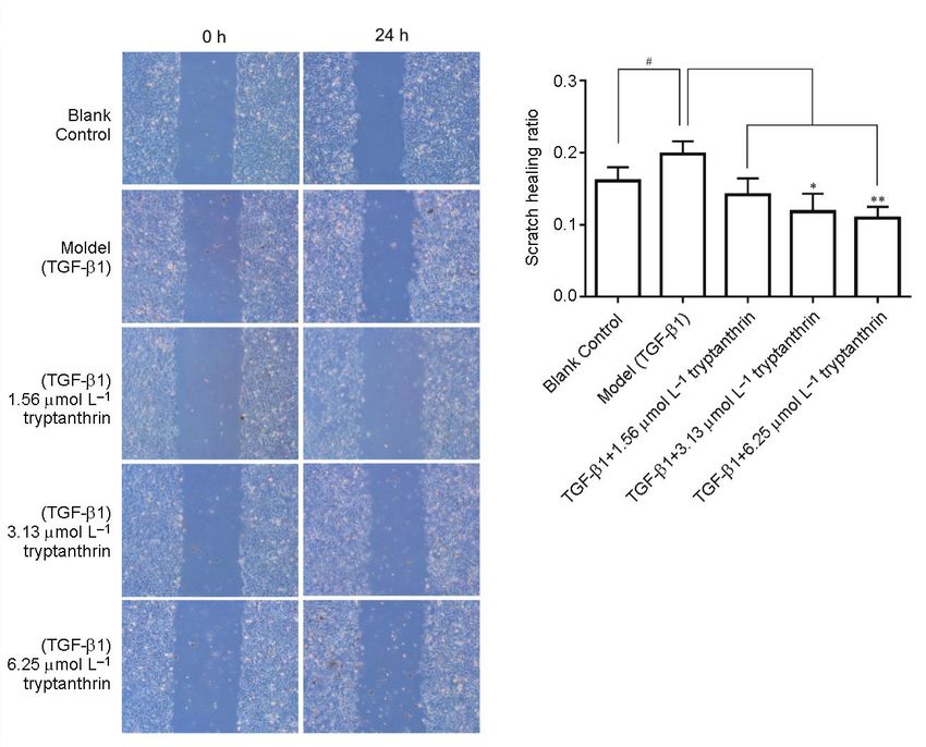
Q. Zeng et al.: Tryptanthrin exerts anti-breast cancer effects both in vitro and in vivo through modulating the inflammatory tumor
microenvironment, Acta Pharm. 71 (2021) ???–???.
which is achieved through inhibiting the lipopolysaccharide (LPS)-induced inflammation,
and down-regulation of pro-inflammatory cytokines (such as TNF-α and IL-6) via the
Nrf2/HO-1 and NF-κB pathways. Additionally, it ameliorates dextran sodium sulfate (DSS)-
-induced colitis in mice (28, 29). In our previous study, tryptanthrin is found to inhibit the
proliferation of human breast cancer MCF-7 cells, and the underlying mechanism of action
may be related to the activation of the MAPK signaling pathways. However, the effect of
tryptanthrin on the inflammatory TME in breast cancer has not been investigated so far, and
a few of the previous studies indicate that tryptanthrin has anti-inflammatory activity and
inhibits breast cancer cell proliferation in vitro (30–32). Therefore, we hypothesize that trypt-
anthrin exerts its anti-breast cancer effects by modulating the inflammatory TME both in vivo
and in vitro. To verify this hypothesis, the current study was carried out to examine the effects
of tryptanthrin on the migration and invasion of human breast cancer MCF-7 cells and the
post-transcriptional activation of EMT associated proteins in vitro. Further, the effects of
tryptanthrin on modulating the inflammatory molecules and proteins, and the pathological
changes in tumor tissues and organs were examined using tumor-bearing mice. Therefore,
this study aimed to explore the mechanisms of action by which tryptanthrin exerted its
antitumor effects through modulating the inflammatory TME both in vitro and in vivo.
EXPERIMENTAL
Chemicals
Tryptanthrin (purity > 99.0 %) was chemically prepared in our laboratory C-2-14 of
School of Pharmacy, Guizhou Medical University, according to the reported method (33),
the detected spectral data of tryptanthrin is as follows (Fig. 1): yellow powder, C15H8N2O2,
1
H NMR (400 MHz, CDCl3) δ: 8.63 (d, J = 8.1 Hz, 1H, Ar–H), 8.44 (dd, J = 7.9, 1.3 Hz, 1H,
Ar–H), 8.04 (d, J = 8.1 Hz, 1H, Ar–H), 7.92 (d, J = 7.5 Hz, 1H, Ar–H), 7.86 (td, J = 7.8, 1.5 Hz,
1H, Ar–H), 7.79 (td, J = 7.9, 1.3 Hz, 1H, Ar–H), 7.68 (m, 1H, Ar–H), 7.43 (t, J = 7.5 Hz, 1H,
Ar–H). 13C NMR (101 MHz, CDCl3) δ: 182.79, 158.29, 146.77, 146.51, 144.49, 138.50, 135.34,
130.91, 130.45, 127.73, 127.40, 125.61, 123.89, 122.08, 118.15. HSMS calcd for C15H9N2O2 (M+H)+
249.0663, found 249.0659.
Cell lines and animals
4T1 cell line and MCF-7 cell line were successively purchased from Wuhan Yipu Bio-
technology Company for two cell passages. 4T1 cells were maintained in 1640 medium
supplemented with 10 % fetal bovine serum (FBS), 100 U mL–l penicillin, and 100 μg mL–l
streptomycin in an incubator under 37 °C and 5 % CO2 conditions. Meanwhile, MCF-7
cells were cultivated in DMEM medium containing 10 % FBS, 100 U mL–l penicillin, and
100 μg mL–l streptomycin. The 4-6-week-old adult Bal b/c female mice weighing 18–22 g
were purchased from the Experimental Animal Center of Guizhou Medical University
(Animal certificate number: SCXK (Beijing) 2016-0002).
Cell viability assay
MCF-7 cells were seeded into 96-well plates at a density of 1.0 × 10⁵ cells mL–l, and
cultured in a cell incubator at 37 °C and 5 % CO2 conditions. Then, the cells were treated
3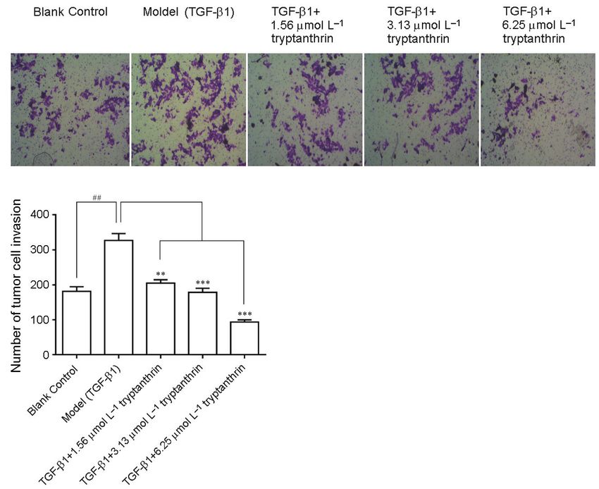
Q. Zeng et al.: Tryptanthrin exerts anti-breast cancer effects both in vitro and in vivo through modulating the inflammatory tumor
microenvironment, Acta Pharm. 71 (2021) ???–???.

                   a)

                   b)

                   c)

Fig. 1. a) 1H NMR spectrum, b) 13C NMR spectrum and c) high-resolution mass spectrum of tryptan-
thrin.

4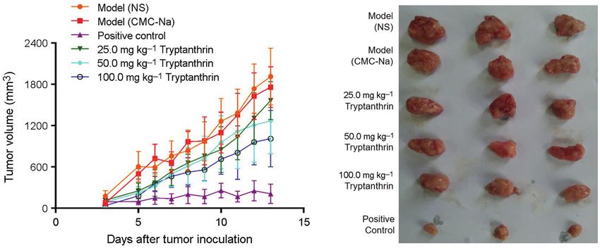
Q. Zeng et al.: Tryptanthrin exerts anti-breast cancer effects both in vitro and in vivo through modulating the inflammatory tumor
microenvironment, Acta Pharm. 71 (2021) ???–???.
with different concentrations of tryptanthrin (1.56, 3.13, 6.25, 12.5, 25.0, 50.0 μmol L–l) for
24, 48 and 72 h, respectively. Afterward, 15 µL of 5 % MTT solution was added into each
well for slight mixing, and the supernatant was discarded after continuous culture at
37 °C for 4 h. Subsequently, 150 µL DMSO was added into each well, and the plate was
shaken at room temperature for 10 min to dissolve crystals. The experiments were repeated
at least three times, the absorbance (OD) value was measured using the microplate reader
at the wavelength of 490 nm, and the average value was calculated based on the experi-
mental data of each group.
Cell colony-forming assay
MCF-7 cells were plated into the 6-well plate at a density of 250 cells mL–l and treated
with either tryptanthrin at 6.25, 3.13 and 1.56 μmol L–l or left untreated as the blank control
group. Three wells were set for each treatment. After 2 weeks of culture, colonies contain-
ing at least 50 cells were stained with Giemsa, photographed and counted. The experiment
was repeated at least three times.
H&E staining
A little amount of culture medium was dropped into the 24-well plate, and then the
24-well plate dedicated cell round-glass crawling slide (WHB-24-CS, ID 14mm, Shanghai
Wohong Biotechnology) was placed horizontally in each well of the 24-well plate. The MCF-7
cell concentration was adjusted to 1.0 × 10⁵ cells mL–l, and then 600 µL cell solution was
added dropwise onto the glass slide surface. After the cell adhesion to the slide, the super-
natants were removed, and the tryptanthrin-containing (6.25, 12.5, 25.0 μmol L–l) culture
medium was added to further culture the MCF-7 tumor cells on the cell-adhered glass slides
at 37 °C for 24 h. Then these glass slides were taken out, then cells were washed twice with
PBS, fixed with 95 % ethanol, and washed with PBS again. Later, cells on the glass slides were
stained with hematoxylin for 5 min and washed with PBS; then, the glass slides were im-
mersed into the 1 % hydrochloric acid alcohol solution to differentiate cells for 10 s and
washed with PBS again. Finally, these slides were transferred into the eosin dye solution for
6 min, washed with PBS, blow/air dried, and photographed under an optical microscope to
obtain the results.
Wound healing assay
A wound healing assay was carried out to detect the migration of tumor cells, MCF-7
cells were inoculated into six-well plates, and the assay was initiated when the cells reached
80~90 % confluence. Tumor cells in the model group were treated with 5 μg L–l TGF-β1 (Lot
no. 0218209-1, PeproTech) for 48 h. Then, a scratch was made in each well of six-well plates
using a 200-µL pipette tip, and the plates were washed twice with PBS. Five cell experimen-
tal groups were set in this experiment, including a blank control group, model group, as well
as high, medium and low concentration of tryptanthrin groups. Of them, cells in model
group were treated with TGF-β1 (containing 1 % FBS); those in the three different concentra-
tion tryptanthrin groups were treated with TGF-β1+6.25 µmol L–l tryptanthrin, TGF-β1+3.13
µmol L–l tryptanthrin and TGF-β1+1.56 µmol L–l tryptanthrin, respectively. Later, cells in
each group were photographed in the same position after 0 and 24 h of the above-mentioned
treatments, respectively, and the scratch width was measured with the ImageJ software.
5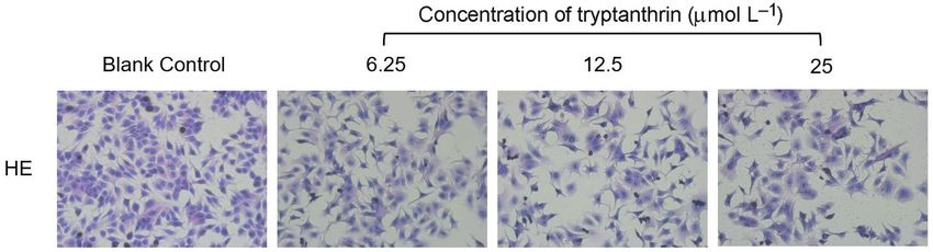
Q. Zeng et al.: Tryptanthrin exerts anti-breast cancer effects both in vitro and in vivo through modulating the inflammatory tumor
microenvironment, Acta Pharm. 71 (2021) ???–???.

Also, the scratch healing rate was calculated according to the following formula to reflect the
migration capacity of tumor cells, and the experiment was repeated at least three times.

                                                       Scratch width(0h) – Scratch width(24h)
                      Scratch healing rate =
                                                                Scratch width(0h)

Transwell chamber assay
     Transwell chamber assay on the invasion of tumor cells, tumor cells were inoculated
into the six-well plates, and five cell experimental groups were set in this experiment,
which were the blank control group, the model group (5 μg L–l TGF-β1), as well as high,
medium and low concentrations of tryptanthrin groups. Specifically, cells in these three
different concentration tryptanthrin groups were treated with TGF-β1+6.25 µmol L–l trypt-
anthrin, TGF-β1+3.13 µmol L–l tryptanthrin and TGF-β1+1.56 µmol L–l tryptanthrin, respec-
tively, for 24 h. Subsequently, 80 µL Matrigel (1:11 dilution, Lot no. 7205008, Corning) was
added into the insert of each 24-well Transwell plate (Lot no. 25717024, Corning) to form
the Matrigel layer on the insert membrane; then, all the above-treated inserts were main-
tained in a clean bench, and air-dried under the hood without UV light. Subsequently, cells
were dissociated and diluted to 5 × 10⁵ cells mL–l, then 200 µL cell suspension was seeded
into each 24-well Transwell plate coated with Matrigel, and 600 µL culture medium con-
taining 10 % FBS was added to the lower layer of each 24-well Transwell plate. Following
incubation for 24 h at 37 °C, the culture medium in each well was discarded, the upper
layer was washed twice with PBS, then both cells and Matrigel outside the permeable
membrane were scraped off with sterilized cotton, and tumor cells invading the mem-
brane were fixed with methanol and stained with crystal violet. Five random fields of view
(FOV) were selected from each membrane of the 24-well Transwell plates under the micro-
scope, and photos were taken to count the invading cells.

Western blotting
     This assay was conducted in the 6-well plates supplemented with MCF-7 tumor cells.
Five cell experiment groups were set, including the blank control group, the model group
treated with 5 μg L–l TGF-β1 for 48 h, and three different concentrations of tryptanthrin-
-treated groups with TGF-β1+25 μmol L–l tryptanthrin, TGF-β1+12.5 μmol L–l tryptanthrin,
and TGF-β1+6.25 μmol L–l tryptanthrin, respectively. After tryptanthrin treatment for 24 h,
all cells were washed twice with the 4 °C PBS and collected by centrifugation. The cellular
proteins were extracted using the Nuclear and Cytoplasmic Protein Extraction Kit (Lot no.
050318180716, Shanghai Beyotime Biotechnology), and the cellular protein concentrations
were determined using the BCA protein concentration determination kit (Lot no. 20180816,
Beijing Solarbio). After sodium dodecyl sulfate-polyacrylamide gel electrophoresis (SDS-
PAGE) vertical plate electrophoresis, the membranes were electrically transferred onto the
polyvinylidene fluoride (PVDF) membrane (Lot no A16954280, GE Healthcare), sealed with
5 % skimmed milk powder for 1 h, and washed with TBST for 5 min three times. Subse-
quently, the membranes were incubated with E-cadherin (1:1000 dilution, Lot no. 3195T, Cell
Signaling Technology), Snail (1:1000 dilution, Lot no. 3879T, Cell Signaling Technology),
MMP-2 (1:1000 dilution, Lot no. GR319128-3, Abcam) and GAPDH (1:8000 dilution, Lot no.

6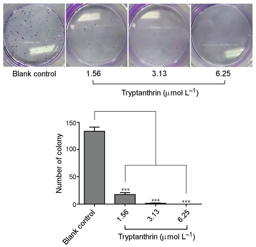
Q. Zeng et al.: Tryptanthrin exerts anti-breast cancer effects both in vitro and in vivo through modulating the inflammatory tumor
microenvironment, Acta Pharm. 71 (2021) ???–???.
10494-1-AP, Wuhan Sanying) antibodies overnight at 4 °C. Then, the membranes were
washed with TBST three times, and then incubated with corresponding horseradish per-
oxidase (HRP)-labeled secondary antibody (1:5000 dilution, Lot no. 134658, Beijing Zhong-
shan Goldenbridge Biotechnology) for 2 h at room temperature. Later, the immunolabeled
bands were visualized using the Immobilon ECL Western HRP substrate (Lot no. 1735601,
Millipore). Digital images of the blots were photographed by the ChemiDoc XRS+ system
and analyzed via the Image Lab Software (Bio-Rad Laboratories). The results were normal-
ized to those of GAPDH, and data from three independent experiments were analyzed.
Establishment of 4T1 mouse breast cancer model
All Bal b/c mice were housed at the Experimental Animal Laboratory of School of
Basic Medical Sciences, Guizhou Medical University. The experimental procedures and
protocols were reviewed and approved by the Animal Care and Use Committee of
Guizhou Medical University and carried out in strict accordance with the Guide for the
Care and Use of Laboratory Animals. 0.1 mL of 1 × 10⁶ mL–l well-grown murine breast
cancer 4T1 cell suspension was injected into the right forelimb armpit of each SPF female
Bal b/c mouse. These model mice were randomly divided into six groups when the tumor
size grew to about 100 mm3 with 9 mice in each experimental group. Groups included
were normal control group, model group (normal saline, NS), model group (0.5 % CMC-
Na, sodium carboxymethyl cellulose), positive control group (40 mg kg–l cyclophospha-
mide), as well as high, medium and low concentration tryptanthrin groups (100.0, 50.0 and
25.0 mg kg–l). The tryptanthrin solutions were administered through oral gavage to mice
once daily for a total of 13 consecutive days. Then, the physical conditions and mental state
of test mice were observed and recorded, the longest diameter (a) and the shortest dia
meter (b) of each tumor were measured with the vernier caliper daily, and the formula for
calculating the tumor volume (V) was as follows, V = ½ × ab2.
Animal tissue harvesting
After continuous tryptanthrin administration for 13 days, the experimental mice
were anesthetized to collect blood samples from the inferior vena cava. Meanwhile, the
serum samples were obtained by low-temperature & low-speed centrifugation and pre-
served at –80 °C for subsequent ELISA. Afterward, the mice were sacrificed by cervical
dislocation, and tumor, liver, spleen and lung were dissected from each mouse, divided
into multiple parts, and finally stored in the –80 °C refrigerator for subsequent analyses.
The computational formula of the organ coefficient was as follows:
Organ coefficient = Organ mass / Body mass × 100 %.
ELISA
The IL-2, IL-6, IL-10, IL-12 and TNF-α levels in mouse serum were measured by the
specific ELISA system kits (Lot no. 201808, Shanghai Jianglai Biology) in accordance with
manufacturer instructions. The nonspecific binding signal was obtained after the incuba-
tion of serum samples and diluted into the appropriate buffer solution in the absence of
the capture antibody. The experiments were repeated three times.
7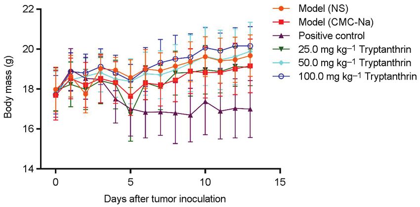
Q. Zeng et al.: Tryptanthrin exerts anti-breast cancer effects both in vitro and in vivo through modulating the inflammatory tumor
microenvironment, Acta Pharm. 71 (2021) ???–???.

H&E staining of tumor tissues and organs
     Pathological changes in tumor tissues and organs from tumor-bearing mice were
observed by means of H&E staining. In brief, tumor tissues and organs were fixed with
the 4 % paraformaldehyde solution, embedded in paraffin, and sliced into the 4 μm paraffin
sections, followed by further deparaffinage and staining according to the standard opera-
tional procedure of H&E. Thereafter, the sections were subjected to methanol gradient
dehydration, xylene transparentizing, and sealing with neutral gum. The results of H&E
staining were presented in the electronic photos through microscopic analysis and photo-
graphs.

IHC and Western blotting of tumor tissues
     IHC experiment was conducted to measure the protein levels of NOS1 and COX-2 in
tumor tissues from tumor-bearing mice. Similarly, the tumor tissues were fixed with 4 %
paraformaldehyde solution, dehydrated, embedded in paraffin, sliced and other opera-
tional steps. The IHC tissue sections were incubated with primary antibody solution and
then secondary antibody solution at room temperature in dark. Afterwards, DAB staining,
hematoxylin counterstaining, staining and sealing of the tested tissue sections were com-
pleted in succession. The IHC staining results were collected finally.
     Mouse tumor tissues were cut into pieces, lysed in the tissue-lysate solution (1 mg:10
μL, RIPA, Lot no. 20170712, Solarbio), and homogenized with the glass homogenizer. Then,
tissue protein concentrations were determined through the BCA method, and the NF-κB
p65 protein expression in the tumor tissues from tumor-bearing mice was analyzed by
Western blotting. Following tumor tissue lysis, the proteins were resolved by SDS–PAGE,
transferred onto the PVDF membranes, and incubated with primary and secondary anti-
bodies. In this experiment, the anti-NF-κB p65 (1:1000 dilution, Lot no. 8242S, Cell Signal-
ing Technology) and anti-GAPDH primary antibodies were used. The protein bands were
visualized by chemiluminescence and quantified through densitometry using Image Lab
Software. The results were normalized to those of GAPDH, and the data from three inde-
pendent experiments were analyzed.

Statistical analysis
    The SPSS 22.0 software was adopted for statistical analysis, and the statistical signifi-
cance was compared using Student’s t-test or one-way analysis of variance (ANOVA), fol-
lowed by post-hoc test. p < 0.05 indicated statistical significance. All results are expressed
as mean ± SD.

                                             RESULTS AND DISCUSSION

Tryptanthrin inhibited the proliferation and colony formation of MCF-7 cells
     Human breast cancer MCF-7 cells were treated with different concentrations of tryptan-
thrin for 24, 48 and 72 h, respectively. The curves regarding cell proliferation inhibition rates
are shown in Fig. 2a. As observed, the inhibition rates of the low, medium and high concentra-
tion tryptanthrin groups remarkably increased (*p < 0.05, ** p < 0.01 or *** p < 0.001) in a time-

8
Q. Zeng et al.: Tryptanthrin exerts anti-breast cancer effects both in vitro and in vivo through modulating the inflammatory tumor
microenvironment, Acta Pharm. 71 (2021) ???–???.
and concentration-dependent manner. Particularly, when MCF-7 cells were treated with 50.0
µmol L–l tryptanthrin for 72 h, the proliferation rate of MCF-7 cells suffered from the most
significant decrease (*** p < 0.001). In the colony formation assay, the number of colonies formed
in MCF-7 cells treated with tryptanthrin at 6.25, 3.13 and 1.56 μmol L–l was dramatically lower
than that in the blank control group (Fig. 2b). These results clearly demonstrated that tryptan-
thrin exerted the antitumor activity on MCF-7 cells partially by decreasing cell viability and
inhibiting proliferation. Results of H&E staining revealed that the in vitro morphologic
changes in MCF-7 cells, induced by tryptanthrin treatment, were more pronounced than those
in the blank control group. Typically, good cell adherence, reddish cytoplasm and irregular
spindle shape were the main characteristics of MCF-7 cells. After tryptanthrin treatment and
killing of tumor cells, the number of MCF-7 cells has decreased, along with poor cell adhe
rence, darkened nuclei, disappeared cell membrane, and diffused cytoplasm (Fig. 2c). These
cellular morphologic changes demonstrated the favorable antitumor effect of tryptanthrin.
Tryptanthrin blocked migration and invasion of MCF-7 cells
The tumor cell mobility in TGF-β1 induced model group substantially increased com-
pared with that in the blank control group (#p < 0.05) (Fig. 3). After treatment with 3.13 and 6.25
µmol L–l tryptanthrin solutions, the tumor cell mobilities in tryptanthrin groups outstand-
ingly decreased relative to that in the model group (*p < 0.05, ** p < 0.01). Fig. 3 shows the mi-
croscopic photographs (×200) and scratch healing rates of MCF-7 tumor cells in the three trypt-
anthrin concentration groups. The results show what tryptanthrin had greatly inhibited the
migration of MCF-7 tumor cells in vitro. In the tumor cell invasion experiment, the high dose
tryptanthrin (6.25 µmol L–l) had maximally reduced the invasion of MCF-7 tumor cells, with
the smallest number of invading cells (***p < 0.001, Fig. 4); therefore, the tryptanthrin concen-
tration was in direct proportion to the number of invading tumor cells. Our results indicated
that tryptanthrin dramatically blocked the migration and invasion of MCF-7 tumor cells.
Tryptanthrin reversed EMT-associated E-cadherin, MMP-2 and Snail
EMT is an embryonic procedure known to participate in breaking down the cell adhe-
sion complexes and enhancing cell migration and invasion. Cancer cells with EMT show
higher aggressiveness, stronger invasiveness, obvious stem cells-like characteristics, and
higher anti-apoptosis capacity (34). E-cadherin initiates a series of signal transduction path-
ways and a major cell framework recombination process, and the deletion of E-cadherin is
considered as a critical step in tumor and EMT development. The post-transcriptional acti-
vation of E-cadherin is an important factor to predict the prognosis for breast cancer, since
this protein is considered as one of the EMT marker proteins. Hence, we evaluated the ex-
pression of E-cadherin in MCF-7 cells induced by TGF-β1 in this study. The results sug-
gested the reduction of the E-cadherin protein expression in the model group (MCF-7 cells
treated with TGF-β1; #p < 0.05). However, when cells were treated with tryptanthrin, the E-
cadherin protein levels in cells treated with 6.25, 12.5, and 25.0 µmol L–l tryptanthrin had
increased (*p < 0.05). Furthermore, the protein expression levels of MMP-2 and Snail in MCF-
7 cells induced by TGF-β1 were markedly upregulated (#p < 0.05). After tryptanthrin treat-
ment, the protein levels of MMP-2 and Snail in 6.25, 12.5 and 25.0 mol L–l tryptanthrin groups
were remarkably downregulated (*p < 0.05, **p < 0.01, Fig. 5). The above results indicated that
tryptanthrin suppressed the EMT process to achieve the anti-tumor effect.
9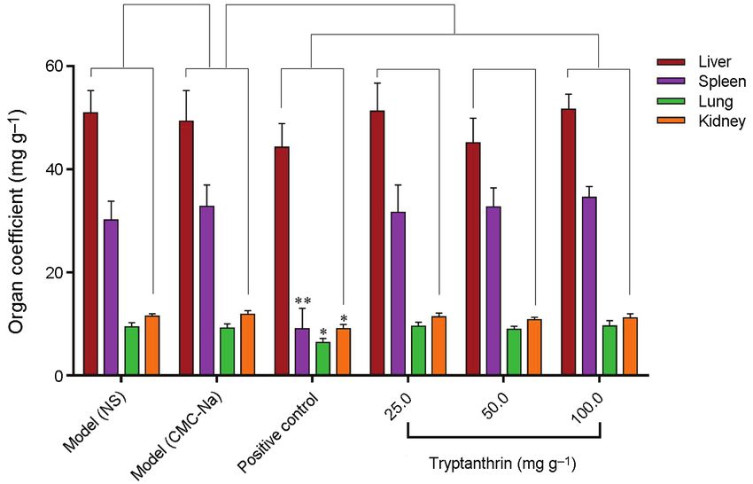
Q. Zeng et al.: Tryptanthrin exerts anti-breast cancer effects both in vitro and in vivo through modulating the inflammatory tumor
microenvironment, Acta Pharm. 71 (2021) ???–???.

                          a)

              b)

              c)

Fig. 2. a) Inhibitory effect of tryptanthrin on MCF-7 tumor cell proliferation, b) the number of MCF-7
cells’ colony formations of and c) HE-stained optical microscopic photographs (×200) of tryptanthrin-
treated MCF-7 cells (compared to the blank control group, *p < 0.05, **p < 0.01, ***p < 0.001, n = 6).

10
Q. Zeng et al.: Tryptanthrin exerts anti-breast cancer effects both in vitro and in vivo through modulating the inflammatory tumor
microenvironment, Acta Pharm. 71 (2021) ???–???.
a) b)
Fig. 3. a) The optical microscopic photographs (×200) and b) scratch healing rates of the effect of dif-
ferent tryptanthrin concentrations on the migration of MCF-7 tumor cells (compared to blank control
group, #p < 0.05; compared to the model group, *p < 0.05, **p < 0.01, n = 6).
a)
b)
Fig. 4. a) The optical microscopic photographs (×200) and b) the quantized histograms of tumor cells
after cell invasion after the treatment with different concentrations of tryptanthrin (compared to
normal control group, # #p < 0.01; compared to the model group, **p < 0.01, ***p < 0.001, n = 6).
11Q. Zeng et al.: Tryptanthrin exerts anti-breast cancer effects both in vitro and in vivo through modulating the inflammatory tumor
microenvironment, Acta Pharm. 71 (2021) ???–???.
Effects of tryptanthrin on body mass, organ coefficients and tumor growth
in tumor-bearing mice
The mental state of all tested mice was reflected indirectly through observing their
daily food intake, physical strength and normal activity, and comparing the differences
between distinct groups. The mice in experimental groups after tryptanthrin treatment had
a better appetite and a greater range of limb motion than those in the morbid model group.
The mean body mass of tumor-bearing mice was not evidently affected by tryptanthrin at
different doses on the 13th day after tryptanthrin administration, and it showed a normal
upward trend compared with the 19.16 ± 1.35 g of the model group (0.5 % CMC-Na) or 19.68
± 1.04 g of the model group (NS). In sharp contrast to the positive group, the mean body mass
of tumor-bearing mice given oral administration of 40 mg kg–l cyclophosphamide for 13 days
was 17.0 ± 1.43 g, which was significantly reduced (Fig. 6). Besides, the organ coefficient of
tumor-bearing mice is another effective physiological index. There were no significant dif-
ferences in the coefficients of the liver, spleen, lung and kidney between the 100.0, 50.0, 25.0
mg kg–l tryptanthrin groups and the model groups (p > 0.05). Similarly to the changes in
mass mentioned above, the organ coefficients of the liver, spleen, lung and kidney in the
positive group were reduced (Fig. 7), especially for the coefficients of lung and kidney (*p <
0.05), as well as spleen (**p < 0.01).
Tumor growth in the tumor-bearing mice of different groups was investigated, and the
change curves of tumor volume during the treatment period were collected, as summarized
in Fig. 8a. The tumor volumes in 100.0, 50.0, 25.0 mg kg–l tryptanthrin-treated groups at the
same treatment time were smaller than those in the two model groups, and the antitumor
efficacy of 100.0 mg kg–l tryptanthrin was quite remarkable, which was only second to the
positive drug-cyclophosphamide in this study. Fig. 8b shows the morphologic changes in
breast cancer tissues from tumor-bearing mice treated with tryptanthrin. As shown by the
results, different concentrations of tryptanthrin exhibited in vivo antitumor activities to
various degrees; additionally, the data from model group and positive group testified the
successful establishment of the in vivo tumor model, thus confirming the reliability of in vivo
anti-tumor effect of tryptanthrin.
Tryptanthrin regulated IL-2, IL-6, IL-10, IL-12 and TNF-α levels in tumor-bearing mice
The serum IL-2 and TNF-α levels of tumor-bearing mice in 100.0, 50.0, 25.0 mg kg–l trypt-
anthrin-treated groups and the positive group had increased in comparison to those in the
model group (0.5 % CMC-Na) (*p < 0.05, **p < 0.01). To be specific, the IL-2 levels in 100.0 and
50.0 mg kg–l tryptanthrin-treated groups were 1.93 and 1.67 as high as that in the model group;
while the TNF-α level in 100.0 mg kg–l tryptanthrin group was 1.37 times of that in model
group. As a soluble leukocyte stimulating factor released by T cells, IL-2 is considered as a
growth factor of T cells and natural killer cells, which exerts an anti-tumor role through pro-
moting the activities of these cells (35, 36). Relevant studies demonstrate that IL-2 has consider-
able therapeutic efficacy in the treatment of cancer. TNF-α induces tumor apoptosis and medi-
ates the occurrence of chronic inflammation, thus initiating the development of the malignant
tumor. The serum IL-10 levels in tumor-bearing mice from tryptanthrin-treated groups were
almost identical to those in the normal group, whereas the levels in the model group (0.5 %
CMC-Na) were relatively high compared to the normal group (#p < 0.05). IL-10 is reported to
promote the tumor immune escape by reducing the anti-tumor immune response in the TME,
12Q. Zeng et al.: Tryptanthrin exerts anti-breast cancer effects both in vitro and in vivo through modulating the inflammatory tumor
microenvironment, Acta Pharm. 71 (2021) ???–???.
Fig. 5. The expression level of E-cadherin, MMP-2 and Snail proteins in MCF-7 tumor cells under the
effect of tryptanthrin (compared to the blank control group, #p < 0.05; compared to the model group,
*p < 0.05, **p < 0.01, n = 3).
13Q. Zeng et al.: Tryptanthrin exerts anti-breast cancer effects both in vitro and in vivo through modulating the inflammatory tumor
microenvironment, Acta Pharm. 71 (2021) ???–???.
Fig. 6. Changes in the body mass of tumor-bearing mice under the effect of tryptanthrin (n = 9).
Fig. 7. Organ coefficients of tumor-bearing mice under the effect of tryptanthrin (compared to model
group, * p < 0.05, **p < 0.01, n = 9).
and IL-10 up-regulation is reported to be related to the poor patient prognosis which plays a
promoting role in breast cancer (37, 38). The serum IL-6 and IL-12 levels in tumor-bearing mice
of all experimental groups did not exhibit a significant trend (Fig. 9).
Tryptanthrin improved histopathological changes in organs and tumor tissues from
tumor-bearing mice
Livers, lungs and spleens were dissected from tumor-bearing mice as the main organs
to evaluate the therapeutic effect of tryptanthrin based on the H&E staining histopatho-
logical images. As shown in Fig. 10, the ratios of H&E staining in the organs of tryptan-
thrin-treated groups showed no significant differences relative to those in the normal control
14Q. Zeng et al.: Tryptanthrin exerts anti-breast cancer effects both in vitro and in vivo through modulating the inflammatory tumor
microenvironment, Acta Pharm. 71 (2021) ???–???.
a) b)
Fig. 8. a) The change curves of tumor volume and b) morphologic changes of breast tumor tissues of
tumor-bearing mice under the effect of tryptanthrin (n = 9).
Fig. 9. Expression of IL-2, IL-6, IL-10, IL-12 and TNF-α in the serum of tumor-bearing mice (compared
to the normal control group, #p < 0.05; compared to the model group, *p < 0.05, **p < 0.01, n = 3).
group and the model group. The tumor in tumor-bearing mice of tryptanthrin-treated
groups showed no difference to the tissue morphology of normal organs, and tryptanthrin
treatments at 25.0~100.0 mg kg–l for once a day for 13 consecutive days had no obvious
15Q. Zeng et al.: Tryptanthrin exerts anti-breast cancer effects both in vitro and in vivo through modulating the inflammatory tumor
microenvironment, Acta Pharm. 71 (2021) ???–???.
toxic effects on these main organs in tumor-bearing mice. By contrast, tumor tissues in
25.0~100.0 mg kg–l tryptanthrin-treated groups were inhibited to various degrees (*p <
0.05), suggesting that tryptanthrin had a favorable anti-breast cancer effect in vivo.
Tryptanthrin inhibited COX-2, NOS1 and NF-κB p65 in tumor tissues from
tumor-bearing mice
IHC analysis demonstrated that the expression levels of COX-2 and NOS1 were down-
regulated in tumor tissues from tumor-bearing mice treated with various doses of trypt-
Fig. 10. HE-staining histopathological images of liver, spleen, lung tissues (×400) and tumor tissue
(×200) of normal mice and tumor-bearing mice under the effect of tryptanthrin.
16Q. Zeng et al.: Tryptanthrin exerts anti-breast cancer effects both in vitro and in vivo through modulating the inflammatory tumor
microenvironment, Acta Pharm. 71 (2021) ???–???.
anthrin or the positive drug (Fig. 11). In the meantime, the expression of COX-2 in tumor-
bearing mice treated with 100.0 mg kg–l tryptanthrin, and that of NOS1 in tumor-bearing
mice treated with 25.0, 50.0, 100.0 mg kg–l tryptanthrin, were dramatically suppressed
(*p < 0.05). COX-2 is overexpressed in the metastatic process of all cancers, which is considered
to be a marker of the dismal prognosis for breast cancer and lung cancer (39). Furthermore,
a) b)
Fig. 11. a) Immunohistochemical staining diagrams and b) quantitative histograms of COX-2 and
NOS1 protein expressions in tumor tissues of tumor-bearing mice (×200) (compared to the 0.5 %
CMC-Na Model group, *p < 0.05, n = 9).
17Q. Zeng et al.: Tryptanthrin exerts anti-breast cancer effects both in vitro and in vivo through modulating the inflammatory tumor
microenvironment, Acta Pharm. 71 (2021) ???–???.
a) b)
Fig. 12. Expression of NF-κB protein in tumor tissues of tumor-bearing mice (compared to the model
group, *p < 0.05, n = 3).
the expression of NF-κB p65 in tumor tissues from tumor-bearing mice was analyzed
through Western blotting, which was high in the model group, but low in the tryptanthrin-
-treated groups and the positive group (*p < 0.05, **p < 0.01) (Fig. 12), indicating that trypt-
anthrin had downregulated NF-κB p65 protein expression in tumor-bearing mice. NF-κB,
as a transcription factor that connects the molecular centers of inflammation and cancer,
is recognized to trigger oncogenesis through activating the NF-κB cascades to enhance cell
proliferation, suppress apoptosis, and promote cell invasion as well as metastasis (40, 41).
CONCLUSIONS
Breast cancer is a malignancy commonly seen among women worldwide. Human
breast cancer MCF-7 cells are a kind of estrogen receptor-positive breast cancer cells. Breast
cancer oncogenesis is related to the inflammatory tumor microenvironment (TME) (42, 43).
In this study, we investigated the modulation of tryptanthrin on the TME both in vitro and
in vivo, and the results demonstrated that tryptanthrin dramatically inhibited the prolife
ration of human breast cancer MCF-7 cells in a time- and concentration-dependent man-
ner. Tryptanthrin treatment induced the poor adherence of MCF-7 cells, resulted in the
darkened nuclei, the loss of cell membrane, and cytoplasm diffusion, indicating a change
in the normal cell morphology. Tryptanthrin treatment not only inhibited the migration
and invasion of MCF-7 cells, but also suppressed the TGF-β1-induced transition of MCF-7
cells, which played an important role in inducing epithelial-mesenchymal transition
(EMT) in vitro. Tryptanthrin had also remarkably enhanced the expression levels of E-cad
herin and depressed those of MMP-2 and Snail in MCF-7 cells stimulated by TGF-β1. This
study demonstrated that tryptanthrin treatment inhibits the proliferation of breast cancer
cells, and suppresses the TME-associated EMT induced by TGF-β1 treatment.
4T1 mouse breast cancer models were applied to evaluate in vivo anti-tumor effect of
tryptanthrin. Body mass of mice administered with the positive drug was dramatically
reduced, whereas in tumor-bearing mice from tryptanthrin treatment groups showed no
marked difference, spleen organ coefficient in the positive group was the most signifi-
18Q. Zeng et al.: Tryptanthrin exerts anti-breast cancer effects both in vitro and in vivo through modulating the inflammatory tumor
microenvironment, Acta Pharm. 71 (2021) ???–???.
cantly reduced. Our results showed that tryptanthrin had considerable inhibitory activity
on tumor growth in mice, much lower toxic and side effects on organisms than those of the
positive drug cyclophosphamide. Serum expression levels of IL-6 and IL-12 in tumor-bear-
ing mice from all groups were not significantly changed, and those of IL-2 and TNF-α in
tumor-bearing mice from tryptanthrin groups were up-regulated. Compared to the normal
control, the serum level of IL-10 in the model group had increased dramatically, whereas the
levels in tumor-bearing mice treated with 100.0 mg kg–l tryptanthrin had decreased. Through
histopathological examinations of organs and tumor tissues from tumor-bearing mice, it
was found that the different doses of tryptanthrin groups partially inhibited the metastasis
and apoptosis of organs and tumor tissues. In addition, the protein expression levels of COX-2,
NOS1 and NF-κB p65 in tumor tissues of tumor-bearing mice from tryptanthrin groups
were notably downregulated, the expression levels of COX-2 and NOS1 are closely associ-
ated with the pro-inflammatory environment of breast cancer.
To sum up, findings of this study suggest that tryptanthrin inhibits the proliferation,
migration and invasion of MCF-7 cells, which is partly achieved through the upregulation
of protein levels of E-cadherin and downregulation of MMP-2 and Snail in MCF-7 cells in
vitro compared to the normal physiological levels. Tryptanthrin also suppresses tumor
growth in mice. The proposed pharmacological mechanism is downregulation of the
NOS1, COX-2 and NF-κB expression in mouse tumor tissues, upregulation of IL-2 and
TNF-α, and recovery of the serum IL-10 levels in mice bearing tumor cells. In conclusion,
tryptanthrin exerts its anti-breast cancer activities through modulating the inflammatory
TEM both in vitro and in vivo.
Acronyms, abbreviations, symbols. – CMC-Na – sodium carboxymethyl cellulose, H&E – hema-
toxylin and eosin, ELISA – enzyme-linked immunosorbent assay, EMT – epithelial-mesenchymal
transition, IHC – immunohistochemistry, TNF-α – tumor necrosis factor α, TME – tumor microenvi-
ronment, HRP – horseradish peroxidase.
Acknowledgements. – This work was financially supported by Guizhou Provincial Natural Sci-
ence Foundation (2020-1Y404), NSFC-Guizhou Karst Scientific Research Center (U1812403), Interna-
tional S&T Cooperation Base for Pharmacy in Guizhou Medical University (2017-5802), Reform Project
of Undergraduate Teaching Content and Course System of Guizhou Medical University (2019-60),
Guizhou Provincial Innovation and Entrepreneurship Training Project for College Students (No.
201510660024) and Doctoral Fund of Guizhou Medical University (No. J[2014]006).
Supplementary material available upon request.
REFERENCES
1. A. H. N. Kamdje, P. F. S. Etet, L. Vecchio, J. M. Muller, M. Krampera and K. E. Lukong, Signaling
pathways in breast cancer: Therapeutic targeting of the microenvironment, Cell. Signal. 26 (2014)
2843–2856; https://doi.org/10.1016/j.cellsig.2014.07.034
2. K. Velaei, N. Samadi, B. Barazvan and J. S. Rad, Tumor microenvironment-mediated chemoresis-
tance in breast cancer, The Breast 30 (2016) 92–100; https://doi.org/10.1016/j.breast.2016.09.002
3. Q. J. Guo, J. Li and H. S. Lin, Effect and molecular mechanisms of traditional chinese medicine on
regulating tumor immunosuppressive microenvironment, BioMed Res. Int. 2015 (2015) 261620;
https://doi.org/10.1155/2015/261620
4. M. Pesic and F. R. Greten, Inflammation and cancer: tissue regeneration gone awry, Curr. Opin.
Cell. Biol. 43 (2016) 55–61; https://doi.org/10.1016/j.ceb.2016.07.010
19Q. Zeng et al.: Tryptanthrin exerts anti-breast cancer effects both in vitro and in vivo through modulating the inflammatory tumor
microenvironment, Acta Pharm. 71 (2021) ???–???.
5. M . Suarez-Carmona, J. Lesage, D. Cataldo and C. Gilles, EMT and inflammation: inseparable ac-
tors of cancer progression, Mol. Oncol. 11 (2017) 805–823; https://doi.org/10.1002/1878-0261.12095
6. X . Y. Li, L. Su, Y. M. Jiang, W. B. Gao, C. W. Xu, C. Q. Zeng, J. Song, Y. Xu, W. C. Weng and W. B.
Liang, The antitumor effect of xihuang pill on treg cells decreased in tumor microenvironment of
4T1 breast tumor-bearing mice by PI3K/AKT~AP-1 signaling pathway, Evid.-Based Compl. Alt. Med.
2018 (2018) 6714829; https://doi.org/10.1155/2018/6714829
7. F. R. Balkwill and A. Mantovani, Cancer-related inflammation: Common themes and therapeutic
opportunities, Semin. Cancer Biol. 22 (2012) 33–40; https://doi.org/10.1016/j.semcancer.2011.12.005
8. Z. T. Li, Y. J. Zhu, C. C. Li, R. Trinh, X. Y. Ren, F. M. Sun, Y. F. Wang, P. Z. Shang, T. Wang, M. Wang,
S. L. Morrison and J. Zhang, Anti-VEGFR2-interferon-α2 regulates the tumor microenvironment
and exhibits potent antitumor efficacy against colorectal cancer, Oncoimmunology 6 (2017) e1290038;
https://doi.org/10.1080/2162402X.2017.1290038
9. F. L. Bai, Z. S. Niu, H. Tian, S. M. Li, Z. Lv, T. Y. Zhang, G. P. Ren and D. S. Li, Genetically engi-
neered Newcastle disease virus expressing interleukin 2 is a potential drug candidate for cancer
immunotherapy, Immunol. Lett. 159 (2014) 36–46; https://doi.org/10.1016/j.imlet.2014.02.009
10. T. van der Heijden, I. Bot and J. Kuiper, The IL-12 cytokine family in cardiovascular diseases,
Cytokine 122 (2019) 154188; https://doi.org/10.1016/j.cyto.2017.10.010
11. K. Singh, M. Roy, P. Prajapati, A. Lipatova, L. Sripada, D. Gohel, A. Singh, M. Mane, M. M. God-
bole, P. M. Chumakov and R. Singh, NLRX1 regulates TNF-α-induced mitochondria-lysosomal
crosstalk to maintain the invasive and metastatic potential of breast cancer cells, BBA-Mol. Basis
Dis. 1865 (2019) 1460–1476; https://doi.org/10.1016/j.bbadis.2019.02.018
12. K. A. Silverio and S. A. Patel, Harnessing antitumor immunity: Employment of tumor recall anti-
gens to optimize the inflammatory response to cancer (Review), Oncol. Lett. 13 (2017) 2015–2020;
https://doi.org/10.3892/ol.2017.5721
13. S . Suman, P. K. Sharma, G. Rai, S. Mishra, D. Arora, P. Gupta and Y. Shukla, Current perspectives
of molecular pathways involved in chronic inflammation-mediated breast cancer, Biochem. Bioph.
Res. Commun. 472 (2016) 401–409; https://doi.org/10.1016/j.bbrc.2015.10.133
14. I . Uehara and N. Tanaka, Role of p53 in the regulation of the inflammatory tumor microenviron-
ment and tumor suppression, Cancers 10 (2018) 219; https://doi.org/10.3390/cancers10070219
15. J . F. Lima, S. Nofech-Mozes, J. Bayani and J. M. S. Bartlett, EMT in breast carcinoma – A review,
J. Clin. Med. 5 (2016) 65; https://doi.org/10.3390/jcm5070065
16. L. Yan, F. Xu and C. L. Dai, Relationship between epithelial-to-mesenchymal transition and the
inflammatory microenvironment of hepatocellular carcinoma, J. Exp. Clin. Cancer Res. 37 (2018)
203; https://doi.org/10.1186/s13046-018-0887-z
17. L. EL-Hajjar, N. Jalaleddine, A. Shaito, K. Zibara, J. M. Kazan, J. El-Saghir and M. El-Sabban, Beva-
cizumab induces inflammation in MDA-MB-231 breast cancer cell line and in a mouse model, Cell.
Signal. 53 (2019) 400–412; https://doi.org/10.1016/j.cellsig.2018.11.007
18. H. N. Chang, S. T. Huang, Y. C. Yeh, H. S. Wang, T. H. Wang, Y. H. Wu and J. S. Pang, Indigo natu-
ralis and its component tryptanthrin exert anti-angiogenic effect by arresting cell cycle and inhibi
ting Akt and FAK signaling in human vascular endothelial cells, J. Ethnopharmacol. 174 (2015)
474–481; https://doi.org/10.1016/j.jep.2015.08.050
19. S . L. Hsuan, S. C. Chang, S. Y. Wang, T. L. Liao, T. T. Jong, M. S. Chien, W. C. Lee, S. S. Chen and J.
W. Liao, The cytotoxicity to leukemia cells and antiviral effects of isatis indigotica extracts on pseu-
dorabies virus, J. Ethnopharmacol. 123 (2009) 61–67; https://doi.org/10.1016/j.jep.2009.02.028
20. Y . H. Liang, H. X. Hou, D. R. Li, J. Qin, L. Qiu and H. H. Wu, Studies on in vitro anticancer activ-
ity of tryptanthrin B, Chin. Tradit. Herbal Drugs 31 (2000) 531–533; https://doi.org/10.3321/j.issn:0253-
2670.2000.07.029
21. J . P. Li, G. H. Zhu, Y. Yuan and M. X. Liu, Anti-tumor and immune function regulation effects of
radix isatidis polysaccharides in vivo, Nat. Prod. Res. Dev. 29 (2017) 2010–2016; https://doi.org/10.163
33/j.1001-6880.2017.12.003
20Q. Zeng et al.: Tryptanthrin exerts anti-breast cancer effects both in vitro and in vivo through modulating the inflammatory tumor
microenvironment, Acta Pharm. 71 (2021) ???–???.
22. L . L. Liu, J. Chen and Y. P. Shi, Advances in studies on antitumor of Chinese materia medica with
heat-clearing and toxin-resolving functions, Chin. Tradit. Herbal Drugs 43 (2012) 1203–1212; https://
www.cqvip.com/qk/80172x/201211/42183551.html
23. G . Honda and M. Tabata, Isolation of antifungal principle tryptanthrin, from Strobilanthes Cusia
O. Kuntze, Planta Med. 36 (1979) 85–86; https://doi.org/10.1055/s-0028-1097245
24. R . Kaur, S. K. Manjal, R. K. Rawal and K. Kumar, Recent synthetic and medicinal perspectives of
tryptanthrin, Bioorg. Med. Chem. 25 (2017) 4533–4552; https://doi.org/10.1016/j.bmc.2017.07.003
25. E . H. Jung, J. Y. Jung, H. L. Ko, J. K. Kim, S. M. Park, D. H. Jung, C. A. Park, Y. W. Kim, S. K. Ku, I.
J. Cho and S. C. Kim, Tryptanthrin prevents oxidative stress-mediated apoptosis through AMP-
activated protein kinase-dependent p38 mitogen-activated protein kinase activation, Arch. Pharm.
Res. 40 (2017) 1071–1086; https://doi.org/10.1007/s12272-017-0947-5
26. S . Lee, D. C. Kim, H. Y. Baek, K. D. Lee, Y. C. Kim and H. Oh, Anti-neuroinflammatory effects of
tryptanthrin from Polygonum tinctorium Lour. in lipopolysaccharide-stimulated BV2 microglial
cells, Arch. Pharm. Res. 41 (2018) 419–430; https://doi.org/10.1007/s12272-018-1020-8
27. S
. T. Yu, J. W. Chern, T. M. Chen, Y. F. Chiu, H. T. Chen and Y. H. Chen, Cytotoxicity and reversal
of multidrug resistance by tryptanthrin-derived indoloquinazolines, Acta Pharmacol. Sin. 31 (2010)
259–264; https://doi.org/10.1038/aps.2009.198
28. Y. W. Kwon, S. Y. Cheon, S. Y. Park, J. Song and J. H. Lee, Tryptanthrin suppresses the activation
of the LPS-treated BV2 microglial cell line via Nrf2/HO-1 antioxidant signaling, Front Cell Neuro-
sci. 11 (2017) 18; https://doi.org/10.3389/fncel.2017.00018
29. M . J. Micallef, K. Iwaki, T. Ishihara, S. Ushio, M. Aga, T. Kunikata, S. Koya-Miyata, T. Kimoto, M.
Ikeda and M. Kurimoto, The natural plant product tryptanthrin ameliorates dextran sodium
sulfate-induced colitis in mice, Int. Immunopharmacol. 2 (2002) 565–578; https://doi.org/10.1016/
S1567-5769(01)00206-5
30. R. Kaur, S. K. Manjal, R. K. Rawal and K. Kumar, Recent Synthetic and Medicinal Perspectives of
Tryptanthrin, Bioorg. Med. Chem. 25 (2017) 4533–4552; https://doi.org/10.1016/j.bmc.2017.07.003
31. W. Zhou, Q. F. Zeng, D. Lai, J. L. Cho, X. Y. Zhang and X. C. Shen, Effect of tryptanthrin on prolife
ration of human breast cancer MCF-7 cells via MAPK signaling pathway, Chin. Pharm. J. 54 (2019)
693–698; https://doi.org/10.11669/cpj.2019.09.005
32. X . M. Liao and K. N. Leung, Tryptanthrin induces growth inhibition and neuronal differentiation
in the human neuroblastoma LA-N-1 cells, Chem. Biol. Interact. 203 (2013) 512–521; https://doi.
org/10.1016/j.cbi.2013.03.001
33. S . Han, D. F. Li, C. M. Wu, X. R. Ma, R. G. Song and Y. Wang, Synthesis and characterization of
indolo quinazoline derivatives, Chem. Reagents 33 (2011) 883–886; https://doi.org/10.13822/j.cnki.
hxsj.2011.10.011
34. J. H. Feng, D. L. Song, S. Y. Jiang, X. H. Yang, T. T. Ding, H. Zhang, J. M. Luo, J. Liao and Q. Yin,
Quercetin restrains TGF-β1-induced epithelial–mesenchymal transition by inhibiting Twist1 and
regulating E-cadherin expression, Biochem. Bioph. Res. Commun. 498 (2018) 132–138; https://doi.
org/10.1016/j.bbrc.2018.02.044
35. M. Mizui, Natural and modified IL-2 for the treatment of cancer and autoimmune diseases, Clin.
Immunol. 206 (2019) 63–70; https://doi.org/10.1016/j.clim.2018.11.002
36. A. Tang and F. Harding, The challenges and molecular approaches surrounding interleukin-
2-based therapeutics in cancer, Cytokine: X 1 (2019) 100001; https://doi.org/10.1016/j.cytox.2018.100001
37. X
. G. Li, P. Lu, B. Li, W. F. Zhang, R. Yang, Y. Chu and K. Y. Luo, Interleukin 2 and interleukin 10
function synergistically to promote CD8+ T cell cytotoxicity, which is suppressed by regulatory T
cells in breast cancer, Int. J. Biochem. Cell Biol. 87 (2017) 1–7; https://doi.org/doi:10.1016/j.bio-
cel.2017.03.003
38. M. H. Mannino, Z. W. Zhu, H. P. Xiao, Q. Bai, M. R. Wakefield and Y. J. Fang, The paradoxical role
of IL-10 in immunity and cancer, Cancer Lett. 367 (2015) 103–107; https://doi.org/10.1016/j.can-
let.2015.07.009
21Q. Zeng et al.: Tryptanthrin exerts anti-breast cancer effects both in vitro and in vivo through modulating the inflammatory tumor
microenvironment, Acta Pharm. 71 (2021) ???–???.
39. R. Liu, H. G. Zheng, W. D. Li, Q. J. Guo, S. L. He, Y. Hirasaki, W. Hou, B. J. Hua, C. H. Li, Y. J. Bao,
Y. B. Gao, X. Qi, Y. X. Pei and Y. Zhang, Anti-tumor enhancement of Fei-Liu-Ping ointment in
combination with celecoxib via cyclooxygenase-2-mediated lung metastatic inflammatory micro-
environment in Lewis lung carcinoma xenograft mouse model, J. Transl. Med. 13 (2015) 366; https://
doi.org/10.1186/s12967-015-0728-1
40. D
. Capece, D. Verzella, A. Tessitore, E. Alesse, C. Capalbo and F. Zazzeroni, Cancer secretome and
inflammation: The bright and the dark sides of NF-κB, Semin. Cell Dev. Bio. 78 (2018) 51–61; https://
doi.org/10.1016/j.semcdb.2017.08.004
41. M
. Patel, P. G. Horgan, D. C. McMillan and J. Edwards, NF-κB pathways in the development and
progression of colorectal cancer, Transl. Res. 197 (2018) 43–56; https://doi.org/10.1016/j.trsl.2018.02.002
42. M. Egue, F. H. R. Gnangnon, M. T. Akele-Akpo and D. M. Parkin, Cancer incidence in Cotonou
(Benin), 2014–2016 First results from the cancer Registry of Cotonou, Cancer Epidemiol. 59 (2019)
46–50; https://doi.org/10.1016/j.canep.2019.01.006
43. L. Su, Y. M. Jiang, Y. Xu, X. Y. Li, W. B. Gao, C. W. Xu, C. Q. Zeng, J. Song, W. C. Weng and W. B.
Liang, Xihuang pill promotes apoptosis of Treg cells in the tumor microenvironment in 4T1
mouse breast cancer by upregulating MEKK1/SEK1/JNK1/AP-1 pathway, Biomed. Pharmacother.
102 (2018) 1111–1119; https://doi.org/10.1016/j.biopha.2018.03.063
22You can also read