Hedgehog Signaling Inhibition Blocks Growth of Resistant Tumors through Effects on Tumor Microenvironment
←
→
Page content transcription
If your browser does not render page correctly, please read the page content below
Published OnlineFirst December 20, 2011; DOI: 10.1158/0008-5472.CAN-11-2681

                                                                                                                                Cancer
Microenvironment and Immunology                                                                                               Research

Hedgehog Signaling Inhibition Blocks Growth of Resistant
Tumors through Effects on Tumor Microenvironment
Emanuela Heller1, Michelle A. Hurchla1, Jingyu Xiang1, Xinming Su1, Sara Chen1, Jochen Schneider4,
Kyu-Sang Joeng2, Marcos Vidal5, Leah Goldberg1, Hongju Deng1, Mary C. Hornick1, Julie L. Prior3,
David Piwnica-Worms3, Fanxin Long2, Ross Cagan6, and Katherine N. Weilbaecher1

Abstract
              Hedgehog (Hh) signaling is implicated in bone development and cellular transformation. Here we show
           that inhibition of Hh pathway activity inhibits tumor growth through effects on the microenvironment.
           Pharmacologic inhibition of the Hh effector Smoothened (Smo) increased trabecular bone in vivo and
           inhibited osteoclastogenesis in vitro. In addition, enhanced Hh signaling due to heterozygosity of the Hh
           inhibitory receptor Patched (Ptch1þ/) increased bone resorption, suggesting direct regulation of osteoclast
           (OC) activity by the Hh pathway. Ptch1þ/ mice had increased bone metastatic and subcutaneous tumor
           growth, suggesting that increased Hh activation in host cells promoted tumor growth. Subcutaneous growth
           of Hh-resistant tumor cells was inhibited by LDE225, a novel orally bioavailable SMO antagonist, consistent
           with effects on tumor microenvironment. Knockdown of the Hh ligand Sonic Hh (SHH) in these cells
           decreased subcutaneous tumor growth and decreased stromal cell production of interleukin-6, indicating
           that tumor-derived Hh ligands stimulated tumor growth in a paracrine fashion. Together our findings show
           that inhibition of the Hh pathway can reduce tumor burden, regardless of tumor Hh responsiveness, through
           effects on tumor cells, OCs, and stromal cells within the tumor microenvironment. Hh may be a promising
           therapeutic target for solid cancers and bone metastases. Cancer Res; 72(4); 1–11. 2011 AACR.

Introduction                                                                 the importance of Hh signaling. Several next-generation ana-
                                                                             logs with increased specificity and tolerability (ref. 7; including
   The Hedgehog (Hh) signaling pathway plays critical roles in               GDC-0449, LDE225, and IPI-926) exert antitumor effects in a
epithelial–mesenchymal transition and cell differentiation                   subset of cancer cells and are currently in clinical trials for a
during embryonic development (1, 2), adult tissue homeosta-                  wide variety of tumors (8–10).
sis, and tumorigenesis (3, 4). In the absence of ligand, the Hh                 The Hh signaling pathway plays a critical role in tumori-
receptor Patched (PTCH) inhibits the activator Smoothened                    genesis and progression in many tumor types. Mutations
(SMO). Upon ligand binding to PTCH, SMO is released, result-                 leading to ligand-independent Hh pathway activation have
ing in pathway activation and transcription of target genes,                 been linked to basal cell carcinoma and medulloblastoma (11,
including Gli1, Gli2, and Ptch1 (5, 6). Cyclopamine, a naturally             12), whereas overexpression of the Hh ligand Sonic Hh (Shh) or
occurring Hh inhibitor and SMO antagonist, first highlighted                  mutations in Hh signaling genes (Smo, Ptch1, Gli1, and Gli2)
                                                                             have been implicated in the emergence and progression of
                                                                             numerous epithelial cancers, including breast, skin, esophagus,
Authors' Affiliations: 1Department of Medicine, Division of Oncology,         stomach, pancreas, liver, lung, and prostate (11, 13, 14). Hh
2
 Department of Medicine, Division of Endocrinology, Metabolism and Lipid     inhibition via cyclopamine suppressed proliferation of breast
Research, 3BRIGHT Institute and Molecular Imaging Center, Mallinkrodt
Institute of Radiology, Washington University School of Medicine, St.        carcinoma cell lines and decreased Gli1 (15, 16). These effects
Louis, Missouri; 4Luxembourg Centre for Systems Biomedicine, University      are not limited to primary tumors, as inhibition of the Hh
of Luxembourg, Luxembourg City, Luxembourg; 5The Beatson Institute for
Cancer Research, Glasgow, United Kingdom; and 6Department of Devel-
                                                                             pathway decreased lung and liver metastases in a mouse
opmental and Regenerative Biology, Mount Sinai School of Medicine, New       pancreatic cancer model together with gemcitabine (17, 18).
York                                                                            In addition to direct effects on tumor cell growth, Hh
Note: Supplementary data for this article are available at Cancer Research   signaling within the host stromal microenvironment also con-
Online (http://cancerres.aacrjournals.org/).                                 trols tumor progression. Mice with a targeted disruption of the
E. Heller and M.A. Hurchla contributed equally to this work.
                                                                             Hh inhibitory receptor Ptch1 develop ductal hyperplasia (15).
                                                                             Interestingly, it was disruption of Ptch1 in mammary stroma,
Corresponding Author: Katherine N. Weilbaecher, Department of Med-
icine, Division of Oncology, Washington University School of Medicine, 660   rather than in mammary epithelium, that led to the ductal
S. Euclid Ave, Box 8069, St. Louis, MO 63110. Phone: 314-454-8858; Fax:      changes, suggesting an indirect effect of Hh signaling on tumor-
314-454-8979; E-mail: kweilbae@dom.wustl.edu                                 initiating cells. Furthermore, paracrine Hh activation in host-
doi: 10.1158/0008-5472.CAN-11-2681                                           derived stromal cells leads to increased tumor growth (19–21)
2011 American Association for Cancer Research.                              and is necessary to support the growth of stromal-dependent B

www.aacrjournals.org                                                                                                                              OF1

            Downloaded from cancerres.aacrjournals.org on September 22, 2015. © 2011 American Association for
                                                   Cancer Research.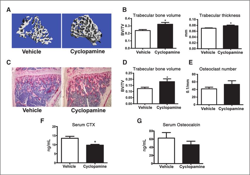
Published OnlineFirst December 20, 2011; DOI: 10.1158/0008-5472.CAN-11-2681

      Heller et al.

      cell lymphoma and multiple myeloma (22). Increased intratu-            mice on C57Bl/6 and mixed backgrounds, respectively,
      moral expression of Hh target gene Gli2 increased production of        were previously described. Animals were housed under
      osteoclast (OC)-activating factor PTHrP in breast cancer cells,        pathogen-free conditions according to the guidelines of the
      linking Hh signaling with tumor-induced osteolysis (23). How-          Division of Comparative Medicine, Washington University,
      ever, in certain breast cancer cell lines that are relatively          St. Louis, MO. The animal ethics committee approved all
      resistant to Hh signaling modulation, Gli2 expression can be           experiments.
      induced through TGFb signaling independent of Hh resulting in
      enhanced osteolysis (24). Together, these studies provide a            Cells
      strong rationale for evaluating Hh signaling as a therapeutic             4T1 BALB/c murine breast cancer (35) and B16-F10
      target for cancer and metastasis.                                      C57Bl/6 murine melanoma cell lines (36) were purchased
         The Hh pathway is critical to osteoblast (OB) differentiation       from the American Type Culture Collection and modified to
      and chondrocyte proliferation during embryonic endochon-               express firefly luciferase as previously described. A bone
      dral bone development (25, 26). Targeted disruption of several         metastatic variant of MDA-MB-231, described in Guise and
      Hh pathway genes result in profound effects on bone devel-             colleagues (37), was a kind gift of T. Guise (Indiana Univer-
      opment (27–29). Postnatal interruption of the Hh pathway               sity, Bloomington, IN). Low passage stocks were utilized and
      leads to trabecular bone abnormalities and disrupted long              regularly tested for Mycoplasma and maintenance of growth
      bone formation (30, 31). The effects of Hh signaling interrup-         characteristics.
      tion on adult bone have not been fully elucidated. Mice with
      conditional deletion of Ptch1 in mature OB using osteocalcin-          Drug compounds and dosing
      Cre show increased production of RANK ligand (RANKL) by                   The following drugs were used as indicated: cyclopamine
      OB, which indirectly increased OC activity and bone loss (32).         (LC Labs), tomatidine (Sigma), GDC-0449 (provided by Dr. Jim
      However, mice with heterozygous germline deletion of Ptch1             Janetka, Washington University, St. Louis, MO), and LDE225
      (Ptch1þ/) show increased bone mass, with enhanced bone                (provided by Novartis Pharmaceuticals). Cyclopamine (25 mg/
      formation being dominant to increased in vivo OC number and            kg) was administer per oral gavage twice daily for 11 to 14 days,
      resorption (33). The discrepancy between the 2 models                  as previously described (17); LDE225 (20 mg/kg) once daily for
      remains to be fully resolved. Interestingly, direct effects of Hh      21 days orally.
      pathway signaling on primary OC have not been reported.
         We hypothesized that disruption of Hh signaling would               Microcomputed tomography
      block tumor growth both directly, by targeting intratumoral              Postmortem, tibiae and femurs were scanned (mCT-40;
      survival signaling, and indirectly, by altering the host micro-        Scanco Medical) and evaluated as described previously
      environment. Indeed, OC- and OB-derived proteins, such as              (36).
      TGFb, can enhance growth of bone-invading tumor cells (34),
      and we propose that the effects of Hh signaling in both tumor          Bone histology and in vitro OC quantification
      and host cells may enhance metastatic growth. In this study, we           Decalcified and paraffin-embedded sections were stained
      show that disruption of the Hh pathway in adult nontumor–              with hematoxylin and eosin (H&E) stain or tartrate-resistant
      bearing mice increased trabecular bone in part through                 acid phosphatase (TRAP). Images were taken with an Eclipse
      reduced OC function and identified a cell-autonomous role               TE300 inverted microscope (Nikon) using the 4 (H&E, TRAP,
      of Hh signaling during osteoclastogenesis. Employing phar-             in vitro OC) or 40 (actin rings, pits) objectives. Histomor-
      macologic inhibitors, we show that disruption of the Hh                phometry was done using BioQuant Osteo.
      pathway decreased subcutaneous and bone tumor burden
      in vivo. Mice heterozygous for Ptch1, resulting in systemically        Serum CTX and osteocalcin
      enhanced Hh signaling, also had increased tumor burden.                   Serum from overnight-fasted mice was measured by ELISA
      Moreover, Hh inhibitors decreased subcutaneous tumor bur-              for CTX (RatLaps; Immunodiagnostic Systems) and osteocal-
      den in a cell line that is resistant to direct cytotoxic effects due   cin (Biomedical Technologies Inc.) according to the manufac-
      to reduced SMO expression (24), showing indirect antitumor             turer's instructions.
      effects of targeting cells of the host microenvironment. Inter-
      estingly, MDA-MB-231 cells produce Hh ligands and knock-               Macrophage and OC generation
      down of SHH decreased tumor growth through paracrine                      To generate macrophages, whole bone marrow cells were
      effects on stromal cell production of growth factors including         cultured in aMEM with 10% FBS and 100 ng/mL M-CSF for 3
      interleukin (IL)-6. Thus, Hh inhibitors represent promising            days. To generate OCs, macrophages were cultured in aMEM,
      therapeutics due to their ability to target both tumor cells           10% FBS, 50 ng/mL M-CSF, and 50 ng/mL RANKL for 6 days
      and the protumor microenvironment.                                     (36, 38). Media was refreshed every 2 days. Cells were fixed and
                                                                             stained for TRAP using the leukocyte acid phosphatase kit
      Materials and Methods                                                  (Sigma).

      Animals                                                                Lentiviral production and infection
        Female BALB/c mice and athymic nude mice (NCr-nude)                    293T cells were transfected with plasmid of interest,
      were obtained from Taconic. Ptch1þ/ (12) and Smofl/fl (25)              pHR'8.2deltaR and pCMV-VSV-G using Xtreme Gene 9

OF2   Cancer Res; 72(4) February 15, 2012                                                                                Cancer Research

              Downloaded from cancerres.aacrjournals.org on September 22, 2015. © 2011 American Association for
                                                     Cancer Research.
Published OnlineFirst December 20, 2011; DOI: 10.1158/0008-5472.CAN-11-2681
Antitumor Effect of Hedgehog Inhibition in Host Microenvironment
(Roche) and supernatant harvested 48 hours later. Cells were In vivo bioluminescence imaging
infected with lentivirus-containing supernatant for 4 hours Imaging was done on a IVIS 100 device (Caliper Life
in the presence of 10 mg/mL protamine sulfate. For Sciences) as previously described (36), except that for subcu-
ex vivo Smo excision, Smofl/fl macrophages were infected taneous tumor image analysis, a software-defined contour
with virus produced from pHR'EF-Cre-WPRE-SIN ('CRE') or region of interest was used to measure total photon flux.
pHR'EF-GFP-WPRE-SIN ('GFP') virus in the presence of 50
ng/mL M-CSF. Forty-eight hours after infection, macro- MTT viability assay
phages were screened for excision efficiency and differenti- A total of 5,000 cells per well were plated in 96-well plates
ated into OC. For SHH knockdown, short hairpin RNA with indicated concentrations of drug. After 48 hours, 10 mL
(shRNA) constructs in pLKOpuro vectors were obtained MTT (Sigma) was added for 4 hours. HCl/isopropanol was
from the Washington University Genome Institute and added to measure absorbance at 570 and 630 nm.
Children's Discovery Institute RNAi Consortium. (shLacZ–
CGCGATCGTAATCACCCGAGT; shSHH-2–GCTGATGACT- Bone marrow chimeras
CAGAGGTGTAA; shSHH-3–CATATCCACTGCTCGGTGAAA). Recipient mice were lethally irradiated (1,000 rads). Twenty-
Transduced cells were selected with 2 mg/mL puromycin. four hours later, 1 106 donor whole bone marrow cells were
transferred intravenously into recipient mice. After 4 weeks,
Quantitative reverse transcription PCR hematopoietic reconstitution with the donor genotype was
RNA was extracted using RNeasy Mini kit (Qiagen), treated confirmed by PCR of peripheral blood and mice were chal-
with DNaseI, and reverse transcribed using iScript (Bio-Rad). A lenged with the B16 tumor.
no-RT control was included in each assay. Quantitative PCR
was done using SsoFast EVA Green Supermix (Bio-Rad). Bone marrow stromal cell culture
Experiments were conducted in duplicate for both the To generate bone marrow stromal cells (BMSC), whole bone
target and the endogenous gene (GAPDH for OC, cyclophilin marrow of WT C57BL/6 mice was cultured in aMEM with 20%
for BMSC and MDA-MB-231) used for normalization. Relative FBS for 7 days and adherent cells replated at 5 105 cells/mL.
quantification of the target gene expression was calculated by At confluence, recombinant murine SHH (Ebioscience) or a
the comparative threshold cycle (Ct) method: 2DDCt in which 50:50 dilution of tumor cell conditioned media (CM, from 3
DCt ¼ Cttarget gene Ctendogenous gene and DDCt ¼ DCtvehicle 106 cells, 24 hours in serum-free media) was added and
DCttreated (see Supplementary Methods for primer sequences). cultured for an additional 72 hours in aMEM with 2.5% FBS.
Actin ring and bone resorption assay Statistical analysis
A total of 3,000 day 3 pre-OCs differentiated as above were Experiments were analyzed using 2-tailed Student t test or
plated on bovine bone slices in 96-well plates. At day 6, actin ANOVA using Prism5 (GraphPad). Errors bars represent SEM.
rings and resorption lacunae were visualized as previously Results were considered to reach significance at P 0.05 and
described (36). are indicated with an asterisk ( ).
BrdUrd proliferation assay Results
A total of 2.5 104 cells/mL were plated with indicated drug
concentrations. Cells were labeled with BrdUrd for 24 hours Cyclopamine increased bone mass and suppressed OC
and processed according to the manufacturer's instructions function in nontumor–bearing mice
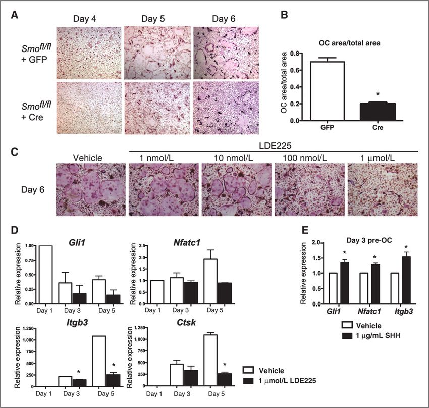
(Cell Proliferation ELISA; Roche). To understand the effect of Hh signaling on adult non-
tumor–bearing bone, we administered the Hh inhibitor
Immunoblotting cyclopamine to adult mice by twice daily by oral gavage for
Fifty micrograms of protein was separated on 8% SDS- 14 days and observed increased trabecular bone volume and
polyacrylamide gels and transferred onto a polyvinylidene thickness (Fig. 1A–D). Despite a nonsignificant increase in
difluoride membrane and incubated with p-AKT-Substrate, OC number (Fig. 1E), serum CTX, a marker of osteoclastic
p-ERK, or total-ERK rabbit antibodies (Cell Signaling Tech- bone resorption, was significantly decreased with cyclopa-
nology), followed by horseradish peroxidase–conjugated anti- mine (Fig. 1F). Serum osteocalcin, a marker of OB activity,
rabbit secondary antibody (Amersham Bioscience). Specific showed a nonsignificant reduction after Hh inhibition (Fig.
bands were developed by enhanced chemiluminescence. Load- 1G). Thus, pharmacologic inhibition of Hh signaling led to
ing control was b-actin (clone AC15; Sigma). increased trabecular bone mass, with evidence of decreased
OC function in vivo.
Tumor models
Intracardiac (1 105 cells) and intratibial (1 104 cells) Disruption of Hedgehog signaling inhibited ex vivo
tumor cell injections were carried out as previously osteoclastogenesis in a cell-autonomous manner
described (36). For subcutaneous injections 1 106 (4T1 To test whether Hh signaling had direct effects on OC
and B16) or 2 106 (MDA-MB-231) tumor cells were injected formation, we disrupted Smoothened by transducing Smofl/fl
in a 1:1 ratio with Matrigel (BD Biosciences) as previously bone marrow macrophages (BMM) with a lentivirus expressing
described (36, 38). Cre-recombinase (Supplementary Fig. S1A) and subjected the
www.aacrjournals.org Cancer Res; 72(4) February 15, 2012 OF3
Downloaded from cancerres.aacrjournals.org on September 22, 2015. © 2011 American Association for
Cancer Research.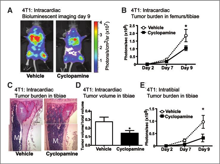
Published OnlineFirst December 20, 2011; DOI: 10.1158/0008-5472.CAN-11-2681

      Heller et al.

      Figure 1. Cyclopamine increased bone mass and suppressed OC function in nontumor–bearing mice. Eight-week-old female C57Bl/6 mice treated with
      vehicle or cyclopamine for 14 days (n ¼ 7 per group). A and B, mCT analysis for calculation of trabecular bone volume and trabecular thickness of
      tibiae. C–E, histomorphometry of tibiae stained for OC marker TRAP (red) to calculate: D, trabecular bone volume and, E, OC number (ns, P ¼ 0.28). F, serum
      CTX and, G, serum osteocalcin (ns, P ¼ 0.37).

      cells to osteoclastogenesis. We observed a decrease in OC size                    BMMs to osteoclastogenesis, Ptch1þ/ OC formed increased
      and number after Smo excision compared with green fluores-                         actin rings and resorption lacunae when plated on bone (Fig.
      cent protein (GFP)-transduced control cells (Fig. 2A and B).                      3B and C), but showed no difference in osteoclastogenesis on
      Furthermore, pharmacologic inhibition of SMO with cyclopa-                        plastic (Supplementary Fig. S2A). We observed increased pro-
      mine (Supplementary Fig. S1B) or LDE225 (Fig. 2C) resulted in                     liferation of Ptch1þ/ BMMs (Fig. 3D) and increased levels of
      a dose-dependent decrease in TRAPþ multinucleated OC                              phospho-AKT substrates (p-AKTs) in day 3 Ptch1þ/ pre-OC
      formation. Likewise, LDE225 reduced the mRNA abundance                            (Fig. 3E). However, we did not observe a difference in the
      of the Hh target Gli1 and of the OC differentiation markers                       expression of phosphorylated ERK in BMMs (Supplementary
      NFATc1 (Nfatc1), b3 integrin (Itgb3), and cathepsin K (Ctsk; Fig.                 Fig. S2B), nor in rate of apoptosis of Ptch1þ/ OCs (Supple-
      2D). Furthermore, treatment of pre-OCs with recombinant                           mentary Fig. S2C). From these data, we concluded that
      murine SHH increased mRNA transcripts of Gli1, Nfatc1, and                        enhanced Hh signaling promoted macrophage proliferation
      Itgb3, suggesting that Hh stimulation enhanced signaling path-                    and OC function.
      ways involved in OC differentiation (Fig. 2E). These results
      suggested that Hh signaling through SMO is critical to normal                     Hedgehog pathway inhibition with cyclopamine
      OC formation in a cell-autonomous fashion.                                        decreased bone metastases in a murine breast cancer
                                                                                        model
      Enhanced Hh signaling due to Ptch1 heterozygosity                                   To test whether Hh inhibition would reduce bone tumor
      increased OC function in a cell-autonomous manner                                 burden, we challenged immunocompetent BALB/c mice
        Adult Ptch1þ/ with enhanced Hh signaling had elevated                          with osteolytic murine 4T1 mammary breast carcinoma
      serum CTX, indicating increased bone resorption (Fig. 3A).                        cells (35). Cyclopamine significantly decreased tumor burden
      Interestingly, subjecting equal numbers of WT and Ptch1þ/                        in bone after either intracardiac (Fig. 4A–D) or intratibial

OF4   Cancer Res; 72(4) February 15, 2012                                                                                                  Cancer Research

              Downloaded from cancerres.aacrjournals.org on September 22, 2015. © 2011 American Association for
                                                     Cancer Research.
Published OnlineFirst December 20, 2011; DOI: 10.1158/0008-5472.CAN-11-2681
Antitumor Effect of Hedgehog Inhibition in Host Microenvironment
fl/fl
Figure 2. Disruption of Hh signaling inhibited ex vivo osteoclastogenesis in a cell-autonomous manner. A, Smo bone marrow–derived macrophages were
lentivirally infected with Cre-recombinase or GFP control to delete Smoothened, differentiated into OCs and stained with TRAP. B, quantification of
above OC area per total area on day 6. C, TRAP staining of OC differentiated in the presence of vehicle or LDE225 for 6 days. D, treatment with 1 mmol/L LDE225
decreased Gli1, Nfatc1, Itgb3, and Ctsk transcripts by qRT-PCR on days 3 and 5 of OC differentiation. Data normalized to gene expression of day 1
vehicle-treated macrophages. E, day 3 pre-OC treated with 1 mg/mL recombinant murine SHH for 24 hours had increased levels of Gli1, Nfatc1, and Itgb3
by qRT-PCR. qRT-PCR, quantitative reverse transcription PCR.
(Fig. 4E) injection. These data showed that Hh antagonism expression of the downstream target Gli1 was also significantly
decreased bone metastatic tumor growth in immunocompe- decreased with cyclopamine (Fig. 5C). The small molecule SMO
tent mice. inhibitor GDC-0449 (Supplementary Fig. S3A) also decreased
4T1 viability, whereas tomatidine, an inactive structural analog
Smo antagonists exert direct cytotoxic effects on 4T1 of cyclopamine, had no effect (Supplementary Fig. S3B). Com-
breast cancer cells pared with vehicle controls, subcutaneous growth of 4T1 cells in
In vitro analyses showed that 4T1 tumor cells have intact BALB/c mice was significantly reduced by cyclopamine (Fig.
Hh signaling pathways that are responsive to SMO antago- 5D). These data were consistent with a direct inhibition of
nism. Cyclopamine decreased the viability and proliferation of tumor growth; however, the specific contribution of host-tar-
4T1 cells in a dose-dependent manner (Fig. 5A and B). mRNA geted SMO antagonism could not be evaluated in this model.
www.aacrjournals.org Cancer Res; 72(4) February 15, 2012 OF5
Downloaded from cancerres.aacrjournals.org on September 22, 2015. © 2011 American Association for
Cancer Research.Published OnlineFirst December 20, 2011; DOI: 10.1158/0008-5472.CAN-11-2681
Heller et al.
Figure 3. Enhanced Hh signaling
due to Ptch1 heterozygosity
increased OC function in a cell-
autonomous manner. A, serum
þ/
CTX was increased in Ptch1
mice compared with WT
littermates (n ¼ 7 per group). B,
increased actin ring formation (top)
and bone resorption lacunae
þ/
(bottom) in Ptch1 OC
differentiated on bovine bone
slices for 6 days. C, quantification
of resorption lacunae area. D,
þ/
proliferation of Ptch1 and WT
macrophages in response to 100
ng/mL MCSF by BrdUrd
incorporation assay. E,
immunoblotting of p-AKT
þ/
substrates and b-actin of Ptch1
and WT day 3 pre-OC starved for 1
hour and stimulated with 50 ng/mL
MCSF or 50 ng/mL RANKL for 30
minutes.
Enhanced Hh signaling due to Ptch1 heterozygosity in subcutaneous B16 tumor burden in Ptch1þ/ mice (Fig. 6E),
indirectly enhanced tumor growth suggesting that the tumor-promoting effects of enhanced Hh
To examine the possibility that Hh signaling may play a role signaling in the host are not specific to the bone environment.
in various tissues through mechanisms independent of direct To test whether this was due to cells of hematopoietic origin
antitumor actions, we evaluated tumor growth in Ptch1þ/ (including myeloid and immune cells), we established radia-
mice. Following intracardiac injection of osteolytic B16 cells tion chimeras of WT and Ptch1þ/ recipient mice reconsti-
into immunocompetent C57Bl/6 mice, metastatic bone tumor tuted with reciprocal WT or Ptch1þ/ bone marrow. In both
burden was significantly increased in Ptch1þ/ mice compared WT and Ptch1þ/ recipients, reconstitution with Ptch1þ/
with WT littermates (Fig. 6A–D). We also observed an increase hematopoietic cells increased B16 subcutaneous tumor
Figure 4. Hh pathway inhibition with
cyclopamine decreased bone
metastases in a murine breast
cancer model. A–D, BALB/c mice
treated with vehicle (n ¼ 6) or
cyclopamine (n ¼ 10) starting 1 day
prior to left ventricular injection of
4T1. A and B, tumor burden in
femurs and tibiae as measured by
in vivo bioluminescence (BLI). C
and D, histomorphometric analysis
of tumor volume per total volume in
tibiae by H&E on day 9. M ¼
marrow; T ¼ tumor. E, cyclopamine
treatment beginning on day 1
also decreased tumor burden
following direct intratibial
inoculation of 4T1 cells as
measured by BLI. BLI,
bioluminescence imaging.
OF6 Cancer Res; 72(4) February 15, 2012 Cancer Research
Downloaded from cancerres.aacrjournals.org on September 22, 2015. © 2011 American Association for
Cancer Research.Published OnlineFirst December 20, 2011; DOI: 10.1158/0008-5472.CAN-11-2681
Antitumor Effect of Hedgehog Inhibition in Host Microenvironment
"sensitive" 4T1 cells could be attributed to direct cytotoxic
effects on the tumor cells (Figs. 4 and 5), effects of SMO
antagonists on growth of "resistant" MDA-MB-231 tumors
would be specifically due to modulation of the host microen-
vironment. In nude mice, subcutaneous growth of MDA-MB-
231 cells was significantly reduced by LDE225 (Fig. 7B), show-
ing that Hh inhibition isolated to host cells can modulate
tumor growth.
Tumor-derived Sonic hedgehog increased tumor growth
through effects on the microenvironment
Although MDA-MB-231 cells are unresponsive to canonical
Hh pathway stimulation, their production of Hh ligands,
particularly SHH (Fig. 7C), could stimulate Hh signaling in
surrounding tissues in a paracrine fashion. To investigate the
effects that tumor-produced Hh ligands have on the microen-
vironment, SHH expression was decreased in MDA-MB-231
cells by approximately 70% using 2 lentivirally expressed
shRNAs (Fig. 7D). Following knockdown, cells maintained in
Figure 5. SMO antagonists exert direct cytotoxic effects on 4T1 breast vitro proliferation rates similar to parental cells (Supplemen-
cancer cells. 4T1 cells treated for 24 hours with cyclopamine had tary Fig. S5C). In vivo, MDA-shSHH-2 and MDA-shSHH-3
decreased: A, viability as measured by MTT assay, B, proliferation as formed significantly smaller subcutaneous tumors than paren-
measured by BrdUrd incorporation, and C, expression of Gli1 by qRT-
tal or control MDA-shLacZ tumors, showing that tumoral
PCR (10 mmol/L cyclopamine). D, tumor weight on day 11 after
subcutaneous injection of 4T1 cells into WT BALB/c mice after vehicle or production of Hh ligands can increase growth of tumors that
cyclopamine (n ¼ 9 per group) treatment starting on day 1. qRT-PCR, fail to respond to canonical Hh stimulation in an autocrine
quantitative reverse transcription PCR. signaling (Fig. 7E).
Stromal cells within the microenvironment produce a
growth compared with reconstitution with WT bone marrow variety of tumor-supporting growth factors. To evaluate the
(Fig. 6F and G). Flow cytometry showed no significant differ- effects of tumor-derived SHH on stromal cells, we added CM
ences in the extent of hematopoietic reconstitution (Supple- from parental MDA or MDA-shLacZ cells to murine BMSCs
mentary Fig. S4A–C). These data suggested that host Hh and found that increased transcription of Gli1 to similar
signaling, in part through hematopoietic cells, influenced levels as recombinant Shh (Supplementary Fig. S5D). In
tumor growth indirectly through the microenvironment, inde- contrast, CM from MDA-shSHH-2 or MDA-shSHH-3 failed
pendent of direct effects on tumor cells. to induce BMSC Gli1, suggesting that Hh signaling was
blunted. BMSC production of IL-6, a protumor and proos-
MDA-MD-231 breast cancer cells are resistant to direct teoclastogenic factor, was increased by recombinant SHH
cytotoxic effects of Smo antagonists (Fig. 7F). CM from parental MDA or MDA-shLacZ further
MDA-MB-231 human breast cancer cells are reported to induced IL-6 transcription (Fig. 7F) and secretion (Supple-
have nondetectable levels of Smo transcripts and to be resis- mentary Fig. S5E) in BMSCs, whereas that of MDA-shSHH-2
tant to killing by SMO antagonists (7, 39). In agreement, we did or MDA-shSHH-3 cells induced it to a lower extent. Although
not observe significantly decreased viability even at micromo- tumor cells produce an abundance of factors that affect
lar concentrations of LDE225 (Fig. 7A). Reports of cytotoxicity numerous stromal cells signaling pathways, this data sug-
in sensitive tumor cell lines are in the nanomolar range (40), gested that BMSC production of IL-6 is due in part to
thus showing relative resistance of this tumor to in vitro stimulation of the Hh pathway. These data suggested that
cytotoxic effects. Furthermore, LDE225 treatment did not Hh inhibition in host microenvironment cells can reduce
decrease the expression of Hh target genes PTCH1 or GLI2 in tumor burden indirectly, even when tumor cells themselves
MDA-MB-231 cells (Supplementary Fig. S5A and B). These data are resistant to direct Hh inhibition.
suggested that MDA-MB-231 cells are resistant to direct effects
of pharmacologic Hh inhibition, allowing for a system in which Discussion
to examine the microenvironment-targeted effects of SMO
antagonists. Although the majority of current pharmaceuticals used in
the treatment of cancer directly target tumor cell growth and
Hh signaling in host microenvironment cells influence survival, a growing body of evidence has shown that many
tumor growth in vivo components of the host microenvironment are critical to
To evaluate the roles of Hh inhibition directly on cells tumorigenesis and represent additional therapeutic targets.
present in the tumor microenvironment, subcutaneous growth Thus, therapeutic manipulation of this pathway has the poten-
of MDA-MB-231 Hh inhibition "resistant" tumor cell lines was tial to decrease tumor growth both through direct and indirect
evaluated. Although the decreased growth of Hh inhibition mechanisms. Intratumoral Hh pathway signaling has been
www.aacrjournals.org Cancer Res; 72(4) February 15, 2012 OF7
Downloaded from cancerres.aacrjournals.org on September 22, 2015. © 2011 American Association for
Cancer Research.Published OnlineFirst December 20, 2011; DOI: 10.1158/0008-5472.CAN-11-2681
Heller et al.
Figure 6. Enhanced Hh signaling
due to Ptch1 heterozygosity
indirectly enhanced tumor growth.
A–D, following intracardiac
injection of B16 cells, tumor burden
þ/
was increased in Ptch1 (n ¼ 6)
mice as compared with WT
littermates (n ¼ 5) by (A and B) BLI
and (C and D) histomorphometric
analysis of H&E-stained tibiae on
day 12. M ¼ marrow; T ¼ tumor. E,
tumor weight on day 11 after
subcutaneous injection of B16 cells
þ/
(WT, n ¼ 4; Ptch1 , n ¼ 5). F and
G, B16 subcutaneous tumor
burden by BLI on day 14 in WT and
þ/
Ptch1 reciprocal bone marrow
chimeras. F, subcutaneous tumors
þ/
of WT recipients of Ptch1 bone
marrow (n ¼ 5) compared with
recipients of WT bone marrow
þ/
(n ¼ 5; P ¼ 0.3009). G, Ptch1
recipients reconstituted with
þ/
Ptch1 bone marrow (n ¼ 5)
showed a trend toward increased
tumor growth compared with those
receiving WT bone marrow (n ¼ 4;
P ¼ 0.0760). BLI, bioluminescence
imaging.
shown to be vital for the growth and maintenance of many growth factors, including, but not limited to, IL-6. We also
tumor types (3, 4). However, a number of tumors have been show that enhanced Hh signaling in the host environment of
shown to be refractory to the direct effects of pharmacologic Ptch1þ/ mice promoted growth of bone metastases and
Hh inhibition with SMO antagonists due to natural or acquired subcutaneous tumors, in part, through contributions of
mutations in Smo (24, 41) or amplification of downstream hematopoietically derived cells, including, but not limited
effector Gli2 (40, 42). to, immune cells and OC. Although the mechanisms under-
Previous reports have shown that tumor growth was lying the described phenotypes still need to be further
blunted when paracrine Hh signaling was inhibited in stro- elucidated, these data underscore a role for Hh inhibition
mal components of the microenvironment, even when the in cells of the microenvironment, even in Hh-unresponsive
tumor itself is SMO independent (19, 22, 43). The Hh tumor cells, and highlights the potential for increased clin-
inhibitor GDC-0449 induced dramatic reductions in the ical utility of Hh inhibition in cancer treatment.
growth of tumors with activating Hh mutations (44, 45). In addition to its role in tumorigenesis, Hh signaling is
Interestingly, GDC-0449 has little direct effect on tumors crucial to proper development and maintenance of many
without Hh mutations; however, it significantly blocked Hh host tissues including bone (25, 26). However, studies into
signaling in tumor stroma and decreased tumor burden (46). the role of Hh signaling in postnatal bone have yielded
In this article, we found that the SMO antagonist LDE225 disparate results. Enhancing Hh signaling through germline
had potent in vivo antitumor activity in MDA-MB-231, an Ptch1 heterozygosity resulted in increased bone density (33),
aggressive breast tumor cell line relatively resistant to Hh whereas conditional homozygous deletion of Ptch1 in
pathway modulation due to undetectable levels of SMO mature OB decreased bone density (32). Both groups
(7, 39). Furthermore, we show that tumoral production of observed increases in bone formation and resorption; how-
the Hh ligand SHH supported growth of subcutaneous ever, the effects of Hh signaling on the OC were attributed to
tumors in vivo. We provide evidence that this effect is due be indirect via increased OB expression of RANKL. Here we
to paracrine stimulation of the Hh signaling pathway in report a previously unrecognized cell-autonomous role for
stromal cells, resulting in the increased production of Hh signaling in the differentiation of bone-resorbing OCs.
OF8 Cancer Res; 72(4) February 15, 2012 Cancer Research
Downloaded from cancerres.aacrjournals.org on September 22, 2015. © 2011 American Association for
Cancer Research.Published OnlineFirst December 20, 2011; DOI: 10.1158/0008-5472.CAN-11-2681
Antitumor Effect of Hedgehog Inhibition in Host Microenvironment
Figure 7. Tumor-derived Sonic Hh increased tumor growth through effects on the microenvironment. A, viability of MDA-MB-231 human breast cancer cells is
not decreased by LDE225 treatment in vitro as assayed by MTT. B, subcutaneous growth of MDA-MB-231 in nude mice treated with LDE225 (n ¼ 6) or
vehicle (n ¼ 4) starting day 2. C, MDA-MB-231 expression of Hh ligands relative to 293T cells by qRT-PCR. D, expression of SHH by qRT-PCR
following lentiviral-based shRNA knockdown of control LacZ (shLacZ) or SHH (2 independent shRNAs: shSHH-2 and shSHH-3) in MDA-MB-231 cells
as compared with parental cells. E, subcutaneous tumor growth of MDA-MB-231 parental (n ¼ 7), -shLacZ (n ¼ 8), -shSHH-2 (n ¼ 7), and –shSHH-3 (n ¼ 8)
cells in nude mice. Repeated measures ANOVA: P 0.05: parental versus shSHH-2; parental versus shSHH-3; shLacZ versus shSHH-3. shLacZ
versus shSHH-2 P ¼ 0.0632. F, murine BMSC expression of Il6 by qRT-PCR following 72-hour culture with 1 mg/mL SHH or CM from MDA-MB-231 -parental,
-shLacZ, -shSHH-2, or –shSHH-3 tumor cells. , P 0.05 versus vehicle-treated BMSC; lines (---) designate P 0.05 between groups. qRT-PCR,
quantitative reverse transcription PCR.
We found that genetic and pharmacologic Hh inhibition istration and/or increased drug dosage could result in more
decreased OC differentiation in vitro. Furthermore, tran- continuous or potent inhibition of Hh and OC function,
scription of target gene Gli1 and key genes involved in OC leading to increased bone density. Similarly, continuous
differentiation (Nfatc1, Itgb3, and Ctsk) were reduced with administration of parathyroid hormone has catabolic effects
Hh inhibition. These results are in agreement with a recent on bone, whereas intermittent dosing is anabolic (48). Hh
report showing that RAW cell differentiation into OCs could signaling in bone homeostasis seems tightly regulated by the
be inhibited with cyclopamine (47). Ptch1þ/ mice, in which strength of signaling, as suggested in discrepant results
Hh signaling is enhanced, are known to have increased bone between heterozygous (33) and homozygous loss of Ptch1
mineral density and OB activity (33). Concurrently, we (32). Overall, our results and those of others (32, 33) suggest
observed that Ptch1þ/ mice also had enhanced OC activity that level and regulation of Hh signaling in the bone micro-
in vitro. Together, these data suggest a direct, cell-autono- environment is important to properly regulate the extent of
mous role for Hh signaling in the OC. bone formation and resorption occurring under nonpatho-
We report that systemic Hh inhibition with cyclopamine logic conditions.
increased bone density of adult WT mice, whereas a previous As OC and tumor cells are both known to produce growth
study found decreased bone density (33). Several differences factors that support the activity of the other, the bone
between treatment protocols (i.e., administration and dos- microenvironment is a hotspot of tumor metastasis, known
ing), and importantly, recognized gender differences in bone as "the vicious cycle" (34). Thus, blunting tumor-driven
biology, may explain these seemingly paradoxical results. manipulation of bone remodeling and turnover can indi-
The previous study dosed male mice with 10 mg/kg cyclo- rectly decrease the expansion of tumor in bone. Here we
pamine intraperitoneally once a day (33), whereas we used show that mice treated with cyclopamine had decreased
25 mg/kg orally twice daily in females. Twice daily admin- tumor burden in bone. However, as subcutaneous tumor
www.aacrjournals.org Cancer Res; 72(4) February 15, 2012 OF9
Downloaded from cancerres.aacrjournals.org on September 22, 2015. © 2011 American Association for
Cancer Research.Published OnlineFirst December 20, 2011; DOI: 10.1158/0008-5472.CAN-11-2681
Heller et al.
growth was also decreased, the antitumor activity of Smo Disclosure of Potential Conflicts of Interest
antagonists was due at least partially to non-bone cell
effects, including those directly on tumor cells and on host No potential conflicts of interest were disclosed.
stromal cells.
Acknowledgments
In conclusion, our data show that components of the Hh
signaling pathway are promising therapeutic targets for The authors thank Steven Teitelbaum, F. Patrick Ross, Deborah Novack,
cancer as they have the ability to decrease tumor growth and Roberta Faccio for helpful discussions; Ozge Uluckan, Crystal Winkeler,
both by exerting direct antitumor effects and by making the Ceren Yalaz, Desiree Floyd, Aixiao Li, Jean Chappel, Wei Zou, Lorena
Salavaggione, Mark Wilkins, Emira Pazzoli, and Georg Feldmann for tech-
host microenvironment less hospitable to tumors. Hh inhi- nical assistance; Jim Janetka for GDC-0449 synthesis; and Crystal Idleburg
bition has now been shown to act on a variety of host for expert histology.
microenvironment cells, including stroma, hematopoietic
cells, and vasculature (49, 50) to contribute to the overall Grant Support
therapeutic effect. In addition to these effects on solid
This research was supported by grants from The Department of Defense
tumors, targeting the Hh pathway is a particularly attractive (W81XWH-07-1-036-KNW, R. Cagan), The St. Louis Men's Group Against Cancer
target for the treatment of bone metastases as it may prove (K.N. Weilbaecher), and the NIH (CA097250, CA100730–K.N. Weilbaecher;
beneficial in interrupting the vicious cycle of OB, OC, and T32CA113275–M.A. Hurchla; DK065789–F. Long; P50CA94056–D. Piwnica-
Worms). MicroCT and histology services were provided by the WU musculo-
tumor cells and effectively decrease both tumor burden and skeletal core (P30AR057235).
tumor-associated osteolysis, which are linked to high rates The costs of publication of this article were defrayed in part by the
payment of page charges. This article must therefore be hereby marked
of mortality and morbidity. As several small-molecule Hh advertisement in accordance with 18 U.S.C. Section 1734 solely to indicate this
signaling inhibitors are currently being clinically evaluated fact.
for efficacy in a variety of tumor types, their effect on Hh
signaling in cells of the tumor microenvironment warrants Received August 8, 2011; revised December 6, 2011; accepted December 6, 2011;
active investigation. published OnlineFirst December 20, 2011.
References
1. McMahon AP, Ingham PW, Tabin CJ. Developmental roles and 14. Al-Hajj M, Wicha MS, Benito-Hernandez A, Morrison SJ, Clarke MF.
clinical significance of hedgehog signaling. Curr Top Dev Biol Prospective identification of tumorigenic breast cancer cells. Proc Natl
2003;53:1–114. Acad Sci U S A 2003;100:3983–8.
2. Echelard Y, Epstein DJ. Sonic hedgehog, a member of a family of 15. Kubo M, Nakamura M, Tasaki A, Yamanaka N, Nakashima H, Nomura
putative signaling molecules, is implicated in the regulation of CNS M, et al. Hedgehog signaling pathway is a new therapeutic target for
polarity. Cell 1993;75:1417–30. patients with breast cancer. Cancer Res 2004;64:6071–4.
3. Jiang J, Hui CC. Hedgehog signaling in development and cancer. Dev 16. Kameda C, Tanaka H, Yamasaki A, Nakamura M, Koga K, Sato N, et al.
Cell 2008;15:801–12. The Hedgehog pathway is a possible therapeutic target for patients
4. Taipale J, Beachy PA. The Hedgehog and Wnt signalling pathways in with estrogen receptor-negative breast cancer. Anticancer Res
cancer. Nature 2001;411:349–54. 2009;29:871–9.
5. Varjosalo M, Taipale J. Hedgehog signaling. J Cell Sci 2007;120:3–6. 17. Feldmann G, Dhara S, Fendrich V, Bedja D, Beaty R, Mullendore M,
6. Rohatgi R, Scott MP. Patching the gaps in Hedgehog signalling. Nat et al. Blockade of hedgehog signaling inhibits pancreatic cancer
Cell Biol 2007;9:1005–9. invasion and metastases: a new paradigm for combination therapy in
7. Zhang X, Harrington N, Moraes RC, Wu MF, Hilsenbeck SG, Lewis MT. solid cancers. Cancer Res 2007;67:2187–96.
Cyclopamine inhibition of human breast cancer cell growth indepen- 18. Bailey JM, Singh PK, Hollingsworth MA. Cancer metastasis facilitated
dent of Smoothened (Smo). Breast Cancer Res Treat 2009;115: by developmental pathways: Sonic hedgehog, Notch, and bone mor-
505–21. phogenic proteins. J Cell Biochem 2007;102:829–39.
8. Tremblay MR, Lescarbeau A, Grogan MJ, Tan E, Lin G, Austad BC, 19. Yauch RL, Gould SE, Scales SJ, Tang T, Tian H, Ahn CP, et al. A
et al. Discovery of a potent and orally active hedgehog pathway paracrine requirement for hedgehog signalling in cancer. Nature
antagonist (IPI-926). J Med Chem 2009;52:4400–18. 2008;455:406–10.
9. De Smaele E, Ferretti E, Gulino A. Vismodegib, a small-molecule 20. Olive KP, Jacobetz MA, Davidson CJ, Gopinathan A, McIntyre D,
inhibitor of the hedgehog pathway for the treatment of advanced Honess D, et al. Inhibition of hedgehog signaling enhances delivery
cancers. Curr Opin Investig Drugs 2010;11:707–18. of chemotherapy in a mouse model of pancreatic cancer. Science
10. Tremblay MR, McGovern K, Read MA, Castro AC. New developments 2009;324:1457–61.
in the discovery of small molecule Hedgehog pathway antagonists. 21. O'Toole SA, Machalek DA, Shearer RF, Millar EK, Nair R, Schofield P,
Curr Opin Chem Biol 2010;14:428–35. et al. Hedgehog overexpression is associated with stromal interactions
11. Oro AE, Higgins KM, Hu Z, Bonifas JM, Epstein EH Jr, Scott MP. Basal and predicts for poor outcome in breast cancer. Cancer Res 2011;
cell carcinomas in mice overexpressing sonic hedgehog. Science 71:4002–14.
1997;276:817–21. 22. Dierks C, Grbic J, Zirlik K, Beigi R, Englund NP, Guo GR, et al. Essential
12. Romer JT, Kimura H, Magdaleno S, Sasai K, Fuller C, Baines H, et al. role of stromally induced hedgehog signaling in B-cell malignancies.
Suppression of the Shh pathway using a small molecule inhibitor Nat Med 2007;13:944–51.
eliminates medulloblastoma in Ptc1(þ/)p53(/) mice. Cancer Cell 23. Sterling JA, Oyajobi BO, Grubbs B, Padalecki SS, Munoz SA, Gupta A,
2004;6:229–40. et al. The hedgehog signaling molecule Gli2 induces parathyroid
13. Ribeiro-Silva A, Ramalho LN, Garcia SB, Brandao DF, Chahud F, hormone-related peptide expression and osteolysis in metastatic
Zucoloto S. p63 correlates with both BRCA1 and cytokeratin 5 in human breast cancer cells. Cancer Res 2006;66:7548–53.
invasive breast carcinomas: further evidence for the pathogenesis of 24. Johnson RW, Nguyen MP, Padalecki SS, Grubbs BG, Merkel AR,
the basal phenotype of breast cancer. Histopathology 2005;47: Oyajobi BO, et al. TGF-beta promotion of Gli2-induced expression of
458–66. parathyroid hormone-related protein, an important osteolytic factor in
OF10 Cancer Res; 72(4) February 15, 2012 Cancer Research
Downloaded from cancerres.aacrjournals.org on September 22, 2015. © 2011 American Association for
Cancer Research.Published OnlineFirst December 20, 2011; DOI: 10.1158/0008-5472.CAN-11-2681
Antitumor Effect of Hedgehog Inhibition in Host Microenvironment
bone metastasis, is independent of canonical Hedgehog signaling. 38. Morgan EA, Schneider JG, Baroni TE, Uluckan O, Heller E, Hurchla
Cancer Res 2011;71:822–31. MA, et al. Dissection of platelet and myeloid cell defects by con-
25. Long F, Zhang XM, Karp S, Yang Y, McMahon AP. Genetic manipu- ditional targeting of the beta3-integrin subunit. FASEB J 2010;24:
lation of hedgehog signaling in the endochondral skeleton reveals a 1117–27.
direct role in the regulation of chondrocyte proliferation. Development 39. Mukherjee S, Frolova N, Sadlonova A, Novak Z, Steg A, Page GP, et al.
2001;128:5099–108. Hedgehog signaling and response to cyclopamine differ in epithelial
26. Long F, Chung UI, Ohba S, McMahon J, Kronenberg HM, McMahon and stromal cells in benign breast and breast cancer. Cancer Biol Ther
AP. Ihh signaling is directly required for the osteoblast lineage in the 2006;5:674–83.
endochondral skeleton. Development 2004;131:1309–18. 40. Buonamici S, Williams J, Morrissey M, Wang A, Guo R, Vattay A, et al.
27. St-Jacques B, Hammerschmidt M, McMahon AP. Indian hedgehog Interfering with resistance to smoothened antagonists by inhibition of
signaling regulates proliferation and differentiation of chondrocytes the PI3K pathway in medulloblastoma. Sci Transl Med 2010;2:51ra70.
and is essential for bone formation. Genes Dev 1999;13:2072–86. 41. Yauch RL, Dijkgraaf GJ, Alicke B, Januario T, Ahn CP, Holcomb T, et al.
28. Yuasa T, Kataoka H, Kinto N, Iwamoto M, Enomoto-Iwamoto M, Smoothened mutation confers resistance to a Hedgehog pathway
Iemura S, et al. Sonic hedgehog is involved in osteoblast differentiation inhibitor in medulloblastoma. Science 2009;326:572–4.
by cooperating with BMP-2. J Cell Physiol 2002;193:225–32. 42. Lauth M, Bergstrom A, Shimokawa T, Toftgard R. Inhibition of GLI-
29. Kinto N, Iwamoto M, Enomoto-Iwamoto M, Noji S, Ohuchi H, Yoshioka mediated transcription and tumor cell growth by small-molecule
H, et al. Fibroblasts expressing Sonic hedgehog induce osteoblast antagonists. Proc Natl Acad Sci U S A 2007;104:8455–60.
differentiation and ectopic bone formation. FEBS Lett 1997;404: 43. Tian H, Callahan CA, DuPree KJ, Darbonne WC, Ahn CP, Scales SJ,
319–23. et al. Hedgehog signaling is restricted to the stromal compartment
30. Maeda Y, Nakamura E, Nguyen MT, Suva LJ, Swain FL, Razzaque MS, during pancreatic carcinogenesis. Proc Natl Acad Sci U S A 2009;
et al. Indian Hedgehog produced by postnatal chondrocytes is essen- 106:4254–9.
tial for maintaining a growth plate and trabecular bone. Proc Natl Acad 44. Von Hoff D, Rudin C, LoRusso P, Borad M, Korn R, Heath E, et al.
Sci U S A 2007;104:6382–7. Efficacy data of GDC-0449, a systemic hedgehog pathway antagonist,
31. Kimura H, Ng JM, Curran T. Transient inhibition of the Hedgehog in a first-in-human, first-in-class phase I study with locally advanced,
pathway in young mice causes permanent defects in bone structure. multifocal or metastatic basal cell carcinoma patients. Proc Am Assoc
Cancer Cell 2008;13:249–60. Cancer Res 2008;49:138.
32. Mak KK, Bi Y, Wan C, Chuang PT, Clemens T, Young M, et al. 45. Lorusso PM, Rudin CM, Reddy JC, Tibes R, Weiss GJ, Borad MJ, et al.
Hedgehog signaling in mature osteoblasts regulates bone formation Phase I trial of Hedgehog pathway inhibitor vismodegib (GDC-0449) in
and resorption by controlling PTHrP and RANKL expression. Dev Cell patients with refractory, locally advanced or metastatic solid tumors.
2008;14:674–88. Clin Cancer Res 2011;17:2502–11.
33. Ohba S, Kawaguchi H, Kugimiya F, Ogasawara T, Kawamura N, Saito 46. Brochure Is. GDC-0449. Genentech, Inc. 2008.
T, et al. Patched1 haploinsufficiency increases adult bone mass and 47. Das S, Samant RS, Shevde LA. Hedgehog signaling induced by breast
modulates Gli3 repressor activity. Dev Cell 2008;14:689–99. cancer cells promotes osteoclastogenesis and osteolysis. J Biol Chem
34. Weilbaecher KN, Guise TA, McCauley LK. Cancer to bone: a fatal 2011;286:9612–22.
attraction. Nat Rev Cancer 2011;11:411–25. 48. Uzawa T, Hori M, Ejiri S, Ozawa H. Comparison of the effects of
35. Smith MC, Luker KE, Garbow JR, Prior JL, Jackson E, Piwnica-Worms intermittent and continuous administration of human parathyroid hor-
D, et al. CXCR4 regulates growth of both primary and metastatic breast mone (1–34) on rat bone. Bone 1995;16:477–84.
cancer. Cancer Res 2004;64:8604–12. 49. Cao X, Geradts J, Dewhirst MW, Lo HW. Upregulation of VEGF-A and
36. Uluckan O, Becker SN, Deng H, Zou W, Prior JL, Piwnica-Worms D, CD24 gene expression by the tGLI1 transcription factor contributes to
et al. CD47 regulates bone mass and tumor metastasis to bone. the aggressive behavior of breast cancer cells. Oncogene 2011; Jun 13
Cancer Res 2009;69:3196–204. [Epub ahead of print].
37. Guise TA, Yin JJ, Taylor SD, Kumagai Y, Dallas M, Boyce BF, et al. 50. Chen W, Tang T, Eastham-Anderson J, Dunlap D, Alicke B, Nannini M,
Evidence for a causal role of parathyroid hormone-related protein in the et al. Canonical hedgehog signaling augments tumor angiogenesis by
pathogenesis of human breast cancer-mediated osteolysis. J Clin induction of VEGF-A in stromal perivascular cells. Proc Natl Acad Sci
Invest 1996;98:1544–9. U S A 2011;108:9589–94.
www.aacrjournals.org Cancer Res; 72(4) February 15, 2012 OF11
Downloaded from cancerres.aacrjournals.org on September 22, 2015. © 2011 American Association for
Cancer Research.Published OnlineFirst December 20, 2011; DOI: 10.1158/0008-5472.CAN-11-2681
Hedgehog Signaling Inhibition Blocks Growth of Resistant
Tumors through Effects on Tumor Microenvironment
Emanuela Heller, Michelle A. Hurchla, Jingyu Xiang, et al.
Cancer Res Published OnlineFirst December 20, 2011.
Updated version Access the most recent version of this article at:
doi:10.1158/0008-5472.CAN-11-2681
Supplementary Access the most recent supplemental material at:
Material http://cancerres.aacrjournals.org/content/suppl/2011/12/20/0008-5472.CAN-11-2681.DC1.ht
ml
E-mail alerts Sign up to receive free email-alerts related to this article or journal.
Reprints and To order reprints of this article or to subscribe to the journal, contact the AACR Publications
Subscriptions Department at pubs@aacr.org.
Permissions To request permission to re-use all or part of this article, contact the AACR Publications
Department at permissions@aacr.org.
Downloaded from cancerres.aacrjournals.org on September 22, 2015. © 2011 American Association for
Cancer Research.You can also read