Clinical application of patient specific 3D printing brain tumor model production system for neurosurgery
←
→
Page content transcription
If your browser does not render page correctly, please read the page content below
www.nature.com/scientificreports
OPEN Clinical application
of patient‑specific 3D printing brain
tumor model production system
for neurosurgery
Yun‑Sik Dho1, Doohee Lee2, Teahyun Ha2, So Young Ji3, Kyung Min Kim4, Ho Kang4,
Min‑Sung Kim4, Jin Wook Kim4, Won‑Sang Cho4, Yong Hwy Kim4, Young Gyu Kim1,
Sang Joon Park2,5* & Chul‑Kee Park4*
The usefulness of 3-dimensional (3D)-printed disease models has been recognized in various medical
fields. This study aims to introduce a production platform for patient-specific 3D-printed brain
tumor model in clinical practice and evaluate its effectiveness. A full-cycle platform was created
for the clinical application of a 3D-printed brain tumor model (3D-printed model) production
system. Essential elements included automated segmentation software, cloud-based interactive
communication tools, customized brain models with exquisite expression of brain anatomy in
transparent material, adjunctive devices for surgical simulation, and swift process cycles to meet
practical needs. A simulated clinical usefulness validation was conducted in which neurosurgeons
assessed the usefulness of the 3D-printed models in 10 cases. We successfully produced clinically
applicable patient-specific models within 4 days using the established platform. The simulated clinical
usefulness validation results revealed the significant superiority of the 3D-printed models in surgical
planning regarding surgical posture (p = 0.0147) and craniotomy design (p = 0.0072) compared to
conventional magnetic resonance images. The benefit was more noticeable for neurosurgeons with
less experience. We established a 3D-printed brain tumor model production system that is ready to
use in daily clinical practice for neurosurgery.
Since 3-dimensional (3D) printing was introduced to the scientific field approximately 25 years ago, remarkable
advances and recent generalizations in technology have enabled 3D printing-related medical devices to be rapidly
applied in many medical fields, proving their necessity1–4. One of the most promising areas for the clinical appli-
cation of 3D printing technology is surgical simulation systems for anatomical disease models. Patient-specific
disease models, guaranteeing precise definition of disease extent and detailed reflection of related anatomical
structures, are undoubtedly useful in planning surgery for individual p atients5–7. To apply this patient-specific
surgical planning system to real-world clinical practice, a well-organized interactive process ranging from design
to production in a reasonable time is a vital element. In the field of neurosurgery, many studies on the clinical
application of 3D printing technology, such as models for cranioplasty, patient-specific spinal instruments, and
cerebral aneurysm models, have been r eported8–14. However, the clinical implementation of a patient-specific
3D-printed brain tumor model system for daily surgical planning has not been established until now.
For brain tumor surgery, total resection of the tumor while preserving functional integrity should be consid-
ered a top priority. To achieve this goal, the selection of an optimal approach with stereoscopic understanding
of related anatomical structures is the core issue. The current best practice in neurosurgical planning largely
depends on the conceptual reconstruction of lesions in the surgeon’s head based on 2-dimensional magnetic
resonance (MR) images or computer-aided image reconstruction incorporated into neuro-navigation, which
1
Department of Neurosurgery, Chungbuk National University Hospital, Chungbuk National University College of
Medicine, Cheongju, Republic of Korea. 2MEDICALIP Co. Ltd., Changgyeong Building, 174, Yulgok‑ro, Jongno‑gu,
Seoul 03127, Republic of Korea. 3Department of Neurosurgery, Seoul National University Bundang Hospital,
Seongnam, Republic of Korea. 4Department of Neurosurgery, Seoul National University Hospital, Seoul National
University College of Medicine, Daehak‑ro 101, Jongno‑gu, Seoul 03080, Republic of Korea. 5Department of
Radiology, Seoul National University Hospital, Daehak‑ro 101, Jongno‑gu, Seoul 03080, Republic of Korea. *email:
lunao78@snu.ac.kr; nsckpark@snu.ac.kr
Scientific Reports | (2021) 11:7005 | https://doi.org/10.1038/s41598-021-86546-y 1
Vol.:(0123456789)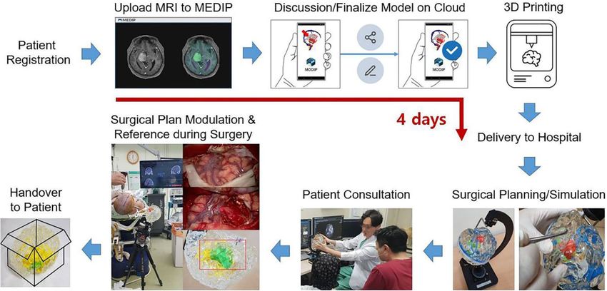
www.nature.com/scientificreports/
is still not intuitive15–19. This kind of skill requires repeated practice and a high level of experience, which is
difficult to achieve in the current training environment with limited o pportunities20. For trainees, case lessons
using a patient-specific 3D-printed disease model can be an effective tool to shorten the learning c urve19–21. In
addition, the complex nature of neurosurgical disease often makes it difficult for patients and caregivers without
any expertise to understand about their situation. Explanations using the 3D-printed model may provide an
intuitive understanding of the disease, surgical procedure, and r isks22,23.
Currently, many medical device companies and medical startups around the world are providing services for
producing clinical 3D-printed models in various fields, and their range and fields are gradually e xpanding4. It has
been accepted that creating 3D-printed brain tumor models is more intricate than creating models of common
cerebrovascular disease due to the anatomical complexity of the brain, diversity of lesions and adjunctive changes,
difficulty in segmentation, and texture r ealization24–27. With the recent development of auto-segmentation and
3D-printed manufacturing technology, it has become practical to create a 3D-printed brain tumor model that
meets all the requirements for clinical a pplication24–29. In the field of neurosurgery, the applications of patient-
specific 3D-printed brain disease models have also expanded, and several studies proved the effectiveness of these
models for trainee education, patient education, surgical planning and simulation24,26,28,29. However, to apply and
commercialize the 3D-printed brain tumor model for practical clinical use, the accuracy of a model that satis-
fies the neurosurgeon’s fickle needs, a production periods tailored to the surgical schedule, and reasonable costs
should be predetermined in a standardized and systematized manner. Here, we present our newly developed
clinical application platform for patient-specific 3D-printed models of brain tumor patients and verification of
its usefulness through a simulated clinical use.
Methods
Brain tumor segmentation. Segmentation is a term used in 3-dimensional modeling and refers to the
extraction of regions that show the same signal in a specific image. The first step was to create software that
can accurately segment the brain tumor and anatomical structures of the brain. In previous studies, manual
drawing methods using threshold differences were unavoidable in some part of the segmentation process due
to the complexity of the brain25,28–30. We automated most of the segmentation process using machine learning-
based thresholds, region growing, and graph-cut algorithms and produced a program that can perform seg-
mentation, reconstruction and rendering (MEDIP, http://medicalip.com/Medip, MEDICALIP, Seoul, Republic
of Korea). When MR DICOM images of patients are registered in MEDIP, the preprocessing step removes the
noise generated in the medical image before full-scale segmentation and is performed to improve image qual-
ity. Subsequently, when the user designates the region of interest (ROI), the specified ROI is replaced with seed
information, and segmentation is performed for the area with the same seed information, such as the specified
ROI, using the graph-cut algorithm (Fig. 1A), which is the core algorithm of MEDIP software. The graph-cut
algorithm is an algorithm that separates the foreground from the background by configuring each pixel with
graph intersections (nodes) and utilizing the difference in energy (flow network) between them. Segmentation is
performed with the seed point specified by the user as the starting point. Using the methods of drawing a solid
line with a pencil via an input device such as a mouse (Sketch method), creating a polygon by clicking on the
coordinates and filling it inside (Polygon method) and filling the inside in a free form (Freedraw method), the
user can directly specify the foreground and background. This technology was patented in the United S tates31.
Since 3D medical images should be segmented within the actual distance (spacing) between each pixel and
based on limited pixel and resolution information, optimization was carried out in the program. Segmentation
is performed by the function Draw-cut on MEDIP, and semi-automated segmentation is carried out through this
function. Using the Multi-Planar Reconstruction (MPR) function (Fig. 1B), the virtual 3D brain tumor model
and the MR image can be overlapped while cross sectioning the desired reference point, so the segmentation can
proceed while confirming that the process has been performed correctly. As a source image for segmentation,
enhanced T1-weighted, T2-weighted, and T2 fluid-attenuated inversion recovery (FLAIR) MR images were used
in combination. All MR image sequences tested in this study were performed using a 3.0-T GE Discovery MRI
system (GE Healthcare), and the images were acquired with a 1.0-mm thick slabs and 1.0-mm spaces. The goal
was to realize the brain tumor and the anatomical structures of the brain in 3 dimensions with the maximum
utilization of the signal differences between the source image and the thin section image in 1-mm intervals.
Virtual 3D model sharing and modification. The virtual 3D brain tumor model (virtual 3D model),
which was primarily rendered after segmentation, can be shared with the requesting neurosurgeon and the
3D model manufacturer in an interactive way by sending the Uniform Resource Locator (URL) of the model
uploaded to the web through mobile MEDIP (MODIP, MEDICALIP, Seoul, Republic of Korea). MODIP is a tool
for real-time communication between the neurosurgeon and manufacturer, through which the prototype of the
rendered virtual 3D model can be shared (example URL; http://147.47.229.147:8080/STLRendering/190513_
brain.html). The neurosurgeon can check the result of the shared model and request modifications by entering
text or drawing pictures using a smartphone or computer. MODIP is equipped with a function to rotate and
resize the 3D model in all directions so that the accuracy of production can be checked in detail from multiple
directions. By expressing the tumor and each anatomical structure in different colors, the anatomical structure
can be clearly identified, and with customization functions such as annotation, color change, addition and sub-
traction of each structure, the neurosurgeon can describe or sketch the preferred modifications, add or remove
the desired brain structure (blood vessels and nerves around the tumor, skull base structure) and change the
color or transparency of the structure. If the tumor was located in the cranial base, a portion of the skull base
structure adjacent to the tumor could be selected to tailor the model to only the desired range. After this interac-
tive process, the neurosurgeon can finally confirm the model to proceed to the 3D printing step.
Scientific Reports | (2021) 11:7005 | https://doi.org/10.1038/s41598-021-86546-y 2
Vol:.(1234567890)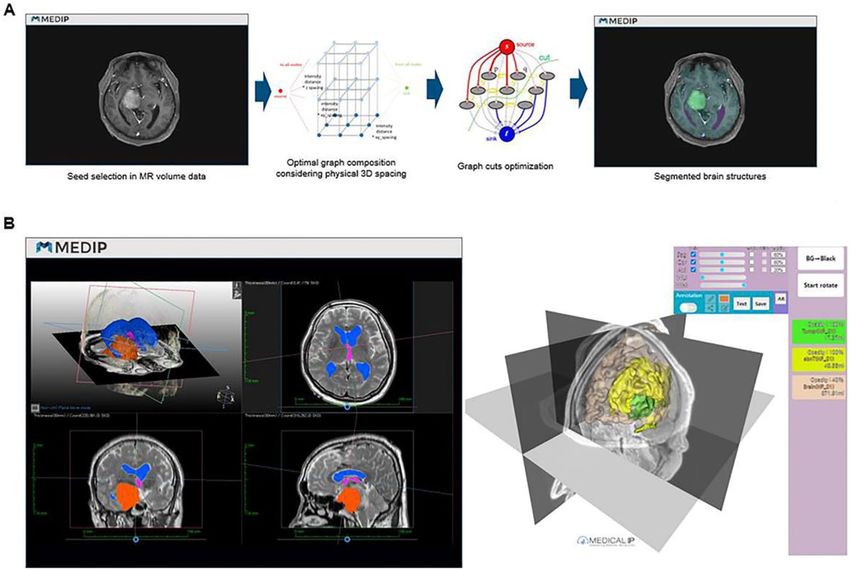
www.nature.com/scientificreports/
Figure 1. (A) Graph-cut algorithm. (B) Multiplanar reconstruction (MPR) function of MEDIP during the
segmentation process. The finished result can be seen at the following link (http://medicalip.synology.me:8082/
191011_NP01.html).
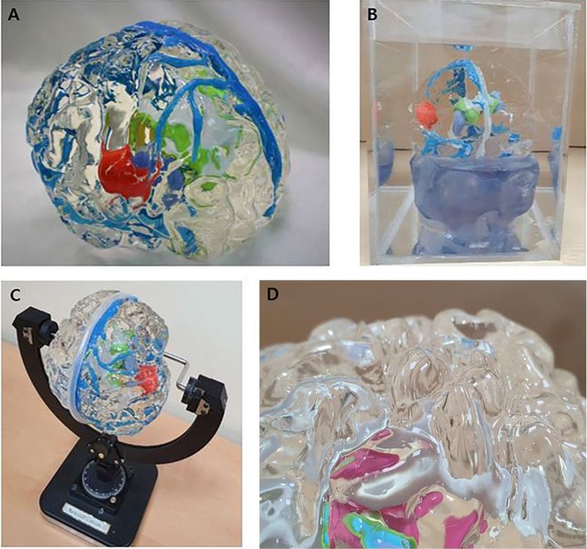
Production of a patient‑specific 3D‑printed brain tumor model. The production of the patient-
specific 3D-printed brain tumor models consisted of three stages: creating a stereolithography (STL) file for 3D
printing, printing physibles using a 3D printer, and performing a post-processing step, which includes manual
editing. Authentic realization of the gyrus and sulcus using transparent silicon materials with a similar brain tex-
ture to see through intra-axial tumors is a key component of our model (Fig. 2A,D). The gyri and sulci-shaped
moldings of the brain surface, brain tumor and associated brain structures are printed. After performing the sub-
traction process using the slicing program, if no overlap was observed among the brain parenchyma, tumor and
other brain structures, these features were extracted into separate STL files and printed independently with the
desired material and color. The brain parenchyma was sliced using the Flash Print program (FlashForge; No.518
XianYuan Road, Jinhua City, ZheJiang Province, China), and the gyri and sulci-shaped moldings were printed
using acrylonitrile butadiene styrene (ABS) copolymers by the fused deposition modeling method (Guider2,
[FlashForge; No.518 XianYuan Road, Jinhua City, ZheJiang Province, China]). The tumor and other associated
structures were sliced using a Grab CAD program (Stratasys; 7665 Commerce Way Eden Prairie, MN 55344,
USA) and printed with photovoltaic resin by the Polyjet method (J750 [Stratasys; 7665 Commerce Way Eden
Prairie, MN 55344, USA]). The supports attached to the outputs were removed after printing, and the brain
parenchyma was fumigated to smoothen the surface. After assembling the gyri and sulci-shaped moldings, out-
putted tumor and internal brain structures, the transparent silicone material with a similar texture to brain when
hardened was injected into the molding and then and dried. After the silicone material was completely dried, the
mold was removed, and the 3D-printed brain tumor model was finished by homogenizing the surface.
In addition, we developed assistive devices, including a rotatable water tank (Fig. 2B) to minimize diffuse
reflection caused by the flexion of the gyri and sulci and a model fixator, which is similar to the head fixator used
in brain surgery (Fig. 2C), to gain a better view of the internal structures and achieve precise surgical planning
and simulation.
Clinical usefulness validation. A simulated clinical validation was conducted so that neurosurgeons
could check the clinical usefulness of a customized 3D-printed brain tumor model. A total of 10 cases of brain
tumors with various pathologies, locations, and depths were retrospectively selected, and 3D-printed brain
tumor models were produced (Table 1). The simulated clinical usefulness validation was executed at an alloca-
tion site of an exhibition booth during the 59th Annual Meeting of the Korean Neurosurgical Society (http://
2019.kns-neurosurgery.or.kr) using an electronic questionnaire scenario with randomly chosen models for
Scientific Reports | (2021) 11:7005 | https://doi.org/10.1038/s41598-021-86546-y 3
Vol.:(0123456789)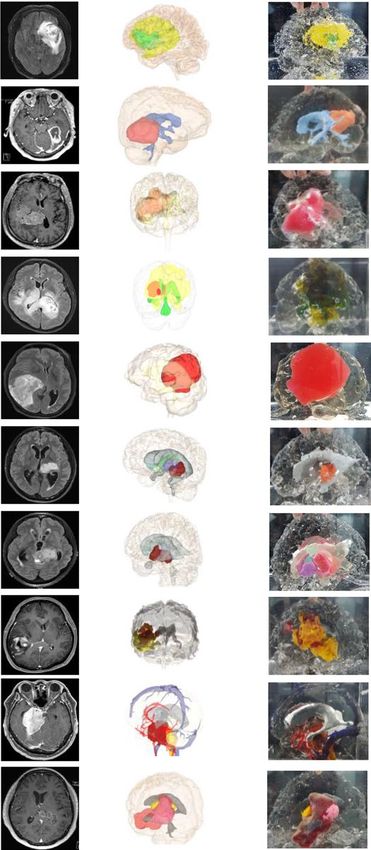
www.nature.com/scientificreports/
Figure 2. (A) The final 3D-printed brain model of right insular glioma. Normal brain parenchyma with
expression of the gyri and sulci was reconstructed using transparent silicon material. The tumor is colored light
red, the ventricle system is light gray, the caudate nucleus is yellow-green, the thalamus is light blue, and the
major venous system is blue. The soft real brain-like texture of the model enables the neurosurgeon to simulate
surgery by incising and excavating the area of interest. (B) The internal structure of the model can be clearly
observed, and diffuse reflections can be eliminated by placing the model in the water tank. (C) The surgical
approach can be fine-tuned with a dedicated fixator that can determine the rotation angle of the model in all
directions. (D) The closure view of the model shows the details of gyri and sulci.
each volunteer (http://147.47.229.147:9090/brain/index.php). These processes were performed under institu-
tional review board (IRB) approval from the 2 institutions (Seoul National University Hospital and Chungbuk
National University Hospital) where the cases originated. The IRB of 2 institutions where the cases originated
waived off the requirement for informed consent for the simulated clinical validation due to the no interaction
with patients and the retrospective nature of the study. To assess the usefulness of the 3D printed brain tumor
model in surgical planning for various tumor types and locations, the tumors were categorized according to the
location depth: cortex, intermediate (outer tumor margin located less than 2 cm from the cortical surface), deep
(outer tumor margin located between 2 and 4 cm from the cortical surface), and very deep (outer tumor margin
located more than 4 cm from the cortical surface). A total of 32 neurosurgeons (14 faculty members, 11 fellows,
and 7 residents) voluntarily participated for 2 days. The participants responded at a table separated by a partition
and the surroundings were quiet. The process was carried out alone with the help of an assistant who presented
MR images and 3D-printed brain tumor models.
Questionnaires were administered about the decisions regarding surgical plans for a certain selected case
based on MR images (enhanced T1, T2, T2 FLAIR sequences in the coronal, axial, sagittal planes) alone, a
perspective 3D reconstructed brain tumor plan before printing (which can be rotated in all directions on a com-
puter), and a 3D-printed brain tumor model (with the aid of a water tank and a fixator). The participants were
repeatedly asked about the proper surgical position, including head rotation direction and degree, the location
and extent of craniotomy, and the goal of the extent of tumor removal after inspecting the given MR images,
perspective plan, and 3D-printed model. The surgical position questionnaire was answered by choosing objec-
tive item from supine, prone, and lateral. The choice of direction and degree of head rotation was determined by
selecting the right/left side and then 0, 10, 30, 45, 60, 90, or 100 degrees. The area and extent of craniotomy were
determined by drawing directly on the screen showing the skull image with the selected direction and degree
Scientific Reports | (2021) 11:7005 | https://doi.org/10.1038/s41598-021-86546-y 4
Vol:.(1234567890)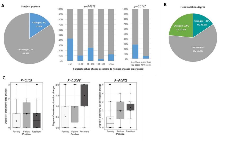
www.nature.com/scientificreports/
Model no. Sex Age Diagnosis Laterality Location Location depth MRI MEDIP/MODIP 3D printed model Associated structures
1 M 71 Glioblastoma left temporal, insular intermediate non-enhancing tumor
2 F 66 Glioblastoma Right temporal, occipital deep ventricles
3 M 69 Meningioma Right intraventricular deep ventricles
parietal, temporal, ventricles, non-enhancing
4 F 55 Glioblastoma Right/Left intermediate
thalamus tumor
Anaplastic parietal, temporal,
5 F 59 Right cortex ventricles
oligodendroglioma occipital
Diffuse midline ventricles, putamen,
6 M 22 Left thalamus very deep
glioma caudate nucleus
Diffuse midline ventricles, putamen,
7 F 26 Left thalamus very deep
glioma caudate nucleus
ventricles, non-enhancing
8 F 25 Glioblastoma Right temporal, parietal intermediate
tumor
major arteries, major
9 M 64 Meningioma Right petroclival very deep sinuses, ventricles, brain
stem, tentorium
temporal, thalamus,
10 F 53 Glioblastoma Left deep ventricles, thalamus
hippocampus
Table 1. Clinical features of 10 fabricated 3D printed brain tumor models. M, male; F, female; MRI, magnetic
resonance image; 3D, 3-Dimensional
of head rotation. To score the changes in degree of craniotomy size, we devised the following scoring system
ranging from 0 to 3 points: 0 for less than a 25% change, 1 for a 25–50% change, 2 for a 0–75% change, and 3 for
a 75–100% change in craniotomy size. Similarly, changes in craniotomy location were scored from 0 to 2 points:
0 for more than a 90% overlap, 1 for more than a 50% overlap, and 2 for less than a 50% overlap in craniotomy
area. The participants were also asked if they would modify the goal of the surgery in terms of the extent of
resection after inspecting the 3D brain tumor model. No time limit was set for determining the answers. An
additional survey consisting of 5 questions was administered to evaluate the usefulness of the MR images and
3D-printed model in establishing surgical plans on a 5-point scale from 1 (not useful) to 5 (very useful). The
statistical significance of the results was also examined according to respondent factors, such as their status and
experience in brain tumor surgery, as well as tumor factors.
Statistical analysis. The chi-square test, Fisher’s exact test, two sample t-test and ANOVA were used to
appropriately compare the results according to the abovementioned factors. All statistical analyses were per-
formed using the R free statistical software package (version 3.4.0; http://www.r-proje ct.org/). A p-value less
than 0.05 was considered statistically significant.
Ethics declarations. The study was conducted in accordance with the Declaration of Helsinki and complies
with the current laws of the countries in which it was performed. An independent ethics committee or institu-
tional review board (Seoul National University Hospital, IRB No. 1811-040-986 and Chungbuk National Uni-
versity Hospital, IRB No. 2019-06-015-001) for each study site approved the study protocol and all procedures
performed in this study were in accordance with the ethical standards as set. The institutional review boards
waived off the requirement for informed consent for the simulated clinical validation due to the no interaction
with patients and the retrospective nature of the study. However, participants those who appeared in the figures
signed an informed consent to publish the images in an online open access publication.
Results
Usefulness of the 3D‑printed brain tumor model. The simulated clinical usefulness validation
acquired responses for 64 cases from 32 neurosurgeons, each of whom evaluated 2 out of the 10 prepared cases.
In total, for 10 out of 64 cases (15.6%), the surgical posture determined by MR images only would be changed
after inspecting the 3D-printed brain tumor model (Fig. 3A). These changes were significantly more common
among respondents with less experience with brain tumor surgery (Fig. 3A and Supplementary Table 1). In
particular, among 7 respondents who had experienced fewer than 10 surgical cases, 42.9% of the decisions were
changed after inspecting the 3D-printed brain tumor model. Moreover, there was a significantly higher propor-
Scientific Reports | (2021) 11:7005 | https://doi.org/10.1038/s41598-021-86546-y 5
Vol.:(0123456789)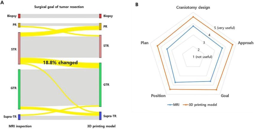
www.nature.com/scientificreports/

                                            Figure 3.  (A) Change in surgical posture from that determined from MR images after inspecting the
                                            3D-printed brain tumor model. The surgical posture change rate shows significant differences among
                                            respondents according to the number of cases treated. (B) Change in head rotation degree from that determined
                                            from MR images after inspecting the 3D-printed brain tumor model. (C) The degree of change in craniotomy
                                            size and location was determined after inspecting the 3D -printed brain tumor model according to the training
                                            level of the respondents.

                                            tion (p = 0.0147) of surgical posture changes in the group who had performed less than 100 cases of brain tumor
                                            surgery (8/28, 28.6%) than in the group who had performed more than 100 surgeries (2/36, 5.6%) (Fig. 3A and
                                            Supplementary Table 1). For a total of 25 out of 64 cases (39.1%), the degree of head rotation for the surgery was
                                            changed after inspecting the 3D brain tumor model (Fig. 3B), and these changes were not related to the respond-
                                            ents’ training level or experience (Supplementary Table 1). The changes in the location and extent of craniotomy
                                            size determined by MR images and the 3D-printed brain tumor model were also analyzed in a similar way.
                                            There were significant changes in craniotomy location only (p = 0.0008) or both location and size (p = 0.0072)
                                            after inspecting the 3D-printed brain tumor model among residents and/or fellows (Fig. 3C and Supplementary
                                            Table 1). The surgical plans made by faculty members were relatively consistent regardless of the information
                                            provided by either MR images or the 3D-printed brain tumor model. Tumor factors had little impact on the
                                            determination of the surgical positions or craniotomy design (Supplementary Table 2).
                                                The surgical goals were modified in terms of the extent of resection after examination of the 3D-printed brain
                                            tumor model in 12 out of 64 cases (18.8%, Fig. 4A). Among them, the resection goal was lessened in 4 cases,
                                            while the resection extent was widened in 8 cases after inspecting the 3D-printed brain tumor model. Five ques-
                                            tions, which were scored on a 5-point scale, on comparing MR images and the 3D-printed brain tumor model
                                            were asked; Q1. Usefulness in predicting and determining the location and extent of craniotomy (craniotomy
                                            design), Q2. Usefulness in determining the appropriate degree of head rotation (approach), Q3. Usefulness in
                                            determining the extent of tumor resection (goal), Q4. Usefulness in determining the surgical posture to remove
                                            the tumor effectively (position), and Q5. Usefulness in envisaging the entire process of the surgical resection
                                            (plan). The average scores showed that the 3D-printed brain tumor model was superior to MR images for surgical
                                            planning surgery in all aspects (Fig. 4B).

                                            Clinical application flow. We built a patient-specific 3D-printed brain tumor model production system
                                            from patient registration to delivery of the final model to the hospital, and this process was completed within
                                            4 days (Fig. 5). This is possible thanks to the interactive online system for making perspective 3D models and
                                            standardized 3D-printed manufacturing processes. If there is a candidate who needs a 3D-printed brain tumor
                                            model produced for surgical planning, a neurosurgeon uploads the DICOM files of the MR images to the MEDIP
                                            after obtaining consent from the patient. The segmentation process is followed, and the result is transferred to a

          Scientific Reports |   (2021) 11:7005 |                https://doi.org/10.1038/s41598-021-86546-y                                                 6

Vol:.(1234567890)
www.nature.com/scientificreports/
Figure 4. (A) Sankey diagram showing the change in the extent of tumor removal between the MRI-based
and 3D-printed brain tumor model-based plans. Overall, for 12 out of 64 cases (18.8%), the surgical goal was
modified after examining the 3D-printed brain tumor model. (PR, partial resection; STR, subtotal resection;
GTR, gross total resection; Supra-TR, supra-total resection). (B) The result of the questions evaluating the
usefulness of MR images and the 3D-printed model in establishing surgical plans, as scored on a 5-point scale
from 1 (not useful) to 5 (very useful). In all questions, the 3D-printed brain tumor model scored higher than the
MR images.
Figure 5. Clinical application flow of the 3D-printed brain tumor model.
cloud-based system using the MODIP interface in approximately one day. The perspective 3D model is finalized
after online discussion and fine tuning between the neurosurgeons and technicians during the following day.
Then, the customized 3D printed brain tumor model is produced, and the finished product is delivered in 2 days.
Using a patient-specific 3D-printed brain tumor model, neurosurgeons can make a surgical plan, simulate
a surgery, and use the model to explain the surgery to the patient (Fig. 5). Neurosurgeons can obtain insights
into the stereoscopic location of the tumor, appropriate surgical corridor or approach to the tumor, effective
craniotomy design, and goal of the surgery using the 3D-printed brain tumor model. The transparent and soft
nature of the model with exquisite realization of the gyri and sulci provides intuitive ideas for surgical planning.
However, diffuse reflection may limit the view of internal structures due to the transparent flexion of the gyri and
sulci, especially when a deep-seated tumor is encountered. To compensate for this diffuse reflection, a rotatable
Scientific Reports | (2021) 11:7005 | https://doi.org/10.1038/s41598-021-86546-y 7
Vol.:(0123456789)www.nature.com/scientificreports/
water tank was devised to clearly see through the internal structures by placing the model in the water. In addi-
tion, more sophisticated surgical planning can be enabled by the model fixator, which can simulate the surgical
position by replicating the angle of head rotation in all directions. By incising and excavating the delicately
implemented gyri and sulci having similar physical properties to the brain, neurosurgeons can determine the
effective cortical window to the tumor, avoiding injury to eloquent areas and critical structures. The model can
also be used for patient consultations before surgery so that patients or caregivers can gain a better understand-
ing of the nature of the disease and surgery.
During surgery, the 3D-printed model can be installed in the operating room according to the planned surgi-
cal position and can be used as a reference for surgical plan modulation and surgical anatomy during surgery
(Fig. 5). After the surgery, the 3D-printed model is retrieved and provided to the patient upon request.
Discussion
Since Spottiswoode et al. reported the first 3D-printed brain tumor model, there have been continuous efforts
in developing 3D-printed brain tumor models25. These pilot models were created for training and education
purposes, surgical simulation, and surgical p lanning20,24,25,28–30,32,33. Tumors in various locations, including the
cerebrum, cerebellum, brain stem and pituitary tumors, have thus far been implemented20,24,28,29,33. Most of the
early models focused on the brain tumor itself without paying much attention to the surrounding brain struc-
tures, which provides insufficient information to create a delicate surgical plan. Although recent models gave
shape to peritumoral neurovascular structures, there are still limitations to designing a surgical plan unless the
whole brain is embodied, especially for intra-axial tumors.
To further progress from the previous phase of the 3D-printed model within academic boundaries, we have
established a full-cycle platform for the clinical application of a 3D-printed brain tumor model system. Although
many efforts have been exerted to introduce 3D-printed brain tumor models to the clinical field, previous stud-
ies have only suggested a possibility for clinical entry or limited educational purposes24–26,28,29. To overcome the
hurdles of the practical application of 3D-printed brain tumor models in daily clinical use, we have standardized
the manufacturing process, shortened the production period, advanced the design and materials, devised aux-
iliary equipment, and reduced the manufacturing cost. These points are the differences and superiorities of our
platform from those of other previously reported models. Implementing precise segmentation in a short time
with an interactive sharing system for the perspective 3D model was the most strenuous challenge in this study.
When the neurosurgeon registers DICOM files to MEDIP, subsequent steps, including image preprocessing for
quality improvement, overall configuration of the graphs based on foreground/background seed settings, and
semiautomated graph-cut segmentation, are executed sequentially without delay. The purpose of the MODIP
platform is to enable the neurosurgeon to check the rendered perspective 3D model immediately on any kind
of device without installing specific applications and communicate with multiple users. This interactive tool
contributes to effective discussions for developing and modifying models in a short time.
The top priority of 3D-printed models for surgical planning is to mimic all anatomical structures as realisti-
cally as possible. Previously published 3D brain tumor models had mainly focused on the tumor itself due to
issues with cost or technical problems and based on the purpose of the study24,26,28,29. We thought that imple-
menting a 3D brain tumor model with the whole brain parenchyma shown in transparent soft material with
precise expression of the gyri and sulcus is important for use in surgical simulation. This is also the differenti-
ated advantages of our models. To this end, we discovered a silicon material that is transparent and has similar
properties to the brain when hardened. On the other hand, it was also not easy to model a gyri and sulci similar
to the real brain. Printing the gyri and sulci similar to the brain using conventional 3D printer properties elicited
a problem: the shape collapsed during printing due to the weight of the product. Through trial and error, we
established a process to make external moldings and inject transparent silicone material into these moldings.
We found that this process not only facilitates the expression of delicate real brain-like gyri and sulci but also
reduces the production time and cost. Another effort to reduce the production time of the 3D printing process
was the application of a polyjet (photopolymer + jetting) printer that sprays materials and solidifies them. When
we used a fused deposition modeling (FDM)-type 3D printer that can only print with a single material with a
single nozzle, the production time was much longer due to postprocessing work, which included removing the
supports, surface-treating and assembling for the individual structures. However, the polyjet printer allows for
simultaneous printing with materials of multiple properties, thus producing the model in the assembled form.
Furthermore, support removal and surface treatment can be performed by the water-jet equipment, which
reduces the production time and improves precision. Although we showed only 10 representative cases of brain
tumor models in with various pathologies, locations, and depth in this study, there are no limitations on anatomi-
cal location of interest or age of patients to produce the model. Even very small and delicate structures such as
blood vessels and cranial nerves can be implemented as soon as they are visible on MRI.
In the simulated clinical validation executed in this study, we confirmed that the 3D-printed brain tumor
model is more helpful for neurosurgeons with less experience in planning surgery. The changes made to the surgi-
cal plans after inspecting the 3D-printed brain tumor models were crucial in terms of patient safety. With the aid
of novel tools such as 3D-printed brain tumor models, novice neurosurgeons can perform surgery confidently
with intuitive information about the tumor, and the learning curve is expected to be shortened. The additional
comments from the participants in the simulated clinical validation revealed that the 3D-printed brain tumor
model helped determine the surgical plan by providing both microscopic views of the tumor as well as views
from multiple directions after the model was fixed and the brain parenchymal portion was dissected out; this
provided insight into the depth of the tumor location. Additionally, the model could help reduce the craniotomy
size and increase the accuracy of the craniotomy site. The participants also mentioned that the 3D-printed model
has merits in transferring tumor information among physicians engaged in patient management.
Scientific Reports | (2021) 11:7005 | https://doi.org/10.1038/s41598-021-86546-y 8
Vol:.(1234567890)www.nature.com/scientificreports/
For neurosurgeons, the 3D-printed brain tumor model will enable an accurate craniotomy, ensure the maxi-
mum level of safety during resection, and minimize neurological deficits caused by surgery, thus, a safe surgery
can be performed within the appropriate range and time. This will eventually result in a lower infection rate, rapid
recovery, and a decrease in the incidence of neurological deficits in patients. Using the model to explain the surgi-
cal procedure could improve the patient’s psychological status and rapport with the doctor by enhancing their
surgical understanding. In addition, sharing the 3D-printed brain tumor model with surgical assistants, nurses,
anesthesiologists, and intensive care unit physicians can improve the quality of care for patients by enhancing
the understanding of the surgery throughout the surgical team. This ultimately leads to benefits for the patients.
Conclusion
We established a 3D-printed brain tumor model production system that is ready to use in daily clinical practice
for neurosurgery. The effectiveness of this system was tested in clinical field and could be confirmed by simulated
clinical validation. We hope that this system will be widely introduced to the neurosurgery clinic as a new gear
for the development of the next step for the future surgery.
Received: 7 December 2020; Accepted: 17 March 2021
References
1. Jones, N. J. N. Science in three dimensions: the print revolution. Nat. News 487, 22–23 (2012).
2. Berman, B. 3-D printing: the new industrial revolution. Bus. Horizons 55, 155–162 (2012).
3. Aoun, R. J. N., Hamade, Y. J., Zammar, S. G., Patel, N. P. & Bendok, B. R. Futuristic three-dimensional printing and personalized
neurosurgery. World Neurosurg. 4, 870–871 (2015).
4. Michalski, M. H. & Ross, J. S. J. J. The shape of things to come: 3D printing in medicine. JAMA 312, 2213–2214 (2014).
5. Ryan, J. R., Almefty, K. K., Nakaji, P. & Frakes, D. H. Cerebral aneurysm clipping surgery simulation using patient-specific 3D
printing and silicone casting. World Neurosurg. 88, 175–181 (2016).
6. Kimura, T. et al. Simulation of and training for cerebral aneurysm clipping with 3-dimensional models. Neurosurgery 65, 719–726
(2009).
7. Anderson, J. R. et al. Three-dimensional printing of anatomically accurate, patient specific intracranial aneurysm models. J. Neu-
rointerventional Surg. 8, 517–520 (2016).
8. Evins, A. I. et al. On-demand intraoperative 3-dimensional printing of custom cranioplastic prostheses. Oper. Neurosurg. 15,
341–349 (2018).
9. Mashiko, T. et al. Development of three-dimensional hollow elastic model for cerebral aneurysm clipping simulation enabling
rapid and low cost prototyping. World Neurosurg. 83, 351–361 (2015).
10. Wang, L. et al. Comparison of two three-dimensional printed models of complex intracranial aneurysms for surgical simulation.
World Neurosurg. 103, 671–679 (2017).
11. Rengier, F. et al. 3D printing based on imaging data: review of medical applications. Int. J. Comput. Assist. Radiol. Surg. 5, 335–341
(2010).
12. Opolski, A. C. et al. Experimental three-dimensional biomodel of complex aortic aneurysms by rapid prototyping technology. 3D
Print. Addit. Manuf. 1, 88–94 (2014).
13. Kim, B.-J. et al. Customized cranioplasty implants using three-dimensional printers and polymethyl-methacrylate casting. J. Korean
Neurosurg. Soc. 52, 541 (2012).
14. Cohen, A. et al. Mandibular reconstruction using stereolithographic 3-dimensional printing modeling technology. Oral Oral Surg.
Oral Med. Pathol. Oral Radiol. Endodontol. 108, 661–666 (2009).
15. Galloway, R. L., Maciunas, R. J. & Edwards, C. Interactive image-guided neurosurgery. IEEE Trans. Biomed. Eng. 39, 1226–1231
(1992).
16. Wendt, M., Bani-Hashemi, A. & Sauer, F. (Google Patents, 2002).
17. Kockro, R. A. et al. A collaborative virtual reality environment for neurosurgical planning and training. Oper. Neurosurg. 61,
ONSE379–ONSE391 (2007).
18. Kikinis, R. et al. Computer-assisted interactive Three-dimensional planning neurosurgical procedures. Neurosurgery 38, 640–651
(1996).
19. Ploch, C. C., Mansi, C. S., Jayamohan, J. & Kuhl, E. Using 3D printing to create personalized brain models for neurosurgical train-
ing and preoperative planning. World Neurosurg. 90, 668–674 (2016).
20. Vakharia, V. N., Vakharia, N. N. & Hill, C. S. Review of 3-dimensional printing on cranial neurosurgery simulation training. World
Neurosurg. 88, 188–198 (2016).
21. Waran, V., Narayanan, V., Karuppiah, R., Owen, S. L. & Aziz, T. Utility of multimaterial 3D printers in creating models with
pathological entities to enhance the training experience of neurosurgeons. J. Neurosurg. 120, 489–492 (2014).
22. Kim, P. S. et al. Obtaining informed consent using patient specific 3D printing cerebral aneurysm model. J. Korean Neurosurg. Soc.
62, 398 (2019).
23. Bernhard, J.-C. et al. Personalized 3D printed model of kidney and tumor anatomy: a useful tool for patient education. World J.
Urol. 34, 337–345 (2016).
24. Lin, J. et al. Using three-dimensional printing to create individualized cranial nerve models for skull base tumor surgery. World
Neurosurg. 120, e142–e152 (2018).
25. Spottiswoode, B. et al. Preoperative three-dimensional model creation of magnetic resonance brain images as a tool to assist
neurosurgical planning. Stereotactic Funct. Neurosurg. 91, 162–169 (2013).
26. Kondo, K. et al. A neurosurgical simulation of skull base tumors using a 3D printed rapid prototyping model containing mesh
structures. Acta Neurochir. 158, 1213–1219 (2016).
27. Pucci, J. U., Christophe, B. R., Sisti, J. A. & Connolly, E. S. Jr. Three-dimensional printing: technologies, applications, and limita-
tions in neurosurgery. Biotechnol. Adv. 35, 521–529 (2017).
28. Panesar, S. S. et al. Patient-specific 3-dimensionally printed models for neurosurgical planning and education. Neurosurg. Focus
47, E12 (2019).
29. Lau, I. et al. Patient-specific 3D printed model in delineating brain glioma and surrounding structures in a pediatric patient. Digit.
Med. 3, 86 (2017).
30. Waran, V. et al. Injecting realism in surgical training—initial simulation experience with custom 3D models. J. Surg. Educ. 71,
193–197 (2014).
31. Park, S. J. & Lee, D. H. (Google Patents, 2019).
Scientific Reports | (2021) 11:7005 | https://doi.org/10.1038/s41598-021-86546-y 9
Vol.:(0123456789)www.nature.com/scientificreports/
32. Thawani, J. P. et al. Three-dimensional printed modeling of diffuse low-grade gliomas and associated white matter tract anatomy.
Neurosurgery 80, 635–645 (2017).
33. Randazzo, M., Pisapia, J. M., Singh, N. & Thawani, J. P. 3D printing in neurosurgery: a systematic review. Surg. Neurol. Int. 7, S801
(2016).
Acknowledgements
We would like to acknowledge all the members from MEDICALIP who devoted themselves developing and
manufacturing models used in this study, and So-Jin Yun, Clinical Researcher Associate (Medical Device Inno-
vation Center, Seoul National University Hospital) for her efforts in the administrative process of this study.
Author contributions
Y-.S.D., D.L., T.H., and C-.K.P. wrote the main manuscript text and prepared Figs. 1, 2, 3, 4 and 5. D.L., T.H.
and S.J.P. created of new software used in the work. Y-.S.D., S.Y.J., K.M.K., H.K., M.S.K., J.W.K., W-.S.C., Y.H.K.,
Y.G.K., and P.C.K. contributed to the acquisition of data. S.J.P. and C-.K.P. supervised the study. All authors
reviewed the manuscript.
Funding
This research was supported by a grant of the Korea Health Technology R&D Project through the Korea Health
Industry Development Institute (KHIDI), funded by the Ministry of Health & Welfare, Republic of Korea (grant
number: HI15C1532), by the Korea Medical Device Development Fund grant funded by the Korea government
(the Ministry of Science and ICT, the Ministry of Trade, Industry and Energy, the Ministry of Health & Welfare,
the Ministry of Food and Drug Safety) (Project Number: 202012E08), and Seoul National University Research
Fund (29–2019-0020).
Competing interests
Sang Joon Park is founder and CEO of MEDICALIP. Chul-Kee Park owns stock options in MEDICALIP. Other
authors have no conflict of interest to declare.
Additional information
Supplementary Information The online version contains supplementary material available at https://doi.org/
10.1038/s41598-021-86546-y.
Correspondence and requests for materials should be addressed to S.J.P. or C.-K.P.
Reprints and permissions information is available at www.nature.com/reprints.
Publisher’s note Springer Nature remains neutral with regard to jurisdictional claims in published maps and
institutional affiliations.
Open Access This article is licensed under a Creative Commons Attribution 4.0 International
License, which permits use, sharing, adaptation, distribution and reproduction in any medium or
format, as long as you give appropriate credit to the original author(s) and the source, provide a link to the
Creative Commons licence, and indicate if changes were made. The images or other third party material in this
article are included in the article’s Creative Commons licence, unless indicated otherwise in a credit line to the
material. If material is not included in the article’s Creative Commons licence and your intended use is not
permitted by statutory regulation or exceeds the permitted use, you will need to obtain permission directly from
the copyright holder. To view a copy of this licence, visit http://creativecommons.org/licenses/by/4.0/.
© The Author(s) 2021
Scientific Reports | (2021) 11:7005 | https://doi.org/10.1038/s41598-021-86546-y 10
Vol:.(1234567890)You can also read