FUNCTIONAL ULTRASOUND (FUS) DURING AWAKE BRAIN SURGERY: THE CLINICAL POTENTIAL OF INTRA-OPERATIVE FUNCTIONAL AND VASCULAR BRAIN MAPPING - REPUB ...
←
→
Page content transcription
If your browser does not render page correctly, please read the page content below
ORIGINAL RESEARCH
                                                                                                                                      published: 09 January 2020
                                                                                                                                   doi: 10.3389/fnins.2019.01384

                                            Functional Ultrasound (fUS) During
                                            Awake Brain Surgery: The Clinical
                                            Potential of Intra-Operative
                                            Functional and Vascular Brain
                                            Mapping
                                            Sadaf Soloukey 1,2 , Arnaud J. P. E. Vincent 1 , Djaina D. Satoer 1 , Frits Mastik 3 ,
                                            Marion Smits 4 , Clemens M. F. Dirven 1 , Christos Strydis 2 , Johannes G. Bosch 3 ,
                                            Antonius F. W. van der Steen 3 , Chris I. De Zeeuw 2,5* , Sebastiaan K. E. Koekkoek 2 and
                                            Pieter Kruizinga 2,3*
                          Edited by:        1
                                              Department of Neurosurgery, Erasmus MC, Rotterdam, Netherlands, 2 Department of Neuroscience, Erasmus MC,
      Kamran Mohammad Avanaki,
                                            Rotterdam, Netherlands, 3 Department of Biomedical Engineering, Thorax Centre, Erasmus MC, Rotterdam, Netherlands,
Wayne State University, United States       4
                                              Department of Radiology and Nuclear Medicine, Erasmus MC, Rotterdam, Netherlands, 5 Netherlands Institute
                      Reviewed by:          for Neuroscience, Royal Dutch Academy for Arts and Sciences, Amsterdam, Netherlands
                    Rayyan Manwar,
Wayne State University, United States
                Zahra Nasiriavanaki,        Background and Purpose: Oncological neurosurgery relies heavily on making
Massachusetts General Hospital and          continuous, intra-operative tumor-brain delineations based on image-guidance.
           Harvard Medical School,
                        United States       Limitations of currently available imaging techniques call for the development of real-
                  *Correspondence:          time image-guided resection tools, which allow for reliable functional and anatomical
                    Chris I. De Zeeuw       information in an intra-operative setting. Functional ultrasound (fUS), is a new mobile
           c.dezeeuw@erasmusmc.nl
                      Pieter Kruizinga
                                            neuro-imaging tool with unprecedented spatiotemporal resolution, which allows for
           p.kruizinga@erasmusmc.nl         the detection of small changes in blood dynamics that reflect changes in metabolic
                                            activity of activated neurons through neurovascular coupling. We have applied fUS
                   Specialty section:
         This article was submitted to
                                            during conventional awake brain surgery to determine its clinical potential for both intra-
              Brain Imaging Methods,        operative functional and vascular brain mapping, with the ultimate aim of achieving
               a section of the journal
                                            maximum safe tumor resection.
            Frontiers in Neuroscience
      Received: 13 September 2019           Methods: During awake brain surgery, fUS was used to image tumor vasculature
      Accepted: 09 December 2019            and task-evoked brain activation with electrocortical stimulation mapping (ESM) as a
        Published: 09 January 2020
                                            gold standard. For functional imaging, patients were presented with motor, language or
                              Citation:
            Soloukey S, Vincent AJPE,       visual tasks, while the probe was placed over (ESM-defined) functional brain areas. For
        Satoer DD, Mastik F, Smits M,       tumor vascular imaging, tumor tissue (pre-resection) and tumor resection cavity (post-
    Dirven CMF, Strydis C, Bosch JG,
                                            resection) were imaged by moving the hand-held probe along a continuous trajectory
   van der Steen AFW, De Zeeuw CI,
Koekkoek SKE and Kruizinga P (2020)         over the regions of interest.
   Functional Ultrasound (fUS) During
    Awake Brain Surgery: The Clinical       Results: A total of 10 patients were included, with predominantly intra-parenchymal
Potential of Intra-Operative Functional     frontal and temporal lobe tumors of both low and higher histopathological grades. fUS
         and Vascular Brain Mapping.
             Front. Neurosci. 13:1384.
                                            was able to detect (ESM-defined) functional areas deep inside the brain for a range of
       doi: 10.3389/fnins.2019.01384        functional tasks including language processing. Brain tissue could be imaged at a spatial

Frontiers in Neuroscience | www.frontiersin.org                                  1                                         January 2020 | Volume 13 | Article 1384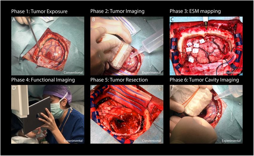
Soloukey et al.                                                                                                                   fUS During Awake Brain Surgery

                                            and temporal resolution of 300 µm and 1.5–2.0 ms respectively, revealing real-time
                                            tumor-specific, and healthy vascular characteristics.
                                            Conclusion: The current study presents the potential of applying fUS during awake
                                            brain surgery. We illustrate the relevance of fUS for awake brain surgery based
                                            on its ability to capture both task-evoked functional cortical responses as well as
                                            differences in vascular characteristics between tumor and healthy tissue. As current
                                            neurosurgical practice is still pre-dominantly leaning on inherently limited pre-operative
                                            imaging techniques for tumor resection-guidance, fUS enters the scene as a promising
                                            alternative that is both anatomically and physiologically informative.
                                            Keywords: brain tumor, functional ultrasound, awake craniotomy, tumor vasculature, neoplasm, imaging

INTRODUCTION                                                                             of eloquent brain areas adjacent to tumors. Intra-operative
                                                                                         use of ESM to remove low- (De Witt Hamer et al., 2012) or
Oncological neurosurgery relies heavily on making continuous,                            HGGs (Gerritsen et al., 2019) has been associated with fewer
intra-operative delineations between tumor and brain tissue. The                         post-operative complications and higher percentages of GTRs.
ultimate surgical aim is reaching maximum-safe tumor resection,                          However, ESM also presents with a range of its own inherent
in which most of the tumor is removed, while preserving                                  limitations, including lack of (sulcus-)depth resolution (Imbault
functional brain areas to prevent post-operative neurological and                        et al., 2017), functional over-estimation due to current leakage
cognitive deficits (Marko et al., 2014; Li et al., 2015). Given the                      (Ritaccio et al., 2018), lack of standardization of stimulation
large heterogeneity in tumor presentation and growth, especially                         protocols (Pouratian et al., 2004; Ritaccio et al., 2018) and
in gliomas (Patel et al., 2014), optimal delineation remains a                           the risk of eliciting epileptic seizures (Su and Ojemann, 2013;
challenging task, even when done through a high-resolution                               Ritaccio et al., 2018).
surgical microscope. Therefore in vivo real-time, functional brain                          Other conventional and experimental imaging techniques
imaging is essential to advance maximum-safe tumor resection                             such as Laser Speckle Contrast Imaging (LSCI) (Klijn et al.,
to the next level.                                                                       2012), Optical Coherence Tomography (OCT) (Valdés et al.,
    Conventional clinical practice makes use of several pre-                             2016; Almasian et al., 2019) Near-Infrared Spectroscopy (NIRS)
operative imaging modalities, including (f)MRI and DTI, which                            (Murata, 2008), HSI (Fabelo et al., 2018), and FGS (Zhang et al.,
are linked to intra-operative neuro-navigation systems. These                            2018) also facilitate intra-operative functional and/or anatomical
imaging modalities allow the surgeon to pre-operatively acquire                          imaging, but they also suffer from critical limitations, such
anatomical and functional data of the tumor and surrounding                              as a limited penetration depth (millimeters), field of view or
eloquent areas. However, due to the inevitable brain shift after                         spatial resolution.
cranio- and durotomy, pre-operative images only provide a                                   These limitations of currently available pre- and intra-
rough estimation of the 3D-locus of the tumor during in vivo                             operative imaging modalities greatly warrant the need for the
surgery following craniotomy (Imbault et al., 2017; Fabelo et al.,                       development of new real-time image-guided resection tools,
2018), complicating tumor delineation as the surgery proceeds. In                        which allow for reliable anatomical and functional information in
some specialized hospitals, MRI is also available intra-operatively                      an intra-operative setting. Recently, such an alternative emerged
(iMRI) in so called ‘MRI surgical suites.’ Although the use of iMRI                      within the field of ultrasound imaging, to which we refer as
can solve initial brain shift-related problems, it does not allow us                     fUS. fUS relies on HFR ultrasound and subsequent Doppler
to readily distinguish surgically manipulated healthy brain tissue,                      processing and interregional correlation analyses (Deffieux et al.,
edema, and tumor-infiltrated brain tissue. Furthermore, iMRI                             2018). As such, it allows for the detection of very small changes
disrupts surgical workflow, is time-consuming, and requires a                            in vascular dynamics including changes in CBV, CBF and
large financial investment.                                                              related processes such as vasodilation, which in turn reflect
    The widespread introduction of awake craniotomy surgery                              changes in metabolic activity of activated neurons through
with ESM has greatly improved the intra-operative identification                         neurovascular coupling (Deffieux et al., 2018). Therefore, fUS
Abbreviations: BOLD, blood oxygenation level dependent; CBF, cerebral blood              can capture brain functionality in real-time by detecting small
flow; CBV, cerebral blood volume; DTI, diffusion tensor imaging; ESM,                    hemodynamic responses with a temporal resolution in the order
electrocortical stimulation mapping; FGS, fluorescence guided surgery; fMRI,             of milliseconds, which is believed to be much higher than the
functional magnetic resonance imaging; FR, frame rate; fUS, functional ultrasound;
GBM, glioblastoma; GTRs, gross total resections; HFR, high-frame-rate; HGG,
                                                                                         vascular dynamics involved in neurovascular coupling (Deffieux
high grade glioma; HIS, hyperspectral imaging; iMRI, intra-operative magnetic            et al., 2018; Logothetis, 2018). In addition to its high temporal
resonance imaging; LGG, low grade glioma; LSI, laser speckle imaging; METC,              resolution, fUS can reach a spatial resolution of approximately
Medical Ethics Review Committee; MRI, magnetic resonance imaging; PCC,                   50 µm (Imbault et al., 2017), rendering the technique in
Pearson correlation coefficient; PDIs, power doppler images; PRF, pulse repetition
frequency; PT, patient; SEM, scanning electron microscope; SVD, singular value           principle valuable for advancing the borders of maximum-safe
decomposition.                                                                           tumor resection.

Frontiers in Neuroscience | www.frontiersin.org                                      2                                     January 2020 | Volume 13 | Article 1384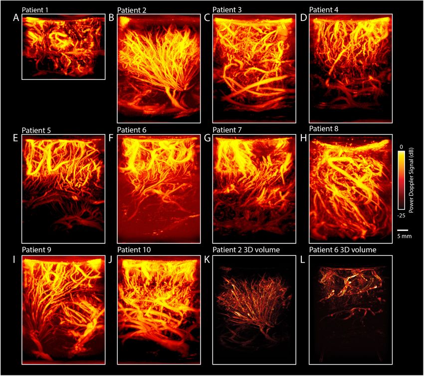
Soloukey et al.                                                                                                     fUS During Awake Brain Surgery

    So far, fUS has been applied successfully in brains and spinal         connected to a cortical stimulation unit (Grass Technologies,
cords of mice (Tiran et al., 2017; Koekkoek et al., 2018; Macé             Astro-Med, Inc.), square-wave pulses were delivered to induce
et al., 2018; Soloukey et al., 2019), rats (Osmanski et al., 2014;         depolarization of relevant cortices. According to standard
Tiran et al., 2017; Song et al., 2019), ferrets (Demené et al.,            protocol, the intensity of the working current was increased from
2016; Bimbard et al., 2018), birds (Rau et al., 2018), swine               6 to maximum 12 mA (60 Hz, 1 ms), depending on whether a
(Song et al., 2019), primates (Dizeux et al., 2019), and humans            functional effect was evoked. In some cases, no functional effect
(Imbault et al., 2016, 2017; Demene et al., 2017), proving to be           was found, even at maximum current intensity. Eloquent areas, if
a powerful tool for studying the dynamics of endogenous brain              found during ESM, were labeled with numbers.
signals. By combining its high spatiotemporal resolution with
a relatively large field of view, fUS can also capture functional          Functional Tasks
brain connectivity across many different brain regions in real             During fUS-imaging, the probe was partially placed over
time. In addition, fUS has shown to be a low-cost, contrast-free,          the (expected location of) the tumor and partially over the
and mobile technique (Deffieux et al., 2018). This unique set of           surrounding functional areas as identified by ESM, ensuring that
features warrants the exploration of the clinical, intra-operative         both types of tissues were in the field of view. If ESM did not
application of fUS.                                                        allow us to identify any functional area, the probe was placed over
    In this work, we apply fUS to a cohort of patients undergoing          the tumor and surrounding tissue most likely to be functional as
awake neurosurgery following craniotomy for the indication                 extrapolated from the local anatomy.
of tumor removal near eloquent areas of the brain. We                          Based on the expected eloquent area under the probe, patients
present a range of anatomically informative images acquired                were presented with appropriate, matching tasks for that area.
intra-operatively, discussing their potential for both clinical            These tasks were anticipated beforehand as much as possible,
applications and functional brain-mapping. More specifically, we           based on both anatomy as well as pre-operative fMRI-data if
present here for the first time whether fUS can be exploited at            available. Each task consisted of a total of 60 s, with alternating
the clinical level to reveal tumor-specific vascular features and          three blocks of tasks (8 s each) and three blocks of resting
at the functional level to delineate healthy brain activity patterns       conditions (10 s each), all preceded by a 6 s baseline. Functional
surrounding the tumor during motor and cognitive (language-                tasks as used in this study ranged from motor tasks (e.g., ‘lip
related) tasks.                                                            pouting’), to language tasks (e.g., ‘word repetition’) (De Witte
                                                                           et al., 2015) and visual tasks (e.g., ‘8 Hz checkerboard’). Motor
                                                                           and visual tasks were presented on a 12 inch tablet to the patient,
MATERIALS AND METHODS                                                      including a progress bar for the patient to follow task pattern-
                                                                           timing. Language tasks were only seen by the clinical linguist and
Inclusion of Participants                                                  communicated verbally to the patient.
After obtaining approval from the METC (MEC-2018-037,                          Patients were neurolinguistically assessed for the task prior to
NL64082.078.17) and a written informed consent accordingly,                surgery and informed about the tasks by a clinical linguist, who
participants were included in the study. All participants were             also presented the tasks to the patients intra-operatively (DS).
recruited from the Department of Neurosurgery of Erasmus                   An overview of the details of all the functional tasks as used per
MC in Rotterdam. Participants were eligible for inclusion when             patient can be found in Supplementary Table S1.
diagnosed with a brain tumor planned for tumor removal using                   After functional imaging, tumor resection was commenced.
awake craniotomy surgery with ESM. Participants were excluded              Finally, the tumor resection cavity was filled with saline and
if they were < 18 years old. Some patients underwent fMRI                  a final acquisition was made by the surgeon to capture the
pre-operatively per conventional clinical protocol.                        remaining tissue vasculature post-resection.

Study Procedure                                                            Image Acquisition
Functional ultrasound-data acquisition was conducted around                For ultrasound data acquisition, we used an experimental
three time-points as indicated in Figures 1B,D,F, which in total           research system (Vantage-256, Verasonics, United States)
prolonged the conventional surgical procedure (Figures 1A,C,E)             interfaced with a 5 MHz, 128 element linear array (ATL L7-4,
by a maximum of 20 min. After conventional craniotomy and                  300 µm pitch) driven with a three cycle burst at 5.2 MHz.
durotomy, the probe (in a sterile cover with ultrasound coupling           Baseband quadrature sampling was applied as implemented
gel) was placed over the tumor (guided by the neuro-navigation             by the Verasonics system in order to reduce the overall data-
and/or based on visual inspection). A 3D-volume of the tumor               rate. For all scans we acquired continuous angled plane wave
was obtained by acquiring 2D-images during a 60 s sweeping                 acquisition (12–16 angles equally spaced between −12 and 12
motion along a continuous trajectory made by the surgeon. Saline           degrees) with a PRF ranging from 6 to 8 kHz depending on the
was added frequently to the operating field by the OR nurse to             imaging depth. The average ensemble size (number of frames
ensure adequate acoustic coupling during imaging.                          used to compute one PDI) ranged between 120 and 140 frames
                                                                           from which the PDIs were computed, providing a live Doppler
Electrocortical Stimulation Mapping (ESM)                                  FR ranging between 3.6 and 4.8 Hz. The PDIs as well as the raw,
After acquisition of tumor vasculature images, the conventional            angle compounded beamformed frames (taken at an FR ranging
ESM procedure was performed. Using a bipolar electrode                     from 500 to 667 Hz, see Supplementary Table S2) were stored

Frontiers in Neuroscience | www.frontiersin.org                        3                                     January 2020 | Volume 13 | Article 1384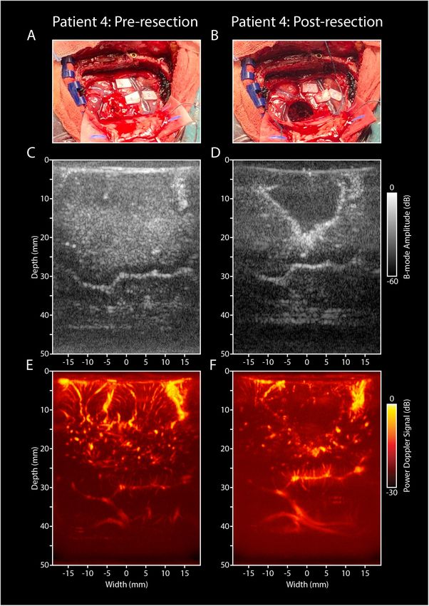
Soloukey et al.                                                                                                                           fUS During Awake Brain Surgery

  FIGURE 1 | Flowchart providing an overview of the study procedure. (A) After conventional craniotomy and durotomy, the tumor was exposed. (B) Imaging
  commenced with a sweeping motion as made by the surgeon across the length of the tumor to capture multiple 2D-images along a continuous trajectory.
  (C) Afterward, conventional ESM was performed to determine eloquent areas of interest surrounding tumor tissue. (D) Based on the ESM-defined areas, appropriate
  functional tasks were presented to the patient. Each functional task consisted of a 60 s task pattern, alternating three functional and rest blocks. Motor and visual
  tasks were presented on a 12 inch tablet to the patient. Language tasks were only seen by the clinical linguist and communicated verbally to the patient. In this case,
  a finger tapping motor task is presented to the patient. (E) After functional imaging, conventional tumor resection was performed. (F) Finally, the resection cavity was
  filled with saline to ensure adequate acoustic coupling, after which a final acquisition of the resection cavity was made [in a similar fashion as the tumor imaging
  explained under (B)]. Written informed consent was obtained for the identifiable image (D). ESM, electrocortical stimulation mapping.

to a fast PCIe SSD hard disk for offline processing purposes.                               For the functional datasets we applied rigid motion
The PDIs were computed using an adaptive SVD clutter filter                              compensation by registering every PDI to the median PDI
(Demené et al., 2015). All processing steps, including Fourier                           using the inbuilt matlab function ‘imregtform.m’. To assess the
domain image reconstruction, compounding, clutter filtering,                             functional signal we bandpass filtered (passband between 0.05
and storage were done using an inhouse built CPU/GPU code                                and 0.5 Hz) the PDI stack over time and computed for every pixel
written in C++/CUDA which was interfaced with the standard                               the PCC r (Macé et al., 2011). The optimal lag (between −2 and
Verasonics Matlab (MathWorks, Inc.) environment using                                    2 s) between the stimulus signal and the recording was chosen
MEX. In all cases, the probe was hand-held by the surgeon,                               empirically based on the overall functional map. Coefficients
and placed inside a sterile cover with ultrasound coupling gel.                          higher than 0.3 were considered as functionally relevant and
For vasculature imaging, the probe was either moved along                                displayed on top of the PDI (displayed in gray) using a red/yellow
a continuous trajectory over the tumor or resection cavity or                            colormap. The mean signal of the functional and remaining
‘fanned’ whereby the imaging plane rotates along the array                               pixels was plotted to confirm the validity of the functional signal.
axis. Instead, for functional imaging, the probe was placed                                 All the post-processing software and the visualization
over the area of interest and kept stable during the functional                          was done in Matlab (MathWorks, Inc.). We used Paraview
measurement. Both vasculature and functional image acquisition                           (Kitware, Inc.), an open-source software tool, for visualizing the
sessions consisted of 60 s each.                                                         3D vascular scans.

Post-processing                                                                          RESULTS
Storage of the raw frames allowed for offline optimization of
the scan-specific processing parameters that yielded the best                            Participant Characteristics
vasculature and functional images. In all cases we mapped the                            Between October 2018 and June 2019, a total of 10 participants
images onto a 100 µm grid using zero-padding in the frequency                            (two females, eight males) were included in the study (Table 1).
domain. The ensemble size was adaptively set to match one                                Included participants had a mean age of 42 years (31–56 years),
cardiac cycle. To allow for a smooth PD signal over time we                              with predominantly intra-parenchymal frontal and temporal
applied a 3/4 overlap between consecutive ensembles.                                     lobe gliomas of WHO grades II–IV. No surgical complications

Frontiers in Neuroscience | www.frontiersin.org                                      4                                             January 2020 | Volume 13 | Article 1384
Soloukey et al.                                                                                                                   fUS During Awake Brain Surgery

TABLE 1 | Clinical characteristics of the six participants in the current study.

Pt. number                Age category                  Tumor location                  Tumor type          Pre-op fMRI?               Others

1                              36–40                    Frontal (left)                  HGG (GBM)                 Y                    Re-operation (7 years prior)
2                              46–50                    Frontal (left)                  LGG                      N
3                              40–45                    Frontal (left)                  LGG                      N
4                              30–35                    Parietal (right)                LGG                       Y                    Intra-operative seizure
5                              56–60                    Temporal (left)                 HGG (GBM)                 Y
6                              40–45                    Occipito-parietal (left)        LGG                       Y
7                              56–60                    Occipito-parietal (left)        HGG (GBM)                N
8                              46–50                    Temporal (right)                HGG                      N                     Pre-operative seizure
9                              30–35                    Frontal (left)                  HGG                      N
10                             30–35                    Frontal (left)                  LGG                      N

HGG, high grade glioma; GBM, glioblastoma; LGG, low grade glioma.

occurred, except for two epileptic seizures in two patients (pt#4                      Language Tasks
and pt#8), one of which was induced by ESM (pt#4).                                     Figure 3 depicts the functional response to a language-
                                                                                       related task in pt.#5, who presented with a GBM in the
                                                                                       left temporal lobe (Figure 3A). Initial pre-operative fMRI
Functional Brain Mapping                                                               data showed an association of the tumor with eloquent areas
A total of 44 functional measurements were performed in 10                             during a conventional verbal fluency fMRI-task (Figure 3B).
patients, ranging from 2 to 5 functional measurements per                              Intra-operatively, ESM also identified several language-related
patient. Of these 44 measurements, 14 involved finger tapping,                         deficits upon stimulation, including phonemic paraphasia.
6 lip pouting, 18 verbal or silent word or sentence repetition,                        After placement of the probe over the relevant markers,
and 6 visual checkerboard stimulation (see Supplementary                               the patient was presented with a word repetition task.
Table S1 for an overview of functional tasks used per                                  The patient was asked to perform this task both verbally
patient). After post-processing, 9 measurements showed an                              (by repeating the words out loud) as well as silently (by
actual functional signal during motor and language-related                             covertly repeating the words without speaking out loud).
tasks. None of the visual functional tasks showed a functional                         This combination of tasks allowed for the interrogation of
signal. An overview of all measurements is presented in                                different parts within the language-related functional brain
Supplementary Table S2.                                                                areas. The fUS-image allowed for a field of view of 3.8 cm
                                                                                       wide and 5.0 cm deep in both versions of the functional
Motor Tasks                                                                            task (Figures 3C,D). In both cases, functional signals were
Figure 2 depicts the fUS-image as acquired from pt.#1 and                              found, with the location and extent of activation visually
pt#4, who both performed a motor task intra-operatively. Pt.#1                         differing between the verbal and silent word repetition (see also
presented with a recurrent left-sided GBM 7 years after primary                        Supplementary Video S3).
surgery. In line with the location of the original and recurrent
tumor as well as the images of the pre-operative fMRI, intra-                          Vascular Mapping
operative ESM confirmed that the primary motor cortex of the                           Across the 10 patients, a cumulative total of 30 measurements
mouth was in close proximity to the previous tumor cavity                              were made of the pre-resection tumor-vasculature, and a total of
(Figure 2A). After placement of the probe over the relevant ESM-                       16 measurements of the post-resection cavity. An overview of all
marker, the patient was asked to perform a 60 s lip pouting                            measurements is presented in Supplementary Table S2.
functional task. The fUS-image allowed for a field of view of
3.8 cm wide and 3.0 cm deep (Figure 2B), in which functional                           Maximum Projection Images
signals related to the task were observed in close proximity to the                    In addition to the regular 2D-images, we also made maximum
resection cavity (see also Supplementary Video S1).                                    projection images, showing an overview of the maximum signal
   Pt.#4 presented with a LGG in the right parietal lobe, also                         per pixel during the imaging session of 60 s. As such, a
in close proximity to the primary motor cortex, which in pre-                          single image with more depth-information can be created.
operative fMRI was confirmed with a bilateral finger tapping                           For each patient, the most vascular-dense pre-resection image
task (Figure 2D). Intra-operatively, ESM again confirmed this                          is highlighted in Figures 4A–J. As becomes clear from the
association. After placement of the probe over the relevant ESM-                       images, there is a rich variety in tissue vascularization patterns
marker, the patient was asked to perform a 60 s finger tapping                         across our patients. In two patients in particular, we saw
task. The fUS-images allowed for a field of view of 3.8 cm wide                        some interesting tumor vasculature (see Figures 4K,L for
and 5.0 cm deep (Figure 2E), in which functional signals related                       reconstructions of 3D-volumes). Tumor-vasculature imaging of
to the task were observed in an area approximately 2.0 cm in                           the LGG tumor in the left frontal lobe of pt.#2 (Figure 4K)
depth (see also Supplementary Video S2).                                               showed an arborous vascular structure, which in 2D-images

Frontiers in Neuroscience | www.frontiersin.org                                    5                                       January 2020 | Volume 13 | Article 1384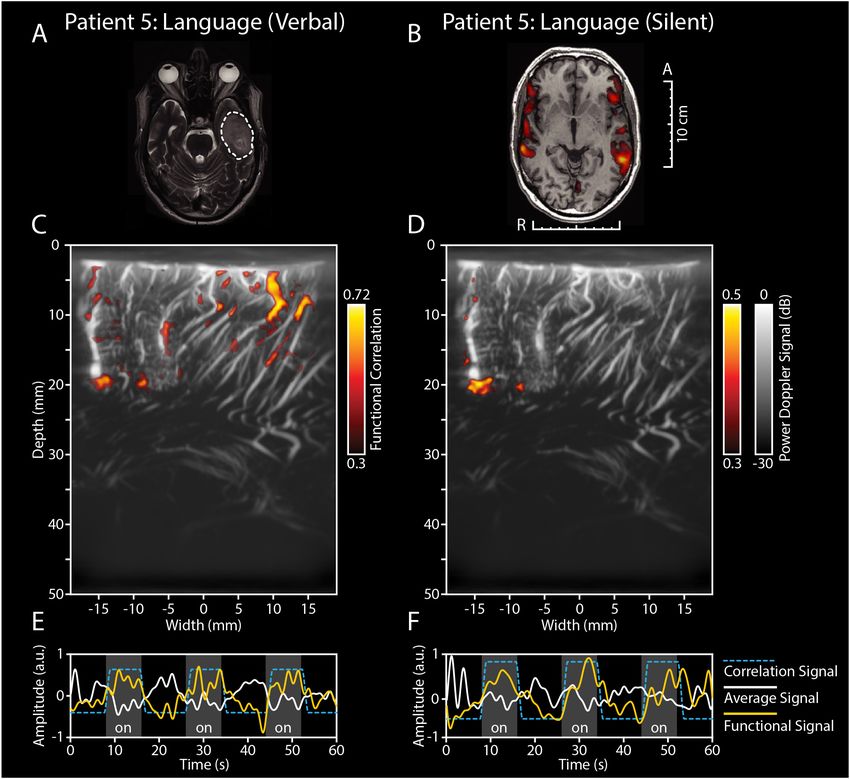
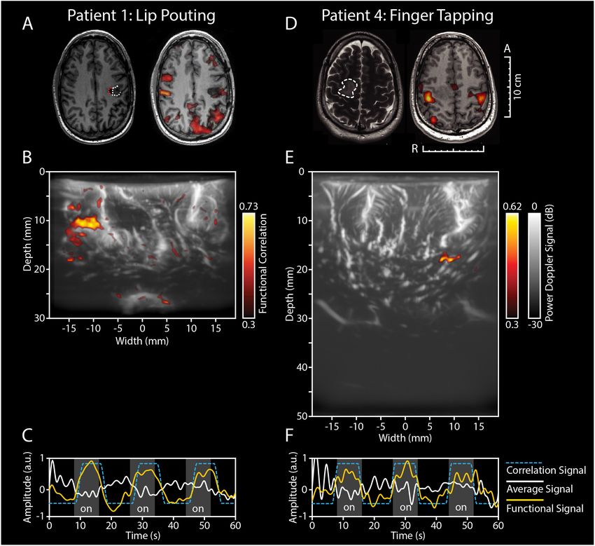
Soloukey et al.                                                                                                                            fUS During Awake Brain Surgery

  FIGURE 2 | Functional ultrasound results of two functional motor tasks in pt#1 and pt#4. (A–C) fUS-results of pt.#1, who presented with a recurrent HGG (GBM) in
  the left temporal lobe. (A) The tumor was located near the left precentral gyrus, as becomes clear from the pre-operative T1-weighted MRI (left-side). The white
  dotted line indicates the tumor borders, with the red dotted line indicating the resection cavity as a result of previous resection. Pre-operative fMRI revealed an
  association of the tumor with the previous tumor cavity during the functional task of lip pouting, indicating activation of the primary motor cortex of the mouth
  (right-side). (B) Intra-operatively, ESM confirmed association with the primary motor cortex. The patient was presented with a 60 s task-video of lip-pouting,
  consisting of three blocks of tasks (8 s each) and three blocks of resting conditions (10 s each), all preceded by a 6 s baseline. The image depicts the functional
  correlation map as made during the lip pouting task. A 3.8 cm wide and 3.0 cm deep image reveals functional activity in brain tissue around the tumor cavity evoked
  by the lip-pouting task. A video of this functional response over time is available as a supplement (Supplementary Video S1). (C) As becomes clear from the time
  traces, the average hemodynamic response in the areas defined as functional in B) follows the task pattern (yellow line). In contrast, non-functional areas do not
  follow this task pattern (white line). Details of this recording session can be found in Supplementary Table S2 (Recording ID 9). (D–F) fUS-results of pt.#4, who
  presented with a LGG in the right parietal lobe. (D) The tumor was located in the right parietal lobe, in close proximity to the primary motor cortex, as becomes clear
  from the pre-operative T2-weighted + GD MRI (left-side). The white dotted line indicates the tumor borders. Pre-operative fMRI revealed an association of the
  overlying motor cortex with the tumor during the functional task of bilateral finger tapping, indicating activation of the primary motor cortex of the hand (right-side).
  (E) Intra-operatively, ESM confirmed association with the primary motor cortex of the hand. The patient was presented with a 60 s task-video of finger-tapping, in the
  same task pattern as described for pt.#1 above. A 3.8 cm wide and 5.0 cm deep image reveals functional activity in deep brain tissue evoked by the lip-pouting
  task. A video of this functional response over time is available as a supplement (Supplementary Video S2). (F) As becomes clear from the time traces, the average
  hemodynamic response in the areas defined as functional in (E) follows the task pattern (yellow line). In contrast, non-functional areas do not follow this task pattern
  (white line). Details of this recording session can be found in Supplementary Table S2 (Recording ID 26). fUS, functional ultrasound; HGG, high grade glioma;
  GBM, glioblastoma; ESM, electrocortical stimulation mapping; LGG, low grade glioma; GD, gadolinium.

seemed to originate from a single point, deemed the vessel of                            tumor vasculature could also be identified. Although not visible
origin. Offline 3D-reconstruction of the 2D-PDIs confirmed the                           to the surgeon on the superficial cortex, the fUS-image pre-
possible existence of a single vessel of origin in this patient.                         resection identified a well-circumscribed oval-shaped region,
In pt#6, presenting with a LGG in the occipito-parietal region,                          where the tumor was to be expected (Figure 4L). After using

Frontiers in Neuroscience | www.frontiersin.org                                      6                                             January 2020 | Volume 13 | Article 1384
Soloukey et al.                                                                                                                            fUS During Awake Brain Surgery

  FIGURE 3 | Functional ultrasound results of two functional language tasks in pt#5. fUS-results of two language tasks in pt.#5, who presented with a GBM in the left
  temporal lobe. (A) The tumor was located in the left temporal lobe, as becomes clear from the pre-operative T2-weighted + GD MRI. The white dotted line indicates
  the tumor borders. (B) Pre-operative fMRI data showed an association of the tumor with eloquent areas during a conventional verbal fluency fMRI-task.
  Intra-operatively, ESM also identified several language-related deficits upon stimulation, including phonemic paraphasia. (C) After placement of the probe over the
  relevant markers, the patient was presented with a word repetition task, which the patient was asked to perform both verbally (by repeating the words out loud) as
  well as silently (by covertly repeating the words without speaking out loud). In (C) the functional correlation map as made during the verbal word repetition task is
  depicted. The fUS-image allowed for a field of view of 3.8 cm wide and 5.0 cm deep with several functional areas found across the cortex in view. Details of this
  recording session can be found in Supplementary Table S2 (Recording ID 36). (D) The functional correlation map as made during the silent word repetition task in
  the exact same field of view as discussed in (C). In comparison to the verbal word repetition task, this activation map shows less functional areas found within the
  field of view. (E,F) As becomes clear from the time traces, the average hemodynamic response in the areas defined as functional in (C,D) follows the task pattern
  (yellow line). In contrast, non-functional areas do not follow this task pattern (white line) (see also Supplementary Video S3). Details of this recording session can be
  found in Supplementary Table S2 (Recording ID 37). fUS, functional ultrasound; GBM, glioblastoma; ESM, electrocortical stimulation mapping; GD, gadolinium.

multiple 2D PDIs of the imaging session for reconstruction of                            Vascular Details
a 3D-volume, we observed an oval, well-defined nature of the                             In addition to the tumor-specific vascular characteristics
tumor. These examples highlight the potential of vasculature                             described above, the pre-resection image acquisition also entailed
mapping for brain-tumor delineations. See also Supplementary                             a rich variety of other high-resolution vascular details in pre-
Videos S4, S5 for the 3D reconstruction and Supplementary                                dominantly healthy tissue, some of which are depicted in
Figure S1 for an overview of similar 3D-reconstructions                                  Figure 5. First, all patients presented with what we dubbed
of all patients.                                                                         ‘feather vessels’ (Figure 5A), vascular structures consisting of a

Frontiers in Neuroscience | www.frontiersin.org                                      7                                             January 2020 | Volume 13 | Article 1384
Soloukey et al.                                                                                                                           fUS During Awake Brain Surgery

  FIGURE 4 | Maximum Projections of all patients (n = 10). (A–J) In addition to the regular 2D-images, we also made maximum projection images, showing an
  overview of the maximum signal per pixel during the imaging session of 60 s. As such, a single image with more depth-information can be created. For each patient,
  the most vascular-dense pre-resection image is highlighted, revealing a rich diversity in vascular patterns across patients. (K) 3D-overview of the pre-resection tumor
  vasculature images as acquired intra-operatively for pt.#2. Pre-operative MRI showed a suspected LGG tumor in the left frontal lobe. The probe was moved over the
  tumor along a continuous trajectory. During linear 2D-acquisition, an arborization structure was observed of the vessels within the tumor. The arborous vascular
  structure seems to originate from a single point, dubbed the vessel of origin. Multiple 2D PDIs acquired during the 60 s measurement session, showed the arborous
  structure. The PDIs were stacked offline in a 3D-volume, which confirms the vessel of origin, as depicted here. See also Supplementary Video S4 for the 3D
  reconstruction. (L) Overview of the pre-resection tumor vasculature images as acquired intra-operatively for pt.#6. Pre-operative MRI showed a suspected LGG in
  the left occipito-parietal region. The probe was moved over the tumor along a continuous trajectory. During linear 2D-acquisition, a well-defined vascular structure
  was observed, delineating the tumor from the rest of the tissue. Multiple 2D PDIs acquired during the 60 s measurement session, were stacked offline in a
  3D-volume, confirming the well-defined vascular area. See also Supplementary Video S5 for the 3D reconstruction. These last two examples highlight the potential
  of vascular mapping for brain-tumor delineations. fUS, functional ultrasound; LGG, low grade glioma; PDI, power doppler image.

single, large vessel hosting multiple orthogonal sprouting vessels.                     larger cortical vessels along the sulci and gyri, some examples
These structures were found both in the superficial cortex layers,                      of which are depicted in Figure 5C. Lastly, several circular
and more in the depth, resembling intra-cortical arteries reported                      vascular structures were observed (Figure 5D), the origins of
previously (Duvernoy et al., 1981). In addition, tortuous vessels                       which remain unknown. The zoomed-in images give examples of
were also observed (Figure 5B). Although vessel tortuosity is a                         the high resolution (300 µm) and level of detail achieved during
potential sign of fast-growing, pathological tumor vasculature,                         image acquisition.
the coiled vessels were also observed in healthy tissue, resembling
physiological vascular patterns known as ‘recurrent arteries’                           Resection-Cavity
(Duvernoy et al., 1981). In those patients where the superficial                        Figure 6 depicts an example of a matched pre- and post-
cortical vessels were exposed, we were often able to follow these                       resection image of tumor and surrounding healthy tissue in

Frontiers in Neuroscience | www.frontiersin.org                                     8                                             January 2020 | Volume 13 | Article 1384
Soloukey et al.                                                                                                                              fUS During Awake Brain Surgery

  FIGURE 5 | Overview of rich vascular characteristics found in our pre-resection datasets (n = 10). (A) All patients presented with feather-like vessels, vascular
  structures consisting of a single, large vessel hosting multiple orthogonal sprouting vessels. These structures were found both in the superficial cortex layers, as well
  as deeper, and resemble intra-cortical arteries reported previously in literature (Duvernoy et al., 1981). (B) Tortuous vessels were also observed. Although the
  tortuous vessels are known to be potential signs of fast-growing, pathological tumor vasculature, the coiled vessels seem to resemble physiological vascular patterns
  known as ‘recurrent arteries,’ as also depicted in the SEM-images presented in Duvernoy et al. (1981). These torturous vessels are also visible in (C) of this figure
  (bottom picture) and Figure 6 (patient 4). (C) In those patients where the superficial cortical vessels were exposed, we were also able to capture these larger vessels
  following, e.g., the sulcus and gyrus patterns of the brain, allowing for lobular distinction. (D) Several circular vascular structures were observed, the origins of which
  remain unknown. The scale bar in the bottom right corner is applicable to all subpanels. SEM, scanning electron microscope.

Frontiers in Neuroscience | www.frontiersin.org                                       9                                              January 2020 | Volume 13 | Article 1384
Soloukey et al.                                                                                                      fUS During Awake Brain Surgery

pt.#4. This figure demonstrates the potential of using vasculature          starting before (see example of pt#7 in Supplementary Figure S2)
characteristics as a guide for tumor-brain delineations intra-              or after the actual the task. As explained in the methods, we aimed
operatively, identifying the tumor’s borders based on its                   to compensate for these differences in timing by allowing a −2 to
vascular characterization.                                                  2 s delay between the stimulus signal and the recording. Reasons
                                                                            for these lags may include: (1) slow hemodynamic response
                                                                            function, (2) inaccurate timing between start of the acquisition
DISCUSSION                                                                  and presented functional task, and (3) delayed or anticipatory
                                                                            response of the patient to the presented task. Determining the
Inherent limitations of currently available neuro-imaging                   potential bias that for example anticipatory effects might have
techniques warrant the development of new real-time image-                  during functional imaging, and how to design proper functional
guided resection tools, which allow for reliable functional                 paradigms to exclude them, will be part of our future studies.
and anatomical information in a neurosurgical setting. The                      The ability of fUS to capture complex functional processes
present study demonstrates the clinical potential of fUS as a new           in the brain opens up possibilities not only for functional
image-guided resection tool during awake craniotomy surgery.                neurosurgery, but also for unraveling brain function in general.
fUS is able to detect the functional areas that were found using            Currently, fUS in humans is still restricted to those circumstances
ESM (the current gold standard) across a range of functional                where the brain is no longer covered by skull [craniotomies,
tasks. As demonstrated by the vascular patterns mapped with                 fontanels in babies (Demene et al., 2017)]. However, future
submillimeter resolution, fUS also provides a potential new                 developments in the field of transcranial-fUS overcoming signal
means for tumor delineation based on vasculature characteristics            blocking by bone (Errico et al., 2016; Tiran et al., 2017) could
of tumor and healthy tissue.                                                open up possibilities for functional studies in humans in a
    The work presented here is the second study to demonstrate              clinical-diagnostic, translational and fundamental setting.
the power of fUS for neurosurgery as a real-time technique                      The current study is the first to highlight the potential of
to asses local brain functionality. With respect to the study               real-time, high-resolution imaging of vasculature as a means of
by Imbault et al. (2016, 2017) we demonstrate the ability of                anatomical delineation. Within clinical oncology, it is widely
fUS to capture not only motor activation (Figure 2) but also                accepted that tumor angiogenesis and as such tumor vasculature,
more complex language-related activation (Figure 3). In one                 is differently developed than that of normal tissue (Gerstner
of our patients, this was demonstrated by performing a double               et al., 2008; Forster et al., 2017). In fact, numerous therapies
word repetition language-task, both verbally and silently, which            such as vascular targeting techniques (Pilat et al., 2004), as
revealed two different activation patterns within the same field            well as histopathological malignancy gradings (Hansen et al.,
of view (Figure 3). Although the exact underlying mechanism                 2002; Jain et al., 2007), are based on these tumor-vasculature
remains speculative at this point, the above-mentioned approach             differences. Using fUS, we have been able to identify tumor-
using fUS does allow for, e.g., interrogation of the motor-                 specific vasculature with up to 300 µm resolution in both low
specific and word production component in language. The ability             and high-grade tumors (finer resolution is achievable at the
to interrogate the system requires the design of appropriate                cost of imaging depth). In one LGG in particular, we were able
sets of functional tasks, which became especially apparent in               to image an arborization structure with originating from one
pt.#2 and pt#7 where a verbal language-related task (sentence               vessel of origin (Figure 4K). Although our current results are
and word repetition respectively), showed an activation map                 still too preliminary to draw general conclusions against the
where language and motor functional response could not be                   backdrop of inherent glioma heterogeneity, they do provide ideas
distinguished (Supplementary Figure S2).                                    for potential fUS applications. For those tumors with vessels of
    In those cases where we did not find functional signals                 origin, for example, fUS-guided targeted tumor therapies would
(n = 35), the use of an inappropriate functional task could                 be an interesting approach. What is more, using our fUS-data
also be a possible explanation. However, problems either due                in real-time during tumor removal could possibly allow for
to shifted brain functionalities in space due to oncogenesis, too           vascularity-guided tumor resection, in parallel to the treatment
much in- and out-of-plane motion of the hand-held probe or                  of meningiomas with so called ‘pedicles,’ where early access to
placement of the probe over an incorrect or non-functional area,            the vascular origin facilitates safe and effective tumor resection
form alternative explanations. The latter two explanations are              (Dowd et al., 2008; Watts et al., 2014; Hilmani et al., 2016).
especially plausible, as the majority of the measurements where             It would be worthwhile to focus future efforts on imaging
we found a functional signal (6 out of 9), were made in patients            heterogeneous groups of brain tumors and comparing and
who did show ESM-related functional deficits intra-operatively.             contrasting vascular structure across histopathological gradings,
Furthermore, almost all (8 out of 9) measurements presenting                not only in awake but also in anesthetized patients.
with functional signal showed below average displacement in the                 Additionally, vascular mapping in our dataset of 10 patients
x-axis (
Soloukey et al. fUS During Awake Brain Surgery FIGURE 6 | Pre- and post-resection tumor and resection cavity imaging in pt.#4. (A) Intra-operative image of a LGG in the right parietal lobe in pt#4, who presented with several eloquent areas in the proximity of the tumor (see ESM-markers). (B) Intra-operative image of the post-resection cavity after tumor removal. (C) Pre-resection B-mode image of the tumor and surrounding structures. (D) Post-resection B-mode image, showing the hypodense resection cavity created after tumor removal. (E) PDI of the pre-resection field of view covering the tumor and surrounding healthy tissue. (F) PDI of the post-resection field of view depicting the resection cavity, filled with saline. Noticeable are the similar vascular structures depicted in the depth (3–4 cm) of both images (E,F), indicating a similar field of view pre- and post-resection. LGG, low grade glioma; PDI, power doppler image. Frontiers in Neuroscience | www.frontiersin.org 11 January 2020 | Volume 13 | Article 1384
Soloukey et al.                                                                                                        fUS During Awake Brain Surgery

also delineation of healthy tissue structures, as becomes clear in             terms of tumor resection, post-operative neurological deficits,
an incidental imaging session of the thalamus (Supplementary                   and survival when performing fUS-guided resections.
Figure S3). This in turn opens up potential applications in, e.g.,                The current study demonstrates that fUS has the potential
neurovascular or functional neurosurgery.                                      to be a highly flexible technique for providing vascular as
   Compared to conventional intra-operative mapping using                      well as functional information in an intra-operative setting
ESM, fUS provides higher spatiotemporal resolution and higher                  with high spatiotemporal resolution. As current neurosurgical
depth-penetration. In our implementation, temporal resolution                  practice is still relying on inherently limited imaging techniques
can be as high as 1.5 ms, while maintaining real-time display for              for tumor resection-guidance, fUS enters the scene with great
the surgeon (up to 4.8 Hz) and continuous raw frame storage                    clinical potential.
(up to 667 Hz). Compared to conventional pre- and intra-
operative imaging techniques such as (f)MRI, fUS proves to
have higher temporal resolution using a more mobile system.                    DATA AVAILABILITY STATEMENT
Surprisingly, several of our functional imaging sessions showed
activation maps also involving concentrated areas at deeper                    The raw data supporting the conclusions of this article will be
levels of the brain, which would otherwise not have been                       made available by the authors upon reasonable request.
interrogated intra-operatively with ESM. In addition, the lack
of need for electrical stimulation eliminates the risk of intra-               ETHICS STATEMENT
operative seizure elicitation. It is also imaginable that several tasks
can be performed at once before resecting the tumor, which could               The studies involving human participants were reviewed and
save time. Lastly, the movability and cost-effectiveness of fUS                approved by the Medical Ethics Review Committee (METC)
make it a highly clinically accessible technique.                              of the Erasmus Medical Center in Rotterdam, Netherlands
   Nevertheless, the technique will need to be further validated               (MEC 2018-037). The patients/participants provided their
and improved to reach clinical maturity. For fUS as used in its                written informed consent to participate in this study.
current form, real-time, automated image classification will be
an important next step, which would allow for, e.g., automated
brain-tumor delineation. In addition, hand-held fUS-imaging is                 AUTHOR CONTRIBUTIONS
inherently prone to problems such as motion, both in- and out-
of-plane, which can either lead to (1) the inability to capture                SS, AV, SK, and PK were involved in the study design. SS, AV, DS,
functional response, or (2) capturing of artifactual ‘functional’              and PK were involved in the determination of appropriate intra-
signal. Using linear stages or other probe-mounting options                    operative functional tasks. DS was involved in the intra-operative
in the OR-setting would be a solution. Future work will also                   guidance of the patients while presenting the functional tasks.
need to focus on the tracking and integration of the probe                     FM, SS, MS, SK, and PK were involved in the data-processing
in currently available neuro-navigation software. Not only will                and -analysis of fMRI and fUS-data. AV included all patients and
this facilitate the validation of fUS with (f)MRI images made                  performed all surgeries and scans. SS, SK, and PK were involved
pre-operatively, but it will also allow for better cross-patient               in drafting the manuscript, with the critical input of AV, DS, FM,
comparisons. Comparison of fUS with fMRI in particular is                      MS, CMD, CS, JB, AS, and CID.
interesting, as both techniques rely on different aspects within the
neurovascular coupling mechanism. Where fMRI relies on the
BOLD-signal, a measure of blood-oxygenation levels influenced
                                                                               FUNDING
by changes in blood volume and flow as well as the rate of                     This study was funded by the Erasmus Medical Center
oxygen consumption (Logothetis, 2018) to determine neuronal                    MRACE-pilot grant (Grant no. 108581). SS, AV, CMD, CS,
activity, fUS relies on changes in vascular dynamics as measured               JB, AS, CID, SK, and PK were supported by the NWO-
by Doppler (Deffieux et al., 2018). How these two measurements                 Groot grant of The Dutch Organization for Scientific Research
of the same phenomenon relate, remains to be elucidated. What                  (NWO) (Grant no. 108845), awarded to CUBE (Center for
is more, it would be worthwhile to critically revisit in future                Ultrasound and Brain-Imaging @ Erasmus MC, see for website:
studies our perhaps simplistic assumption of an almost one-                    www.ultrasoundbrainimaging.com). CID was supported by the
to-one correlation between the functional task pattern and the                 Dutch Organization for Medical Sciences (ZonMw), Life Sciences
changes in blood dynamics as measured by fUS.                                  (Grant no. 854.10.004), the Neurotime, ERC-advanced and
   In addition, replacing the currently used linear array                      ERC-PoC programs of the European Community (Grant nos.
with a 3D-probe will allow for intra-operative fUS-imaging                     294775 and 768914).
of 3D volumes. Both in terms of functional as well as
vascular anatomical information, this could constitute a huge
improvement. Furthermore, with a 3D-vascular map, vascular-                    ACKNOWLEDGMENTS
based calibration instead of the usual bone-based calibration
for neuro-navigation, could also potentially solve the post-                   The authors would like to express great gratitude to Dr. Evy
craniotomy brain shift problem. Most importantly, future work                  Visch-Brink for her contribution to the neurolinguistic tasks and
will also have to center around the actual patient outcomes in                 intra-operative measurements.

Frontiers in Neuroscience | www.frontiersin.org                           12                                    January 2020 | Volume 13 | Article 1384
Soloukey et al. fUS During Awake Brain Surgery
SUPPLEMENTARY MATERIAL complicates the ability to identify between the functional areas in response to the
language task vs. the motor activation of the mouth. This in contrast to the
functional measurements explicated in Figure 3 with pt.#5. (F) The time-traces
The Supplementary Material for this article can be found
display an unexpected average hemodynamic response in the areas defined as
online at: https://www.frontiersin.org/articles/10.3389/fnins. functional in (E), starting before the actual task pattern (yellow line). In contrast,
2019.01384/full#supplementary-material non-functional areas do not follow this task pattern (white line). Details of this
recording session can be found in Supplementary Table S2 (Recording ID 59).
FIGURE S1 | Overview of 3D-volume stacks of patient 1–10. (A–J) Multiple 2D HGG, high grade glioma; GBM, glioblastoma; ESM, electrocortical stimulation
PDIs (ranging from n = 80–297) acquired during 60 s measurement sessions for mapping; LGG, low grade glioma.
each individual patient were stacked offline in a 3D-volume. PDI, power doppler
image. FIGURE S3 | Vascular imaging of the thalamus in pt.#10. Incidental imaging
session of the thalamus in pt.#10, who presented with a LGG in the left frontal
FIGURE S2 | Functional ultrasound results of two unusual functional responses to lobe. (A) Conventional B-mode image in a sagittal plane, showing (from top to
tasks in pt#2 and pt#7 (language). (A–C) Functional results of a language task bottom) the corpus callosum, the ventricle, and the thalamus. (B) The PDI of the
(sentence repetition) in a patient with a LGG in the left frontal lobe. (A) same field of view as described under (A), reveals a rich vascular pattern in the
Pre-operative MRI showing the extent of the LGG in the left frontal lobe. The white thalamus, allowing for a clear delineation of the nucleus from the surrounding brain
dotted line indicates the tumor borders. (B) Intra-operatively, ESM identified both tissue. This would open up possibilities for monitoring of vascularization of critical
language-related functional areas (anomia), as well as motor related functional structures such as the thalamus during neurosurgical procedures. (C) An overlay
areas (primary motor cortex of the mouth) in close proximity to each other. When of B-mode and PDI, reveals the vascular pattern in relation to the tissue as would
presented with a verbal functional language task (sentence repetition), multiple be displayed in conventional echography. LGG, low grade glioma; PDI, power
functional areas could be defined within the field of view. The lack of a silent doppler image.
language task as a counter-part in this particular patient, complicates the ability to
TABLE S1 | Overview of functional tasks as used in the current study.
identify between the functional areas in response to the language task vs. the
motor activation of the mouth. This in contrast to the functional measurements TABLE S2 | Overview of all recordings (n = 90) as performed in the context of
explicated in Figure 3 with pt.#5. (C) As becomes clear from the time traces, the the current study.
average hemodynamic response in the areas defined as functional in (B) follows
the task pattern (yellow line). In contrast, non-functional areas do not follow this VIDEO S1 | Functional response of pt.#1 during the ‘lip pouting’ functional
task pattern (white line). Details of this recording session can be found in motor task.
Supplementary Table S2 (Recording ID 13). (D–F) Functional results of a VIDEO S2 | Functional response of pt.#4 during the ‘finger tapping’ functional
language task (word repetition, verbal) in a patient with a HGG (GBM) in the motor task.
occipito-parietal region. (D) Pre-operative T2-weighted MRI showing the GBM in
the left hemisphere. The white dotted line indicates the tumor borders. (E) VIDEO S3 | Functional response of pt.#5 during the ‘word repetition’ functional
Intra-operative ESM did not identify any clear functional areas. Based on the language task (verbal and silent).
anatomical location, the probe was placed partially over a tumor area and partially
VIDEO S4 | 3D vascular tumor reconstruction of a LGG in the left frontal
over a potential functional area related to language. The patient was presented
lobe of pt.#2.
with a verbal functional language task (word repetition), which resulted in an
activation map with strong response in the upper left corner of the field of view. VIDEO S5 | 3D vascular tumor reconstruction of a LGG in the occipito-parietal
Again, the lack of a silent language task as a counter-part in this patient, region of pt.#6.
REFERENCES Demené, C., Deffieux, T., Pernot, M., Osmanski, B. F., Biran, V., Gennisson,
J. L., et al. (2015). Spatiotemporal clutter filtering of ultrafast ultrasound data
Almasian, M., Wilk, L. S., Bloemen, P. R., van Leeuwen, T. G., ter Laan, M., highly increases doppler and fultrasound sensitivity. IEEE Trans. Med. Imag.
and Aalders, M. C. G. (2019). Pilot feasibility study of in vivo intraoperative 34, 2271–2285. doi: 10.1109/TMI.2015.2428634
quantitative optical coherence tomography of human brain tissue during Dizeux, A., Gesnik, M., Ahnine, H., Blaize, K., Arcizet, F., Picaud, S., et al. (2019).
glioma resection. J. Biophotonics. 12:e201900037. doi: 10.1002/jbio.201900037 Functional ultrasound imaging of the brain reveals propagation of task-related
Bimbard, C., Demene, C., Girard, C., Radtke-Schuller, S., Shamma, S., Tanter, M., brain activity in behaving primates. Nat. Commun. 10:1400. doi: 10.1038/
et al. (2018). Multi-scale mapping along the auditory hierarchy using high- s41467-019-09349-w
resolution functional UltraSound in the awake ferret. eLife 7:e35028. doi: 10. Dowd, C. F., Halbach, V. V., and Higashida, R. T. (2008). Meningiomas: the
7554/elife.35028 role of preoperative angiography and embolization. Neurosurg. Focus 15:E10.
De Witt Hamer, P. C., Robles, S. G., Zwinderman, A. H., Duffau, H., and Berger, doi: 10.3171/foc.2003.15.1.10
M. S. (2012). Impact of intraoperative stimulation brain mapping on glioma Duvernoy, H. M., Delon, S., and Vannson, J. L. (1981). Cortical blood vessels of the
surgery outcome: a meta-analysis. J. Clin. Oncol. 30, 2559–2565. doi: 10.1200/ human brain. Brain Res. Bull. 7, 519–579. doi: 10.1016/0361-9230(81)90007-1
JCO.2011.38.4818 Errico, C., Osmanski, B. F., Pezet, S., Couture, O., Lenkei, Z., and Tanter, M. (2016).
De Witte, E., Satoer, D., Robert, E., Colle, H., Verheyen, S., Visch-Brink, E., et al. Transcranial functional ultrasound imaging of the brain using microbubble-
(2015). The dutch linguistic intraoperative protocol: a valid linguistic approach enhanced ultrasensitive Doppler. Neuroimage 124(Pt A), 752–761. doi: 10.1016/
to awake brain surgery. Brain Lang. 140, 35–48. doi: 10.1016/j.bandl.2014.10. j.neuroimage.2015.09.037
011 Fabelo, H., Ortega, S., Lazcano, R., Madroñal, D., Callicó, G. M., Juárez, E.,
Deffieux, T., Demene, C., Pernot, M., and Tanter, M. (2018). Functional ultrasound et al. (2018). An intraoperative visualization system using hyperspectral
neuroimaging: a review of the preclinical and clinical state of the art. Curr. Opin. imaging to aid in brain tumor delineation. Sensors 18:E430. doi: 10.3390/s1802
Neurobiol. 50, 128–135. doi: 10.1016/j.conb.2018.02.001 0430
Demene, C., Baranger, J., Bernal, M., Delanoe, C., Auvin, S., Biran, V., et al. Forster, J., Harriss-Phillips, W., Douglass, M., and Bezak, E. (2017). A review
(2017). Functional ultrasound imaging of brain activity in human newborns. of the development of tumor vasculature and its effects on the tumor
Sci. Transl. Med. 9:eaah6756. doi: 10.1126/scitranslmed.aah6756 microenvironment. Hypoxia 5, 21–32. doi: 10.2147/hp.s133231
Demené, C., Bimbard, C., Gesnik, M., Radtke-Schuller, S., Shamma, S., Boubenec, Gerritsen, J. K. W., Arends, L., Klimek, M., Dirven, C. M. F., and Vincent, A. J. P. E.
Y., et al. (2016). “Functional Ultrasound Imaging of the thalamo-cortical (2019). Impact of intraoperative stimulation mapping on high-grade glioma
auditory tract in awake ferrets using ultrafast Doppler imaging,” in Proceedings surgery outcome: a meta-analysis. Acta Neurochir. 161, 99–107. doi: 10.1007/
of the IEEE International Ultrasonics Symposium, IUS, Tour. s00701-018-3732-4
Frontiers in Neuroscience | www.frontiersin.org 13 January 2020 | Volume 13 | Article 1384You can also read