Aberrant Intraregional Brain Activity and Functional Connectivity in Patients With Diarrhea-Predominant Irritable Bowel Syndrome - Frontiers
←
→
Page content transcription
If your browser does not render page correctly, please read the page content below
ORIGINAL RESEARCH
                                                                                                                                      published: 03 September 2021
                                                                                                                                     doi: 10.3389/fnins.2021.721822

                                            Aberrant Intraregional Brain Activity
                                            and Functional Connectivity in
                                            Patients With Diarrhea-Predominant
                                            Irritable Bowel Syndrome
                                            Xiao-Fei Chen 1 , Yun Guo 2 , Xing-Qi Lu 1 , Le Qi 1,3 , Kuang-Hui Xu 3 , Yong Chen 3 ,
                                            Guo-Xiong Li 2 , Jian-Ping Ding 1,3 and Jie Li 1*
                                            1
                                             Department of Radiology, The Affiliated Hospital of Hangzhou Normal University, Hangzhou, China, 2 Department
                                            of Gastroenterology, The Affiliated Hospital of Hangzhou Normal University, Hangzhou, China, 3 Medical College, Hangzhou
                                            Normal University, Hangzhou, China

                                            Background and Purpose: The appearance and aggravation of diarrhea-predominant
                                            irritable bowel syndrome (IBS-D) have proven to be closely related to psychosocial
                                            factors. We aimed to measure altered spontaneous brain activity and functional
                                            connectivity (FC) in patients with IBS-D using resting-state functional magnetic
                          Edited by:        resonance imaging (RS-fMRI) and to analyze the relationship between these parameters
                         Mingrui Xia,
      Beijing Normal University, China      and emotional symptoms.
                     Reviewed by:           Methods: Thirty-six adult IBS-D patients and thirty-six demographic-matched healthy
                    Ruiwang Huang,
South China Normal University, China
                                            controls (HCs) underwent RS-fMRI scans. After processing RS-fMRI data, the values of
                       Peiyu Huang,         the amplitude of low-frequency fluctuation (ALFF) and regional homogeneity (ReHo) of
           Zhejiang University, China
                                            the two groups were compared. The abnormal regions were selected as the regions of
                   *Correspondence:
                                            interest to compare whole-brain seed-based FC between the groups. The relationships
                               Jie Li
                jie_sweethz@163.com         between RS-fMRI data and mood and gastrointestinal symptoms were analyzed using
                                            correlation and mediation analyses.
                   Specialty section:
         This article was submitted to      Results: Compared with HCs, IBS-D patients showed increased ALFF in the right
              Brain Imaging Methods,        cerebellum posterior lobe, the right lingual gyrus/calcarine, the right postcentral gyrus,
               a section of the journal
            Frontiers in Neuroscience       the right superior frontal gyrus (SFG), and middle frontal gyrus (MFG), with decreased
           Received: 07 June 2021           ALFF in the right inferior parietal lobule, the right striatum, the right anterior cingulated
        Accepted: 09 August 2021            cortex, the right insula, the right hippocampus, the right thalamus, the right midbrain,
     Published: 03 September 2021
                                            and the left precuneus. IBS-D patients showed increased ReHo in the bilateral lingual
                               Citation:
       Chen X-F, Guo Y, Lu X-Q, Qi L,
                                            gyrus/calcarine, the bilateral SFG, the right MFG, and the right postcentral gyrus,
Xu K-H, Chen Y, Li G-X, Ding J-P and        with decreased ReHo in the orbital part of the left inferior frontal gyrus and the right
     Li J (2021) Aberrant Intraregional     supplementary motor area. Patients showed enhanced FC between the left precuneus
           Brain Activity and Functional
          Connectivity in Patients With     and the bilateral orbitofrontal cortex (OFC). There was a positive correlation between
 Diarrhea-Predominant Irritable Bowel       increased ALFF values in the right midbrain and anxiety-depression symptoms in IBS-
                             Syndrome.
           Front. Neurosci. 15:721822.
                                            D patients, and the mediating effect of gastrointestinal symptoms indirectly caused
      doi: 10.3389/fnins.2021.721822        this correlation.

Frontiers in Neuroscience | www.frontiersin.org                                   1                                     September 2021 | Volume 15 | Article 721822
Chen et al.                                                                                                        Brain Spontaneous Dysfunctions in IBS-D

                                            Conclusion: IBS-D patients had dysregulated spontaneous activity and FC in regions
                                            related to pain regulation and emotional arousal involved in prefrontal–limbic–midbrain
                                            circuit and somatosensory processing. The development of mood disorders in IBS-
                                            D patients may be partly related to the dysfunction of components in the dopamine
                                            pathway (especially the midbrain, OFC) due to visceral pain.
                                            Keywords: irritable bowel syndrome, amplitude of low-frequency fluctuation, regional homogeneity, functional
                                            connectivity, resting-state fMRI

INTRODUCTION                                                                      fMRI (RS-fMRI) experimental modes. During rectal dilation,
                                                                                  the brain regions associated with the fronto–limbic circuit for
Irritable bowel syndrome (IBS) is one of the most common                          regulating emotion, particularly the insula, the amygdala, and the
functional gastrointestinal disorders (FGID), characterized by                    prefrontal cortex (PFC), were abnormally activated in patients
recurrent abdominal pain or discomfort and changes in bowel                       with IBS (Elsenbruch et al., 2010; Labus et al., 2013; Hong
habits (Mearin et al., 2016). IBS is classified into diarrhea type                et al., 2016). RS-fMRI can detect spontaneous neural activity
(IBS-D), constipation type (IBS-C), mixed type, and undefined                     without specific tasks with a high spatial resolution (Fox and
type according to the predominant stool pattern. Patients with                    Raichle, 2007). RS-fMRI studies showed that IBS patients have
IBS-D account for about one-third of all patients, and their                      abnormal spontaneous activities in brain regions related to
quality of life is lower than that of other subtypes; their daily                 pain processing [i.e., the amygdala, anterior cingulated cortex
activities are more disturbed, and they commonly avoid eating                     (ACC), thalamus, and insula] and emotion regulation (i.e., the
(Rome Foundation, 2006; Singh et al., 2015). IBS is often                         amygdala, cingulate cortex, hippocampus, and hypothalamus),
accompanied by psychological disorders, among which anxiety                       and partly attributable to anxiety and depression (Ma et al.,
and depression are the most common, with a prevalence of up to                    2015; Qi et al., 2016). This association between abnormalities
50% (Ford et al., 2020). Patients with IBS-D have loose stools and                in brain activity and psychological symptoms was analyzed by
frequent bowel movements, especially accompanied by urgency                       comparing outcomes with and without the inclusion of anxiety
and fear of urinary incontinence, making them more prone to                       and depression symptoms as covariates. Based on independent
panic and anxiety (Singh et al., 2015). Treatment options for                     component analysis or graph theory analysis, IBS was found to
IBS-D are limited (Lucak et al., 2017; Dothel et al., 2018).                      be associated with dysfunction of functional connectivity (FC)
   The pathogenesis of IBS is not entirely understood; however,                   in several networks, including the salience/executive control
it is thought to be related to visceral hypersensitivity,                         network, the attention network, the emotion regulation network,
psychosocial factors, intestinal infections, and intestinal                       and the default mode network (DMN) (Mayer et al., 2019). Few
flora disturbance (Ford et al., 2020). Psychosocial factors                       fMRI studies on IBS-specific subtypes and only two task-fMRI
have recently attracted particular attention because of their                     studies observed differences in brain activation patterns between
essential role in the brain-gut axis. Specifically, psychosocial                  IBS-C and IBS-D when receiving rectal dilation stimulation. The
factors affect the central processing of visceral stimulation                     activation of the pain matrix related brain regions in IBS-D
through the emotional circuit of the central nervous system,                      patients was higher than that in IBS-C patients and was related to
leading to further amplification of central pain in a top-                        the higher sensitivity of IBS-D patients to rectal dilation (Wilder-
down approach (Mudyanadzo et al., 2018). Patients with                            Smith et al., 2004; Guleria et al., 2017). Therefore, it is necessary
IBS have insufficient secretion of neurotransmitters related                      to observe the patterns of brain function changes during resting-
to gastrointestinal function [e.g., 5-hydroxytryptamine                           state and their correlation with emotional symptoms in patients
(5-HT) and dopamine], which affects the operation of                              with specific subtypes of IBS.
neurotransmitter-related circuits in the brain from bottom                            The amplitude of low-frequency fluctuations (ALFF) (Zang
to top, further aggravating emotional disorders (Gershon                          et al., 2007; Zuo et al., 2010) and regional homogeneity (ReHo)
and Tack, 2007; Stasi et al., 2012). Nevertheless, the exact                      (Zang et al., 2004) are commonly used RS-fMRI methods for local
neuropathological mechanisms of IBS-induced emotional                             neural activity. The ALFF measures the regional intensity of a
disturbance remain unclear. Psychosocial factors might impact                     voxel in a low-frequency range (Zang et al., 2007; Zuo et al., 2010).
pain thresholds differently according to the subtype of IBS.                      The ReHo calculated the synchronization of low-frequency
Affective and emotional symptoms should be considered                             fluctuations between a given voxel with neighboring voxels (Zang
specific and parts of the syndrome. Recognizing their impact                      et al., 2004). Therefore, ALFF and ReHo complement each other
on different IBS subtypes may have implications for treatment                     and provide a more comprehensive characterization of local brain
options and clinical outcomes (de Medeiros et al., 2008;                          abnormalities (An et al., 2013; Lv et al., 2019; Li et al., 2021).
Muscatello et al., 2010).                                                             The functional brain patterns of IBS-D patients during
   Functional magnetic resonance imaging (fMRI) is a powerful                     resting-state and their relationship to emotional symptoms
tool for evaluating brain function by detecting relative changes                  remain unclear. Based on previous research and theoretical
in the content of local deoxyhemoglobin when measuring                            results, we selected IBS-D patients as the research subjects,
neuronal activity, including in task-fMRI and resting-state                       utilized ALFF and ReHo methods for data-driven analysis,

Frontiers in Neuroscience | www.frontiersin.org                               2                                 September 2021 | Volume 15 | Article 721822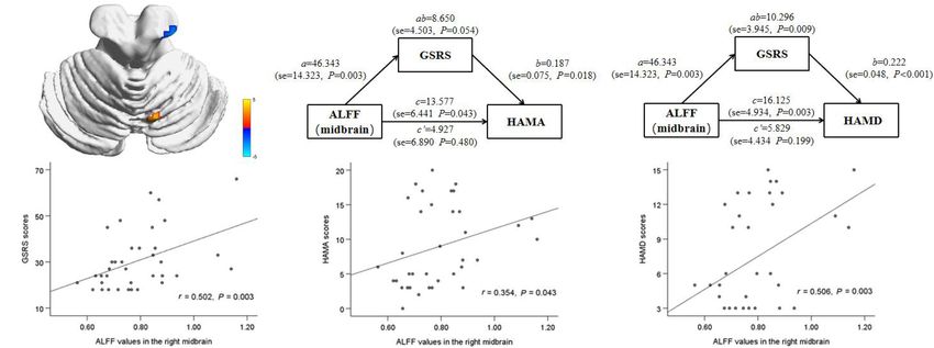
Chen et al.                                                                                                        Brain Spontaneous Dysfunctions in IBS-D

and combined with FC analysis to explore changes in                         bed, wear special non-magnetic headphones, and use sponge
whole-brain spontaneous neural activity and FC in patients                  cushions to fix both sides of the head to reduce head movement.
with IBS-D. We hypothesized that there would be aberrant                    During the image acquisition process, the participants need to
brain function in emotion or pain-related processing regions                remain awake, relax with their eyes closed, breathe smoothly, and
in IBS-D patients, and the partly neuroimaging changes                      keep their heads motionless as much as possible. The scanning
would be associated with emotional symptoms. In particular,                 sequences included the whole-brain three-dimensional high-
gastrointestinal manifestations with pain as the core symptom               resolution T1 weighted image (3D T1WI) obtained through the
may mediate this correlation.                                               three-dimensional spoiled gradient recalled-echo sequence and
                                                                            the RS-fMRI image obtained by the blood oxygenation level
                                                                            dependent-gradient echo-echo planar imaging sequence. The
MATERIALS AND METHODS                                                       parameters of 3D T1WI are repetition time (TR) = 8.16 ms,
                                                                            echo time (TE) = 3.18 ms, flip angle = 8◦ , the field of
Thirty-eight patients with IBS-D and 36 age-, sex- and education            view (FOV) = 256 mm × 256 mm, Acquisition matrix
level- matched healthy controls (HCs) were recruited from the               (matrix) = 256 × 256, layer thickness = l mm, 176 layers. The
Gastroenterology Clinic and Physical Examination Center of the              scan parameters of RS-fMRI are TR = 2,000 ms, TE = 30 ms,
Affiliated Hospital of Hangzhou Normal University from June                 flip angle = 90◦ , FOV = 192 × 192 mm, Matrix = 64 × 64,
2016 to July 2018. All patients were diagnosed simultaneously               layer thickness = 4 mm, interval = 0 mm. The scan range
by two senior gastroenterologists. All subjects provided written            includes the whole brain, and the scan time is 8 min in total.
informed consent before participating. The Research Ethics                  T1WI and T2 fluid-attenuated inversion recovery were collected
Committee of the Affiliated Hospital of Hangzhou Normal                     simultaneously to rule out abnormal anatomical structures
University approved the study that we performed according to                and organic brain diseases by visual inspection; subjects with
the Declaration of Helsinki of the World Medical Association.               abnormal signs such as intracranial space-occupying lesions and
   All patients with IBS-D underwent a complete medical history,            cerebral infarction were excluded.
physical examination, blood biochemistry, and colonoscopy. The
inclusion criteria included age 18–55 years and meeting the
Rome III diagnostic criteria. The exclusion criteria included
                                                                            MRI Data Preprocessing
taking antidepressants and gastrointestinal motility drugs in the           Data preprocessing was implemented using the Data Processing
previous 2 weeks, a history or current diagnosis of psychosocial            and Analysis for (Resting-State) Brain Imaging (DPABI) software
disorders (e.g., major depression, psychosis, or drug and alcohol           package,1 which is based on Statistical Parametric Mapping
abuse as defined by the DSM-IV-TR criteria), a history of brain             version 8 (SPM 8).2 Data were converted from the DICOM
trauma or craniocerebral surgery, and pregnancy or lactation.               format to the NIFTI format using the MRI CONVERT software.3
The age range of HCs was 18–55 years, and the exclusion criteria            The first ten volumes were deleted to achieve signal balance
were the same as those of the IBS-D patients. All research                  and participant adaptation to the scan. Then slice-timing and
subjects were right-handed according to the Edinburgh Hand-                 head motion correction were performed. Subjects with head
Handling Scale.                                                             motion exceeding 3.0 mm translation or 3.0 rotation in any
                                                                            direction were excluded. Each T1WI was co-registered to the
                                                                            mean motion-corrected function images. We used DARTEL
Questionnaire Scale Assessment
                                                                            (Diffeomorphic Anatomical Registration using Exponentiated
Before the MRI, the clinical data of all IBS patients were collected,
                                                                            Lie algebra) for registration, normalization, and modulation
including the course of the disease and the gastrointestinal
                                                                            (Ashburner, 2007). A customized template was generated using
symptom rating scale (GSRS) (Svedlund et al., 1988). The GSRS
                                                                            the average tissue probability maps across all participants, and
scale includes 15 common gastrointestinal symptoms, and each
                                                                            the segmented map from each participant was warped into the
item is divided into seven levels (range 1–7 points), and the total
                                                                            template. This procedure was repeated until the best study-
score range of the scale is 15–105 points. All subjects completed
                                                                            specific template was generated. The images were then modulated
the 17-item Hamilton Depression Rating Scale (HAMD-17)
                                                                            based on Jacobian determinants. Thereafter, the functional
(Hamilton, 1960) and the 14-item Hamilton Anxiety Rating
                                                                            images were registered to the Montreal Neurological Institute
Scale (HAMA-14) (Thompson, 2015) questionnaires under the
                                                                            (MNI) space using the registration information of the structural
guidance of two psychiatrists. These two questionnaires are the
                                                                            image (resampling voxel size = 3 mm × 3 mm × 3 mm).
most used scales for clinical evaluation of depression and anxiety.
                                                                            Finally, spatial smoothing with a Gaussian kernel with 6-mm
Scores greater than seven suggest depression or anxiety. Before
                                                                            full-width at half-maximum (FWHM) (smoothing was applied
and after the scan, the visual analog scale (VAS) (Huskisson,
                                                                            after ReHo calculation). Nuisance covariates were regressed out,
1974) was used to evaluate the patient’s pain intensity at baseline
                                                                            including the head movement parameters, cerebrospinal fluid
and during the scan.
                                                                            signals, and white matter using the Friston 24-parameter model.
MRI Data Acquisition
GE Discovery-750 3.0T MRI (GE Discovery MR-750, Milwaukee,                  1
                                                                              http://rfmri.org/DPABI
WI) and 8-channel head coil were used for data acquisition.                 2
                                                                              https://www.fil.ion.ucl.ac.uk/spm/software/spm8/
Subjects are required to lie on their backs on the MRI scanning             3
                                                                              http://lcni.uoregon.edu/~jolinda/MRIConvert/

Frontiers in Neuroscience | www.frontiersin.org                         3                                      September 2021 | Volume 15 | Article 721822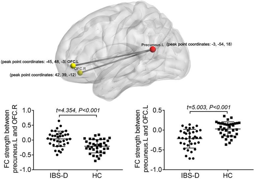
Chen et al.                                                                                                        Brain Spontaneous Dysfunctions in IBS-D

Temporal band-pass filtering (0.01–0.08 Hz) was performed for            RESULTS
ReHo and FC analyses.
                                                                         Demographic Characteristics and
ALFF, ReHo, and FC Calculation                                           Clinical Results
The time series of a given voxel was converted into a frequency
                                                                         Two patients were excluded due to excessive head movement,
domain using the fast Fourier transform. The ALFF value was
                                                                         leaving 36 IBS-D patients and 36 HCs for further analysis.
calculated by obtaining the square root of the power spectrum
                                                                         Demographic details of all participants and their clinical
and then averaging it in the range of 0.01–0.08 Hz. The ALFF
                                                                         characteristics are summarized in Table 1.
of each voxel was divided by the global mean ALFF value to
                                                                            There were no significant differences between the groups
standardize data across subjects.
                                                                         concerning sex (t = 2.17, P = 0.15), age (t = 1.24, P = 0.22), or
    Kendall’s coefficient of concordance was used to calculate
                                                                         education level (t = –1.57, P = 0.12). Mean disease duration of
the local synchronization between a voxel and the nearest 26
                                                                         IBS-D patients was 19.11 months. Mean GSRS degree was 30.5.
voxels. The standardized ReHo map was then obtained by
                                                                         The depression and anxiety scores of patients were higher than
dividing the ReHo value of each voxel by the global mean ReHo
                                                                         those of HCs (t = 8.09, P < 0.001; t = 6.96, P < 0.001). The VAS
value (Zang et al., 2004). Spatial smoothing (FWHM = 6 mm)
                                                                         score of patients during the MRI scan was greater than the score
was then performed.
                                                                         before the scan (t = –2.10, P = 0.04).
    FC analysis was performed to investigate the connectivity
patterns between the regions of interest (ROI) and the voxels of
the rest brain. Based on the results of ALFF and ReHo analysis,
                                                                         fMRI Data Analyses
the changed brain regions [FWE-corrected with threshold-free             Compared to the HCs, IBS-D patients had significantly higher
cluster enhancement (TFCE), P < 0.05] in IBS-D were chosen               ALFF values in the right cerebellum posterior lobe, the right
as the ROIs (spherical regions with a radius of 5 mm centered            lingual gyrus/calcarine, the right postcentral gyrus, the right SFG,
on the peak coordinates), including the right lingual gyrus, the         and the right MFG, with lower ALFF values in the right IPL,
right superior frontal gyrus (SFG), the right postcentral gyrus,         the right striatum, the right ACC, the right insula, the right
the right inferior parietal lobule (IPL), the right supplementary        hippocampus, the right thalamus, the right midbrain, and the
motor area (SMA), the right cerebellar posterior lobe, the right         left precuneus (Figure 1 and Table 2). IBS-D patients had higher
supramarginal gyrus, the right midbrain, the orbital part of the         ReHo values in the bilateral lingual gyrus/calcarine, the bilateral
left inferior frontal gyrus (orb_IFG), and the left precuneus. The       SFG, the right MFG, and the right postcentral gyrus, with lower
mean time series of each ROI was extracted, Pearson’s correlation        ReHo values in the left orb_IFG and the right SMA (Figure 2
coefficients were calculated between the mean time series of each        and Table 3). Compared to the HCs, IBS-D patients showed
ROI and each voxel’s time series of the whole brain to generate a        enhanced connectivity between the left precuneus [the peak point
correlation map. Finally, Fisher’s r-to-z transform was performed        coordinates was (–3, –54, 18)] and the bilateral orbitofrontal
to improve the normality of the correlation coefficient.                 cortex (OFC) (right OFC: the peak point coordinates was [42,
                                                                         39, –12], the peak t-value was 4.262, and the number of voxels
Statistical Analysis                                                     was 31; left OFC: The peak point coordinates was [–45, 48, –3],
SPSS software version 18 (SPSS, Chicago, Illinois, United States)        the peak t-value was 4.809, and the number of voxels was
was adopted to analyzed demographic and clinical data. Two-              86) (Figure 3). These brain regions were described according
sample t-tests were used to explore the ALFF, ReHo, and FC               to the Anatomical Automatic Labeling (AAL) templates. We
differences between the two groups. Age, sex, education level,           also conducted ROI-based whole-brain FC analysis with other
and mean head movement parameters were used as covariates
in these comparisons. Multiple comparison correction with                TABLE 1 | Demographic and clinical characteristics of participants.
TFCE was performed with 5,000 permutation tests (P < 0.05
after correction) (Smith and Nichols, 2009). Partial correlation         Variables                      IBS-D              HCs        t-values P-values
                                                                                                       (n = 36)          (n = 36)
analyses were then conducted between altered ALFF values,
ReHo values, FC z-values and mood/gastrointestinal symptoms              Sex(male/female)               16/20              10/26         2.17     0.15a
in the IBS-D group. Gender, age, and education level were                Age (years)                34.36 ± 9.53      31.67 ± 8.85       1.24     0.22b
included as covariates, and P < 0.05 was considered statistically        Education (years)          12.08 ± 3.12      13.28 ± 3.32      –1.57     0.12b
significant. If the correlations were significant, a tentative           HAMD-17                     7.33 ± 4.60        0.42 ± 0.77      8.90
Chen et al. Brain Spontaneous Dysfunctions in IBS-D
FIGURE 1 | Clusters showing ALFF changes in IBS-D patients as compared to that in healthy controls (TFCE, FWE-corrected P < 0.05). Blue color denotes
relatively lower ALFF values in IBS-D group, red color denotes relatively higher ALFF values in IBS-D group, and the color bar in dicates the t-value from two-sample
t-test between IBS-D group and HCs. L, left; R, right.
TABLE 2 | Brain regions with abnormal ALFF in IBS-D patients compared to HCs.
MNI coordinates of maximum voxel Cluster size (voxels) BA Peak voxel t-value Anatomical region
x y z
IBS-D > HC
12 –72 –15 17 4.325 Right cerebellum posterior lobe
21 –63 0 18 30 4.725 Right lingual gyrus
3 –75 6 22 18/30 4.384 Right calcarine
51 –15 54 16 3 4.673 Right postcentral gyrus
9 33 63 304 6/8 4.974 Right superior and middle frontal gyrus (peak
region: right medial superior frontal gyrus)
IBS-D < HC
42 –39 36 730 13/24/40 –4.939 Right putamen, right supramarginal gyrus, right
caudate, right anterior cingulated gyrus, right insula,
right hippocampus, right thalamus, right pallidum,
left caudate (peak region: right supramarginal gyrus)
6 –21 –12 22 –3.400 Right midbrain
–3 –54 18 40 23/31 –4.026 Left precuneus
42 –42 48 23 40 –3.899 Right inferior parietal lobule
IBS-D, diarrhea-predominant irritable bowel syndrome; HCs, healthy controls; MNI, Montreal Neurological Institute; BA, Brodmann area.
different brain regions as ROI, and no significant difference was by gastrointestinal symptoms. The statistical values of the
found after multiple comparison corrections between the groups. specific mediation model were as follows: Model 1 Path c,
P = 0.028; Path a, P = 0.002; Path b, P < 0.001; Path
Correlation and Mediation Analyses c, not significant; Standardization indirect effect = 22.932,
In IBS-D patients, ALFF values in the right midbrain positively 95% confidence interval: 8.238, 43.578. Model 2 Path c,
correlated with HAMD, HAMA, and GSRS score (r = 0.506, P = 0.006; Path a, P = 0.002; Path b, P < 0.001; Path c’,
P = 0.003; r = 0.349, P = 0.047; r = 0.501, P = 0.003). not significant; Standardization indirect effect = 15.118, 95%
Mediation analysis revealed that the ALFF value of the confidence interval: 5.086, 24.785 (Figure 4). These results
right midbrain had a significant overall effect on anxiety suggest that ALFF in the right midbrain tends to have
(critical statistical significance, P = 0.054) and depression an indirect effect on psychological symptoms predominantly
(P = 0.009) (Models 1 and 2) and was completely mediated through visceral pain.
Frontiers in Neuroscience | www.frontiersin.org 5 September 2021 | Volume 15 | Article 721822Chen et al. Brain Spontaneous Dysfunctions in IBS-D
FIGURE 2 | Clusters showing ReHo changes in IBS-D patients as compared to that in healthy controls (TFCE, FWE-corrected P < 0.05). Blue color denotes
relatively lower ReHo values in IBS-D group, red color denotes relatively higher ReHo values in IBS-D group, and the color bar in dicates the t-value from two-sample
t-test between IBS-D group and HCs. L, left; R, right.
TABLE 3 | Brain regions with abnormal ReHo in IBS-D patients compared to HCs.
MNI coordinates of maximum voxel Cluster size (voxels) BA Peak voxel t-value Anatomical region
x y z
IBS-D > HC
9 –84 –3 236 18/17 4.555 Bilateral calcarine, bilateral lingual gyrus
(peak region: right lingual gyrus)
9 30 63 276 6/8 5.073 Bilateral superior frontal gyrus, right middle frontal
gyrus(peak region: right medial superior frontal gyrus)
33 0 63 19 6 4.058 Right superior frontal gyrus
30 –30 66 15 3 4.417 Right postcentral gyrus
IBS-D < HC
–30 39 –6 16 47 –4.489 Left inferior frontal gyrus (orbital part)
15 –3 51 50 6 –4.651 Right supplementary motor area
IBS-D, diarrhea-predominant irritable bowel syndrome; HCs, healthy controls; MNI, Montreal Neurological Institute; BA, Brodmann area.
DISCUSSION have implications for understanding the functional mechanisms
of IBS-D and its related mood disorders.
To the best of our knowledge, this is the first study applying
ALFF, ReHo, and FC simultaneously to detect abnormalities of Altered Brain Activity Implicated in Regions of the
spontaneous brain activity in IBS-D patients, and explored the Pain Regulation and Emotional Arousal Within the
correlations between these abnormalities and clinical variables. Prefrontal–Limbic–Midbrain Circuit in IBS-D Patients
We identified significant regional brain functional alterations Prefrontal Cortex
in the PFC (mainly the bilateral dorsolateral prefrontal cortex The PFC is essential for higher-order cognitive, language,
[DLPFC]), the limbic system (including the right hippocampus, and emotional processing (Wiech et al., 2008). IBS symptoms
the right striatum, the right thalamus, the right insula, and the characterized by chronic visceral pain can be manifested at
right ACC), the midbrain, the sensorimotor cortex, and the several levels, including visceral hyperalgesia and lowered pain
visual cortex. Based on the abnormal brain regions in ALFF and threshold in the sensory dimension, emotional symptoms such as
ReHo, FC analysis showed enhanced connectivity between the anxiety and depression induced by chronic pain, and cognitive
left precuneus (seed region) and the bilateral OFC. These findings dysfunction such as decreased learning ability and memory loss.
Frontiers in Neuroscience | www.frontiersin.org 6 September 2021 | Volume 15 | Article 721822Chen et al. Brain Spontaneous Dysfunctions in IBS-D FIGURE 3 | Comparing to HCs, IBS-D patients showed enhanced connectivity between the seed region in the left precuneus and the bilateral orbital frontal cortex (TFCE, FWE-corrected P < 0.05) (A). Distribution of FC values between left precuneus and right OFC (B)/left OFC (C) in the two groups and differences between groups. L, left; R, right; FC, functional connectivity; OFC, orbital frontal cortex; IBS-D, diarrhea-predominant irritable bowel syndrome; HC, healthy controls. FIGURE 4 | Correlation and mediation analyses between ALFF value in the right midbrain and emotional symptoms. The area in blue color shows a reduction in ALFF (midbrain) in IBS-D patients compared to the healthy controls (A). The gastrointestinal symptoms completely mediated the relationship between ALFF in the right midbrain and HAMA (critical statistical significance), HAMD scale (B,C). The positive correlations were observed between ALFF value in right midbrain and gastrointestinal symptoms and HAMA, HAMD scale in IBS-D patients (D–F). HAMD, Hamilton depressive scale; HAMA, Hamilton anxiety scale; GSRS, gastrointestinal symptoms rating scale. Frontiers in Neuroscience | www.frontiersin.org 7 September 2021 | Volume 15 | Article 721822
Chen et al.                                                                                              Brain Spontaneous Dysfunctions in IBS-D

The sensory and emotional abnormalities caused by pain are               the hippocampal-amygdala circuit when performing cognitive
regulated by cognitive function. Cognitive regulation of pain can        tasks (Aizawa et al., 2012). Niddam et al. (2011) found
be categorized as attention, expectation, and reevaluation (Wiech        a decreased concentration of glutamate complex of the
et al., 2008), all of which involve different regions of the PFC,        left hippocampus in IBS patients and negatively correlated
especially the DLPFC and OFC (Jensen et al., 2012).                      with anxiety. The striatum is a vital nucleus of the basal
   As the core component of the central control network, the             ganglia and a core element of the pain matrix, which is
DLPFC plays a regulatory role in pain perception through                 involved in the integration of information in the cortex,
the descending inhibitory pathway (Lorenz et al., 2003).                 thalamus and three specific pain processing regions (sensory,
Our previous and other brain morphometric studies reported               emotional/cognitive, and endogenous/regulatory) (Legrain et al.,
atrophied cortical thickness or gray matter volume of DLPFC in           2011). Our previous study showed that the reduction of gray
patients with IBS and other chronic pain syndromes (Schmidt-             matter volume in the ACC, insula, and basal ganglia was
Wilcke et al., 2006; Hubbard et al., 2016; Li et al., 2020). The         significantly associated with visceral hypersensitivity in patients
DLPFC participates in “keeping pain out of mind,” especially             with IBS, which was highly consistent with other studies (Bhatt
in chronic pain states (Lorenz et al., 2003; Wiech et al., 2008).        et al., 2019; Li et al., 2020). This study found that patients
The higher pain scores of patients undergoing MRI in this study          with IBS-D have decreased ALFF values in regions of the
may be related to the amplification of pain perception during            limbic system involved in pain regulation and pain-related
the scan. In addition, the ALFF and ReHo values of DLPFC in              emotional processing, including ACC, insula, hippocampus,
IBS-D patients showed a consistent increase, further suggesting          basal ganglia, and thalamus, which may be related to the
that DLPFC plays a vital role in visceral hypersensitivity and the       long-term visceral hypersensitivity, visceral pain, emotional and
reduction of pain threshold.                                             cognitive impairment.
   OFC is thought to play a vital role in adjusting emotions,
decision-making, pain adjustment, and the integration of visceral        Midbrain
sensorimotor information (Kringelbach and Rolls, 2004; Price,            The periaqueductal gray matter (PAG) and dorsal raphe
2007; Moont et al., 2011). Jarcho et al. (2008) found that               nucleus play essential roles in the descending pain inhibition
activity in the bilateral OFC could be used to predict IBS               pathway at the midbrain level, which may be related to
symptom improvement by 5-HT subtype 3 general receptor                   the activation of dopamine neurons and the high expression
antagonists. A study combining positron emission tomography              of opioid receptors (Takada et al., 2013; Li et al., 2016).
(PET) and RS-fMRI found that dopamine receptor availability              Tominaga et al. (2015) found that upregulation of serotonin
was higher in individuals with more robust connectivity between          transporter levels in the midbrain and thalamus may underlie
OFC and DMN (Cole et al., 2012). The left precuneus is the               the pathogenesis of FGID such as abdominal and psychological
core node of the DMN, which maintains self-awareness and                 symptoms through brain-gut interactions. In a quantitative
episode-memory retrieval during the resting state (Cavanna               meta-analysis of task-fMRI studies related to rectal stimulation,
and Trimble, 2006; Zhang and Li, 2012). IBS patients showed              IBS subjects showed consistent activation of a large midbrain
activation of the precuneus and adjacent brain regions under             region (Tillisch et al., 2011). Consistent with this, our study
painful rectal dilation stimulation (Kano et al., 2017). The             showed that decreased ALFF values in the midbrain during
present study showed that IBS-D patients had the abnormal                the resting state and was positively correlated with abdominal
local neural activity of OFC and the precuneus, as well as               and psychological symptoms in IBS-D patients. By applying
enhanced FC between the bilateral OFC and the precuneus,                 mediation analysis, we found that the relationship between ALFF
which we speculated might be related to the dysfunction                  in the right midbrain and psychological symptoms was entirely
of the dopamine pathway (such as changes in dopamine                     mediated by abdominal symptoms, although the analysis of
receptor availability), further leading to the corresponding mood        anxiety symptoms was only statistically critical significant. These
and pain symptoms.                                                       results are consistent with previous studies on pathogenesis
                                                                         biology, suggesting that primary intestinal disorders may be
Limbic System                                                            the underlying driving factors of mood disorders (Jones et al.,
The insula and ACC belong to the medial system of the                    2012; Koloski et al., 2012). The decrease of ALFF in the right
pain matrix and are associated with the affective-motivational           midbrain might indicate the dysfunction of the descending
component, which is essential for visceral sensation, autonomous         pain inhibitory pathway in IBS-D patients, leading to visceral
visceral movement, pain and emotional processing (Nakata                 hypersensitivity and abdominal pain, and further aggravating
et al., 2014). These two regions have been the most consistently         emotional symptoms.
abnormal functional and structural regions in IBS patients and
were significantly associated with negative emotions (Blankstein         Altered Brain Activity Implicated in Somatic and
et al., 2010; Jiang et al., 2013; Piché et al., 2013; Qi et al.,         Sensory Processing in Patients With IBS-D
2016; Kano et al., 2020). The hippocampus is the region                  The postcentral gyrus belongs to the parietal lobe, also known
involved in memory formation, memory organization, and                   as the somatosensory center, part of the pain matrix. It may
memory storing, inhibiting feedback on the hypothalamic-                 play a crucial role in adjusting pain perception, including the
pituitary-adrenal axis (Tasker and Herman, 2011). IBS patients           positioning and recognition of pain intensity. Our finding of
have abnormal hippocampus activation and dysregulation of                increased ALFF and ReHo in the postcentral gyrus paralleled

Frontiers in Neuroscience | www.frontiersin.org                      8                                September 2021 | Volume 15 | Article 721822
Chen et al. Brain Spontaneous Dysfunctions in IBS-D
that reported by Ke et al. (2015), resulting from adaptive and reproducibility of these results across multiple cohorts
neuronal changes caused by long-term pain stimuli and biological for test-retest.
responses. The SMA is part of the sensorimotor region that
receives information from the basal ganglia. It is also thought to
be part of the ventral prefrontal parietal attention network, which CONCLUSION
is involved in detecting and directing attention to behavior-
related sensory stimuli while ignoring irrelevant, competitive By combining ReHo, ALFF, and FC analysis, we found abnormal
stimuli (Vossel et al., 2014). An RS-fMRI study suggested that spontaneous activity and FC dysregulation in areas related to
SMA may play a role in suppressing the abnormal emotional pain regulation and emotional arousal in the prefrontal–limbic–
effects caused by physical symptoms such as abdominal midbrain circuit and areas related to somatosensory processing.
distension (Zhu et al., 2016). The abnormal local neural activity The development of mood disorders in IBS-D patients may be
in the brain areas related to somatic and sensory processing related to the dysfunction of dopamine pathway components
(e.g., posterior central gyrus and SMA) may be closely related (especially midbrain and OFC) caused by visceral pain.
to the degree of pain and visceral hypersensitivity in patients
with chronic visceral pain, and affect emotional and cognitive
functions to a certain extent. DATA AVAILABILITY STATEMENT
Our study also found increased local activity in the cerebellum
posterior lobe and the visual cortex. The cerebellum is one of the The raw data supporting the conclusions of this article will be
elements of the pain matrix, which can transmit pain information made available by the authors, without undue reservation.
upward through the fiber connection with the cerebral cortex
and participate in pain regulation by affecting the downward
inhibitory pathway of the brainstem nociceptive (Naegel et al., ETHICS STATEMENT
2014). Transcranial cerebellar direct current stimulation can
effectively regulate the perception and reception of pain (Wiech The studies involving human participants were reviewed
et al., 2010; Bocci et al., 2019). The occipital cortex is traditionally and approved by the Research Ethics Committee of the
regarded as a region related to visual information processing. Affiliated Hospital of Hangzhou Normal University. The
However, neuroimaging studies reported changes in the signal patients/participants provided their written informed consent to
intensity of the occipital lobe in patients with chronic pain (Klug participate in this study.
et al., 2011; Li et al., 2018). A task-fMRI study found that IBS
patients showed increased activity in the visual cortex during the
expectation of rectal pain, suggesting that it is easier to promote AUTHOR CONTRIBUTIONS
the attention process under uncertain conditions (Lee et al.,
2012). Increased local neural activity in these two posterior brain X-FC and JL designed and supervised the project. YG, X-QL,
regions in IBS-D patients may be associated with hypervigilance K-HX, YC, G-XL, and LQ collected the data. X-FC, J-PD, and
for pain perception and regulation. JL processed and analyzed the data. All authors revised the
Although both ReHo and ALFF analysis methods are reliable manuscript and approved the final version.
means to assess the functional activity of the brain, most of the
brain regions obtained by these two methods in this study did
not overlap, except for the right DLPFC, the right postcentral FUNDING
gyrus, and the right lingual gyrus. These results suggest that, in
addition to the abnormal activity of local neurons in these regions This work was supported by the Social Development Project
during the resting state of IBS-D patients, more of these region- of Zhejiang Public Welfare Technology Research (Grant No.
related neurons are involved. The difference in results between LGF20H180016), the Project of Zhejiang Medical and health
the two methods may indicate different forms of functional Science and Technology (Grant No. 2020KY710), and the
dysfunction in neuronal activity in multiple brain regions in Project of Hangzhou Health, Science and Technology Plan
IBS-D, which may also contribute to the complexity of clinical (Grant No. 0020190644).
manifestations of the disease.
A few limitations should be noted in this study. First, the
sample size was small. Second, a longitudinal investigation is ACKNOWLEDGMENTS
needed to verify the indirect effect of functional activity in
brain regions such as the midbrain as a possible cause of IBS- We thank all patients and healthy volunteers for their
D on emotional scores. Future studies will explore the reliability participation.
REFERENCES in patients with irritable bowel syndrome, based on FMRI and dynamic causal
modeling. Gastroenterology 143, 1188–1198. doi: 10.1053/j.gastro.2012.07.104
Aizawa, E., Sato, Y., Kochiyama, T., Saito, N., Izumiyama, M., Morishita, J., et al. An, L., Cao, Q. J., Sui, M. Q., Sun, L., Zou, Q. H., Zang, Y. F., et al. (2013). Local
(2012). Altered cognitive function of prefrontal cortex during error feedback synchronization and amplitude of the fluctuation of spontaneous brain activity
Frontiers in Neuroscience | www.frontiersin.org 9 September 2021 | Volume 15 | Article 721822Chen et al. Brain Spontaneous Dysfunctions in IBS-D
in attention-deficit/hyperactivity disorder: a resting-state fMRI study. Neurosci. Jensen, K. B., Berna, C., Loggia, M. L., Wasan, A. D., Edwards, R. R., and Gollub,
Bull 29, 603–613. doi: 10.1007/s12264-013-1353-8 R. L. (2012). The use of functional neuroimaging to evaluate psychological and
Ashburner, J. (2007). A fast diffeomorphic image registration algorithm. other non-pharmacological treatments for clinical pain. Neurosci. Lett. 520,
Neuroimage 38, 95–113. doi: 10.1016/j.neuroimage.2007.07.007 156–164. doi: 10.1016/j.neulet.2012.03.010
Bhatt, R. R., Gupta, A., Labus, J. S., Zeltzer, L. K., Tsao, J. C., Shulman, R. J., et al. Jiang, Z., Dinov, I. D., Labus, J., Shi, Y., Zamanyan, A., Gupta, A., et al. (2013).
(2019). Altered Brain Structure and Functional Connectivity and Its Relation to Sex-related differences of cortical thickness in patients with chronic abdominal
Pain Perception in Girls With Irritable Bowel Syndrome. Psychosom. Med. 81, pain. PLoS One 8:e73932. doi: 10.1371/journal.pone.0073932
146–154. doi: 10.1097/psy.0000000000000655 Jones, M. P., Oudenhove, L. V., and Talley, N. J. J. G. (2012). Mo1007 functional
Blankstein, U., Chen, J., Diamant, N. E., and Davis, K. D. (2010). Altered brain gastrointestinal disorders (FGIDs) and psychological disorders: strong evidence
structure in irritable bowel syndrome: potential contributions of pre-existing that the link is bidirectional, but psychological distress is more likely to precede
and disease-driven factors. Gastroenterology 138, 1783–1789. doi: 10.1053/j. a new diagnosis of an FGID. Gastroenterology 142, S–570–S–570.
gastro.2009.12.043 Kano, M., Muratsubaki, T., Morishita, J., Kono, K., Mugikura, S., Takase, K., et al.
Bocci, T., De Carolis, G., Ferrucci, R., Paroli, M., Mansani, F., Priori, A., et al. (2017). Influence of Uncertain Anticipation on Brain Responses to Aversive
(2019). Cerebellar transcranial direct current stimulation (ctDCS) ameliorates Rectal Distension in Patients With Irritable Bowel Syndrome. Psychosom. Med.
phantom limb pain and non-painful phantom limb sensations. Cerebellum 18, 79, 988–999. doi: 10.1097/psy.0000000000000484
527–535. doi: 10.1007/s12311-019-01020-w Kano, M., Muratsubaki, T., Yagihashi, M., Morishita, J., Mugikura, S., Dupont,
Cavanna, A. E., and Trimble, M. R. (2006). The precuneus: a review of its functional P., et al. (2020). Insula Activity to Visceral Stimulation and Endocrine Stress
anatomy and behavioural correlates. Brain 129(Pt 3), 564–583. doi: 10.1093/ Responses as Associated With Alexithymia in Patients With Irritable Bowel
brain/awl004 Syndrome. Psychosom. Med. 82, 29–38. doi: 10.1097/psy.0000000000000729
Cole, D. M., Beckmann, C. F., Searle, G. E., Plisson, C., Tziortzi, A. C., Nichols, T. E., Ke, J., Qi, R., Liu, C., Xu, Q., Wang, F., Zhang, L., et al. (2015). Abnormal
et al. (2012). Orbitofrontal connectivity with resting-state networks is associated regional homogeneity in patients with irritable bowel syndrome: A resting-state
with midbrain dopamine D3 receptor availability. Cereb Cortex 22, 2784–2793. functional MRI study. Neurogastroenterol. Motil. 27, 1796–1803. doi: 10.1111/
doi: 10.1093/cercor/bhr354 nmo.12692
de Medeiros, M. T., Carvalho, A. F., de Oliveira Lima, J. W., Dos Santos, Klug, S., Anderer, P., Saletu-Zyhlarz, G., Freidl, M., Saletu, B., Prause, W., et al.
A. A., de Oliveira, R. B., Nobre, E., et al. (2008). Impact of depressive (2011). Dysfunctional pain modulation in somatoform pain disorder patients.
symptoms on visceral sensitivity among patients with different subtypes of Eur. Arch. Psychiatry Clin. Neurosci. 261, 267–275. doi: 10.1007/s00406-010-
irritable bowel syndrome. J. Nerv. Ment. Dis. 196, 711–714. doi: 10.1097/NMD. 0148-4
0b013e318183f896 Koloski, N. A., Jones, M., Kalantar, J., Weltman, M., Zaguirre, J., and Talley,
Dothel, G., Barbaro, M. R., Raschi, E., Barbara, G., and De Ponti, F. (2018). N. J. (2012). The brain–gut pathway in functional gastrointestinal disorders is
Advancements in drug development for diarrhea-predominant irritable bowel bidirectional: a 12-year prospective population-based study. Gut 61, 1284–1290.
syndrome. Expert Opin. Investig. Drugs 27, 251–263. doi: 10.1080/13543784. doi: 10.1136/gutjnl-2011-300474
2018.1442434 Kringelbach, M. L., and Rolls, E. T. (2004). The functional neuroanatomy
Elsenbruch, S., Rosenberger, C., Enck, P., Forsting, M., Schedlowski, M., and of the human orbitofrontal cortex: evidence from neuroimaging and
Gizewski, E. R. (2010). Affective disturbances modulate the neural processing neuropsychology. Prog. Neurobiol. 72, 341–372. doi: 10.1016/j.pneurobio.2004.
of visceral pain stimuli in irritable bowel syndrome: an fMRI study. Gut 59, 03.006
489–495. doi: 10.1136/gut.2008.175000 Labus, J. S., Hubbard, C. S., Bueller, J., Ebrat, B., Tillisch, K., Chen, M., et al.
Ford, A. C., Sperber, A. D., Corsetti, M., and Camilleri, M. (2020). Irritable bowel (2013). Impaired emotional learning and involvement of the corticotropin-
syndrome. Lancet 396, 1675–1688. doi: 10.1016/s0140-6736(20)31548-8 releasing factor signaling system in patients with irritable bowel syndrome.
Fox, M. D., and Raichle, M. E. (2007). Spontaneous fluctuations in brain activity Gastroenterology 145, .e1251–.e1253. doi: 10.1053/j.gastro.2013.08.016
observed with functional magnetic resonance imaging. Nat. Rev. Neurosci. 8, Lee, H. F., Hsieh, J. C., Lu, C. L., Yeh, T. C., Tu, C. H., Cheng, C. M.,
700–711. doi: 10.1038/nrn2201 et al. (2012). Enhanced affect/cognition-related brain responses during visceral
Gershon, M. D., and Tack, J. (2007). The serotonin signaling system: from placebo analgesia in irritable bowel syndrome patients. Pain 153, 1301–1310.
basic understanding to drug development for functional GI disorders. doi: 10.1016/j.pain.2012.03.018
Gastroenterology 132, 397–414. doi: 10.1053/j.gastro.2006.11.002 Legrain, V., Iannetti, G. D., Plaghki, L., and Mouraux, A. (2011). The pain matrix
Guleria, A., Karyampudi, A., Singh, R., Khetrapal, C. L., Verma, A., Ghoshal, U. C., reloaded: a salience detection system for the body. Prog. Neurobiol. 93, 111–124.
et al. (2017). Mapping of Brain Activations to Rectal Balloon Distension Stimuli doi: 10.1016/j.pneurobio.2010.10.005
in Male Patients with Irritable Bowel Syndrome Using Functional Magnetic Li, C., Sugam, J. A., Lowery-Gionta, E. G., McElligott, Z. A., McCall, N. M.,
Resonance Imaging. J. Neurogastroenterol. Motil. 23, 415–427. doi: 10.5056/ Lopez, A. J., et al. (2016). Mu Opioid Receptor Modulation of Dopamine
jnm16148 Neurons in the Periaqueductal Gray/Dorsal Raphe: A Role in Regulation of
Hamilton, M. (1960). A rating scale for depression. J. Neurol. Neurosurg. Psychiatry Pain. Neuropsychopharmacology 41, 2122–2132. doi: 10.1038/npp.2016.12
23, 56–62. doi: 10.1136/jnnp.23.1.56 Li, J., Huang, X., Sang, K., Bodner, M., Ma, K., and Dong, X. W. (2018). Modulation
Hayes, A. (2013). Introduction to Mediation, Moderation, and Conditional Process of prefrontal connectivity in postherpetic neuralgia patients with chronic pain:
Analysis, Vol. 51. New York,NY: Guilford Press, 335–337. doi: 10.1111/jedm. a resting-state functional magnetic resonance-imaging study. J. Pain Res. 11,
12050 2131–2144. doi: 10.2147/jpr.S166571
Hong, J. Y., Naliboff, B., Labus, J. S., Gupta, A., Kilpatrick, L. A., Ashe-McNalley, Li, J., Yuan, B., Li, G., Lu, X., Guo, Y., Yang, Y., et al. (2020). Convergent
C., et al. (2016). Altered brain responses in subjects with irritable bowel syndromic atrophy of pain and emotional systems in patients with irritable
syndrome during cued and uncued pain expectation. Neurogastroenterol. Motil. bowel syndrome and depressive symptoms. Neurosci. Lett. 723:134865. doi:
28, 127–138. doi: 10.1111/nmo.12710 10.1016/j.neulet.2020.134865
Hubbard, C. S., Becerra, L., Heinz, N., Ludwick, A., Rasooly, T., Wu, R., et al. Li, L., Ma, J., Xu, J. G., Zheng, Y. L., Xie, Q., Rong, L., et al. (2021). Brain functional
(2016). Abdominal Pain, the Adolescent and Altered Brain Structure and changes in patients with Crohn’s disease: A resting-state fMRI study. Brain
Function. PLoS One 11:e0156545. doi: 10.1371/journal.pone.0156545 Behav. 14:2243. doi: 10.1002/brb3.2243
Huskisson, E. C. (1974). Measurement of pain. Lancet 2, 1127–1131. doi: 10.1016/ Lorenz, J., Minoshima, S., and Casey, K. L. (2003). Keeping pain out of mind: the
s0140-6736(74)90884-8 role of the dorsolateral prefrontal cortex in pain modulation. Brain 126(Pt 5),
Jarcho, J. M., Chang, L., Berman, M., Suyenobu, B., Naliboff, B. D., Lieberman, 1079–1091. doi: 10.1093/brain/awg102
M. D., et al. (2008). Neural and psychological predictors of treatment response Lucak, S., Chang, L., Halpert, A., and Harris, L. A. (2017). Current and
in irritable bowel syndrome patients with a 5-HT3 receptor antagonist: a pilot emergent pharmacologic treatments for irritable bowel syndrome with
study. Aliment Pharmacol. Ther. 28, 344–352. doi: 10.1111/j.1365-2036.2008. diarrhea: evidence-based treatment in practice. Therap. Adv. Gastroenterol. 10,
03721.x 253–275. doi: 10.1177/1756283X16663396
Frontiers in Neuroscience | www.frontiersin.org 10 September 2021 | Volume 15 | Article 721822Chen et al. Brain Spontaneous Dysfunctions in IBS-D
Lv, Y., Li, L., Song, Y., Han, Y., Zhou, C., Zhou, D., et al. (2019). The Local Brain Takada, T., Yamashita, A., Date, A., Yanase, M., Suhara, Y., Hamada, A., et al.
Abnormalities in Patients With Transient Ischemic Attack: A Resting-State (2013). Changes in the circadian rhythm of mRNA expression for µ-opioid
fMRI Study. Front. Neurosci. 13:24. doi: 10.3389/fnins.2019.00024 receptors in the periaqueductal gray under a neuropathic pain-like state.
Ma, X., Li, S., Tian, J., Jiang, G., Wen, H., Wang, T., et al. (2015). Altered brain Synapse 67, 216–223. doi: 10.1002/syn.21633
spontaneous activity and connectivity network in irritable bowel syndrome Tasker, J. G., and Herman, J. P. (2011). Mechanisms of rapid glucocorticoid
patients: A resting-state fMRI study. Clin. Neurophysiol. 126, 1190–1197. doi: feedback inhibition of the hypothalamic-pituitary-adrenal axis. Stress 14, 398–
10.1016/j.clinph.2014.10.004 406. doi: 10.3109/10253890.2011.586446
Mayer, E. A., Labus, J., Aziz, Q., Tracey, I., Kilpatrick, L., Elsenbruch, S., et al. Thompson, E. (2015). Hamilton Rating Scale for Anxiety (HAM-A). Occup. Med.
(2019). Role of brain imaging in disorders of brain-gut interaction: a Rome 65:601. doi: 10.1093/occmed/kqv054
Working Team Report. Gut 68, 1701–1715. doi: 10.1136/gutjnl-2019-318308 Tillisch, K., Mayer, E. A., and Labus, J. S. (2011). Quantitative meta-
Mearin, F., Lacy, B. E., Chang, L., Chey, W. D., Lembo, A. J., Simren, M., et al. analysis identifies brain regions activated during rectal distension in irritable
(2016). Bowel Disorders. GaJstroenterology 2016:31. doi: 10.1053/j.gastro.2016. bowel syndrome. Gastroenterology 140, 91–100. doi: 10.1053/j.gastro.2010.
02.031 07.053
Moont, R., Crispel, Y., Lev, R., Pud, D., and Yarnitsky, D. (2011). Temporal changes Tominaga, K., Tsumoto, C., Ataka, S., Mizuno, K., Takahashi, K., Yamagami,
in cortical activation during conditioned pain modulation (CPM), a LORETA H., et al. (2015). Regional brain disorders of serotonin neurotransmission are
study. Pain 152, 1469–1477. doi: 10.1016/j.pain.2011.01.036 associated with functional dyspepsia. Life Sci. 137, 150–157. doi: 10.1016/j.lfs.
Mudyanadzo, T. A., Hauzaree, C., Yerokhina, O., Architha, N. N., and 2015.07.023
Ashqar, H. M. (2018). Irritable Bowel Syndrome and Depression: A Shared Vossel, S., Geng, J. J., and Fink, G. R. (2014). Dorsal and ventral attention systems:
Pathogenesis. Cureus 10:e3178. doi: 10.7759/cureus.3178 distinct neural circuits but collaborative roles. Neuroscientist 20, 150–159. doi:
Muscatello, M. R., Bruno, A., Pandolfo, G., Micò, U., Stilo, S., Scaffidi, M., et al. 10.1177/1073858413494269
(2010). Depression, anxiety and anger in subtypes of irritable bowel syndrome Wiech, K., Lin, C. S., Brodersen, K. H., Bingel, U., Ploner, M., and Tracey, I.
patients. J Clin Psychol Med Settings 17, 64–70. doi: 10.1007/s10880-009-9182-7 (2010). Anterior insula integrates information about salience into perceptual
Naegel, S., Holle, D., Desmarattes, N., Theysohn, N., Diener, H. C., Katsarava, decisions about pain. J. Neurosci. 30, 16324–16331. doi: 10.1523/jneurosci.2087-
Z., et al. (2014). Cortical plasticity in episodic and chronic cluster headache. 10.2010
Neuroimage Clin. 6, 415–423. doi: 10.1016/j.nicl.2014.10.003 Wiech, K., Ploner, M., and Tracey, I. (2008). Neurocognitive aspects of pain
Nakata, H., Sakamoto, K., and Kakigi, R. (2014). Meditation reduces pain-related perception. Trends Cogn. Sci. 12, 306–313. doi: 10.1016/j.tics.2008.05.005
neural activity in the anterior cingulate cortex, insula, secondary somatosensory Wilder-Smith, C. H., Schindler, D., Lovblad, K., Redmond, S. M., and Nirkko,
cortex, and thalamus. Front. Psychol. 5:1489. doi: 10.3389/fpsyg.2014.01489 A. (2004). Brain functional magnetic resonance imaging of rectal pain and
Niddam, D. M., Tsai, S. Y., Lu, C. L., Ko, C. W., and Hsieh, J. C. activation of endogenous inhibitory mechanisms in irritable bowel syndrome
(2011). Reduced hippocampal glutamate-glutamine levels in irritable bowel patient subgroups and healthy controls. Gut 53, 1595–1601. doi: 10.1136/gut.
syndrome: preliminary findings using magnetic resonance spectroscopy. Am. 2003.028514
J. Gastroenterol. 106, 1503–1511. doi: 10.1038/ajg.2011.120 Zang, Y., Jiang, T., Lu, Y., He, Y., and Tian, L. (2004). Regional homogeneity
Piché, M., Chen, J. I., Roy, M., Poitras, P., Bouin, M., and Rainville, P. (2013). approach to fMRI data analysis. Neuroimage 22, 394–400. doi: 10.1016/j.
Thicker posterior insula is associated with disease duration in women with neuroimage.2003.12.030
irritable bowel syndrome (IBS) whereas thicker orbitofrontal cortex predicts Zang, Y. F., He, Y., Zhu, C. Z., Cao, Q. J., Sui, M. Q., Liang, M., et al. (2007).
reduced pain inhibition in both IBS patients and controls. J. Pain 14, 1217–1226. Altered baseline brain activity in children with ADHD revealed by resting-state
doi: 10.1016/j.jpain.2013.05.009 functional MRI. Brain Dev. 29, 83–91. doi: 10.1016/j.braindev.2006.07.002
Price, J. L. (2007). Definition of the orbital cortex in relation to specific connections Zhang, S., and Li, C. S. (2012). Functional connectivity mapping of the human
with limbic and visceral structures and other cortical regions. Ann. N Y Acad. precuneus by resting state fMRI. Neuroimage 59, 3548–3562. doi: 10.1016/j.
Sci. 1121, 54–71. doi: 10.1196/annals.1401.008 neuroimage.2011.11.023
Qi, R., Liu, C., Ke, J., Xu, Q., Zhong, J., Wang, F., et al. (2016). Intrinsic brain Zhu, Q., Cai, W., Zheng, J., Li, G., Meng, Q., Liu, Q., et al. (2016). Distinct resting-
abnormalities in irritable bowel syndrome and effect of anxiety and depression. state brain activity in patients with functional constipation. Neurosci. Lett. 632,
Brain Imag. Behav. 10, 1127–1134. doi: 10.1007/s11682-015-9478-1 141–146. doi: 10.1016/j.neulet.2016.08.042
Rome Foundation. (2006). Guidelines–Rome III Diagnostic Criteria for Functional Zuo, X. N., Di Martino, A., Kelly, C., Shehzad, Z. E., Gee, D. G., Klein, D. F., et al.
Gastrointestinal Disorders. J. Gastrointestin. Liver Dis. 15, 307–312. (2010). The oscillating brain: complex and reliable. Neuroimage 49, 1432–1445.
Schmidt-Wilcke, T., Leinisch, E., Gänssbauer, S., Draganski, B., Bogdahn, U., doi: 10.1016/j.neuroimage.2009.09.037
Altmeppen, J., et al. (2006). Affective components and intensity of pain correlate
with structural differences in gray matter in chronic back pain patients. Pain Conflict of Interest: The authors declare that the research was conducted in the
125, 89–97. doi: 10.1016/j.pain.2006.05.004 absence of any commercial or financial relationships that could be construed as a
Singh, P., Staller, K., Barshop, K., Dai, E., Newman, J., Yoon, S., et al. potential conflict of interest.
(2015). Patients with irritable bowel syndrome-diarrhea have lower disease-
specific quality of life than irritable bowel syndrome-constipation. World J. Publisher’s Note: All claims expressed in this article are solely those of the authors
Gastroenterol. 21, 8103–8109. doi: 10.3748/wjg.v21.i26.8103 and do not necessarily represent those of their affiliated organizations, or those of
Smith, S. M., and Nichols, T. E. (2009). Threshold-free cluster enhancement: the publisher, the editors and the reviewers. Any product that may be evaluated in
addressing problems of smoothing, threshold dependence and localisation this article, or claim that may be made by its manufacturer, is not guaranteed or
in cluster inference. Neuroimage 44, 83–98. doi: 10.1016/j.neuroimage.2008. endorsed by the publisher.
03.061
Stasi, C., Rosselli, M., Bellini, M., Laffi, G., and Milani, S. (2012). Altered neuro- Copyright © 2021 Chen, Guo, Lu, Qi, Xu, Chen, Li, Ding and Li. This is an open-
endocrine-immune pathways in the irritable bowel syndrome: the top-down access article distributed under the terms of the Creative Commons Attribution
and the bottom-up model. J. Gastroenterol. 47, 1177–1185. doi: 10.1007/s00535- License (CC BY). The use, distribution or reproduction in other forums is permitted,
012-0627-7 provided the original author(s) and the copyright owner(s) are credited and that the
Svedlund, J., Sjödin, I., and Dotevall, G. (1988). GSRS–a clinical rating scale for original publication in this journal is cited, in accordance with accepted academic
gastrointestinal symptoms in patients with irritable bowel syndrome and peptic practice. No use, distribution or reproduction is permitted which does not comply
ulcer disease. Dig Dis Sci 33, 129–134. doi: 10.1007/bf01535722 with these terms.
Frontiers in Neuroscience | www.frontiersin.org 11 September 2021 | Volume 15 | Article 721822You can also read