A circadian clock regulates efflux by the blood-brain barrier in mice and human cells
←
→
Page content transcription
If your browser does not render page correctly, please read the page content below
ARTICLE
                  https://doi.org/10.1038/s41467-020-20795-9                 OPEN

                  A circadian clock regulates efflux by the
                  blood-brain barrier in mice and human cells
                  Shirley L. Zhang 1,2 ✉, Nicholas F. Lahens 3, Zhifeng Yue1,2, Denice M. Arnold1, Peter P. Pakstis1,
                  Jessica E. Schwarz 1 & Amita Sehgal 1,2 ✉
1234567890():,;

                  The blood-brain barrier (BBB) is critical for neural function. We report here circadian reg-
                  ulation of the BBB in mammals. Efflux of xenobiotics by the BBB oscillates in mice, with
                  highest levels during the active phase and lowest during the resting phase. This oscillation is
                  abrogated in circadian clock mutants. To elucidate mechanisms of circadian regulation, we
                  profiled the transcriptome of brain endothelial cells; interestingly, we detected limited cir-
                  cadian regulation of transcription, with no evident oscillations in efflux transporters. We
                  recapitulated the cycling of xenobiotic efflux using a human microvascular endothelial cell
                  line to find that the molecular clock drives cycling of intracellular magnesium through tran-
                  scriptional regulation of TRPM7, which appears to contribute to the rhythm in efflux. Our
                  findings suggest that considering circadian regulation may be important when therapeutically
                  targeting efflux transporter substrates to the CNS.

                  1 Chronobiology and Sleep Institute, Perelman School of Medicine at the University of Pennsylvania, Philadelphia, PA, USA. 2 Howard Hughes Medical

                  Institute, Perelman School of Medicine at the University of Pennsylvania, Philadelphia, PA, USA. 3 Institute for Translational Medicine and Therapeutics
                  (ITMAT), Perelman School of Medicine, University of Pennsylvania, Philadelphia, PA, USA. ✉email: zhangshi@pennmedicine.upenn.edu;
                  amita@pennmedicine.upenn.edu

                  NATURE COMMUNICATIONS | (2021)12:617 | https://doi.org/10.1038/s41467-020-20795-9 | www.nature.com/naturecommunications                                    1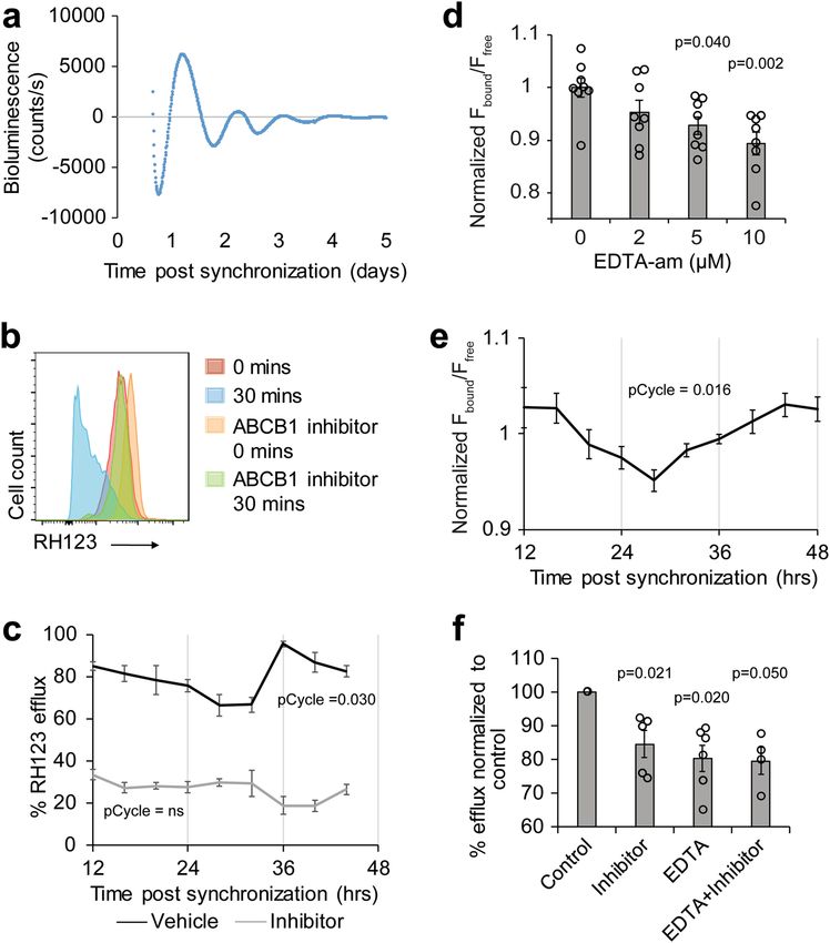
ARTICLE                                                                NATURE COMMUNICATIONS | https://doi.org/10.1038/s41467-020-20795-9

T
       he blood–brain barrier (BBB) is a structure in the CNS
       vasculature that insulates the brain from the periphery. A
       key function of the BBB is to protect the brain from
xenobiotics and other potentially harmful environmental insults;
however, it also prevents diagnostic and therapeutic drugs from
entering the brain1,2. Understanding regulation of the BBB is
important for addressing effects of the periphery on the brain and
also for improving methods of drug delivery. The BBB is com-
prised of capillary endothelial cells, surrounded by astrocytic
endfeet and pericytes3,4. BBB endothelial cells constitute a
polarized cell layer held together with tight junctions and char-
acterized by low transcytosis, which together restrict paracellular
and transcellular passage of molecules into the brain5,6. The
luminal membrane of the BBB has high concentrations of efflux
transporters, notably ABCB1 (ATPase binding cassette also
known as permeability glycoprotein [Pgp] and multi-drug resis-
tant protein 1 [MDR1]), which is an ATP-dependent pump with
broad substrate specificity7,8.
   Circadian clocks are endogenous timekeeping processes that
cycle with a period of ~24 h and drive rhythms in most physio-
logical processes. A synchronizing central clock resides in the
suprachiasmatic nucleus in the hypothalamus; however, the core
molecular clock is present in nearly every cell in mammals9,10.
These peripheral clocks share the same clock machinery as
the central clock, but regulate tissue-specific genes and func-
tions11–14. The basic mechanism of the clock is a transcription-
translation feedback loop in which the Period (Per) and
Cryptochrome (Cry) genes are rhythmically transcribed and the
two proteins negatively regulate their transcriptional activators,
BMAL1 (brain and muscle aryl hydrocarbon receptor nuclear                    Fig. 1 Efflux of ABCB1 substrate from the brain oscillates over the course
translocator-like protein 1) and either CLOCK (circadian loco-               of the day. a Schematic of experiment. The ABCB1 substrate RHB was
motor output cycles kaput) or NPAS2 (neuronal PAS domain                     intravenously injected via jugular vein into mice. Mice were allowed 90 min
protein)15. Because of the pervasiveness of circadian rhythms in             to recover and brains and sera were collected. Fluorescence was read at
cellular processes16,17, they are likely involved in the tissue-             ex540/em590nm using a plate reader. b Brain RHB levels are regulated by
response to circulating factors, including drugs; however, the               ABCB1-mediated efflux. RHB or RHB and the ABCB1-inhibitor tariquidar was
extent to which the circadian clock regulates access of molecules/           intravenously injected into WT mice. Individual mice are shown with
drugs to different mammalian tissues, in particular to the brain, is         triangle markers and means ± SEM are shown (n = 6; 2 independent
largely unknown.                                                             experiments). c RHB injection was performed on WT mice at indicated time
   To address time-of-day influences on the BBB, we eliminated                points. Individual points are shown with triangle markers and lines
the molecular clock specifically in the mouse endothelium and                 represent the mean. pCycle value was calculated by JTKCycle analysis (n =
used a xenobiotic to measure ABCB1-mediated efflux from the                   19; 3 independent experiments). d Absence of vascular leakage in both day
brain. We find that the BBB endothelium has a cell autonomous                 and night. Evans Blue was intravenously injected into mice untreated or
circadian clock that is required for the generation of rhythms of            treated with LPS 24 h prior. Animals were sacrificed and brains and liver
efflux through ABCB1. We then examined molecular mechan-                      were collected after 30 min. Evans Blue was extracted from tissue and
isms of clock-mediated control by conducting transcriptomic                  amount was measured at absorbance 620 nm. Individual data points and
analysis using RNA-seq. Surprisingly, unlike the robust rhythms              means are shown. n = 8 control, 6 LPS-treated; 2 independent experiments.
of RNA expression noted in other tissues, very few transcripts               p-value was determined by Student’s T-test.
cycle in the endothelium and these do not include efflux trans-
porters. We recapitulated efflux cycling in human microvascular               lipophilic, fluorescent compound that is a known substrate of
endothelial cells and demonstrate oscillations in the level of               ABCB1 (p-glycoprotein) into the jugular vein of the mouse. After
intracellular free magnesium, a required cofactor for robust efflux.          90 min, we harvested the blood, sacrificed and perfused the
We find binding of BMAL1 to the promoter of the highly                        mouse, and harvested the brain. The amount of RHB fluorescence
expressed magnesium transporter TRPM7 as well as cycling of                  was measured in the serum and brain homogenate (Fig. 1a). To
TRPM7 transcript and protein. Together these data suggest a                  verify that the level of RHB measured in the brain was a result of
model in which BMAL1 regulates TRPM7 transcription, which                    biological processes rather than experimental error (such as
affects intracellular magnesium homeostasis in a rhythmic fash-              contamination by blood), we used the ABCB1-specific non-
ion and produces time-of-day changes in efflux. Our data show                 competitive inhibitor tariquidar (Kd = 5.1 ± 0.9 nM) to block
the interaction of the molecular clock with BBB function in                  efflux of RHB. We also controlled for possible differences in
mammalian cells.                                                             blood concentrations of RHB, by normalizing RHB levels in the
                                                                             brain to serum levels. We found that administering tariquidar
                                                                             increased the level of RHB in the brain relative to the serum
Results                                                                      (Fig. 1b), verifying that RHB is effluxed by ABCB1 at the mouse
Efflux of xenobiotics at the mouse BBB is gated by the circa-                 BBB.
dian clock. Activity of efflux transporters at the BBB is an                     To determine whether efflux cycles through the circadian day,
important determinant of BBB permeability18. To measure brain                we intravenously injected RHB into WT mice at 6 time points
efflux, we intravenously injected Rhodamine B (RHB), a small,                 throughout the circadian day (zeitgeber [ZT] 2, 6, 10, 14, 18, 22

2               NATURE COMMUNICATIONS | (2021)12:617 | https://doi.org/10.1038/s41467-020-20795-9 | www.nature.com/naturecommunications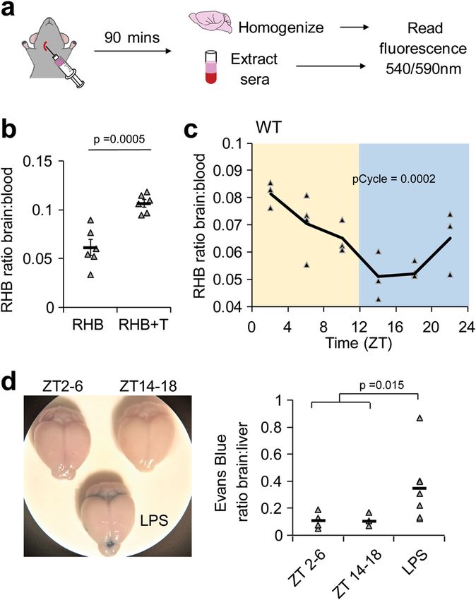
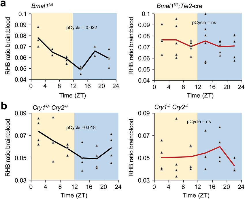
NATURE COMMUNICATIONS | https://doi.org/10.1038/s41467-020-20795-9                                                                     ARTICLE

Fig. 2 A circadian clock in the BBB regulates brain permeability of RHB. RHB was intravenously injected via jugular vein into mice. Mice were allowed
90 min to recover and brains and sera were collected. Fluorescence was read at ex540/em590nm using a plate reader. a Clock ablation in endothelial cells
disrupts the brain permeability rhythm to RHB. RHB was injected into control (n = 18; 4 independent experiments) or endothelial-specific Bmal1-deficient
(n = 31; 4 independent experiments) mice at indicated time points. b Global circadian disruption ablates brain permeability rhythm to RHB in mice. RHB
was injected into Cry1/2 het (n = 24; 3 independent experiments) and Cry1/2 DKO (n = 23; 4 independent experiments) mice at indicated time points.
Individual mice are shown with triangle markers, and lines represent the mean. pCycle values were calculated by JTKCycle analysis.

with lights on and off corresponding to ZT0 and ZT12,                           the mice given LPS exhibited Evans Blue staining in the brain, but
respectively) and measured the brain–serum ratio 90 min later.                  uninflamed mice did not exhibit BBB vascular leakage in the
We find that the level of RHB in the brain oscillates throughout                 morning or night (Fig. 1d), indicating that rhythms of RHB in the
the day with the peak early after lights on and trough after lights             brain are not generated by vascular leakage.
off, indicating that p-glycoprotein-mediated efflux is highest at                   To determine whether this oscillation was due to a circadian
night, when the animals are active (Fig. 1c). Levels of RHB in the              clock within the blood–brain barrier, we used an endothelial-
serum did not show differences over time (Supplementary                         specific (Tie2) cre recombinase to delete a floxed exon of Bmal1/
Fig. 1a).                                                                       Arntl (Bmal1fl/fl), which is required for the generation of
   Higher blood flow due to increased cardiac output and blood                   circadian rhythms. Control mice (Bmal1fl/fl) have a robust
pressure during the active period of the mice may increase RHB                  rhythm in RHB efflux; however, in the absence of a functional
circulation to the brain19; however, in our experiments we find                  clock in the BBB endothelium (Bmal1fl/fl; Tie2-cre), the rhythms
decreased RHB levels in the brain at night, suggesting that                     are lost and retention of RHB in the brain is high (Fig. 2a,
increased blood circulation does not explain these results.                     Supplementary Fig. 2a). We then injected Evans Blue to assay for
Nevertheless, to ensure that our measurements reflect efflux over                 vascular leak and did not observe any differences between control
time, we sought to determine the amount of RHB lost from the                    and mutant mice (Supplementary Fig. 2b). To confirm circadian
brain at different times of day. We first measured the RHB                       control, we obtained mice mutant for the Cryptochrome 1,
brain–serum ratio over the circadian day at 60 min post-injection               Cryptochrome 2 proteins (Cry1−/−Cry2−/−), which repress
and found, as expected, that RHB accumulated in the brain to                    Bmal1 function. We find that Cry1−/−Cry2−/− also lack RHB
higher levels than at 90 min and the RHB brain–serum ratio                      efflux rhythms compared to the heterozygous (Cry1+/−Cry2+/−)
trended toward a 24-h cycle (JTK pCycle = 0.1) (Supplementary                   controls (Fig. 2b). Because BMAL1 is part of the positive arm of
Fig. 1b). By comparing the RHB blood-serum ratio at 60 and                      the circadian clock while CRY1 and CRY2 are part of the negative
90 min post-injection, we extrapolated the rate of efflux per                    arm, we expected that deletion of Bmal1 or Cry1/2 would
minute for each ZT time and found that the highest rate was                     have opposing effects on the level of RHB in the brain and indeed
indeed at night (Supplementary Fig. 1c).                                        Cry1−/−Cry2−/− mice trended toward lower levels of RHB in
   To determine whether gross vascular leakage changes over the                 the brain (Supplementary Fig. 2c). The high variability of the
day and night, which would contribute to RHB accumulation in                    Cry1−/−Cry2−/− mice may be due to some paravascular leakage
the brain, we tracked the level of serum albumin in the brain                   from increased inflammation in the mice20. Together, these data
using the high affinity Evans Blue dye. Wild-type mice were given                suggest that the circadian clock in the BBB regulates RHB efflux
intravenous injections of Evans Blue in the morning (ZT2-6),                    from the brain.
night (ZT14-18), or 24 h after treatment with vascular leak-
inducing inflammatory stimulus, lipopolysaccharide (LPS), and                    Few circadian transcripts in endothelial cells in the mouse
the brain and liver were assessed 30 min after injection. Half of               brain. To determine the mechanism of circadian clock regulation,

NATURE COMMUNICATIONS | (2021)12:617 | https://doi.org/10.1038/s41467-020-20795-9 | www.nature.com/naturecommunications                               3
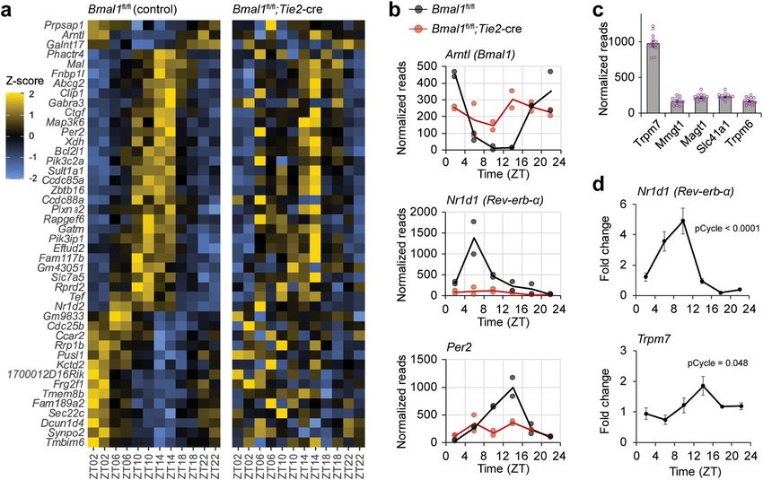
ARTICLE                                                                  NATURE COMMUNICATIONS | https://doi.org/10.1038/s41467-020-20795-9

Fig. 3 Cycling of circadian clock genes and a highly expressed magnesium transporter in brain endothelial cells. Control or endothelial-cell specific
Bmal1-deficient mice were collected at ZT2, ZT6, ZT10, ZT14, ZT18, and ZT22 (n = 12; 6 time points, 2 independent experiments); brains were dissected
and endothelial cells were isolated by FACS using CD31 antibody. RNA was extracted and sequenced with HiSeq. a Few transcripts cycle in BBB endothelial
cells. Heatmap of cycling transcripts of control animals (Meta2d, q < 0.6) is organized by peak and trough with corresponding transcripts of Bmal1-deficient
endothelial cells. b Expression of core circadian genes shows robust rhythms in mouse BBB. Transcripts of controls (black) are rhythmic (pCycle
NATURE COMMUNICATIONS | https://doi.org/10.1038/s41467-020-20795-9                                                                    ARTICLE

controls, such as the amino acid transporter Slc7a5a, were also                 hCMEC/D3. We measured the ratio of YFP/CFP in the cells
rhythmic in the mutants suggesting that rhythms in other tissues                for up to 48 h following synchronization with dexamethasone
can compensate or that the rhythms are dependent on oscillatory                 and found that the level of intracellular magnesium was
factors like sleep or light. Since ABC transporters are known to                rhythmic (Supplementary Fig. 5a). Use of a magnesium chelator
regulate xenobiotic efflux, we examined the transcripts of ABC-                  (EDTA-am) verified that the FRET sensor was sensitive to
family transporters and found no consistent phases over time                    intracellular magnesium, although the sensitivity was much
(Supplementary Fig. 4a). Abcb1a, the transporter relevant for                   lower than that of Magfura2-AM (Supplementary Fig. 5b). To
RHB efflux and most homologous to human ABCB1 is highly                          determine whether magnesium levels affect efflux, we incubated
expressed in the BBB, but was not rhythmic or dependent on                      cells with EDTA-am and/or verapamil and performed a RH123
Bmal1 (Supplementary Fig. 4b). One ABC efflux transporter,                       efflux assay. We found similar reduction of efflux if either drug
Abcg2, was rhythmic by Meta2d, but it was not dependent on                      or both were applied to the cells, suggesting that changing
Bmal1 (Supplementary Fig. 4b). Together these results suggest                   concentrations of intracellular magnesium affect the activity of
that the circadian clock does not regulate ABC transporters at the              the ABCB1 transporter (Fig. 4f). Together these results suggest
level of transcription.                                                         that oscillations of intracellular magnesium levels regulate the
   Previous reports have suggested that circadian clocks regulate               cycling of efflux activity.
intracellular magnesium concentrations27. Since magnesium is a
necessary cofactor in the regulation of ABC transporters, we
examined whether its regulation was circadian clock-dependent.                  Magnesium transporter TRPM7 is a direct target of the cir-
We evaluated the RNA-seq results in control animals for all the                 cadian clock. Our next objective was to understand how the
known magnesium transporters and found Transient receptor                       circadian clock affects levels of intracellular magnesium. To
potential cation channel, subfamily M, member 7 (Trpm7) to be                   determine whether the clock regulates magnesium transporters,
highly expressed (Fig. 3c). Trpm7 transcripts in control animals                we examined transcript levels in hCMEC/D3 cells following
trended toward having a rhythm, although this was not                           dexamethasone synchronization. We found both BMAL1 and
statistically significant (Supplementary Fig. 4c). We then sorted                TRPM7 transcripts cycle relative to tubulin (Fig. 5a, b). Because
BBB from WT mice, extracted RNA, and used real-time PCR to                      the acrophase of BMAL1 preceded the acrophase of TRPM7, we
quantify the level of Trpm7 transcripts, verifying that the mRNA                assessed whether BMAL1 directly binds the TRPM7 promoter by
levels are indeed rhythmic (Fig. 3d). This suggested the possibility            using an anti-BMAL1 antibody for chromatin immunoprecipi-
that the clock regulates intracellular magnesium homeostasis to                 tation (ChIP). Using the Eukaryotic Promoter Database to
affect efflux.                                                                   identify putative BMAL1-binding e-box sites in the TRPM7
                                                                                promoter, we then used qPCR to determine the fold enrichment
                                                                                of BMAL1-binding relative to that of IgG controls. The increase
Oscillations of intracellular magnesium levels likely regulate                  in BMAL1 binding to the TRPM7 e-box is comparable to the level
efflux in a cultured human brain endothelial cell (BEC) line.                    of BMAL1 binding to a known e-box in the PER2 promoter
We were unable to measure magnesium levels in the mouse BBB                     (Fig. 5c). ChIP-seq data sets have also identified the promoter
because tissue penetrance of magnesium indicators is poor; thus,                region of TRPM7 to be a target of BMAL131,32. By contrast,
we used an immortalized human cerebral microvascular endo-                      ABCB1 transcript remained at comparable levels over a 24-hour
thelial (hCMEC/D3) cell line28. First, we validated that the                    period (Fig. 5d) and negligible enrichment of BMAL1 binding
hCMEC/D3 cell line has a circadian rhythm by transducing the                    was found for two putative e-boxes on ABCB1 promoter (Sup-
cell line with PER2-luciferase and measuring luminescence over                  plementary Fig. 6). Together these results are consistent with the
several days. We find that the cells have a period of ~23 h                      idea that TRPM7, but not ABCB1, is a direct target of the
(Fig. 4a). To determine whether circadian clocks in the cell lines              molecular clock.
produce oscillations in efflux, we treated hCMEC/D3 cells with                      We then determined whether the transcriptional oscillation of
dexamethasone to synchronize their endogenous circadian                         TRPM7 translates to oscillations in protein levels. Using an
clocks29. Twelve to forty-eight hours after synchronization, cells              antibody against TRPM7 to assess protein expression by
were incubated with the xenobiotic rhodamine 123 (RH123), a                     immunoblot following dexamethasone synchronization, we found
previously validated ABCB1 substrate of hCMEC/D3 cells30.                       that, indeed, protein levels of TRPM7 oscillate in phase with the
Fluorescence retained in the cells after 30 min at 37 ˚C compared               mRNA levels (Fig. 5e). Immunoblot of ABCB1 protein shows that
to fluorescence retained in cells on ice was measured by flow                     levels do not cycle (Fig. 5f), suggesting that the activity of ABCB1
cytometry. Increased retention of fluorescence by incubation with                is independent of the level of protein.
verapamil indicates that the fluorescence loss reflects ABCB1-                       To directly manipulate the level of TRPM7 in the hCMEC/D3
mediated efflux of RH123 from the hCMEC/D3 (Fig. 4b). The                        line, we created stable cell lines using lentiviral vectors containing
amount of RH123 efflux by hCMEC/D3 cells oscillates over time                    GFP and scrambled siRNA or siRNA targeting TRPM7.
and the rhythm is lost when efflux is inhibited with verapamil                   Interestingly, siRNA knockdown of TRPM7 resulted in >50%
(Fig. 4c).                                                                      cell loss as well as lower copy numbers (reduced GFP expression),
   To measure the level of intracellular magnesium over time, we                suggesting that TRPM7 is required for survival (Supplementary
synchronized the hCMEC/D3 culture and used a ratiometric                        Fig. 7a). The resultant siTRPM7 cell line had 4-fold reduction in
intracellular magnesium indicator Magfura2-AM. We verified                       TRPM7 transcript levels (Supplementary Fig. 7b), which was
that the indicator was sensitive to the level of intracellular                  sufficient to reduce intercellular free magnesium and abrogate the
magnesium in the hCMEC/D3 culture by using EDTA-AM to                           24-h oscillation (Fig. 6a). Because of the presence of GFP in our
chelate magnesium (Fig. 4d), and then measured magnesium at                     stable cell lines, we could not use RH123 and thus used RHB for
different times following dexamethasone synchronization. The                    ABCB1-dependent efflux experiments. Consistent with the
level of intracellular magnesium cycles roughly in phase with the               intracellular magnesium levels, the siTRPM7 cell line also showed
cycle of efflux (Fig. 4e). Because the amplitude of the change in                lower levels and loss of oscillation of RHB efflux compared to the
magnesium levels was low in this assay, we also stably expressed                control cell line (Fig. 6b). These results demonstrate that TRPM7
a genetically encoded magnesium FRET sensor, MARIO                              can regulate the levels of intracellular magnesium and ABCB1-
(magnesium ratiometric indicator for optical imaging) in                        mediated efflux.

NATURE COMMUNICATIONS | (2021)12:617 | https://doi.org/10.1038/s41467-020-20795-9 | www.nature.com/naturecommunications                             5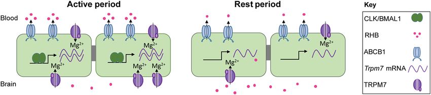
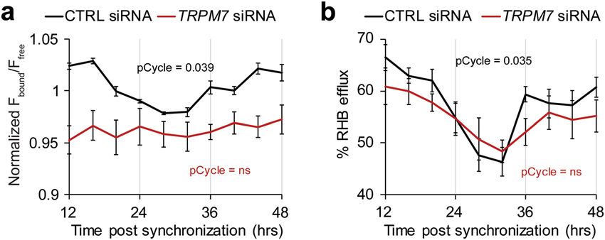
ARTICLE                                                                  NATURE COMMUNICATIONS | https://doi.org/10.1038/s41467-020-20795-9

Fig. 4 Cyclic efflux in human BECs is likely driven by intracellular magnesium oscillations. a Human BEC contain a circadian clock. A stable line was
established from hCMEC/D3 cells containing Per2-dLuc. Cells were synchronized with a 30 min pulse of dexamethasone and bioluminescent counts were
measured over 5 days with a luminometer (LumiCycle 32). Data were analyzed with LumiCycle software and representative detrended plot is shown.
b Verapamil inhibits RH123 efflux. Suspended cells were incubated with RH123 with or without the ABCB1-inhibitor verapamil on ice for 15 min. Excess
RH123 was removed and half of the culture was incubated at 37 °C for 30 min to allow for optimal efflux conditions while the rest remained on ice. The
amount of intracellular RH123 was determined by flow cytometry. Representative histograms of each treatment condition are shown. c Efflux of RH123
from BBB cells is rhythmic. Cells were synchronized with dexamethasone and efflux assay was performed at the indicated time point. The percentage of
RH123 fluorescence effluxed in 30 min comparing the level of fluorescence in cultures with or without 37 °C incubation is shown as means ± SEM. Cells
were incubated with vehicle (n = 81; 9 time points, 9 experiments) or the ABCB1-inhibitor verapamil (n = 45; 9 time points, 5 experiments). pCycle values
were calculated by JTKCycle analysis. d Chelating magnesium reduces ratio of bound to free MagFura2. hCMEC/D3 cells were incubated with MagFura2-
AM and indicated doses of EDTA-AM. MagFura was measured at ex330/em490nm (free) ex369/em490nm (bound). Means of normalized fluorescence
of bound Magfura2 (Fbound) over the fluorescence of free Magfura2 (Ffree) ± SEM are shown. n = 4, representative of 2 independent experiments. One-way
repeat measures mixed effect analysis with Dunnett’s multiple comparisons test was used to compare each experimental group to control. e Intracellular
magnesium levels oscillate in phase with efflux cycles. hCMEC/D3 cells were incubated with intracellular magnesium indicator Magfura2-AM at the
indicated time points after dexamethasone synchronization and measured at ex330/em490 (free) ex369/em490 (bound) using a plate reader. Means of
normalized fluorescence of bound Magfura2 (Fbound) over the fluorescence of free Magfura2 (Ffree) ± SEM are shown (n = 50; 10 time points; 5
independent experiments.). pCycle values were calculated by JTKCycle analysis. f Reducing intracellular magnesium inhibits efflux. Verapamil and/or
EDTA was added to hCMEC/D3 cells and RH123 was measured by flow cytometry. The percent of RH123 of efflux was normalized to control (n = 5–8 from
4 independent experiments). One-way ANOVA was used to compare all experimental groups to control with Dunnett’s multiple comparisons test.

Discussion                                                                     also serve to regulate brain levels of endogenous hormones and
We report circadian regulation of xenobiotic efflux in both mouse               cytokines through the activity of transporters. Previous reports
BBB and human cultured brain endothelial cells. The circadian                  show concentrations of leptin, interleukin-6, and tumor necrosis
regulation of BBB efflux explains previous findings in which levels              factor α have different phases between blood and brain34–36.
of an ABCB1 substrate, quinidine, in the rat brain depended on the             Together, a growing body of work suggests that the BBB is not
timing of administration33. The molecular clock in the BBB may                 only physically restrictive, but also temporally restrictive.

6                 NATURE COMMUNICATIONS | (2021)12:617 | https://doi.org/10.1038/s41467-020-20795-9 | www.nature.com/naturecommunications
NATURE COMMUNICATIONS | https://doi.org/10.1038/s41467-020-20795-9                                                                      ARTICLE

Fig. 5 TRPM7 is a clock-controlled gene in human brain endothelial cell culture. a, b BMAL1 and TRPM7 cycle in phase following dexamethasone
synchronization. hCMEC/D3 mRNA was extracted at indicated time points post-synchronization. Real-time PCR analysis was performed for BMAL1 and
TRPM7. Data are shown as means ± SEM (n = 40; 10 time points from 4 independent experiments.) pCycle values were calculated by JTKCycle analysis.
c BMAL1 binds TRPM7 e-box site. TRPM7 gDNA from hCMEC/D3 cells were immunoprecipitated with either IgG or BMAL1 antibody. Primers for PER2,
TRPM7 e-boxes were assessed by real-time PCR. Data are shown as mean fold change of sites binding to BMAL1 normalized to IgG (n = 6 plates from 3
independent experiments). d mRNA from hCMEC/D3 cultures was extracted at indicated time points post-synchronization. Real-time PCR analysis was
performed for ABCB1. Data are shown as means ± SEM (n = 40 plates; 10 time points; 4 independent experiments). pCycle values were calculated by
JTKCycle analysis. e, f Protein levels of TRPM7, but not ABCB1 oscillate in hCMEC/D3 cultures. Cell lines were synchronized with a pulse of dexamethasone.
Cell lysates were collected between 12 and 48 h later. Lysates were blotted with antibodies against TRPM7 or ABCB1 and ACTIN. Representative
immunoblot of TRPM7 (n = 4; 4 independent experiments) or ABCB1 (n = 3; 3 independent experiments) and ACTIN and means ± SEM of quantifications
are shown. Quantification of blots was performed in ImageJ. P-values were determined using one-way ANOVA to compare among time points.

   A seemingly simple mechanism of clock-control of BBB efflux                      Our results suggest that the efficiency of xenobiotic efflux of
would be to directly regulate the cycling of transporter tran-                  the BBB is regulated, at least in part, by oscillating free
scripts; however, we did not observe a consistent phase among the               magnesium levels. This is consistent with previous reports of
ABC transporter transcripts. In fact, very few transcripts are                  oscillating free magnesium concentrations and magnesium
clock-controlled genes in the mouse brain endothelium, which is                 transporters in other cell types27,40. Because Mg2+ is a cofactor
surprising given the extent of circadian regulation of the tran-                for hundreds of MgATP-dependent enzymes, even modest
scriptome in other tissues24 as well as previous reports of cycling             changes in cytosolic Mg2+ may serve to regulate cellular energy
ABC-family transporters in the gut37,38 and liver39. Yet the                    expenditure over the course of a day41. Indeed, we find that
impact of the clock on cell physiology in the BBB is dramatic. We               inhibition of intracellular Mg2+ in the range of the daily var-
find the clock regulates intracellular Mg2+ levels, which may                    iation can reduce efflux. In the BBB, such changes in intracel-
affect cell physiology more broadly than transcriptional changes.               lular free magnesium may also regulate the activity of other BBB
For instance, changing Mg2+ or translational machinery may                      transporters, such as those for amino acids and glucose trans-
affect the activity or levels of many proteins; however, to achieve             porters, if Mg2+ is rate-limiting for transport. In addition to
the same effect at the transcriptional level, clock proteins would              magnesium transporter, changes in intracellular calcium may
need to bind and regulate each individual transcript.                           contribute to oscillations of Mg2+.

NATURE COMMUNICATIONS | (2021)12:617 | https://doi.org/10.1038/s41467-020-20795-9 | www.nature.com/naturecommunications                                 7
ARTICLE                                                                  NATURE COMMUNICATIONS | https://doi.org/10.1038/s41467-020-20795-9

Fig. 6 Knockdown of TRPM7 reduces and dampens rhythms of intracellular Mg2+ and xenobiotic efflux. Stable hCMEC/D3 cell lines containing
scrambled siRNA and GFP or TRPM7 siRNA and GFP were made with Lentiviral transduction. Stable lines were synchronized with dexamethasone and
assayed at the indicated time points. a Knockdown of TRPM7 results in reduced Mg2+ and loss of cycling. Generated cell lines were incubated with
intracellular magnesium indicator Magfura2-AM at the indicated time points after dexamethasone synchronization and measured at ex330/em490 (free)
ex369/em490 (bound) using a plate reader. Means of normalized fluorescence of bound Magfura2 (Fbound) over the fluorescence of free Magfura2 (Ffree)
± SEM are shown (n = 50; 10 time points, 5 independent experiments). p < 0.0001 comparing control to TRPM7 siRNA cell line by paired T-test, showing
less intracellular Mg2+ in the siTRPM7 cell line across time points. pCycle values were calculated by JTKCycle analysis. b Knockdown of TRPM7 reduces
xenobiotic efflux and dampens rhythms. Cells were synchronized with dexamethasone and RHB efflux assay was performed at the indicated time point.
Control or TRPM7 siRNA-treated suspended cells were incubated with RHB on ice for 15 min. Excess RHB was removed and half of the culture was
incubated at 37 °C for 30 min to allow for optimal efflux conditions while the rest remained on ice. The amount of intracellular RHB was determined by flow
cytometry. The percentage of RH123 fluorescence effluxed in 30 min comparing the level of fluorescence in cultures with or without 37 °C incubation is
shown as means ± SEM (n = 40; 10 time points, 5 independent experiments. pCycle values were calculated by JTKCycle analysis. p = 0.0085 comparing
control to TRPM7 siRNA cell line by paired T-test, showing reduced RHB efflux in the siTRPM7 cell line across time points.

Fig. 7 Model of clock regulation of xenobiotic efflux at the BBB. Core circadian clock transcription factors BMAL1 and CLOCK activate transcription of
Trpm7. Increased TRPM7 allows for higher intracellular free magnesium, which regulate the activity of xenobiotic transporters. Higher transporter activity
decreases the level of xenobiotics in the brain. In the absence of BMAL1/CLOCK, TRPM7 and therefore free intracellular magnesium is low. Lower levels of
magnesium are associated with decrease the activity of the xenobiotic transporters, thus xenobiotics are retained in the brain.

   Together, our data suggest a model in which BMAL1 in the                    xenobiotic efflux, we find differences in mechanism between flies
BBB endothelium drives transcript and protein expression of                    and mammals. In flies, the circadian clock uses a non-cell
TRPM7 during active periods (Fig. 7). The increase in                          autonomous mechanism to regulate efflux activity of the BBB;
TRPM7 channel likely results in higher concentrations of intra-                however, in mammals the endothelial BBB-clock drives cycling of
cellular free magnesium, which increases the activity of                       efflux within the same cell type. Interestingly, though, both sys-
the ABCB1 transporter and thereby the level of xenobiotic efflux,               tems include an important role for magnesium. In flies, rhythmic
reducing the amount of RHB in the brain. During resting periods,               expression of gap junctions restricts the passage of magnesium
BMAL1 is low, reducing the level of TRPM7 and subsequently                     between two layers of the BBB to specific times of day; at these
reducing the level of free magnesium in the BBB. Lower levels of               times, magnesium is depleted from the subperineurial layer,
intracellular magnesium decrease xenobiotic efflux, resulting in                which contains efflux transporters and normally has very high
greater brain retention of RHB. In support of this model, loss of              magnesium, thereby resulting in a rhythm of magnesium43. In
the CRY proteins, which are negative regulators of BMAL1,                      mammals, on the other hand, our data suggest that magnesium
results in lower retention of RHB. However, we cannot exclude                  rhythms are driven by cell-autonomous clock control of a mag-
the possibility that other factors, such as the recently proposed              nesium transporter.
regulation of the BBB by neuronal activity, contribute to rhythms                 The cycling of ABCB1 activity may have implications in disease
in efflux42.                                                                    settings; for instance in Alzheimer’s disease, where ABCB1 con-
   Our results show that accumulation of xenobiotics in the brain              tributes to the clearance of amyloid beta from extracellular spaces
following intravenous injection of mice occurs with an inverted                of the brain44,45. Typically, amyloid beta is either broken down in
cycle relative to that of a previously described efflux cycle in                the brain or cleared through the BBB endothelium or the cere-
Drosophila43. This aligns with the rest:activity rhythms of the                brospinal fluid. Interestingly, amyloid beta clearance is higher
organism with highest efflux during the active period and lowest                during sleep46, whereas here we show the peak activity of ABCB1
efflux during the resting period. High levels of efflux would be                 during active hours. However, efficient clearance of amyloid beta
most advantageous during periods of activity because of the                    through the endothelium requires both ABCB1 and LRP1 for
presumed increase in exposure to toxins from foraging or injury.               phosphatidylinositol binding clathrin assembly protein
Despite the evolutionary conservation of circadian regulation of               (PICALM)-mediated transcytosis through BBB endothelium47.

8                 NATURE COMMUNICATIONS | (2021)12:617 | https://doi.org/10.1038/s41467-020-20795-9 | www.nature.com/naturecommunications
NATURE COMMUNICATIONS | https://doi.org/10.1038/s41467-020-20795-9                                                                                     ARTICLE

Despite lower activity of ABCB1, it is possible that transcytosis is                  Isolation of BBB endothelium. Brains were harvested from mice at time points
higher during sleep, as shown for endocytosis in the Drosophila                       around the circadian day (ZT2, 6, 10, 14, 18, 22). Brains were finely minced in
                                                                                      RPMI media containing 10% CCS, incubated at 37 °C with 2 mg/mL collagenase IV
BBB48. Thus, increasing ABCB1 activity at this time could facil-                      (Gibco) and 200 μg/mL DNase I (Roche) for 30 min with shaking at 250 rpm,
itate amyloid beta clearance.                                                         pipetting up and down once during incubation. Brain homogenate was demyeli-
   A therapeutic application of this work is the possibility of                       nated using myelin removal beads II (Miltenyi Biotec) as instructed by the man-
improving drug delivery to the central nervous system (CNS).                          ufacturer’s protocol. Cell suspensions were filtered using a 100 μm cell strainer.
Given that many drug targets are clock-controlled genes, cells and                    Cells were labeled with anti-CD90 (clone M5/49.4.1, BioXell) for 15 min on ice and
                                                                                      incubated with BioMag Goat Anti-Rat IgG (Qiagen) for 30 min with rotation, and
tissues have different drug sensitivities throughout the circadian                    magnetically separated (Easy Sep). Supernatant was removed and spun down at
day49. Utilizing circadian timing in medicine, also known as                          600 × g. Cells were resuspended in 250 μl, labeled with fluorescence-conjugated
chronotherapy or chronomedicine, can improve outcomes and                             anti-CD31 (390, BioLegend), anti-CD90 (53-2.1, BioLegend) antibodies sorted
decrease side effects due to lower required doses to achieve the                      using either a BD FACSAria or BD FACSMelody (BD Biosciences). Dead cells were
                                                                                      excluded through 4′,6-diamidino-2-phenylindole uptake. Doublets were excluded
same efficacy50. Here, we show that ABCB1 substrates are subject                       through FSC-H by FSC-W and SSC-H by SSC-W parameters. Between 10,000 and
to circadian regulation at the BBB, indicating an additional level                    20,000 cells were sorted from a single mouse at >95% purity verified by post-sort
of circadian control for many drugs targeted to the CNS; how-                         analysis. Processing of tissue and BBB isolation takes ~4 h. Data from sort were
ever, other mechanisms of oscillatory regulation could also                           analyzed using FlowJo V10.6 software (TreeStar).
influence drug concentrations in the brain. Sleep has been shown
to increase removal of metabolites from the brain46, and robust                       RNA sequencing and analysis. RNA was extracted from sorted cells using RNeasy
circadian rhythms in the choroid plexus increase cerebrospinal                        Plus Micro Kit (Qiagen) following the manufacturer’s protocol. RNA quality was
                                                                                      assessed using 2100 Bioanalyzer Instrument (Agilent). RNA integrity numbers
fluid (CSF) production and turnover during the resting                                 (RIN) of 6–8 were used to construct libraries. Strand-specific libraries were con-
period51,52. These mechanisms may allow brain retention of other                      structed using the SMARTer Stranded Total RNA-Seq Kit v2 - Pico Input Mam-
classes of drugs in a time-of-day-dependent matter, which could                       malian (Clontech) and sequenced across three lanes of a HiSeq 4000 (Illumina),
inform decisions on optimal chronotherapy. Our results suggest                        generating 150 bp paired-end reads. All of the following analyses used the GRCm38
                                                                                      build of the mouse reference genome and gene models from Ensembl v91, both
that optimizing circadian timing of drug delivery in regards to                       downloaded from the Ensembl portal57. Reads were aligned to the reference gen-
BBB permissiveness can achieve increased CNS drug efficacy and                         ome using STAR v2.5.3a23 with the following command line options: --out-
should be taken into consideration in developing therapeutic                          SAMtype BAM Unsorted; --outSAMunmapped Within KeepPairs;
regimens.                                                                             --outFilterMismatchNmax 33; --seedSearchStartLmax 33; --alignSJoverhangMin 8.
                                                                                      In addition, STAR was provided with the gene models in GTF format during
                                                                                      alignments and the “—outSAMattrRGline” was used to add a unique read group to
                                                                                      each of the resulting BAM files (required to merge files below). Note that the three
Methods                                                                               pairs of FASTQ files per library (two for each sequencing lane) were aligned
Animal care and husbandry. C57BL/6J (#000664), Bmal1fl/fl (B6.129S4(Cg)-                separately. Following alignment, the three BAM files for each library were com-
Arntltm1Weit/J #007668)53, and Tie2cre (B6.Cg-Tg(Tek-cre)1Ywa/J, #008863)54           bined using the SAMtools v1.3.158 merge command with the “-n” argument, which
mice were purchased from Jackson Laboratory animal facility. Cry1,Cry2 double-        maintains the BAM files in “unsorted” order (i.e., sorted by read ID). The Pipeline
deficient mice were a gift from Katja Lamia originally from Aziz Sancar55. Animals     Of RNA-seq Transformations (PORT) v0.8.5-beta (https://github.com/itmat/
were fed ad libitum and entrained to 12 h:12 h light:dark cycles and given at least   Normalization) was used to perform gene-level normalization and quantification of
1 day per hour of phase shift prior to experiments. All live animal experiments       the aligned data. Cycling transcripts with 24 h rhythmicity were identified using the
were performed according to protocols approved by the Institutional Animal Care       meta2d function from the MetaCycle v1.1.0R package59. The meta2d function was
and Use Committee of the University of Pennsylvania (Philadelphia, PA) in             run with the following arguments: outIntegration = “both”, adjustPhase = “pre-
accordance with guidelines set by the NIH (Protocol #806387). Male and female         dictedPer”, combinePvalue = “fisher”, weightedPerPha = FALSE, cycMethod = c
mice were used for gene expression data (RNAseq and qPCR). Only male mice             (“JTK”, “LS”), analysisStrategy = “selfUSE”, minper = 24, maxper = 24. For the
were used in functional experiments due to the decreased risk for surgical failures   MetaCycle analyses, we applied a minimum expression filter to remove unex-
with their larger size. Mice from the same litters were randomly distributed to       pressed genes. For a gene to be included, it must have a normalized read count of
environmental cabinets for entrainment for different circadian time points.           five or greater in at least control six samples, or six Bmal1 mutant samples.

                                                                                      hCMEC/D3 cell cultures. Immortalized human brain capillary endothelial cells
RHB permeability assay in live mice. Mice between 8 and 12 weeks were used for
                                                                                      (Millipore SCC066) were cultured in EndoGRO-MV Complete media kit
permeability experiments. The surgeon was blinded to the genotype and circadian
                                                                                      (SCME004) as recommended by the manufacturer. Cells were tested every 3–5
time of the mice. Mice were anesthesized with continuous isoflurane to maintain
                                                                                      passages for mycoplasma contamination by DNA Hoechst stain. Cultureware was
surgical plane and given meloxicam (5 mg/kg) and bupivacaine (0.1 mg/kg) sub-
                                                                                      coated with rat tail collagen type I solution at a concentration of 0.1 mg/mL and
cutaneously. RHB (Sigma) was dissolved in 10% water and 90% PBS and was
                                                                                      was incubated for >1 h at 25 °C. Cells were cultured in an incubator at 37 °C with
injected (5 mg/kg) into the jugular vein. Tariquidar (Selleck Chemicals) was sus-
                                                                                      5% CO2. For synchronization of culture, cells were grown to ~60% confluence and
pended in 30% Propylene glycol and 5% Tween 80 and 65% of 5% dextrose in
                                                                                      incubated in 200 nM dexamethasone for 30 min. Circadian time points were
water and coinjected (30 mg/kg) into the jugular vein. Animals that did not receive
                                                                                      obtained by staggering the synchronization of different cultures and assaying them
a proper injection were sacrificed and excluded from analysis (~5–10% of the
                                                                                      at the same time.
animals). The mice were allowed 90 min to recover prior to tissue harvest. Animals
were anesthetized with isoflurane and blood was collected by retro-orbital bleed
and serum was extracted after 50% transfection efficiency was
                                                                                      confirmed via fluorescent microscopy to assess GFP positivity, and the media was
                                                                                      exchanged. Forty-eight hours following transduction, the supernatant was collected
Vascular leakage assay. Evans Blue vascular leakage assay was adapted from            from the transfected 293T cells, and the supernatant was centrifuged at 1000 × g for
previous studies56. Briefly, mice were anethesized with isoflurance and adminis-        5 min to pellet any 293T cells. Finally, stable hCMEC/D3 cell lines were generated
tered 50 mg/kg Evans Blue via retro-orbital injection. After 30 min, mice were        by infecting 70% confluent cultures with the recombinant lentiviral vectors with
sacrificed and perfused with 10 units/mL heparin in PBS. Brain and liver were          10 mg/mL polybrene (Sigma-Aldrich) twice over 2 consecutive days, and then
harvested and Evans Blue was extracted with formamide and the absorbance of the       individual GFP+ cells were sorted on the FACSMelody (BD Biosciences).
supernatant was measured at 620 nm using a spectrophotometer (Cytation 5,
BioTek). Concentration of Evans Blue was determined using standard curve. For         hCMEC/D3 expressing siRNA against TRPM7. Stable hCMEC/D3 cell lines
induction of vascular leak, 3 mg/kg LPS was administered to the mice by intra-        expressing either scrambled siRNA (control) or TRPM7 siRNA with GFP were
peritoneal injection 24-h prior to injection of Evans blue.                           generated via lentiviral vector transduction using commercially available

NATURE COMMUNICATIONS | (2021)12:617 | https://doi.org/10.1038/s41467-020-20795-9 | www.nature.com/naturecommunications                                                 9
ARTICLE NATURE COMMUNICATIONS | https://doi.org/10.1038/s41467-020-20795-9
lentiviruses (LVP015-G and iV026186, abm). GFP+ cells were sorted in bulk on (Promega) on ice. Lysates were boiled for 10 min and run on 4–12% gradient Tris-
the FACSMelody (BD Biosciences). Due to the amount of cell death (50%) visually Glycine gels (Life Technologies), transferred to PVDF membranes, and probed
observed in the TRPM7 siRNA infected cells, we opted to group the cells to avoid with the following antibodies: anti-PGP (1:1000, MDR-1 c219, ThermoFisher),
mutations that may be present in individual clones. anti-TRPM7 (1:500, EPR4582, Abcam), anti-BMAL1 (1:1000, A302-616A, Bethyl
Laboratories), and anti-beta-Actin (1:20,000, mAbcam8224, Abcam). Blots were
digitalized with a photo scanner (Canon) and bands were quantified using ImageJ
Lumicycle assay. Cells containing Per2-dLuc-GFP were seeded in 35 mm dishes at
software.
80% confluence. A 30 min 100 nM dexamethasone (Sigma) pulse was used to
synchronize the cells. Real-time bioluminescence of the cells were monitored in
LumiCycle recording media (hCMEC/D3 media with 10% FBS, 25 mM Hepes, Statistical analysis. Circadian statistical analysis was performed in R using
4.2 mM sodium bicarbonate pH 7.0) with 200 µM beetle luciferin potassium salt JTK_CYCLEv3.164. Sample size estimates for mouse experiments were based on
using LumiCycle luminometer (Actimetrics) as previously described60. Lumines- previous circadian studies; no fewer than n = 12 at 6 time points per 24 h. Variance
cence data were analyzed using LumiCycle software (Actimetrics). of data for each time point was similar. Student’s T-tests and ANOVAs were
performed in Excel (Microsoft) and/or Prism (GraphPad). Determination of
sample size and statistical power was performed with powerandsamplesize.com.
Cell culture efflux assay. For measuring efflux of hCMEC/D3 cells, an assay was
adapted from Löscher Lab61. Cells were cultured and synchronized as described
above. Cells were preloaded with 10 µM RH123 (Sigma-Aldrich) in Opti-MEM Reporting summary. Further information on research design is available in the Nature
(Invitrogen) for 15 min on ice. Cells were washed twice to remove excess RH123 Research Reporting Summary linked to this article.
and resuspended in Opti-MEM without RH123. Half of the cells were incubated at
37 °C with 5% CO2 and the decay of intracellular florescence was measured after
30 min. To control for the amount of RH123 preloaded, half of the culture was
Data availability
The data that support the findings of this study are available upon request. Source data
harvested and remained on ice. Fluorescence was measured using a BD FACSCanto
II (BD Biosciences). For inhibition of efflux, 200 µM verapamil was added to the are available for Figs. 1b–d, 2, 4c–f, 5, 6. The RNA-sequencing datasets have been
cells during the 15 min of RH123 preloading and the 30 min of incubation. For deposited in GEO database under accession code GSE135874.
inhibition of magnesium, 10 µM of EDTA-am (Setareh Biotech) was added to the
cells for the duration of 37 °C incubation. Received: 23 August 2019; Accepted: 18 December 2020;
Measuring magnesium in cell culture
Indicator (MagFura2). Cells were plated in a 96-well plate 2–3 days prior to the
assay. hCMEC/D3 media was removed and cells were incubated in 150 μl Opti-
MEM for 10 min (Gibco). Magfura2-AM was suspended in DMSO to make a 5 mM
stock solution. A dispersion solution was made by adding 4 μl stock solution and References
5 μl of 20% (w/v) Pluronic F-127 (Life Tech) to 1 mL Opti-MEM. Fifty microliters of 1. Abbott, N. J., Patabendige, A. A. K., Dolman, D. E. M., Yusof, S. R. & Begley,
dispersion solution was added to the culture for 30 min at 37 °C. Cells were washed D. J. Structure and function of the blood–brain barrier. Neurobiol. Dis. 37,
2× with PBS and incubated in Opti-MEM for 30 min. Magfura2 was measured at 13–25 (2010).
ex330/em490nm and ex369/em490nm using a Cytation plate reader. 2. Pardridge, W. M. The blood-brain barrier: bottleneck in brain drug
development. NeuroRx 2, 3–14 (2005).
Genetic sensor (MARIO). A plasmid encoding a magnesium FRET sensor, MARIO 3. Abbott, N. J., Rönnbäck, L. & Hansson, E. Astrocyte-endothelial interactions
(magnesium ratiometric indicator for optical imaging) was obtained from Dr. at the blood-brain barrier. Nat. Rev. Neurosci. 7, 41–53 (2006).
Takeharu Nagai62. MARIO was cloned into pBABE retroviral vector using BamHI/ 4. Armulik, A. et al. Pericytes regulate the blood-brain barrier. Nature 468,
EcoRI. pBABE-MARIO was transfected into ΦNIX cells using Lipofectamine 3000 557–561 (2010).
PLUS (Life Tech). Twenty-four hours following transfection, the media was 5. Cereijido, M., Valdés, J., Shoshani, L. & Contreras, R. G. Role of tight junctions
replaced. Forty-eight hours following transfection, the supernatant was collected in establishing and maintaining cell polarity. Annu. Rev. Physiol. 60, 161–177
and applied with 10 mg/mL polybrene to a culture of hCMEC/D3. Culture was (1998).
sorted for YFP+ cells using a FACSMelody (BD Biosciences). The ratio of YFP 6. Schneeberger, E. E. & Lynch, R. D. The tight junction: a multifunctional
(ex440/em528nm) and CFP (ex440/em480nm) were measured with a fluorescent complex. Am. J. Physiol. Cell Physiol. 286, C1213–C1228 (2004).
plate reader. 7. Cordon-Cardo, C. et al. Multidrug-resistance gene (P-glycoprotein) is
expressed by endothelial cells at blood-brain barrier sites. Proc. Natl Acad. Sci.
Chromatin immunoprecipitation. Chromatin immunoprecipitation of BMAL1 USA 86, 695–698 (1989).
protocol was adapted from previous report63. 1 × 10⁶ cells were fixed with 1% 8. Schinkel, A. H. et al. Disruption of the mouse mdr1a P-glycoprotein gene
formalin for 10 min. 125 mM glycine was added to stop the cross-linking reaction. leads to a deficiency in the blood-brain barrier and to increased sensitivity to
Cells were washed with PBS and collected by scraping. Cell pellet was resuspended drugs. Cell 77, 491–502 (1994).
in swelling buffer (5 mM PIPES pH 8.0, 85 mM KCl, 1% NP40, and protease 9. Mohawk, J. A., Green, C. B. & Takahashi, J. S. Central and peripheral circadian
inhibitor cocktail), homogenized, and collected by centrifugation. Nuclei were clocks in mammals. Annu Rev. Neurosci. 35, 445–462 (2012).
resuspended in nuclear lysis buffer (50 mM Tris-HCl pH 8.0, 10 mM EDTA, 1% 10. Hastings, M. H., Reddy, A. B. & Maywood, E. S. A clockwork web: circadian
SDS, and protease inhibitor cocktail) and sonicated to an average length of about timing in brain and periphery, in health and disease. Nat. Rev. Neurosci. 4,
300–500 base pairs, which was confirmed with agarose gel electrophoresis. Samples 649–661 (2003).
were diluted 10-fold with IP dilution buffer (16.7 mM Tris-HCl pH 8.0, 0.01% SDS, 11. Yoo, S.-H. et al. PERIOD2::LUCIFERASE real-time reporting of circadian
1.1% Triton X-100, 1.2 mM EDTA, 167 mM NaCl, and protease inhibitor cocktail) dynamics reveals persistent circadian oscillations in mouse peripheral tissues.
and incubated with anti-BMAL1 antibody (5 μg, ab3350, Abcam) or IgG control Proc. Natl Acad. Sci. USA 101, 5339–5346 (2004).
antibody overnight at 4 °C. DNA complexes were collected on Dynabeads Protein 12. Yan, J., Wang, H., Liu, Y. & Shao, C. Analysis of gene regulatory networks
A and serial washed with dialysis buffer (50 mM Tris-HCl pH 8.0, 2 mM EDTA, in the mammalian circadian rhythm. PLoS Comput. Biol. 4, e1000193
and 0.2% sarkosyl) and IP wash buffer (100 mM Tris-HCl pH 9.0, 500 mM LiCl, (2008).
1% NP40, 1% Deoxycholate, and protease inhibitor cocktail). Samples were 13. Panda, S. et al. Coordinated transcription of key pathways in the mouse by the
removed from beads using elution buffer (50 mM NaHCO3 and 1% SDS). Cross- circadian clock. Cell 109, 307–320 (2002).
linking was reversed by overnight incubation with 0.2 M NaCl at 67 °C and samples 14. Lamia, K. A., Storch, K.-F. & Weitz, C. J. Physiological significance of a
were then RNase A treated and purified. Real-time PCR was performed using PER2 peripheral tissue circadian clock. Proc. Natl Acad. Sci. USA 105, 15172–15177
and ABCB1 primers. Percentage of input was calculated and normalized to IgG. (2008).
15. Dunlap, J. C. Molecular bases for circadian clocks. Cell 96, 271–290
Real-time PCR. RNA was extracted using RNeasy mini kit (Qiagen) and reverse (1999).
transcribed to cDNA using random hexamers and Superscript II (Invitrogen). 16. McCarthy, J. J. et al. Identification of the circadian transcriptome in adult
Real-time polymerase chain reaction (PCR) was performed using Sybr Green PCR mouse skeletal muscle. Physiol. Genomics 31, 86–95 (2007).
Master Mix (Applied Biosystems) with the oligonucleotides described in Supple- 17. Storch, K. F. et al. Extensive and divergent circadian gene expression in liver
mentary Table 4. Assays were run on ViiA7 Real-Time PCR system (Applied and heart. Nature 417, 78–83 (2002).
Biosystems). Relative gene expression was calculated using the ΔΔCt method 18. Löscher, W. & Potschka, H. Blood-brain barrier active efflux transporters:
normalizing to tubulin. ATP-binding cassette gene family. NeuroRx 2, 86–98 (2005).
19. Curtis, A. M. et al. Circadian variation of blood pressure and the vascular
Immunoblot. hCMEC/D3 cells were harvested at the indicated time points fol- response to asynchronous stress. Proc. Natl Acad. Sci. USA 104, 3450–3455
lowing dexamethasone synchronization, and lysed with 1x Passive Lysis Buffer (2007).
10 NATURE COMMUNICATIONS | (2021)12:617 | https://doi.org/10.1038/s41467-020-20795-9 | www.nature.com/naturecommunicationsNATURE COMMUNICATIONS | https://doi.org/10.1038/s41467-020-20795-9 ARTICLE
20. Narasimamurthy, R. et al. Circadian clock protein cryptochrome regulates the 50. Lévi, F., Zidani, R. & Misset, J. L. Randomised multicentre trial of
expression of proinflammatory cytokines. Proc. Natl Acad. Sci. USA 109, chronotherapy with oxaliplatin, fluorouracil, and folinic acid in metastatic
12662–12667 (2012). colorectal cancer. Lancet 350, 681–686 (1997).
21. Kalucka, J. et al. Single-cell transcriptome atlas of murine endothelial cells. Cell 51. Nilsson, C. et al. Circadian variation in human cerebrospinal fluid production
180, 764–779 (2020). measured by magnetic resonance imaging. Am. J. Physiol. - Regul. Integr.
22. Chen, M. B., Yang, A. C., Lehallier, B., Quake, S. R. & Wyss-Coray, T. Brain Comp. Physiol. 262, R20–R24 (1992).
endothelial cells are exquisite sensors of age-related circulatory cues. https:// 52. Myung, J. et al. The choroid plexus is an important circadian clock
doi.org/10.1016/j.celrep.2020.03.012 (2020). component. Nat. Commun. 9, 1–13 (2018).
23. Dobin, A. et al. STAR: ultrafast universal RNA-seq aligner. Bioinformatics 29, 53. Storch, K.-F. & Weitz, C. J. Daily rhythms of food-anticipatory behavioral
15–21 (2013). activity do not require the known circadian clock. Proc. Natl Acad. Sci. USA
24. Zhang, R., Lahens, N. F., Ballance, H. I., Hughes, M. E. & Hogenesch, J. B. A 106, 6808–6813 (2009).
circadian gene expression atlas in mammals: Implications for biology and 54. Kisanuki, Y. Y. et al. Tie2-Cre transgenic mice: a new model for endothelial
medicine. Proc. Natl Acad. Sci. USA 111, 16219–16224 (2014). cell-lineage analysis in vivo. Dev. Biol. 230, 230–242 (2001).
25. Takeda, N. et al. Thrombomodulin is a clock-controlled gene in vascular 55. Thresher, R. J. et al. Role of mouse cryptochrome blue-light photoreceptor in
endothelial cells. J. Biol. Chem. 282, 32561–32567 (2007). circadian photoresponses. Science 282, 1490–1494 (1998).
26. Wen, S. et al. Spatiotemporal single-cell analysis of gene expression in the 56. Radu, M. & Chernoff, J. An in vivo assay to test blood vessel permeability. J.
mouse suprachiasmatic nucleus. Nat. Neurosci. 23, 456–467 (2020). Vis. Exp. https://doi.org/10.3791/50062 (2013).
27. Feeney, K. A. et al. Daily magnesium fluxes regulate cellular timekeeping and 57. Zerbino, D. R. et al. Ensembl 2018. Nucleic Acids Res. 46, D754–D761 (2018).
energy balance. Nature 532, 375–379 (2016). 58. Li, H. et al. The sequence alignment/Map format and SAMtools.
28. Weksler, B. B. et al. Blood-brain barrier-specific properties of a human adult Bioinformatics 25, 2078–2079 (2009).
brain endothelial cell line. FASEB J. 19, 1872–1874 (2005). 59. Wu, G., Anafi, R. C., Hughes, M. E., Kornacker, K. & Hogenesch, J. B.
29. Balsalobre, A. et al. Resetting of circadian time in peripheral tissues by MetaCycle: an integrated R package to evaluate periodicity in large scale data.
glucocorticoid signaling. Science 289, 2344–2347 (2000). Bioinformatics 32, 3351–3353 (2016).
30. Tai, L. M. et al. Polarized P-glycoprotein expression by the immortalised 60. Ramanathan, C., Khan, S. K., Kathale, N. D., Xu, H. & Liu, A. C. Monitoring
human brain endothelial cell line, hCMEC/D3, restricts apical-to-basolateral cell-autonomous circadian clock rhythms of gene expression using
permeability to rhodamine 123. Brain Res. 1292, 14–24 (2009). luciferase bioluminescence reporters. J. Vis. Exp. https://doi.org/10.3791/4234
31. Koike, N. et al. Transcriptional architecture and chromatin landscape of the (2012).
core circadian clock in mammals. Science 338, 349–354 (2012). 61. Noack, A. et al. Drug-induced trafficking of p-glycoprotein in human brain
32. Beytebiere, J. R. et al. Tissue-specific BMAL1 cistromes reveal that rhythmic capillary endothelial cells as demonstrated by exposure to mitomycin C. PLoS
transcription is associated with rhythmic enhancer-enhancer interactions. ONE 9, e88154 (2014).
Genes Dev. 33, 294–309 (2019). 62. Maeshima, K. et al. A transient rise in free Mg2+ ions released from ATP-Mg
33. Kervezee, L. et al. Diurnal variation in P-glycoprotein-mediated transport and hydrolysis contributes to mitotic chromosome condensation. Curr. Biol. 28,
cerebrospinal fluid turnover in the brain. AAPS J. 16, 1029–1037 (2014). 444–451.e6 (2018).
34. Pan, W. & Kastin, A. J. Diurnal variation of leptin entry from blood to brain 63. Miki, T., Matsumoto, T., Zhao, Z. & Lee, C. C. p53 regulates Period2
involving partial saturation of the transport system. Life Sci. 68, 2705–2714 expression and the circadian clock. Nat. Commun. 4, 2444 (2013).
(2001). 64. Hughes, M. E., Hogenesch, J. B. & Kornacker, K. JTK_CYCLE: an efficient
35. Agorastos, A. et al. Circadian rhythmicity, variability and correlation of nonparametric algorithm for detecting rhythmic components in genome-scale
interleukin-6 levels in plasma and cerebrospinal fluid of healthy men. data sets. J. Biol. Rhythm. 25, 372–380 (2010).
Psychoneuroendocrinology 44, 71–82 (2014).
36. Pan, W., Cornélissen, G., Halberg, F. & Kastin, A. J. Selected Contribution:
Circadian rhythm of tumor necrosis factor-α uptake into mouse spinal cord. J. Acknowledgements
Appl. Physiol. 92, 1357–1362 (2002). This work was supported by grants from the NIH (1K99HL147212) (to S.L.Z.),
37. Stearns, A. T., Balakrishnan, A., Rhoads, D. B., Ashley, S. W. & R37NS048471 (to A.S.), and 5UL1TR000003 (to Institute for Translational Medicine and
Tavakkolizadeh, A. Diurnal rhythmicity in the transcription of jejunal drug Therapeutics at University of Pennsylvania) and from the Howard Hughes Medical
transporters. J. Pharmacol. Sci. 108, 144–148 (2008). Institute (to A.S.). We thank the Next-Generation Sequencing Core at University of
38. Okyar, A. et al. Sex-, feeding-, and circadian time-dependency of P- Pennsylvania for construction of libraries for RNA-sequencing, High Performance
glycoprotein expression and activity - implications for mechanistic Computing at Penn Medicine (PMACS HPC; funded by 1S10OD012312-NIH) for
pharmacokinetics modeling. Sci. Rep. 9, 10505 (2019). cluster computing support.
39. Hughes, M. E. et al. Harmonics of circadian gene transcription in mammals.
PLoS Genet. 5, e1000442 (2009). Author contributions
40. Pizarro, A., Hayer, K., Lahens, N. F. & Hogenesch, J. B. CircaDB: a database of S.L.Z. and A.S. planned and designed the project. S.L.Z. performed and directed
mammalian circadian gene expression profiles. Nucleic Acids Res. 41, experiments, processed and analyzed data, and wrote the manuscript. N.F.L. processed
D1009–D1013 (2012). and analyzed RNA-seq dataset. Z.Y. performed mouse experiments and molecular
41. de Baaij, J. H. F., Hoenderop, J. G. J. & Bindels, R. J. M. Magnesium in man: biology experiments. D.M.A. performed and analyzed molecular biology experiments.
implications for health and disease. Physiol. Rev. 95, 1–46 (2015). P.P.P. and J.E.S. performed cell culture experiments. A.S. managed the project and edited
42. Pulido, R. S. et al. Neuronal activity regulates blood-brain barrier efflux the manuscript with contributions from all co-authors.
transport through endothelial circadian genes. Neuron https://doi.org/
10.1016/j.neuron.2020.09.002 (2020).
43. Zhang, S. L., Yue, Z., Arnold, D. M., Artiushin, G. & Sehgal, A. A circadian Competing interests
clock in the blood-brain barrier regulates xenobiotic efflux. Cell 173, 130–139 The authors declare no competing interests.
(2018).
44. Lam, F. C. et al. beta-Amyloid efflux mediated by p-glycoprotein. J.
Neurochem. 76, 1121–1128 (2001).
Additional information
Supplementary information is available for this paper at https://doi.org/10.1038/s41467-
45. Cirrito, J. R. et al. P-glycoprotein deficiency at the blood-brain barrier
020-20795-9.
increases amyloid- deposition in an Alzheimer disease mouse model. J. Clin.
Invest. 115, 3285–3290 (2005).
Correspondence and requests for materials should be addressed to S.L.Z. or A.S.
46. Xie, L. et al. Sleep drives metabolite clearance from the adult brain. Science
342, 373–377 (2013).
Peer review information Nature Communications thanks Karyn Esser and the other
47. Storck, S. E. et al. The concerted amyloid-beta clearance of LRP1 and ABCB1/
anonymous reviewer(s) for their contribution to the peer review of this work. Peer
P-gp across the blood-brain barrier is linked by PICALM. Brain. Behav.
reviewer reports are available.
Immun. 73, 21–33 (2018).
48. Artiushin, G., Zhang, S. L., Tricoire, H. & Sehgal, A. Endocytosis at the
Reprints and permission information is available at http://www.nature.com/reprints
Drosophila blood–brain barrier as a function for sleep. Elife 7, e43326
(2018).
Publisher’s note Springer Nature remains neutral with regard to jurisdictional claims in
49. Levi, F. & Schibler, U. Circadian rhythms: mechanisms and therapeutic
published maps and institutional affiliations.
implications. Annu. Rev. Pharmacol. Toxicol. 47, 593–628 (2007).
NATURE COMMUNICATIONS | (2021)12:617 | https://doi.org/10.1038/s41467-020-20795-9 | www.nature.com/naturecommunications 11You can also read