Identification of the Potential Biomarkers Involved in the Human Oral Mucosal Wound Healing: A Bioinformatic Study - Hindawi.com
←
→
Page content transcription
If your browser does not render page correctly, please read the page content below
Hindawi
BioMed Research International
Volume 2021, Article ID 6695245, 16 pages
https://doi.org/10.1155/2021/6695245

Research Article
Identification of the Potential Biomarkers Involved in the Human
Oral Mucosal Wound Healing: A Bioinformatic Study

          Wanchen Ning ,1 Xiao Jiang ,2 Zhengyang Sun ,3 Anthony Chukwunonso Ogbuehi ,4
          Wenli Gu ,2 Aneesha Acharya ,5 Zhaobi Fang ,6 Xiongjie Zhu ,6 Qianhua Ou ,6
          Muhui Zeng ,6 Cong Li ,6 Shiting Hua ,6 Prabhakar Mujagond ,6 Xiangqiong Liu ,7
          Yupei Deng ,7 Hongying Pan ,8 Shaonan Hu ,9 Xianda Hu ,7 and Simin Li 2
          1
            Department of Conservative Dentistry and Periodontology, Ludwig-Maximilians-University of Munich, Goethestrasse 70,
            80336 Munich, Germany
          2
            Stomatological Hospital, Southern Medical University, 366 South Jiangnan Ave, Haizhu district, 510280 Guangzhou, China
          3
            Faculty of Mechanical Engineering, Chemnitz University of Technology, Reichenhainer Str. 70, 09126 Chemnitz, Germany
          4
            Faculty of Physics, University of Münster, Wilhelm-Klemm-Straße 9, 48149 Münster, Germany
          5
            Dr. D.Y. Patil Dental College and Hospital, Dr. D.Y. Patil Vidyapeeth, Pimpri, Pune, India
          6
            Zhujiang Hospital, Southern Medical University, Guangzhou 510282, China
          7
            Beijing Tibetan Hospital, China Tibetology Research Center, 218 Anwaixiaoguanbeili Street, Chaoyang, Beijing 100029, China
          8
            School of Dentistry, University of Michigan, 1011 N University Ave, Ann Arbor, MI 48109, USA
          9
            Innovation Center Computer Assisted Surgery (ICCAS), University Leipzig, Semmelweisstraße 14, 04103 Leipzig, Germany

          Correspondence should be addressed to Simin Li; simin.li.dentist@gmail.com

          Received 7 December 2020; Revised 16 December 2020; Accepted 25 January 2021; Published 12 February 2021

          Academic Editor: Burak Durmaz

          Copyright © 2021 Wanchen Ning et al. This is an open access article distributed under the Creative Commons Attribution License,
          which permits unrestricted use, distribution, and reproduction in any medium, provided the original work is properly cited.

          Objective. To identify the key genetic and epigenetic mechanisms involved in the wound healing process after injury of the oral
          mucosa. Materials and Methods. Gene expression profiling datasets pertaining to rapid wound healing of oral mucosa were
          identified using the Gene Expression Omnibus (GEO) database. Differential gene expression analysis was performed to identify
          differentially expressed genes (DEGs) during oral mucosal wound healing. Next, functional enrichment analysis was performed
          to identify the biological processes (BPs) and signaling pathways relevant to these DEGs. A protein-protein interaction (PPI)
          network was constructed to identify hub DEGs. Interaction networks were constructed for both miRNA-target DEGs and
          DEGs-transcription factors. A DEGs-chemical compound interaction network and a miRNA-small molecular interaction
          network were also constructed. Results. DEGs were found significantly enriched in several signaling pathways including
          arachidonic acid metabolism, cell cycle, p53, and ECM-receptor interaction. Hub genes, GABARAPL1, GABARAPL2, HDAC5,
          MAP1LC3A, AURKA, and PLK1, were identified via PPI network analysis. Two miRNAs, miR-34a-5p and miR-335-5p, were
          identified as pivotal players in the miRNA-target DEGs network. Four transcription factors FOS, PLAU, BCL6, and RORA were
          found to play key roles in the TFs-DEGs interaction network. Several chemical compounds including Valproic acid,
          Doxorubicin, Nickel, and tretinoin and small molecular drugs including atorvastatin, 17β-estradiol, curcumin, and vitamin
          D3 were noted to influence oral mucosa regeneration by regulating the expression of healing-associated DEGs/miRNAs.
          Conclusion. Genetic and epigenetic mechanisms and specific drugs were identified as significant molecular mechanisms and
          entities relevant to oral mucosal healing. These may be valuable potential targets for experimental research.

1. Introduction                                                        dental implant surgery [1]. Oral mucosal regeneration is an
                                                                       area of increasing research focus and includes the application
Oral mucosal injury is inherent to several disorders and               of regenerative modalities such as 3D cell sheets consisting of
conditions such as trauma, periodontal disease, cancer, and            oral mucosal cells (keratinocytes, fibroblasts, and endothelial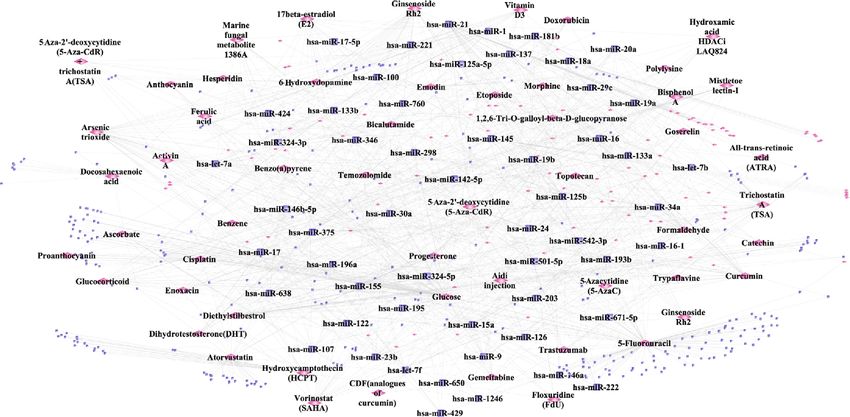
2                                                                                              BioMed Research International

cells) [2, 3]. Genetic modification of such cell-based con-        group where oral mucosal injury was created and the exper-
structs may potentially further accelerate their healing capac-   imental group where wound healing was complete. The
ity [4, 5] and provide a novel strategy to promote wound          model of oral mucosa injury was created by a sterile uniform
healing in oral mucosa injury. Therefore, investigating the       punch biopsy (McKesson) in the mucosa of the cheek [14].
genetic mechanisms involved in the wound healing process          The inclusion criteria pertaining to this dataset were as
of oral mucosa injury could be of significant translational        follows: [1] all participants being nonsmokers without any
value.                                                            systemic diseases; [2] uniform biopsy at less than 5 mm,
    Wound healing of oral mucosa is unique because it is          which was within self-regenerative capacity of the human
typically faster and shows a low tendency for scarring [6].       oral mucosa; and [3] datasets with at least 3 pairs of sam-
The superior healing characteristics of the oral mucosa           ples (3 controls and 3 experimental samples).
might be owing to various factors including the specific
anatomy of the oral cavity and environmental factors such         2.2. Differential Gene Expression Analysis. RNAseq data were
as microbial flora, saliva secretion, and pH balance. Some         processed using TopHat software [17], and RPKM (Reads
other or reasons plausible causes include growth factor pro-      Per Kilobase Million) was calculated by using Partek software
duction, stem cell levels, cellular proliferation capacity, and   [18]. Differential expression analysis was conducted by
genetic and epigenetic factors [7, 8]. As genetic and epige-      using the “DESeq” package in R environment [19]. The
netic regulation plays a pivotal role in the unique healing       differentially expressed mRNAs (genes) with p value < 0.05
characteristics of the oral mucosa, significant research atten-    and ∣log FC ∣ ≥1 were selected as DEGs. The DEGs with log
tion [9–12] has focused on the involvement of genetic and          FC ≥ 1 were considered upregulated DEGs, while DEGs with
epigenetic factors (e.g., genes, signaling pathways, transcrip-   log FC ≤ −1 were considered downregulated DEGs.
tion factors, and miRNAs) in the wound healing process of
oral mucosa injury. In this context, vascular endothelial         2.3. Functional Enrichment Analysis. Functional enrichment
growth factor (VEGF) has been found to promote palatal            analysis of the DEGs involved in the wound healing process
mucosal wound healing by accelerating neovascularization          of oral mucosa injury was performed using the “clusterProfi-
and reepithelialization [9]. In another report, intrinsic         ler” package in R [20]. The functions of the DEGs were
apoptosis pathways Casp2 and Trp53 were found activated           explored by investigating their enriched Gene Ontology
during oral mucosal wound healing [10]. The transcription         (GO) terms, in particular, biological processes (BPs) and
factor forkhead box O1 (FOXO1) has been found as                  Kyoto Encyclopedia of Genes and Genomes (KEGG) path-
increased in the nucleus of keratinocytes and promote ree-        ways. The GO terms and KEGG pathways with p value <
pithelialization during wound healing of oral mucosa [11].        0.05 were regarded as significant functions. If the number
The overexpression of miR-31 has been found to promote            of enriched BPs and pathways was more than 30, the top 30
oral mucosal wound closure by enhancing the proliferation         were chosen to be visualized in the bar plot. If the number
and migration of keratinocytes [12].                              of enriched BPs and pathways was less than 30, then all of
    Apart from these experimental studies, high-throughput        the BPs and pathways were visualized in the bar plot.
sequencing investigations have shown alterations of mRNAs         2.4. Construction of Protein-Protein Interaction (PPI)
or noncoding RNA expression profiles during oral mucosa            Network. Experimentally validated PPI pairs were down-
healing by establishing paired injury models in rats              loaded from a variety of databases including HPRD [21],
(GSE23006 [7] and GSE121996 [13]) and humans                      BIOGRID [22], DIP [23], MINT [24], mentha [25], PINA
(GSE97615 [14]). To the authors’ knowledge, there is no           [26], InnateDB [27], and Instruct [28]. PPI pairs of DEGs
comprehensive bioinformatic investigation leveraging the          were extracted from these databases and further used for
publicly available datasets pertaining to genetic and epige-      constructing the PPI network. The constructed PPI network
netic mechanisms involved in oral mucosa regeneration.            was plotted using Cytoscape Version 3.7.2 [29]. The topolog-
Bioinformatics can enable the interpretation and visualiza-       ical characteristics of the nodes in the PPI network were
tion of large datasets through network construction [15].         determined.
Plausibly, the important factors critical to a specific patho-
physiological process can be identified through bioinformatic      2.5. Construction of miRNA-Target DEGs Interaction
analysis of relevant gene expression datasets. The present        Network. Experimentally validated miRNA-target interac-
study is aimed at analyzing the data in the GSE97615 [14]         tion pairs were downloaded from three databases, including
using multiple bioinformatic techniques in order to identify      “miRTarBase” [30], “miRecords” [31], and “TarBase” v.8
specific molecular mechanisms that are key to the regulation       [32]. miRNAs targeting DEGs were extracted from these
of the wound healing processes in human oral mucosa injury.       databases and used for constructing a miRNA-target DEGs
                                                                  interaction network using the Cytoscape platform [29]. The
2. Materials and Methods                                          topological characteristics of the nodes in the miRNA-
                                                                  target DEGs network were determined.
2.1. Sourcing of Gene Expression Data. Datasets investigating
genetic alterations using paired models of rapid wound heal-      2.6. Construction of Transcription Factor- (TF-) Target DEGs
ing of oral mucosa were searched for in the Gene Expression       Interaction Network. The TF-target gene interaction pairs
Omnibus (GEO) database in NCBI [16]. The study design of          were downloaded from many databases, including “TRANS-
the included datasets consisted of two groups, the control        FAC” [33], “HTRIdb” [34], “TRRUST” [35], “ORTI” [36],
BioMed Research International                                                                                                                                3

                                                                           GSE97615 dataset

                                                                       Identification of DEGs

                                                                                            TF- target DEGs                 DEGs-chemical compound
               Functional enrichment                        PPI network
                                                                                          interaction network                 interaction network

                   Signaling pathways:                       Hub genes:
             arachidonic acid metabolism,                                                                                        Chemical compounds:
                                                       GABARAPL1, GABARAPL1,             Major transcription factors:
               ECM-receptor interaction,                                                                                      Valpronic acid, Doxorubicin,
                                                         HDAC5, MAP1LC3A,                FOS, PLAU, BCL6, RORA
            cell cycle, p53 signaling pathway                                                                                      Nickel, Tretinoin
                                                           AURKA, PLK1

                                                                          miRNAs targeting DEGs

                                    miRNA-target interaction network                              miRNA-small molecule drug interaction network

                                                                                                                                          Small molecule
                                                                                                                                               drugs:
                                                                   miRNAs:                                                                 Atorvastatin,
                                                                  miR-34a-5p,                                                              17β-estradiol,
                                                                  miR-335-5p                                                              Curcumin, and
                                                                                                                                            Vitamin D3

Figure 1: Study flowchart. The GSE97615 dataset was analyzed using a series of bioinformatic analyses: differential expression analysis;
functional enrichment analysis; and construction of networks including PPI network, TF-target DEGs network, DEGs-chemical
compound interaction network, miRNA-target interaction network, and miRNA-small molecular drug interaction network.

and “cGRNB” [37]. The TFs targeting DEGs were extracted                                SM2miR database [39]. Only data associated with human
and used for constructing the TFs-target DEGs interaction                              species were extracted, and 2,756 interaction pairs between
network using Cytoscape [29]. The topological characteris-                             known miRNAs and small molecular drugs were obtained.
tics of the nodes in the TFs-target DEGs network were                                  Among these pairs, miRNAs that target DEGs were
determined.                                                                            extracted, and the small molecule drugs that impact the
                                                                                       expression levels of these miRNAs were further obtained.
2.7. Construction of the DEGs-Chemical Compound                                        Therefore, 1,074 interaction pairs between small molecular
Interaction Network. Data pertaining to the interactions                               drugs and miRNAs that target DEGs were studied. In order
between genes and chemical compounds were downloaded                                   to visualize these interaction pairs, Cytoscape [29] was used
from CTD (Comparative Toxicogenomics Database) [38].                                   to construct the miRNA-small molecule drug interaction
Only the data regarding the human species were extracted.                              network.
858,086 interaction pairs between genes and chemical com-
pounds were obtained. Among these interaction pairs, chem-                             3. Results
ical compounds that correspond to DEGs were extracted, and
thus, 41,634 interaction pairs between DEGs and chemical                               3.1. Included Gene Expression Dataset. The dataset GSE97615
compounds were derived. Thereafter, Cytoscape [29] was                                 was included in this analysis [14]. This dataset examined
employed to visualize the interaction relationships between                            gene expression profiles using high-throughput sequencing
DEGs and their interacting chemical compounds. Following                               technology on the GPL11154 platform of Illumina HiSeq
the construction of the DEGs-chemical compound interac-                                2000. In the experimental group, oral buccal mucosal
tion network, a topological analysis was performed to calcu-                           injury was generated on day 1, and wound healing was
late the characteristics of the nodes in the network.                                  found to be almost complete on day 3. As nonsignificant
                                                                                       differences in gene expression were noted between biopsies
2.8. Construction of the miRNA-Small Molecule Drug                                     obtained on day 1 versus day 6, the oral biopsy samples of
Interaction Network. The data regarding known miRNAs                                   day 1 (GSM2573167, GSM2573168, GSM2573169, and
and small molecular drugs were downloaded from the                                     GSM2573170) and day 3 (GSM2573171, GSM2573172,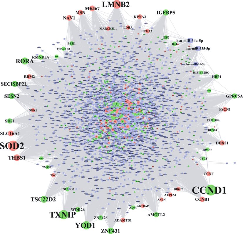
4                                                                                                      BioMed Research International

         Table 1: The top 30 DEGs involved in the process of oral mucosa regeneration, ranked by the ascending order of p value.

Name                 Base mean       Base meanA       Base meanB       Fold change      log2 fold change        p val              p adj
DEFB4A                40.12592        2.388343          77.8635          32.60148           5.026865         1:05E − 25      7:32E − 22
S100A7                310.8578        17.06716          604.6484         35.42758           5.146801         1:33E − 23      6:19E − 20
DEFB4B                13.92444         0.57421          27.27467         47.49947           5.56984          3:75E − 23      1:31E − 19
FLG                   21.40743         40.1873          2.627564         0.065383           -3.93494         3:94E − 22      1:10E − 18
KRT76                 8.60035         16.71021          0.490492         0.029353           -5.09036         4:74E − 19      1:10E − 15
CES1                  9.660384        18.36781          0.952953         0.051882           -4.26863         7:36E − 19      1:47E − 15
CCL19                 11.84998        1.619644          22.08032         13.63283           3.769013         2:64E − 18      4:61E − 15
ALOX12                101.6108        186.2771          16.94443         0.090964           -3.45857         5:61E − 17      8:69E − 14
CYP4F35P              13.31745        24.28438          2.350513         0.096791           -3.36898         8:10E − 17      1:03E − 13
ETNK2                 25.88451          47.104          4.665033         0.099037           -3.33589         8:10E − 17      1:03E − 13
TUBB3                 9.802023        1.010702          18.59334         18.39647           4.201357         8:86E − 17      1:03E − 13
DPT                   13.10894        23.69376          2.524124         0.106531           -3.23065         1:35E − 16      1:45E − 13
KPRP                  12.68058         23.2699          2.091252         0.089869           -3.47603         2:49E − 16      2:48E − 13
CLEC3B                16.77294        29.85829          3.68758          0.123503           -3.01739         7:10E − 16      6:60E − 13
ANKRD37               55.14818        100.8559          9.44044          0.093603            -3.4173         9:28E − 16      8:09E − 13
CYP4F29P              22.09975        39.23028          4.969215         0.126668           -2.98088         2:17E − 15      1:78E − 12
KRT3                  46.56198        82.43067          10.6933          0.129725           -2.94647         8:72E − 15      6:76E − 12
CRISP3                47.9651         84.80899          11.12121         0.131132            -2.9309         6:57E − 14      4:83E − 11
PPP1R3C               50.11791        89.58462          10.65119         0.118895           -3.07224         9:76E − 14      6:80E − 11
SOCS3                 7.850827        1.029401          14.67225         14.2532            3.833214         7:74E − 13      5:14E − 10
KRT16P2               25.30934        6.520768          44.09791         6.762687           2.757597         8:42E − 13      5:34E − 10
KLK6                  49.11907        12.67537          85.56277         6.750319           2.754956         9:84E − 13      5:97E − 10
MFAP4                 14.13919        23.95185          4.326524         0.180634           -2.46886         3:25E − 12      1:89E − 09
CD177                 6.239805        0.532066          11.94754         22.45498           4.488964         6:07E − 12      3:38E − 09
CDH3                  20.64668        5.975959          35.3174          5.909913           2.563137         7:13E − 12      3:82E − 09
ANKRD20A5P            6.083548        11.44316          0.723938         0.063264           -3.98248         4:05E − 11      2:02E − 08
PSCA                  36.97691        63.83741          10.11641         0.158471           -2.65771         4:88E − 11      2:35E − 08
SFRP2                 27.36962        45.66674          9.072498         0.198668           -2.33157         5:22E − 11      2:43E − 08
DUSP5                 32.6366         55.26694          10.00626         0.181053           -2.46551         5:51E − 11      2:48E − 08
CLDN17                30.11305         49.5288          10.69729         0.215981           -2.21102         2:50E − 10      1:09E − 07

GSM2573173, and GSM2573174) were selected as repre-                   by targeting the greatest number of DEGs. Thereafter, a
sentatives of injured versus healed oral mucosal tissue for           miRNA-target interaction network was constructed based
comparative analysis in this study.                                   on DEGs and miRNAs which target these DEGs. Finally, in
                                                                      order to explore the chemical compounds and small molecu-
                                                                      lar drugs that can regulate the healing process of oral mucosa
3.2. Analytical Approaches. A series of bioinformatic analyses        injury, a DEGs-chemical compound interaction network and
were performed and are depicted as a flowchart (Figure 1).             miRNA-small molecular drug interaction network were each
Firstly, differential gene expression analysis was performed           constructed.
to identify DEGs during the healing process of oral mucosa
injury. Subsequently, functional enrichment analysis was
performed to explore the signaling pathways in which DEGs             3.3. Identification of DEGs Involved in the Oral Mucosa
were enriched. In addition, a protein-protein interaction             Regeneration. By performing differential expression analysis,
(PPI) network was constructed to identify the hub proteins            a total of 650 DEGs consisting of 448 downregulated DEGs
that are likely to play critical roles in the healing process.        and 202 upregulated DEGs were identified as significantly
Additionally, a transcription factor- (TF-) target DEGs inter-        involved in the oral mucosa regeneration (File S1). Table 1
action network was constructed to identify the transcription          shows the top 30 DEGs with the most significant p values,
factors that might be key to regulation of the healing process        including defensin beta 4A (DEFB4A), C-C motif chemokine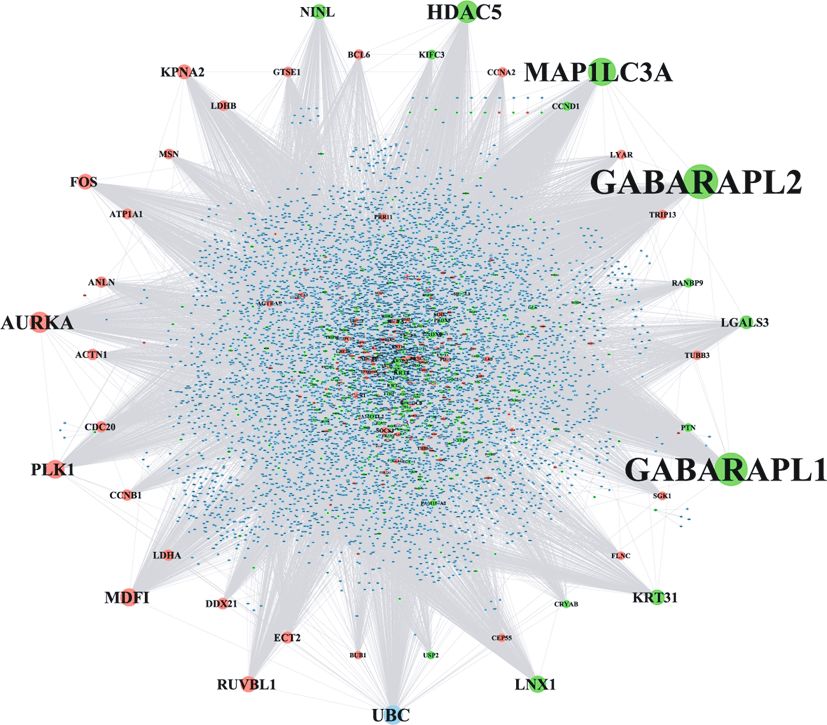
BioMed Research International 5
−1⁎ log10 (pvalue)
Cornification
Epidermis development 18
Skin development
Keratinocyte differentiation
Keratinization
Epidermal cell differentiation
Peptide cross−linking
Extracellular matrix organization
15
Extracellular structure organization
Regulation of chromosome segregation
Mitotic nuclear division
Regulation of nuclear division
Regeneration
Biological process
Aging
Regulation of mitotic nuclear division 12
Regulation of mitotic sister chromatid separation
Metaphase/anaphase transition of mitotic cell cycle
Regulation of sister chromatid segregation
Mitotic sister chromatid separation
Metaphase/anaphase transition of cell cycle
Organelle fission
9
Nuclear division
Spindle checkpoint
Spindle assembly checkpoint
Mitotic spindle checkpoint
Mitotic spindle assembly checkpoint
Regulation of chromosome separation
Negative regulation of mitotic sister chromatid separation 6
Negative regulation of mitotic metaphase/anaphase transition
Negative regulation of metaphase/anaphase transition of cell cycle
5 10 15
−Log10 (pvalue)
Count
10 40
20
50
30
(a)
⁎
–1 log10 (pvalue)
5.5
Arachidonic acid metabolism
5.0
ECM−receptor interaction
4.5
Pathway
Cell cycle 4.0
3.5
p53 signaling pathway
0e+00 2e–04 4e–04 6e–04 8e–04
pvalue
Count
8 11
9 12
10 13
(b)
Figure 2: The enriched functions of DEGs involved in the oral mucosa regeneration process: (a) GO terms-biological processes; (b) signaling
pathways. The count represents the number of genes enriched in a BP term, and -log10(p value) represents the enrichment score. The bigger
size of the dots corresponding to a BP term means more genes were enriched in this term. The coloured dots represent the term enrichment:
green indicates low enrichment, and red indicates high enrichment.6 BioMed Research International
ligand 19 (CCL19), filaggrin (FLG), cysteine-rich secretory
protein 3 (CRISP3), and suppressor of cytokine signaling 3
(SOCS3).
3.4. Functional Terms Enriched by DEGs. File S2 and S3 show
the significantly enriched biological processes and signaling
pathways associated with DEGs, respectively. Figure 2(a)
shows that DEGs involved in oral wound healing were
implicated in many biological processes such as epidermis
development, keratinocyte differentiation, keratinization,
epidermal cell differentiation, extracellular matrix organi-
zation, biological processes related to cell cycle, and spin-
dle checkpoint. Figure 2(b) shows that DEGs involved in
oral wound healing were implicated in arachidonic acid
metabolism, ECM-receptor interaction, cell cycle, and the
p53 signaling pathway.
DEG_Up
3.5. Identification of Hub Genes in the PPI Network. The PPI DEG_Down
network (shown in Figure 3) consisted of 7,055 nodes and Other_Gene
16,851 edges (File S4). As shown in Table 2, the top 30 nodes
with the highest degree were listed, such as GABA type A Figure 3: The PPI network of DEGs involved in the oral mucosa
receptor-associated protein-like 1 (GABARAPL1), GABA regeneration process. The nodes with different colours represent
the upregulated and downregulated DEGs and other genes that
type A receptor-associated protein-like 2 (GABARAPL2),
are not DEGs.
microtubule-associated protein 1 light chain 3 alpha
(MAP1LC3A), histone deacetylase 5 (HDAC5), aurora S7. Specific chemical compounds targeting the DEGs that
kinase A (AURKA), and polo-like kinase 1 (PLK1). These were dysregulated in the healing process of oral mucosa
genes with the highest degree were deemed to be of the high- injury included Valproic acid, Doxorubicin, Nickel, and
est significance in the PPI network by interacting with the tretinoin.
greatest number of genes.
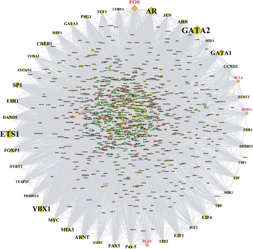
3.6. Identification of the Significant miRNAs Targeting DEGs. 3.9. Identification of Small Molecular Drugs That Regulate the
The miRNA-target DEGs interaction network shown in Expression of miRNAs. A miRNA-small molecular drug
Figure 4 consisted of 2,995 nodes and 20,362 edges (File interaction network was constructed and is shown in
S5). Table 3 shows the topological characteristics of the top Figure 7. This network is comprised of 522 nodes and 1,074
30 nodes in the network. Two miRNAs (miR-34a-5p and edges (File S8). Several small molecular drugs were found
miR-335-5p) played the most significant roles in this net- to regulate the expression of miRNAs targeting the DEGs
work by targeting the greatest number of DEGs. By tracking dysregulated during healing of oral mucosa injury including
the target relationship between a certain miRNA and its atorvastatin, 17β-estradiol, curcumin, and vitamin D3.
target DEGs within the top 30 nodes, it was determined that
miR-34a-5p targets several DEGs including AMOTL2, 4. Discussion
CCNB1, CCND1, DDX21, FSCN1, HBP1, ID4, KPNA2,
MKI67, MSN, NAV1, RRM2, SECISBP2L, SESN2, THBS1, Bioinformatic analysis revealed several genetic and epige-
and TXNIP. miR-335-5p was found to target several DEGs netic regulatory mechanisms implicated in oral mucosa
including DDX21, HBP1, RMND5A, RORA, SECISBP2L, wound healing including specific mRNAs, miRNAs, path-
SESN2, TXNIP, and YOD1. ways, TFs, and targeted drugs.
Hub genes identified as central to the PPI network that
3.7. Identification of Hub Transcription Factors That Target interacted with the greatest number of genes included
DEGs. The TFs-target DEGs interaction network shown in GABARAPL1, GABARAPL2, HDAC5, MAP1LC3A, AURKA,
Figure 5 consisted of 1,054 nodes and 946 edges (File S6). and PLK1. Gamma-aminobutyric acid receptor-associated
Table 4 shows the top TFs with the highest degree that are protein-like 1 (GABARAPL1) and GABARAPL2 are proteins
pivotal to this network. Protooncogene c-Fos (FOS), B-cell associated with gamma-aminobutyric acid (GABA), a non-
lymphoma 5 protein (BCL6), and urokinase-type plasmino- protein amino acid, and the overexpression of GABA has been
gen activator (PLAU) were not only TFs but also upregulated shown to suppress inflammation by inhibiting inflammatory
DEGs. RAR-related orphan receptor A (RORA) was TF and a mediators (iNOS, IL-1beta, and TNF-alpha), promoting fibro-
downregulated DEGs. blast cell proliferation, and stimulating reepithelialization,
suggesting that GABA could accelerate the healing process at
3.8. Identification of Chemical Compounds That Regulate the the early stage of cutaneous wound healing [40]. Histone
Expression of DEGs. A DEGs-chemical compound interac- deacetylase 5 (HDAC5) belongs to class IIa HDACs and plays
tion network was constructed and is shown in Figure 6. This a key role in angiogenesis, inflammation, and immunity [41].
network is comprised of 3,612 nodes and 27,426 edges (File HDAC was shown to inhibit fibroblast proliferation byBioMed Research International 7
Table 2: The topological characteristics of the top 30 hub genes in the PPI network.
Average shortest Betweenness Closeness Clustering Topological
Name Label Degree
path length centrality centrality coefficient coefficient
GABARAPL2 Down 583 2.689935 0.071254 0.371756 0.005399 0.005164
GABARAPL1 Down 559 2.711402 0.057754 0.368813 0.006036 0.005542
MAP1LC3A Down 463 2.683537 0.053743 0.372643 0.007429 0.005812
HDAC5 Down 363 2.837504 0.045007 0.352422 0.001614 0.007383
AURKA Up 332 2.768126 0.049883 0.361255 0.003904 0.007122
UBC Other 302 2.217088 0.270483 0.451042 0.005258 0.007895
PLK1 Up 288 2.75462 0.047684 0.363026 0.006259 0.0075
MDFI Up 277 2.965596 0.057747 0.3372 1:91E − 04 0.005234
LNX1 Down 265 3.016776 0.047711 0.33148 1:50E − 04 0.006827
RUVBL1 Up 243 2.846034 0.034842 0.351366 0.002092 0.010082
KRT31 Down 235 2.960904 0.039526 0.337735 0.002633 0.006649
FOS Up 232 2.932897 0.035432 0.34096 0.001416 0.010324
NINL Down 226 2.914984 0.038519 0.343055 0.002989 0.007416
KPNA2 Up 216 2.815326 0.032401 0.355199 0.003874 0.009379
LGALS3 Down 201 2.922377 0.033476 0.342187 0.002459 0.007997
ECT2 Up 168 2.89238 0.025887 0.345736 0.003302 0.010711
DDX21 Up 161 2.89437 0.0152 0.345498 0.008063 0.014814
LDHA Up 156 2.850299 0.0153 0.35084 0.013245 0.013064
ACTN1 Up 155 2.871908 0.021745 0.348201 0.003848 0.011807
CDC20 Up 155 2.823003 0.017314 0.354233 0.018812 0.012271
CCNB1 Up 155 2.90816 0.014414 0.34386 0.017677 0.014428
ANLN Up 153 2.993034 0.02056 0.334109 0.00517 0.012216
ATP1A1 Up 144 2.873472 0.018625 0.348011 0.00483 0.012658
LDHB Up 142 2.954933 0.010182 0.338417 0.012589 0.016739
MSN Up 142 2.969719 0.015946 0.336732 0.00402 0.015602
BCL6 Up 138 2.938442 0.022118 0.340316 0.004055 0.011744
GTSE1 Up 138 2.953512 0.017776 0.33858 0.007634 0.012333
KIFC3 Down 136 2.949957 0.018258 0.338988 0.006904 0.012187
CCNA2 Up 132 2.961189 0.013698 0.337702 0.011937 0.016085
CCND1 Down 128 2.925931 0.013883 0.341772 0.00427 0.016102
repressing the PDGF-receptor (R)-α [42], which aligns with is involved in the formation and/or stabilization of microtu-
our finding implicating downregulation of HDAC in the oral bules [45]. Targeting microtubules could potentially enhance
mucosal wound healing processes. Microtubule-associated the wound healing process by promoting cell migration [46];
protein 1 light chain 3 alpha (MAP1LC3A), also named thus, AURKA might impact oral mucosa wound repair by
LC3, encodes a central protein in the autophagy pathway influencing cell migration. PLK1 is a family member of the
and is therefore widely used as a marker for detecting autoph- polo-like kinase (Plks) family, which has been demonstrated
agy and autophagic cell death [43]. As the activation of the to be a key regulator of the cell cycle [47]. PLK1 plays a role
autophagy pathway is reported as essential for myofibroblast in regulating mitotic entry, spindle assembly, anaphase entry,
differentiation and collagen deposition during wound healing and cytokinesis in the mitotic phase and DNA checkpoints
[44], the MAP1LC3A gene as an autophagy-related marker is [48]. Since cell cycle regulatory genes are important for the
plausible as integral to oral mucosal wound healing. Besides, reepithelialization of oral mucosal wounds by influencing cel-
another family member of microtubule-associated protein- lular proliferation [49], the overexpression of PLK1 possibly
microtubule-associated protein 4 (MAP4) has been shown to enhances the oral mucosal wound healing by promoting cell
regulate the migration and proliferation of epidermal kerati- proliferation. While existing evidence supports the role of
nocytes and further influence epidermal wound healing by most of the hub genes’ involvement in oral mucosal wound
activating the p38/MAPK pathway. However, the function of healing, their specific functions have not yet been verified by
the microtubule-associated protein family has not been inves- previous research. Future experimental studies should be
tigated in the context of oral mucosal injury healing. Aurora designed to validate their roles during the healing of oral
kinase A (AURKA) encodes a cell cycle-regulated kinase that mucosal injury.8 BioMed Research International
the cell migration of dendritic cells [59], which is in agree-
ment with the expression pattern of FSCN1 noted in the
present study. These genes may be considered targets of
further research focused on gene modulation to enhance oral
mucosal wound healing.
miR-335-5p was also identified as an important factor.
The overexpression of miR-335 has been reported to promote
diabetic wound healing by repressing the Sp1 transcription
factor (Sp1) and downregulating matrix metallopeptidase-
(MMP-) 9 [60]. Figure 3 in this study shows the DEGs targeted
by miR-335-5p, including YOD1, TXNIP, and HBP1. Their
relevance to wound healing is supported by earlier reports.
For example, YOD1 (also known as OTUD2) is known to
encode a protein that belongs to the ovarian tumor (OUT)
family of deubiquitinating enzymes (DUBs) [61]. The over-
expression of YOD1 was shown to promote the migration
of oral keratinocytes by activating the TGF-β3 signaling
pathway [62]. Contradictory to this observation, the present
DEG_Up
study indicated a downregulation of YOD1 during the muco-
DEG_Down
miRNA
sal healing process. Another identified gene, thioredoxin
interacting protein (TXNIP), also known as vitamin D3
Figure 4: The miRNA-target interaction network involved in oral upregulated protein, is a major regulator of redox signaling
mucosa regeneration. The round nodes with different colours, and endoplasmic reticulum (ER) stress [63]. A recent study
respectively, represent the upregulated and downregulated DEGs, showed that the supplementation of vitamin D could signifi-
while the square nodes represent the miRNAs that target DEGs. cantly accelerate wound healing by suppressing ER stress
[64]; however, the present study noted a downregulation of
Two miRNAs, miR-34a-5p and miR-335-5p, were identi- TXNIP in mucosal regeneration. Another gene targeted by
fied as pivotal in the miRNA-target DEGs network, by target- miR-335-5p, HMG-box transcription factor 1 (HBP1), is a
ing the greatest number of DEGs. miR-34a-5p, derived from repressor of the cyclin D1 gene and can inhibit the Wnt-
5 ′ ends of pre-miR-34a, is a member of the miR-34 family, mediated beta-catenin signaling pathway. The activation of
and miR-34a is a well-known tumor suppressor [50], shown Wnt signaling has been shown to promote mucosal repair
to mediate endothelial progenitor cell-mediated angiogenesis [65]. Our results indicated the downregulation of HBP1 dur-
[51]. The knockout of miR-34a has been shown to impair ing oral mucosal repair, suggesting that downregulating
wound healing by augmenting inflammatory response and HBP1 might promote Wnt signaling and thereby augment
delaying reepithelialization [52], implying that miR-34a oral mucosal repair.
upregulation could be a potential novel strategy for enhanc- Four signaling pathways (arachidonic acid metabolism,
ing mucosal regeneration. As shown in Figure 4, miR-34a- cell cycle, p53, and extracellular matrix- (ECM-) receptor
5p was noted to target several DEGs including AMOTL2, interaction) were found significantly enriched during the
THBS1, and FSCN1. Among these, Angiomotin-Like 2 healing of oral mucosa. Arachidonic acid (AA) is a fatty acid
(AMOTL2) has been previously reported as downregulated that is abundantly expressed after tissue injury [66]. AA and
during wound healing after oral mucosal injury. The its metabolites have been demonstrated to facilitate wound
AMOTL2 gene has also been found to regulate the polarity, healing by inducing the adhesion and migration of endothe-
migration, and proliferation of endothelial cells [53]. Thus, lial cells, as well as angiogenesis [66]. The effects of induction
it may be considered a potential target for gene therapy in of AA on cell motility were found regulated by membrane
cell-based regenerative strategies, as its upregulation in oral type 3-matrix metalloproteinase- (MT3-MMP-) dependent
mucosa endothelial cells could facilitate angiogenesis during fibronectin degradation [67]. The arrest of the cell cycle has
wound healing. Thrombospondin 1 (THBS1), also named been shown to impair wound healing, which is characterized
TSP1, is documented to be a major activator of transforming by a significant number of senescent fibroblasts that are
growth factor- (TGF-) β1 [54]. Previous studies have shown irreversible and refractory to growth factor stimulation,
that the knockdown of TSP1 could decrease the rate of unless genetically modified [68]. In addition, it has been
reepithelization, wound neovascularization, and migration shown that periodontal pathogens (Fusobacterium nuclea-
of fibroblast and endothelial cells, thereby impairing the tum and Porphyromonas gingivalis) reduce wound healing
wound healing process [55–57]. Our results indicated that by downregulating the cell cycle genes cyclin1, cyclin-
TSP1 was upregulated during oral mucosal wound healing, dependent kinase 1 (CDK1), and CDK4 that are critical for
which corroborates previous observations. The fascin actin- G1/S transition and initiating the further process of DNA
bundling protein 1 (FSCN1), also called fascin, encodes an replication in S phase [69]. Concerning the cell cycle, the
actin-bundling protein that regulates the focal adhesion tumor suppressor gene p53 is of importance, being a master
dynamics of cytoskeletal structures during cell migration cell cycle regulator [70] and linked to the intrinsic apoptosis
[58]. The overexpression of FSCN1 is reported to promote pathway, which is predominantly involved in the woundBioMed Research International 9
Table 3: The topological characteristics of the top 30 nodes of the miRNA-target network involved in the oral mucosa regeneration.
Average shortest Betweenness Closeness Topological
Name Label Degree
path length centrality centrality coefficient
CCND1 Down 388 2.593186 0.081676 0.385626 0.035366
SOD2 Up 363 2.631931 0.071979 0.379949 0.032418
TXNIP Down 322 2.646627 0.053959 0.377839 0.033412
LMNB2 Up 297 2.706747 0.049038 0.369447 0.029849
YOD1 Down 282 2.674015 0.046754 0.37397 0.04023
RORA Down 231 2.725451 0.028034 0.366912 0.043835
TSC22D2 Down 225 2.752171 0.030429 0.36335 0.04615
THBS1 Up 209 2.738811 0.02335 0.365122 0.047046
ZNF431 Down 203 2.790247 0.028431 0.358391 0.040197
IGFBP5 Down 193 2.775551 0.023039 0.360289 0.042372
SECISBP2L Down 186 2.755511 0.022275 0.362909 0.048889
SESN2 Down 186 2.738811 0.023606 0.365122 0.04734
MKI67 Up 178 2.782231 0.016852 0.359424 0.051973
SLC16A1 Up 176 2.834335 0.020942 0.352816 0.037253
NAV1 Up 168 2.784903 0.019746 0.359079 0.045105
MSN Up 162 2.804275 0.020327 0.356598 0.042947
CCNB1 Up 159 2.798931 0.0135 0.357279 0.0552
GPRC5A Down 155 2.818303 0.018532 0.354823 0.052165
SIK1 Down 153 2.806947 0.012722 0.356259 0.061204
AMOTL2 Down 149 2.815631 0.015657 0.35516 0.042484
WDR26 Down 148 2.814295 0.01461 0.355329 0.053959
DDX21 Up 147 2.765531 0.012253 0.361594 0.057614
RMND5A Down 146 2.777555 0.012082 0.360029 0.054446
hsa-miR-34a-5p miRNA 144 2.359051 0.030631 0.423899 0.028501
ZNF426 Down 142 2.831663 0.016397 0.353149 0.041514
RRM2 Up 139 2.812291 0.013288 0.355582 0.058531
FSCN1 Up 136 2.843687 0.015655 0.351656 0.042006
HBP1 Down 135 2.795591 0.01225 0.357706 0.06161
KPNA2 Up 135 2.817635 0.010852 0.354908 0.062084
hsa-miR-335-5p miRNA 130 2.58016 0.032712 0.387573 0.022844
healing of oral mucosa, in contrast to that of skin [10]. The tics of oral mucosa wound healing that are distinct from skin
inhibition of p53 was reported to accelerate the early epithe- wound healing.
lization and neovascularization during wound healing [71], Four transcription factors, FOS, PLAU, BCL6, and
which supports a previous report showing that the expres- RORA, were identified as highly crucial to oral mucosa
sion level of p53 on the 3rd-day postwounding was close to wound healing and as differentially expressed during the
that with prewounding levels and very low [10]. Further- healing process. The transcription factor FOS (also called
more, the interaction between ECM and cellular adhesion c-Fos) has been found to stimulate cell proliferation and
receptors (e.g., integrin and growth factor receptors) has been regulate cell growth by interacting with c-Jun and further
shown to regulate cellular activities of progenitor stem cells, activating the activator protein- (AP-) 1 complex [74]. FOS
including migration, differentiation, proliferation, and sur- was also found to be rapidly and specifically expressed by epi-
vival, during all phases of cutaneous wound healing [72]. In dermal cells at the margin of the wound, thereby acting as an
addition, fibronectin (FN) and chondroitin sulphate (CS), early molecular component during the wound healing pro-
two components of ECM, are found as significantly elevated cess [75]. Another identified factor, plasminogen activator,
in oral mucosa as compared with skin [73], which could urokinase (PLAU), encodes a secreted serine protease that
account for the faster healing of oral mucosa wounds than can convert plasminogen to plasmin. The plasminogen
skin wounds [14]. These findings, together with our results, activator/plasmin system has been shown to modulate cell
suggest that p53 signaling and ECM (especially FN and CS) adhesion and cellular migration by activating MMPs and
interaction signaling can be regarded as specific characteris- TGF-β, thereby providing the important link between PLAU10 BioMed Research International
DEG_Up DEG_Up&TF
DEG_Down DEG_Down&TF
TF
Figure 5: The transcription factor-target interaction network involved in oral mucosa regeneration. The round nodes with different colours
represent the up- and downregulated DEGs; the diamond nodes with no-colour edges represent TFs; the diamond nodes with red-edges
represent that these nodes are not only upregulated DEGs but also TFs; the diamond nodes with emerald colour represent that these
nodes are not only downregulated DEGs but also TFs.
expression and wound healing [76]. B-cell lymphoma 6 pro- mer research suggested that DXR significantly delayed the
tein (BCL6) (also named zinc finger protein 51 (ZNF51)) has restoration of tensile strength, resulting in skin thinning
been validated as a target of miR-155 [77]. The downregula- and degeneration of skin appendage after surgery [82].
tion of BCL6 mRNA expression induced by the depletion of Nickel (Ni), a metallic element routinely utilized for ortho-
miR-155 has been noted to enhance dermal wound healing dontic and prosthetic device construction, improves chemi-
[77]. Retinoid-related orphan receptor alpha (RORA) is a cal stabilization, corrosion resistance, and durability [83].
transcription factor demonstrated to regulate the differentia- However, Ni can cause side effects under inflammatory con-
tion/survival of keratinocytes under hypoxic conditions [78]. ditions [84] and in turn induce immune responses along with
Since oral mucosa wounds heal under significantly lower exacerbating inflammation, thus impairing oral wound heal-
hypoxic conditions than skin wounds [79], the overexpres- ing [85]. A high concentration of Ni was noted to promote
sion of RORA might be considered a specific feature of oral inflammatory processes and proliferation via toll-like recep-
mucosa wound healing. tor- (TLR-) 4/nuclear factor-kappa B (NF-κB) activation in
Four significant chemical compounds, Valproic acid, Interleukin- (IL-) 1β-induced human gingival fibroblasts
Doxorubicin, Nickel, and tretinoin, were identified to regu- (HGFs) [85]. Tretinoin, also known as all-trans retinoic acid
late the expression of DEGs that were dysregulated during (ATRA), has been applied as a useful treatment for epithelial
oral mucosal wound healing. Valproic acid (VPA) has been wound healing and keratinization disorders, because of its
commonly prescribed to treat neurological disorders for sev- role in stimulating angiogenesis and collagen production
eral decades owing to its neuroprotective effects [80]. VPA [86]. However, a previous study demonstrated that tretinoin
has the potential to enhance cutaneous wound healing as it retarded reepithelialization in open wounds [87]. In contrast,
exerts differential effects on cell proliferation and migration, collagen production was found increased by tretinoin [88] in
via activating Wnt/β-catenin, extracellular signal-regulated recent experimental research [89]. Therefore, the impact of
kinase (ERK), and phosphatidylinositol 3-kinase (PI3K)/Akt tretinoin on wound healing remains controversial.
signaling pathways [81]. Doxorubicin (DXR) is a typical che- Four small molecules, atorvastatin, 17β-estradiol, curcu-
motherapeutic agent that is widely used in cancer treatment min, and vitamin D3, were identified as primary drug
and can act as an inhibitor of wound healing processes. For- candidates relevant to oral mucosa regeneration. Statins areBioMed Research International 11
Table 4: The topological characteristics of the top 30 nodes in the TF-target interaction network.
Average shortest Betweenness Closeness Clustering Topological
Name Label Degree
path length centrality centrality coefficient coefficient
GATA2 TF 253 2.191833 0.082064 0.456239 0.001663 0.04192
ETS1 TF 241 2.178538 0.08846 0.459024 0.001729 0.040046
AR TF 227 2.194682 0.07873 0.455647 0.00191 0.041171
YBX1 TF 202 2.298196 0.068148 0.435124 7:88E − 04 0.044113
GATA1 TF 168 2.426401 0.030482 0.412133 0.001853 0.055186
SP1 TF 154 2.312441 0.036965 0.432444 0.00365 0.050859
FOS Up_TF 139 2.025641 0.069117 0.493671 0.060369 0.045021
ARNT TF 128 2.417854 0.018538 0.41359 0.002338 0.06006
MIA3 TF 127 2.48433 0.017272 0.402523 3:75E − 04 0.068054
ESR1 TF 124 2.458689 0.021245 0.406721 0.001967 0.053796
CREB1 TF 113 2.439696 0.016546 0.409887 0.006637 0.059282
FOXP3 TF 113 2.603039 0.021352 0.384166 6:32E − 04 0.065569
AHR TF 109 2.504274 0.012008 0.399317 0.003058 0.068831
MYC TF 108 2.5717 0.018731 0.388848 6:92E − 04 0.068219
PAX5 TF 105 2.623932 0.011361 0.381107 1:83E − 04 0.080365
E2F4 TF 104 2.518519 0.024369 0.397059 0.002801 0.052057
Pax-5 TF 102 2.683761 0.009227 0.372611 0 0.091831
E2F1 TF 99 2.477683 0.015085 0.403603 0.00268 0.062549
DAND5 TF 98 2.622982 0.007305 0.381245 0.002525 0.078717
PSG1 TF 97 2.645774 0.006395 0.377961 0.002577 0.081277
JUN TF 91 2.509972 0.014222 0.398411 0.016361 0.063318
CCND1 Down 91 2.195632 0.057784 0.45545 0.01514 0.05193
STAT1 TF 89 2.51377 0.012112 0.397809 0.006639 0.062582
GATA3 TF 87 2.593542 0.009757 0.385573 0.002673 0.062089
TP53 TF 87 2.60019 0.015924 0.384587 0.001069 0.059128
Delta-CREB TF 85 2.717949 0.005681 0.367925 0 0.096063
TCF3 TF 82 2.630579 0.009266 0.380144 0.002409 0.071966
PRDM14 TF 73 2.65812 0.006654 0.376206 0.003044 0.059795
PLAU Up_TF 73 2.189934 0.029773 0.456635 0.082572 0.056671
TBP TF 72 2.595442 0.016121 0.385291 0.003912 0.058185
recognized as therapeutic agents for treating different inflam- a novel therapeutic for improving wound regeneration and
matory conditions and wound healing and display diverse is characterized by its anti-infectious, anti-inflammatory,
pleiotropic effects including anti-inflammatory, antioxida- and antioxidant properties [95]. Different topical formula-
tion, immunomodulation, antibacterial, and neoangiogenesis tions of curcumin, including nanoformulations, have been
effects [90]. Both oral and topical administration of atorva- applied for wound healing activities (e.g., inflammation,
statin produced a significant enhancement in wound healing proliferation, and remodelling) and play a vital role in
[91, 92], which accelerated tissue repair by modulating scavenging free radicals, alleviating proinflammatory activi-
inflammatory cytokines and cell growth-associated proteins. ties, improving neovascularization and extracellular matrix
The female sex hormone 17β-estradiol (estrogen/E2) is (ECM) synthesis, and inhibiting apoptosis and scar matu-
found to exert differential effects on females and males ration [96]. Vitamin D3 is a fat-soluble secosteroidsone
during wound healing [93]. 17β-Estradiol treatments are well and exerts various effects on biological activities, including
established to have a beneficial influence on female cutaneous immunomodulation, antiaging, anticarcinogenesis, and anti-
physiology: 17β-estradiol promotes anti-inflammatory (M2) infection effects [97]. Previous studies have suggested that
macrophage actions and the migration of keratinocytes and vitamin D has a potent anti-inflammatory and immunomo-
fibroblasts while decreasing the expression of proinflamma- dulating effect on dental caries [98] and oral candidiasis
tory cytokines and wound protease [93]. However, 17β- [99]. Specifically, vitamin D is shown to promote oral muco-
estradiol is found to delay wound repair in male mice, since sal wound healing in many autoimmunologic oral diseases
it retards reepithelialization and inhibits collagen secretion such as recurrent aphthous stomatitis, Behcet syndrome,
[94]. Curcumin, a highly pleiotropic molecule, is considered and Sjögren syndrome [97].12 BioMed Research International
DEG_Up
DEG_Down
Chemical
Figure 6: The DEGs-chemical compound interaction network involved in oral mucosa regeneration. The round nodes with different colours
represent up- and downregulated DEGs; the pink diamond nodes represent the chemical compounds.
miRNA
Small_molecule
Figure 7: The miRNA-small molecular drug interaction network involved in oral mucosa regeneration. The blue square nodes represent the
miRNAs, while the pink diamond nodes represent the small molecular drugs.
This bioinformatic study provides an overall perspective the study is subject to several limitations. First, results from
of the genetic and epigenetic mechanisms underpinning oral the bioinformatic analysis were not validated in clinical
mucosa wound healing, deduced from the comprehensive experiments, and these should be performed in further
analyses performed, including functional enrichment analy- research. Only one dataset was found related to oral mucosa
sis, PPI network construction, miRNA-target network con- wound healing and thus included for the analysis. The avail-
struction, and DEG-TF network construction. However, ability of more datasets with similar study designs and withBioMed Research International 13
larger sample sizes is essential to replicate these preliminary Conflicts of Interest
results. Furthermore, datasets examining the noncoding
RNA (e.g., miRNAs, lncRNAs, and circRNAs) expression The authors declare that they have no conflicts of interest.
profiles were not found in public databases; therefore, the
identification of the miRNAs in this study was through pre- Authors’ Contributions
diction based on the targeting interactions between DEGs
and miRNAs. Studies investigating the epigenetic mecha- Wanchen Ning (Email: wanchen.ning@campus.lmu.de) is
nisms involved in oral mucosa wound healing by examining the first author and senior author. Dr. Shaonan Hu (Email:
noncoding RNA expression profiles would be valuable in shaonan.hu@medizin.uni-leipzig.de), Prof. Dr. Xianda Hu
this context. (Email: hellocean@hotmail.com), and Dr.rer.med. Simin Li
Despite these limitations, the findings of our study may (Email: simin.li.dentist@gmail.com) are equal to the corre-
serve as the theoretical basis directing future experiments sponding author.
concerning oral wound healing models and related target
genes. They also provide insights into potential therapeutic
targets and strategies for treating chronic and nonhealing
Acknowledgments
oral mucosa wounds. These results could also plausibly be Dr. Wanchen Ning received doctoral study support from
applied to genetic and epigenetic modification of stem cells the China Scholarship Council (CSC) for Wanchen Ning
utilized for tissue regeneration and optimize the design of (CSC No: 201608080112) at Ludwig-Maximilians-Universität
the tissue-engineered 3-dimensional (3D) culture models of München. Dr. Simin Li was funded by the Science Research
oral mucosa regeneration. Further, these findings could Cultivation Program of Stomatological Hospital, Southern
advance the development of topical drugs targeting relevant Medical University (Guangdong Provincial Stomatological
gene targets for treating oral mucosa lesions. Challenges in Hospital) (No. PY2020004).
clinical translation include that of synergistic application
with conventionally used topical drugs accruing anti-inflam-
matory, analgesic, anesthetic, and antimicrobial effects. As Supplementary Materials
such, these results should be considered preliminary and a
directive for future research design. Supplementary 1. File S1. The DEGs dysregulated in the
self-healing process of human oral mucosa injury.
5. Conclusion Supplementary 2. File S2. The GO terms enriched by DEGs.
Supplementary 3. File S3. The KEGG pathways enriched by
This study identified several genes (GABARAPL1, GABAR-
DEGs.
APL2, HDAC5, MAP1LC3A, AURKA, and PLK1), two miR-
NAs (miR-34a-5p and miR-335-5p), four transcription factors Supplementary 4. File S4. The topological characteristics of
(FOS, PLAU, BCL6, and RORA), and signaling pathways (ara- the nodes in the PPI network.
chidonic acid metabolism, cell cycle, p53, and extracellular Supplementary 5. File S5. The topological characteristics of
matrix- (ECM-) receptor interaction) to be the most significant the nodes in the miRNA-target network.
biomarkers in the wound repair process of oral mucosa injury.
This study also identified significant chemical compounds Supplementary 6. File S6. The topological characteristics of
(Valproic acid, Doxorubicin, Nickel, and tretinoin), as well as the nodes in the TF-target network.
small molecular drugs (atorvastatin, 17β-estradiol, curcumin, Supplementary 7. File S7. The topological characteristics of the
and vitamin D3), which could influence the oral mucosal nodes in the DEGs-chemical compound interaction network.
wound healing by regulating the expression of healing-
associated DEGs/miRNAs. These entities could be important Supplementary 8. File S8. The topological characteristics of
targets for genetic and epigenetic modification of oral the nodes in the miRNA-small molecular drug interaction
mucosa-derived cells for tissue engineering and other treatment network.
approaches for promoting oral mucosal wound healing.
References
Data Availability [1] M. Koray and T. Tosun, “Oral mucosal trauma and injuries,”
in Trauma in Dentistry, IntechOpen, London, UK, 2019.
The data used to support the findings of this study are
[2] J. Lee, D. Shin, and J.-L. Roh, “Use of a pre-vascularised oral
available from the corresponding author upon request. mucosal cell sheet for promoting cutaneous burn wound
healing,” Theranostics, vol. 8, no. 20, pp. 5703–5712, 2018.
Ethical Approval [3] J. L. Roh, J. Lee, H. Jang, E. H. Kim, and D. Shin, “Use of oral
mucosal cell sheets for accelerated oral surgical wound heal-
As this study only applied bioinformatic techniques based on ing,” Head & neck, vol. 40, no. 2, pp. 394–401, 2018.
computational analyses, all of the data from oral buccal [4] J. W. Sessions, D. G. Armstrong, S. Hope, and B. D. Jensen, “A
mucosa biopsies were obtained from the public datasets, review of genetic engineering biotechnologies for enhanced
and original samples were not obtained. Therefore, this study chronic wound healing,” Experimental Dermatology, vol. 26,
did not require ethical approval. no. 2, pp. 179–185, 2017.14 BioMed Research International
[5] S.-B. Yong, J. Y. Chung, Y. Song, and Y.-H. Kim, “Recent chal- [22] R. Oughtred, C. Stark, B.-J. Breitkreutz et al., “The BioGRID
lenges and advances in genetically-engineered cell therapy,” interaction database: 2019 update,” Nucleic acids research,
Journal of Pharmaceutical Investigation, vol. 48, no. 2, vol. 47, no. D1, pp. D529–D541, 2019.
pp. 199–208, 2018. [23] I. Xenarios, D. W. Rice, L. Salwinski, M. K. Baron, E. M.
[6] K. Mak, A. Manji, C. Gallant-Behm et al., “Scarless healing of Marcotte, and D. Eisenberg, “DIP: the database of interacting
oral mucosa is characterized by faster resolution of inflamma- proteins,” Nucleic acids research, vol. 28, no. 1, pp. 289–291, 2000.
tion and control of myofibroblast action compared to skin [24] A. Chatr-aryamontri, A. Ceol, L. M. Palazzi et al., “MINT: the
wounds in the red Duroc pig model,” Journal of dermatological Molecular INTeraction database,” Nucleic acids research,
science, vol. 56, no. 3, pp. 168–180, 2009. vol. 35, Supplement 1, pp. D572–D574, 2007.
[7] L. Chen, Z. H. Arbieva, S. Guo, P. T. Marucha, T. A. Mustoe, [25] A. Calderone and G. Cesareni, “Mentha: the interactome
and L. A. DiPietro, “Positional differences in the wound tran- browser,” EMBnet.journal, vol. 18, no. A, p. 128, 2012.
scriptome of skin and oral mucosa,” BMC Genomics, vol. 11, [26] M. J. Cowley, M. Pinese, K. S. Kassahn et al., “PINA v2. 0: min-
no. 1, p. 471, 2010. ing interactome modules,” Nucleic acids research, vol. 40,
[8] A. Turabelidze, S. Guo, A. Y. Chung et al., “Intrinsic differ- no. D1, pp. D862–D865, 2012.
ences between oral and skin keratinocytes,” PLoS One, vol. 9, [27] K. Breuer, A. K. Foroushani, M. R. Laird et al., “InnateDB: sys-
no. 9, article e101480, 2014. tems biology of innate immunity and beyond—recent updates
[9] S. G. Keswani, S. Balaji, L. D. Le et al., “Role of salivary vascular and continuing curation,” Nucleic acids research, vol. 41,
endothelial growth factor (VEGF) in palatal mucosal wound no. D1, pp. D1228–D1233, 2013.
healing,” Wound Repair and Regeneration, vol. 21, no. 4, [28] M. J. Meyer, J. Das, X. Wang, and H. Yu, “INstruct: a database
pp. 554–562, 2013. of high-quality 3D structurally resolved protein interactome
[10] A. Johnson, M. Francis, and L. A. DiPietro, “Differential apo- networks,” Bioinformatics, vol. 29, no. 12, pp. 1577–1579,
ptosis in mucosal and dermal wound healing,” Advances in 2013.
wound care, vol. 3, no. 12, pp. 751–761, 2014. [29] P. Shannon, A. Markiel, O. Ozier et al., “Cytoscape: a software
[11] D. T. Graves and T. N. Milovanova, “Mucosal immunity and environment for integrated models of biomolecular interac-
the FOXO1 transcription factors,” Frontiers in Immunology, tion networks,” Genome Research, vol. 13, no. 11, pp. 2498–
vol. 10, p. 2530, 2019. 2504, 2003.
[12] L. Chen, A. Simões, Z. Chen et al., “Overexpression of the oral [30] H.-Y. Huang, Y.-C.-D. Lin, J. Li et al., “miRTarBase 2020:
mucosa-specific microRNA-31 promotes skin wound closure,” updates to the experimentally validated microRNA–target
International journal of molecular sciences, vol. 20, no. 15, interaction database,” Nucleic acids research, vol. 48, no. D1,
p. 3679, 2019. pp. D148–DD54, 2020.
[13] A. Simões, L. Chen, Z. Chen et al., “Differential microRNA [31] F. Xiao, Z. Zuo, G. Cai, S. Kang, X. Gao, and T. Li, “miRecords:
profile underlies the divergent healing responses in skin and an integrated resource for microRNA–target interactions,”
oral mucosal wounds,” Scientific Reports, vol. 9, no. 1, 2019. Nucleic acids research, vol. 37, Supplement 1, pp. D105–
[14] R. Iglesias-Bartolome, A. Uchiyama, A. A. Molinolo et al., D110, 2009.
“Transcriptional signature primes human oral mucosa for [32] D. Karagkouni, M. D. Paraskevopoulou, S. Chatzopoulos et al.,
rapid wound healing,” Science translational medicine, vol. 10, “DIANA-TarBase v8: a decade-long collection of experimen-
no. 451, 2018. tally supported miRNA–gene interactions,” Nucleic acids
[15] Y. Tao, Y. Liu, C. Friedman, and Y. A. Lussier, “Information research, vol. 46, no. D1, pp. D239–D245, 2018.
visualization techniques in bioinformatics during the postge- [33] E. Wingender, P. Dietze, H. Karas, and R. Knüppel, “TRANS-
nomic era,” Drug Discovery Today: BIOSILICO, vol. 2, no. 6, FAC: a database on transcription factors and their DNA bind-
pp. 237–245, 2004. ing sites,” Nucleic acids research, vol. 24, no. 1, pp. 238–241,
[16] E. Clough and T. Barrett, “The Gene Expression Omnibus 1996.
Database,” in Statistical genomics, pp. 93–110, Springer, [34] L. A. Bovolenta, M. L. Acencio, and N. Lemke, “HTRIdb: an
2016. open-access database for experimentally verified human tran-
[17] C. Trapnell, L. Pachter, and S. L. Salzberg, “TopHat: discover- scriptional regulation interactions,” BMC Genomics, vol. 13,
ing splice junctions with RNA-Seq,” Bioinformatics, vol. 25, no. 1, p. 405, 2012.
no. 9, pp. 1105–1111, 2009. [35] H. Han, J. W. Cho, S. Lee et al., “TRRUST v2: an expanded
[18] T. Downey, “[13] Analysis of a multifactor microarray study reference database of human and mouse transcriptional regu-
using Partek genomics solution,” Methods in enzymology, latory interactions,” Nucleic Acids Research, vol. 46, no. D1,
vol. 411, pp. 256–270, 2006. pp. D380–D386, 2018.
[19] S. Anders and W. Huber, “Differential expression analysis [36] F. Vafaee, J. R. Krycer, X. Ma, T. Burykin, D. E. James, and
for sequence count data,” Genome Biology, vol. 11, no. 10, Z. Kuncic, “ORTI: an open-access repository of transcriptional
2010. interactions for interrogating mammalian gene expression
[20] G. Yu, L.-G. Wang, Y. Han, and Q.-Y. He, “clusterProfiler: an data,” PLoS One, vol. 11, no. 10, article e0164535, 2016.
R package for comparing biological themes among gene clus- [37] H. Xu, H. Yu, K. Tu et al., “cGRNB: a web server for building
ters,” Omics: a journal of integrative biology, vol. 16, no. 5, combinatorial gene regulatory networks through integrated
pp. 284–287, 2012. engineering of seed-matching sequence information and gene
[21] S. Peri, J. D. Navarro, R. Amanchy et al., “Development of expression datasets,” BMC Systems Biology, vol. 7, Supplement
human protein reference database as an initial platform for 2, 2013.
approaching systems biology in humans,” Genome Research, [38] C. J. Mattingly, M. C. Rosenstein, G. T. Colby, J. N. Forrest Jr.,
vol. 13, no. 10, pp. 2363–2371, 2003. and J. L. Boyer, “The Comparative Toxicogenomics DatabaseYou can also read