Selinexor versus doxorubicin in dedifferentiated liposarcoma PDXs: evidence of greater activity and apoptotic response dependent on p53 nuclear ...
←
→
Page content transcription
If your browser does not render page correctly, please read the page content below
Zuco et al. Journal of Experimental & Clinical Cancer Research              (2021) 40:83
https://doi.org/10.1186/s13046-021-01886-x

 RESEARCH                                                                                                                                          Open Access

Selinexor versus doxorubicin in
dedifferentiated liposarcoma PDXs:
evidence of greater activity and apoptotic
response dependent on p53 nuclear
accumulation and survivin down‐regulation
Valentina Zuco1†, Sandro Pasquali1†, Monica Tortoreto1, Silvia Brich3, Stefano Percio1, Gian Paolo Dagrada3,
Chiara Colombo2, Roberta Sanfilippo4, Calogero Lauricella5, Mrinal Gounder6, Rihan El Bezawy1, Marta Barisella3,
Angelo Paolo Dei Tos7, Paolo Giovanni Casali4,8, Alessandro Gronchi2, Silvia Stacchiotti4‡ and Nadia Zaffaroni1*‡

  Abstract
  Background: Dedifferentiated liposarcoma (DDLPS), a tumor that lacks effective treatment strategies and is
  associated with poor outcomes, expresses amplified MDM2 in the presence of wild-type p53. MDM2 ubiquitination
  of p53 facilitates its XPO1-mediated nuclear export, thus limiting p53 tumor suppressor functions. Consequently,
  nuclear export is a rational target in DDLPS. We directly compared the antitumor activity of the first-in class XPO1
  inhibitor selinexor and doxorubicin, the standard front-line therapy in sarcomas, in DDLPS patient-derived
  xenografts (PDXs) and primary cell lines.
  Methods: Drug activity was assessed in three PDXs (and two corresponding cell lines) established from the
  dedifferentiated component of primary untreated retroperitoneal DDLPS with myogenic (N = 2) and
  rhabdomyoblastic (N = 1) differentiation from patients who underwent surgery. These models were marked by
  amplification of MDM2, CDK4 and HMGA2 genes.
  Results: Selinexor was moderately active in the three PDXs but achieved greater tumor response compared to
  doxorubicin (maximum tumor volume inhibition: 46–80 % vs. 37–60 %). The PDX harboring rhabdomyoblastic
  dedifferentiation showed the highest sensitivity to both agents. PDX response to selinexor and doxorubicin was not
  associated with the extent of MDM2 and CDK4 gene amplification. Interestingly, the most chemosensitive PDX
  model showed the lowest extent of HMGA2 amplification. Selinexor was also more efficient than doxorubicinin in
  inducing an apoptotic response in PDXs and cell lines. Consistently, an increased nuclear accumulation of p53 was
  (Continued on next page)

* Correspondence: nadia.zaffaroni@istitutotumori.mi.it
‡
 Silvia Stacchiotti and Nadia Zaffaroni are senior authors.
†
 Valentina Zuco and Sandro Pasquali contributed equally to this work.
1
 Molecular Pharmacology Unit, Department of Applied Research and
Technological Development, Fondazione IRCCS Istituto Nazionale Tumori, Via
Amadeo 42, 20133 Milan, Italy
Full list of author information is available at the end of the article

                                       © The Author(s). 2021 Open Access This article is licensed under a Creative Commons Attribution 4.0 International License,
                                       which permits use, sharing, adaptation, distribution and reproduction in any medium or format, as long as you give
                                       appropriate credit to the original author(s) and the source, provide a link to the Creative Commons licence, and indicate if
                                       changes were made. The images or other third party material in this article are included in the article's Creative Commons
                                       licence, unless indicated otherwise in a credit line to the material. If material is not included in the article's Creative Commons
                                       licence and your intended use is not permitted by statutory regulation or exceeds the permitted use, you will need to obtain
                                       permission directly from the copyright holder. To view a copy of this licence, visit http://creativecommons.org/licenses/by/4.0/.
                                       The Creative Commons Public Domain Dedication waiver (http://creativecommons.org/publicdomain/zero/1.0/) applies to the
                                       data made available in this article, unless otherwise stated in a credit line to the data.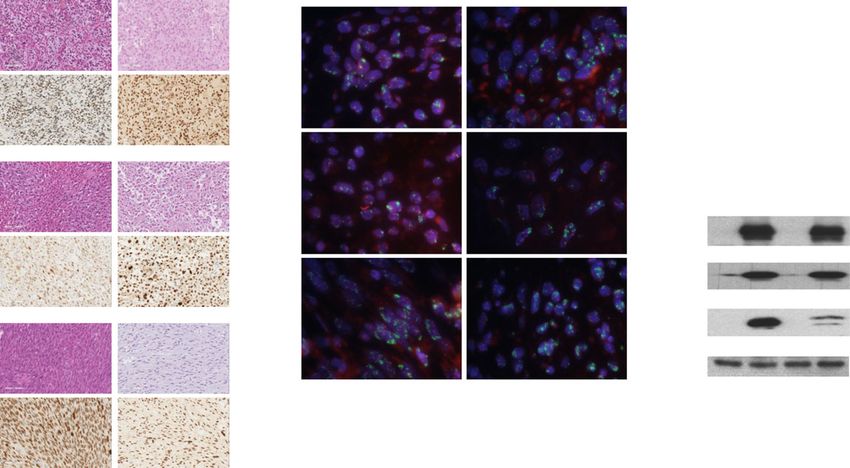
Zuco et al. Journal of Experimental & Clinical Cancer Research   (2021) 40:83                                          Page 2 of 12

 (Continued from previous page)
 seen in all selinexor-treated models. In addition, a time-dependent decrease of survivin expression, with an almost
 complete abrogation of the cytoplasmic anti-apoptotic pool of this protein, was observed as a consequence of the
 decreased acetylation/activation of STAT3 and the increased ubiquitination of nuclear survivin.
 Conclusions: Selinexor showed a moderate antitumor activity in three DDLPS PDXs, which was, however,
 consistently higher than doxorubicin across all different models regardless the extent of MDM2 amplification and
 the histological differentiation. The depletion of survivin protein seems to significantly contribute to the induction
 of apoptosis through which selinexor exerts its antitumor activity.
 Keywords: Dedifferentiated liposarcoma, PDX, Primary cell culture, XPO1, Selinexor, Doxorubicin, Survivin,

Background                                                              DDLPS, the heterogeneous clinical course of the disease,
Well differentiated (WD)/dedifferentiated (DD) liposar-                 which is also determined by different patterns of differ-
coma (LPS) is the most frequent soft-tissue-sarcoma                     entiation, and the limited availability of translatable
arising in the retroperitoneal space, accounting for                    experimental models able to properly recapitulate clin-
approximately 50 % of all retroperitoneal sarcomas [1].                 ical tumor biology and response to treatment strongly
WDLPS is a low-grade disease composed of adipocytic                     impair the development of innovative and effective
cells that may recur locally, while DDLPS is a more                     treatments.
aggressive non-lipogenic malignancy characterized by                       DDLPS harboring MDM2 amplification usually
multifocal recurrences and, less frequently, distant site               expresses wild-type p53. MDM2 ubiquitination of p53
metastasis. Among others, DDLPS may undergo myo-                        facilitates its XPO1-mediated nuclear export, thus limit-
genic and rhabdomyoblastic heterologous differentiation,                ing p53 tumor suppressor functions [10]. Consequently,
which conveys an aggressive phenotype to the tumor,                     nuclear export is a rational target in DDLPS. Indeed, the
mainly evident when rhabdomyoblastic traits are                         first-in-class XPO1 inhibitor selinexor has shown activity
acquired [2]. Recent studies suggest that WDLPS and                     in established cell line-based preclinical models of soft
DDLPS divergently evolve from a common precursor                        tissue sarcoma, including DDLPS [11–13], and a phase
harboring 12q13–q15 chromosomal sub-region amplifi-                     III randomized trial of selinexor versus placebo in
cation that results in the abnormal expression of genes                 patients with metastatic DDLPS (ClinicalTrials.gov iden-
such as MDM2, CDK4 and HMGA2, which are the                             tifier: NCT02606461) has recently completed the accrual
drivers of oncogenic transformation [3]. DDLPS has an                   [14]. A prospective study comparing selinexor and doxo-
overall more complex genomic profile than WDLPS,                        rubicin, the standard front-line therapy for metastatic
which may sustain its more aggressive phenotype, al-                    DDLPS, has not been conducted.
though specific drivers of the DDLPS distinct evolution-                   In this study, we report the development and
ary pattern are to be clarified.                                        characterization of three patient-derived xenografts
  Surgery remains the primary treatment for localized                   (PDXs), obtained from primary, treatment naïve, retro-
DDLPS, but approximately 40 % of newly diagnosed                        peritoneal DDLPS displaying myogenic and rhabdomyo-
patients will eventually die from advanced disease and,                 blastic heterologous differentiation, which have been
in particular, from local inoperable relapse. When sur-                 used to directly compare the antitumor activity of seli-
gery fails to achieve local tumor control, few effective                nexor and doxorubicin, the standard front-line therapy
therapeutic options are currently available [4]. No pro-                in sarcomas. Results are reported herein.
spective trials focusing on doxorubicin-based chemo-
therapy have been conducted in DDLPS, while                             Materials and Methods
retrospective studies on anthracycline containing regi-                 Assessment of XPO1 gene expression in DDLPS clinical
mens have shown a limited antitumor effect [5, 6]. A                    samples
higher degree of activity is seen when doxorubicin is                   Frozen tissues of the WD component, the DD compo-
combined to ifosfamide, which in the daily practice can                 nent and the normal fat from each DDLPS were col-
also be used as monotherapy with a high-dose regimen                    lected from 15 patients with a retroperitoneal, primary,
[7]. Other options in further lines are represented by tra-             untreated DDLPS. Specular formalin-fixed paraffin-
bectedin or eribulin [8]. However, in all cases the antitu-             embedded (FFPE) sections of each tumor were assessed
mor effect of these anti-cancer agents remains limited.                 for histology confirmation and quality check by a
  The poor response of DDLPS to conventional systemic                   sarcoma-dedicated pathologist. Total RNA was extracted
chemotherapy emphasizes the need for novel actionable                   from frozen tissues with an RNeasy mini kit (Qiagen,
targets and new therapies [9]. Indeed, the rarity of                    Hilden, Germany). Gene expression profiles were
Zuco et al. Journal of Experimental & Clinical Cancer Research   (2021) 40:83                                         Page 3 of 12

assessed using the GeneChip Clariom S Human Arrays                      and based on growth characteristics. The 3 models used
platform. Raw data was normalized according to the                      in the study are part of a DDLPS PDX panel, consisting
RMA algorithm of oligo Bioconductor package [15].                       of 6 models we generated from the DD component of
XPO1 differential expression was estimated using the                    the 15 DDLPS (engraftment rate: 40 %) above
non-parametric Kruskal-Wallis statistic among the three                 mentioned.
tissue types. Nemeyi post-hoc test was used to assess                     Tumor fragments of established PDX models were
significant differences (0.05 as threshold) in pair-wise                engrafted into CD1-Foxn1nu (nude) mice (Charles River)
comparisons. All analyses were performed using the                      for preclinical pharmacology studies. SCID and nude
computing microenvironment R.                                           mice were maintained in a pathogen-free facility where
   The dataset generated in the study was deposited in                  temperature and humidity were kept constant, and had
the GEO database (GSE159659).                                           free access to food and water.
                                                                          To establish DDLPS cell lines, surgical tumor samples
Development of PDXs and patient‐derived cell lines                      were cut into pieces of ~ 1 mm3 and enzymatically
Each DD component of DDLPS clinical samples suitable                    digested with collagenase (200 U/mL) (Sigma, St. Louis,
for mouse implantation (based on dimension and tumor                    MO, USA) for 3 h at 37°C. Cells were resuspended in
cellularity) was obtained from retroperitoneal primary                  DMEM supplemented with 10 % fetal bovine serum to
tumors of three patients with retroperitoneal, treatment-               inactivate collagenase (Merck) and applied to a cell
naïve, MDM2 positive, FNCLCC (Fédération Nationale                      strainer (100 µm, Corning), centrifuged at 500×g for 5
des Centres de Lutte Contre Le Cancer) grade 3 DDLPS.                   minutes and resuspended in DMEM supplemented with
Myogenic and rhabdomyoblastic differentiation were de-                  10 % fetal bovine serum. Tumor cell lines were then
fined as previously described [2]. Briefly, a DDLPS was                 propagated in DMEM F-12 medium (Lonza, Treviglio,
characterized as having a myogenic differentiation when                 Italy), supplemented with 10 % fetal bovine serum and
at least one myogenic immunohistochemistry marker                       maintained in a 37°C humidified 5 % CO2 incubator.
among actin 14A, desmin and caldesmin was detected in                     The origins of both PDXs and corresponding cell lines
> 10 % of neoplastic cells, while the presence of a posi-               were authenticated through microsatellite analysis by the
tive immunostaining for myogenin was required to con-                   AmpFISTR Identifiler PCR amplification kit (Applied
firm the presence of rhabdomyoblastic differentiation.                  Biosystems, PN4322288, Foster City, CA, USA).
   A human tumour (LS-GD-1) showed histopathological
and immunohistochemical features of rhabdomyoblastic                    Characterization of PDX models
heterologous dedifferentiation, as it contained rhabdoid                Four micrometer sections of FFPE tumor tissue,
cells and was positive for desmin, actin 1A4 and myo-                   obtained from sacrificed mice when tumor nodules were
genin staining. The other two tumours displayed fea-                    about 400 mm3, were stained with hematoxylin and
tures of myogenic dedifferentiation, being LS-BP-1                      eosin (H&E) for morphological evaluation and immuno-
positive for actin 14A and LS-BZ-1 positive for desmin.                 stained with MDM2 (anti-MDM2, Ab-1, mouse, MAb,
The use of patient material to generate PDXs and corre-                 IF2, #OP46, San Diego, CA, USA; dilution 1:40), CK
sponding cell lines from consented patients was autho-                  AE1/AE3 (clone AE1-AE3, mouse, Dako, Santa CA,
rized by Fondazione IRCCS Istituto Nazionale Tumori                     USA; dilution 1:100), EMA (clone E29, mouse, Dako;
(Milan, Italy) (INT) Ethics Committee (Project approval                 dilution 1:250), myogenin (clone F5D, mouse, Ready-to-
code: INT 139/17).                                                      use, Dako), actin (clone 1A4, mouse, Dako; dilution 1:
   For establishment of PDX models, fresh tumor speci-                  250), desmin (clone D33, mouse, Dako; dilution 1:400),
mens were collected immediately after surgical resection,               H3 trimethyl K27 (Ab H3K27me3, Clone C36B11, Cell
aseptically dissected and cut into small fragments (~ 3                 Signaling, Beverly, MA, USA; dilution 1:400) and Ki67
mm3). At least 3 fragments were grafted subcutaneously                  (Ab Ki67, Clone Mib1, #GA626 Dako; dilution 1:400)
into the right flank of 6-week old female CB17/lcr-                     antibodies for further characterization. Immunohisto-
Prkdcscid (SCID) mice (Charles River, Calco, IT). The lag               chemical (IHC) analysis was performed at room
time for tumor growth after the first inoculum ranged                   temperature on the Dako Autostainer Link 48 AS480
from 45 to 68 days for the different models. Tumor                      (Agilent, Santa Clara, CA, USA), as previously described
growth was followed by biweekly measurement of tumor                    [12]. Ki67 labeling index (number of Ki67-positive nu-
diameters with a Vernier calliper. Tumor volume (TV)                    clei/overall number of nuclei x 100) was quantified using
was calculated according to the formula: TV (mm3) = d2                  ImageJ 1.47q software (). FISH assay was carried out to
× D/2, where d and D are the shortest and the longest                   assess MDM2 gene amplification in the DDLPS clinical
diameters, respectively. Tumors were transplanted into                  tumors and corresponding PDX models using ZytoLight
other mice when reached a TV of 600 mm3. A PDX was                      SPEC MDM2/CEN 12 Dual Color Probe (ZytoVision
considered established after the third passage in mice                  GmbH. Bremerhaven Germany).
Zuco et al. Journal of Experimental & Clinical Cancer Research (2021) 40:83 Page 4 of 12
MDM2, CDK4, HMGA2 copy number variation was of adherent cells in treated samples compared with con-
assessed by droplet digital PCR (ddPCR) on genomic trol samples. The in vitro drug activity was assessed in
DNA extracted from PDX tissue sections using Maxwell® terms of concentrations able to inhibit cell growth by
CSC FFPE DNA AS1350. Twenty µl ddPCR reaction 50 % (IC50).
mixture was loaded into the Bio-Rad DG8 droplet gener-
ator cartridge that was then placed into the QX200 Western blotting
droplet generator. The generated droplets were manually Lysates were obtained from frozen PDX tumors
transferred to an Eppendorf 96-well PCR plate that was collected immediately after (and, when indicated, after
then heat-sealed with a pierceable foil in the PX1 PCR 12 days from) the end of mouse treatment with selinexor
Plate Sealer, and then amplified on the ProFlex PCR sys- or doxorubicin as well as from DDLPS cell lines at dif-
tem. The thermal-cycling conditions were: 95°C for 10 ferent intervals after exposure to different concentra-
min, 40 cycles of 94°C for 30 s, 60°C for 1 min, 98°C for tions of each drug. Nuclear and cytosolic fractions were
10 min and a final step at 4°C. After amplification, the obtained from DDLPS cells using NE-PER™ nuclear and
96-well PCR plate was loaded on QX200 droplet reader cytoplasmic extraction reagents (#78,835; Thermo Fisher
and ddPCR data were analyzed with QuantaSoft analysis Scientific) following the manufacturer’s protocol.
software version 1.7.4. For the assessment of the ubiquitinated form of survi-
vin, the nuclear fraction of the protein was immunopre-
Evaluation of drug activity cipitated with the anti-human survivin antibody (#ab469;
Nude mice were randomized to receive either selinexor Abcam) and eluted as described [17]. Total/fractioned
or doxorubicin which were administered when xeno- cellular lysates and immunoprecipitates were separated
grafts were approximately 150 mm3. Eight mice for each by SDS-PAGE, transferred onto nitrocellulose mem-
experimental group were used. The treatment dose and branes and incubated with primary monoclonal anti-
schedule for each drug were selected from the literature bodies: anti-MDM2 (#86,934, Cell Signaling
(Table 1). After dilution in sterile water, doxorubicin Technology), anti-HMGA2 (#5269, Cell Signaling Tech-
(Adriblastina, Pfizer Italia) was administered intraven- nology), anti-cleaved CCP32 (#9661, Cell Signaling
ously (i.v.) every 7 days for three times (q7d x 3). Seli- Technology), anti-STAT3 (#4904; Cell Signaling Tech-
nexor (Selleck Chemicals, USA) was prepared as nology), anti-acetyl-STAT3 (#2523; Cell Signaling Tech-
previously described [16] and delivered orally (p.o.) twice nology), anti-HDAC2 (#5113; Cell Signaling
a week for 8 times (q3-4d x 8). Drug treatment activity Technology), anti-LC3B (#2775 Cell Signaling), anti-
was assessed in terms of TV inhibition percentage survivin (#ab469; Abcam), anti-XPO1 (#ab24189;
(TVI%) in treated versus control mice and expressed as Abcam), anti-LC3B (#2775 Cell Signaling Technology),
TVI% = 100-[(mean TV treated/mean TV control) x anti-ubiquitin (#ab7780, Abcam), anti-α-tubulin (T5168,
100]. Treatment toxicity was determined in terms of Merck), anti-p53 (sc-126, Santa Cruz Biotechnology, CA,
body weight loss and lethal toxicity. USA), anti-CDK4 (sc-601, Santa Cruz Biotechnology),
anti-p16 (sc-6579, Santa Cruz Biotechnology) and anti-
In vivo drug-based experiments were authorized by β-actin (A2066, Merck). Band intensities were quantified
the Italian Ministry of Health according to the national by scanning films and processing image intensities with
law (Project approval code: 234/2018-PR) and in compli- the ImageJ 1.47v Software.
ance with international policies and guidelines.
The growth inhibitory effect of selinexor and doxo- Apoptosis analysis
rubicin, singly administered at concentrations ranging Apoptosis was assessed in tumors excised from
from 1.0 nM to 1.0 µM for 72 h, was assessed by cell untreated and drug-treated mice using the SignalStain®
counting using a particle counter (Beckman Coulter, Apoptosis IHC Detection Kit (Cell Signaling), which
Luton, UK). The results were expressed as the number detects the presence of cleaved caspase-3, according to
Table 1 Pharmacological treatments and tumor responses of DDLPD PDX models
Treatment Route Schedule Dose (mg/kg) Max TVI % (day) a Tumor growth delay (days)b
LS-GD-1 LS-BZ-1 LS-BP-1 LS-GD-1 LS-BZ-1 LS-BP-1
Doxorubicin i.v. q7d×3 4 60 (49)* 40 (17)* 37 (46)* 20 5 16
Selinexor p.o. q3/4d×8 10 80 (49)** 55 (14)* 46 (55)* > 46 7 23
a
Maximum tumor volume inhibition (TVI) % in treated versus control mice. In parentheses, the day on which it was assessed. *P < 0.05, **P < 0.005 versus
control tumors
b
The tumor growth delay is calculated as the median time (days) in excess required for the tumor of the drug treated groups mice to reach 1000 mm3 compared
to the diluents treated miceZuco et al. Journal of Experimental & Clinical Cancer Research (2021) 40:83 Page 5 of 12
the manufacturer’s instructions. Sections were counter- PDX and cell model characterization
stained with hematoxylin. Number of caspase-3 positive Patient-derived tumor samples used for the generation
cells/overall number of nuclei x 100 was quantified using of PDXs were consistent with the histopathological diag-
ImageJ 1.47v Software. nosis of DDLPS (Fig. 2A). Two human tumors showed
Floating and adherent cells were harvested 72 h after myogenic dedifferentiation (LS-BZ-1 and LS-BP-1) and a
treatment and processed for apoptosis evaluation with In tumor showed rhabdomyoblastic dedifferentiation (LS-
Situ Cell Death Detection Kit, Fluorescein (#11,684,795, GD-1). The histomorphology of PDX models was
910, Merck), according to manufacturer’s instructions, consistent with the paired human samples (Fig. 2A).
and analyzed by BD™ Accuri C6 (Becton Dickinson, Immunophenotypic profile of models differed from
Mountain View, CA, USA). A specific software (Cell- the paired clinical tumours as LS-BP-1 and LS-BZ-1
QuestPro, Becton Dickinson) was used to estimate the PDXs lost expression of the myogenic markers Actin
percentage of apoptotic cells. 14A and desmin, respectively. Detailed morphological
and immunohistochemical characterization of PDXs is
provided in Table 2.
Statistical analysis
Analyses by two-sided Student’s t-test were performed All clinical samples and corresponding PDXs were
using the GraphPad Prism software, version 4.0 (Graph- marked by MDM2 gene amplification, as detected by
Pad Prism Inc., San Diego, CA, USA). A P value of ≤ IHC (Fig. 2A) and by FISH (Fig. 2B), and over-expressed
0.05 was considered statistically significant. MDM2 protein compared to normal cells (Fig. 2 C).
Quantitative analysis of gene copy number by ddPCR
showed a copy number gain for MDM2, CDK4 and
Results HMGA2 genes in all PDX models, although to a variable
XPO1 is over-expressed in clinical DDLPS samples extent (Fig. 2 C). Consistently, the two patient-derived
Gene expression profiles comparatively assessed on cell lines were characterized by an enhanced expression
paired samples –including DD component, WD compo- of MDM2, CDK4 and HMGA2 proteins compared to
nent and normal adipose tissue– obtained from 15 retro- the corresponding normal adipose tissues, as detected by
peritoneal primary tumors of treatment-naïve DDLPS western blotting (Fig. 2D).
patients showed a significantly increased expression of
XPO1 in DD and WD compared to healthy tissue (Fig. 1). Selinexor is more active that doxorubicin and induces
These data confirmed a previous observation indicating apoptotic response in all PDX and cell models
a significantly higher XPO-1 expression in DDLPS com- In DDLPS animal models, selinexor and doxorubicin as
pared to lipoma samples [13]. single agents induced a tumor growth delay which was
more pronounced for the XPO1 inhibitor than the
This finding, together with MDM2 amplification [3], anthracycline in all PDXs (maximum TVI%: 46–80 % vs.
strongly supports XPO1-mediated nuclear export as a 37–60 %) (Fig. 3a; Table 1). The difference between seli-
rational target in DDLPS. nexor and doxorubicin antitumor activity was particu-
larly evident in the LS-GD-1 model, as also indicated by
the significantly increased time for selinexor-treated ani-
mals to reach 1000 mm3 tumor burden compared to
doxorubicin-treated and untreated animals (Table 1).
This PDX model, which is characterized by rhabdo-
myoblastic dedifferentiation, was the most sensitive to
both drugs.
The extent of selinexor and doxorubicin activity did
not seem to be influenced by the level of MDM2 and
CDK4 gene amplification. Interestingly, the most chemo-
sensitive PXD model displayed the lowest extent of
HMGA2 amplification (Table 1; Fig. 2 c). No sign of
toxicity, in terms of tumor weight loss and occurrence of
Fig. 1 XPO1 expression in paired samples, including normal adipose toxic death, was registered with either drug.
tissue (A), WD component (WD) and DD component (DD), obtained In vitro cytotoxicity experiments carried out on DDLP
from the retroperitoneal primary tumors of 15 treatment-naïve DDLP
S cell models showed that doxorubicin was active at
S patients. *P < 0.05, **P < 0.005
lower concentrations than selinexor. The two cellZuco et al. Journal of Experimental & Clinical Cancer Research (2021) 40:83 Page 6 of 12
Fig. 2 Characterization of patient-derived DDLPS models. a, Representative pictures of PDX models and corresponding clinical tumors. The
histology was assessed on hematoxylin and eosin (H&E)-stained slides. MDM2 amplification was detected at the protein level by MDM2
immunostaing. b, FISH analysis: Spectrum Orange labeled Chromosome 12 centromere and Spectrum Green labeled MDM2. High level of MDM2
amplification with clustering of gene copies are observed in both patient tissues and PDXs. c, Quantification of MDM2, CDK4 and HMGA2 copy
number variation in PDXs by ddPCR. d, Assessment of MDM2, CDK4 and HMGA2 protein expression in cell lines by western blotting. A
representative blot of three independent experiments is shown. For each protein, band intensity was quantified using Image J normalized to
loading control reported below
models exhibited a comparable sensitivity to the anthra- appreciably affect cell proliferation of any model
cycline (IC50 values: 45 ± 16 nM and 46 ± 9 nM for LS- (Fig. 3 c).
GD-1 and LS-BZ-1, respectively). Interestingly, consist- Western blot analysis, which was performed at the end
ent with in vivo data, LS-GD-1 cells displayed a higher of treatment with each drug and also after 12 days for
susceptibility to selinexor than LS-BZ-1 cells (IC50 selinexor, revealed that the XPO1 inhibitor –but not
values: 116 ± 54 nM and 187 ± 59 nM, respectively). doxorubicin– reduced the expression of the anti-
Histopathological evaluation of drug-treated tumors apoptotic protein survivin and that, in 2 out of 3 PDX
excised from mice immediately after the end of treat- models, such a decrease was still appreciable 12 days
ment with selinexor or doxorubicin showed no marked after the end of treatment (Fig. 4a). Selinexor also
differences at the morphological level, but only a slight induced an increased abundance of p53 protein, mainly
and focal reduction in cellularity with an increased loose appreciable at the end of treatment in two PDX models
stroma in post-selinexor PDXs (Fig. 3b). The prolifera- and after 12 days in the third model (Fig. 4a). In LS-BZ-
tion rate, as detected by Ki67 index, was significantly 1 and LS-BP-1 models, the over-expression of p53 was
lower in selinexor-treated compared to untreated tumors paralleled by an enhanced accumulation of p21/
in all PDXs. Conversely, doxorubicin exposure did not CDKN1A protein (Fig. 4a).
Table 2 Histomorphology and immunophenotypic profile of DDLPS PDX models
LS-BZ-1 LS-GD-1 LS-BP-1
Human DDLPS, G3, myogenic DDLPS, G3, rhabdmyoblastic dedifferentiation DDLPS, G3, myogenic
tumor dedifferentiation dedifferentiation
H&E Spindle cells and epitheliod cells Spindle cells organized in fascicles, and rhabdoid cells Spindle cells with small nuclei
CK1 Negative Negative Negative
CK3 Negative Negative Negative
EMA Negative Negative Negative
Myogenin Negative Positive, diffuse Negative
Actin 1A4 Negative Positive, focal Negative
Desmin Negative Positive, diffuse Negative
H3K27me3 Positive Positive Positive
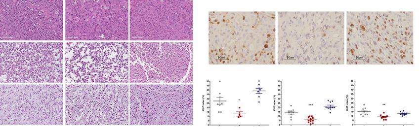
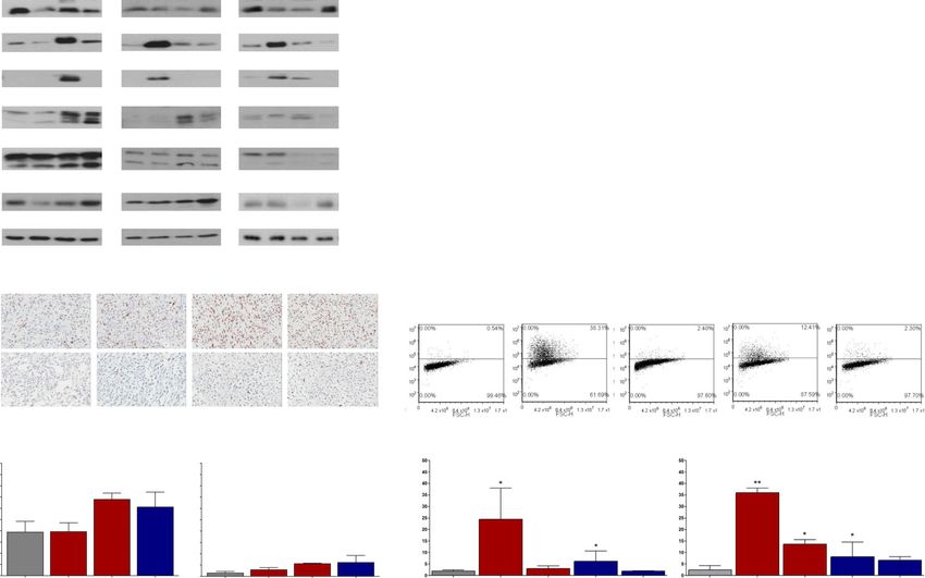
MDM2 Positive Positive PositiveZuco et al. Journal of Experimental & Clinical Cancer Research (2021) 40:83 Page 7 of 12 Fig. 3 Antitumor activity of selinexor and doxorubicin in DDLPS PDXs. a, Growth curves report the average tumor volume (±S.E.M.) in control and drug-treated animal groups (8 mice/group). The arrows in the figure indicate when drugs were administered. b, Pathologic evaluation of tumors obtained from untreated (Ctrl) and selinexor- or doxorubicin-treated mice at the end of treatment. c, Ki67 immunostaining of tumors obtained from untreated (Ctrl) and selinexor- or doxorubicin-treated mice at the end of treatments (upper panel) and quantification of Ki67 index (lower panel). Two tumors for each experimental group were analyzed. The symbols reported in the lower panel represent counted fields. *P < 0.05, ** P < 0.01, ***P < 0.005 The induction of apoptosis, assessed in terms of Selinexor induces survivin down‐regulation by inhibiting cleaved caspase-3 expression by western blotting, was STAT3 acetylation and promoting nuclear survivin observed in all selinexor-treated PDXs, although to a ubiquitination very different extent, and was mainly appreciable 12 The effect of drug treatment was assessed in more days after treatment. Caspase-3 cleavage was also de- details in the two patient-derived DDLPS cell lines. tected at the end of doxorubicin treatment, though in Western blot analysis carried out on the whole protein LS-GD-1 and LS-BZ-1 models (Fig. 4a). Consistent extract showed that, consistently with in vivo findings, a results were obtained when the presence of cleaved 24 h exposure to 1 µM selinexor caused an enhanced caspase-3 was detected by immunohistochemistry accumulation of p53 protein in both cell models (Fig. 4b). (Fig. 5a, b). Interestingly, drug treatment also induced In DDLPS cell models, flow cytometric detection of the expression –mainly appreciable in the LS-GD-1 TUNEL-positive cells indicated a dose-dependent induc- model (Fig. 5A,B)– of MDM2-p60, a MDM2 fragment tion of apoptosis by both drugs, although the extent of generated by caspase-3-mediated cleavage, which loses the apoptotic response was consistently greater for seli- the ring domain and, consequently, cannot target p53 nexor than doxorubicin (Fig. 4b). to proteasome degradation [18]. Moreover, an almost A slightly increased abundance of the autophagy complete abrogation of XPO1 and survivin expression marker LC3B-II was only observed 12 days or immedi- was observed in both cell models following selinexor ately after treatment with selinexor and doxorubicin, exposure (Fig. 5a, b). respectively, in the LS-GD-1 PDX, while a slightly The assessment of protein sub-cellular localization indi- enhanced expression of the senescence-associated cated that the increased expression of p53 in selinexor- marker p16 was appreciable after treatment with doxo- treated cells was mainly ascribable to their enhanced rubicin in all models (Fig. 4a). nuclear accumulation (Fig. 5a, b). As regards survivin,
Zuco et al. Journal of Experimental & Clinical Cancer Research      (2021) 40:83                                                         Page 8 of 12

 Fig. 4 Induction of apoptosis by selinexor and doxorubicin in DDLPS models. a, Western blot analysis of survivin, p53, p21, and cleaved caspase 3
 on tumors obtained from untreated (Ctrl) and selinexor- or doxorubicin-treated mice at different intervals from the end of treatment. The
 expression of autophagy (LC3B) and senescence (p16) markers was also assessed. A representative blot of three independent experiments is
 shown. For each protein, band intensity was quantified using Image J normalized to loading control reported below and referred to respective
 untreated control. The band intensity of LC3B-I and LC3B-II were quantified separately (above and below, respectively). b, Cleaved caspase-3
 immunostaining of tumors obtained from untreated (Ctrl) and selinexor- or doxorubicin-treated mice at the end of treatments (upper panel) and
 quantification of the percentage of cleaved caspase-3 positive cells (lower panel). c, Flow cytometric assessment of TUNEL-positive cells after 72 h
 exposure to equimolar concentration of drugs in DDPLS cells (upper panel) and quantification of TUNEL-positive cells (lower panel). Results
 represent the mean values ± SD of 3 independent experiments. *P < 0.05, **P < 0.01, ***P < 0.005

whose sub-cellular localization is known to determine                        activator also in DDLPS cells. Since we and others previ-
protein function [19], selinexor induced an early nuclear                    ously showed that the forced retention of survivin in the
accumulation appreciable at 4 h, which was followed by a                     nucleus promotes its clearance by the ubiquitin-
progressive decline of the protein in the nucleus and par-                   proteasome proteolytic pathway [21, 22], we checked
alleled by a time-dependent reduction of cytoplasmic pro-                    whether selinexor might cause the ubiquitination of sur-
tein levels in both cell models (Fig. 5a, b).                                vivin nuclear fraction. Western blot results on nuclear
  Based on a previous report indicating that in triple-                      survivin immunoprecipitates indicated that exposure of
negative breast cancer cells the inhibition of XPO1 by                       LS-BZ-1 cells to selinexor caused protein ubiquitination
selinexor was able to repress BIRC5/survivin gene tran-                      (Fig. 5 c). This finding suggests that proteasome-
scription by inhibiting STAT3 acetylation and blocking                       dependent degradation of survivin might concur to the
STAT3 binding to the survivin promoter [20], we                              overall reduction of protein expression induced by seli-
assessed STAT3 protein expression and acetylation in                         nexor also in DDLPS cells.
LS-BZ-1 cells. Western blot showed a time-dependent
decrease of acetyl-STAT3 levels (Fig. 5 c), suggesting                       Discussion
that survivin inhibition is related, at least in part, to                    In this preclinical study, we directly compared the activ-
post-translational modifications of its transcriptional                      ity of the XPO1 inhibitor selinexor and doxorubicin, the
Zuco et al. Journal of Experimental & Clinical Cancer Research         (2021) 40:83                                                           Page 9 of 12

 Fig. 5 Effects of selinexor on the expression and subcellular localization of survivin. Western blot analysis of survivin, MDM2, p53 and XPO1 in
 whole protein extracts (left panel) and fractionated protein extracts (right panel) of LS-GD-1 cells a, and LS-BZ-1 cells b, at different intervals of
 exposure to selinexor. c, Western blot analysis of STAT3 and acetyl-STAT3 expression on whole protein extracts (left panel) and survivin
 ubiquitination in protein nuclear fraction (right panel) of LS-BZ-1 cells at different intervals of exposure to selinexor. Representative blots of three
 independent experiments are shown. For each protein, band intensity was quantified using Image J normalized to loading control reported
 below and referred to respective untreated control

standard front-line medical therapy in sarcomas, on in-                         we found that, although our DDLPS PDXs maintained
house developed DDLPS PDXs and corresponding cell                               the histological features as well as the amplification of
lines. The results showed a moderate antitumor activity                         driver genes, which characterize the originating tumors,
of selinexor, which was, however, consistently higher                           two of them do not express some of the immunopheno-
than doxorubicin in the different PDXs, irrespective of                         typic features of the corresponding clinical tumors. This
the extent of MDM2 amplification and the heterologous                           finding could possibly reflect the sampling of DDLPS
differentiation subtype, although the most robust evi-                          which are characterized by tumor heterogeneity, as indi-
dence of a superior activity of the XPO1 was observed in                        cated by differences in histomorphology, which may also
the PDX model showing rhabdomyoblastic differenti-                              reflect in genetic heterogeneity [24].
ation. The depletion of anti-apoptotic survivin protein,                          PDXs are more reliable than cell line-derived xeno-
consistently observed in in vivo and in vitro models,                           grafts in predicting drug response [23]. Based on this
seemed to significantly contribute to the induction of                          premise, they are particularly useful for directly compar-
apoptosis through which selinexor exerts its antitumor                          ing standard-of-care agents and newer therapies in rare
activity.                                                                       tumors, especially when prospective comparative clinical
   PDXs are robust preclinical models which retain the                          trials are lacking or challenges related to disease rarity
main characteristics of clinical tumors [23]. In this study,                    question their feasibility. In this context, we already
Zuco et al. Journal of Experimental & Clinical Cancer Research   (2021) 40:83                                             Page 10 of 12

reported the consistency between preclinical data ob-                      XPO1 is the sole nuclear exporter for some of the
tained on PDXs of ultra-rare sarcomas, such as solitary                 major tumor suppressors (i.e., p53), cell cycle regulators
fibrous tumor and epithelioid sarcoma, and clinical                     (i.e., p21/CDKN1A) and growth promoting/anti-apop-
results concerning the activity of different cytotoxic,                 totic proteins (i.e., survivin). The superior activity of seli-
anti-angiogenic and epigenetic agents [25–28]. The                      nexor compared to doxorubin observed in this study
preclinical data showing effectiveness of the targeted                  seems to be sustained by a greater ability to induce an
drugs axitinib and pazopanib in our PDX models of                       apoptotic response in both PDXs and cell lines. Consist-
solitary fibrous tumors were also instrumental to                       ently with previous reports [22, 40], apoptosis was found
design new successfully conducted prospective phase                     to be dependent on nuclear accumulation of p53. In
II clinical trials [29, 30].                                            addition, selinexor caused marked down-regulation of
   Previous studies on DDLPS established cell models                    survivin, with an almost complete abrogation of the pro-
showed the antiproliferative effect of selinexor and, in                tein cytoplasmic pool, which is known to be responsible
some instances, in vitro data were corroborated by                      for survivin anti-apoptotic function [19]. Survivin down-
in vivo evidence of antitumor activity of this drug in                  regulation was already reported by us and others in ex-
xenografts [11, 13]. This is the first report showing the               perimental models of different human tumor types ex-
effect of the XPO1 inhibitor on DDLPS patient-derived                   posed to selinexor [22, 41, 42]and also observed in a
models, which also take into account the heterologous                   DDLPS cell line [11]. Survivin plays a vital role in onco-
differentiation of PDXs as well as the extent of MDM2,                  genesis being involved in both cell cycle control and re-
CDK4 and HMGA2 gene amplification. Differently from                     sistance to apoptosis [43]. As regards liposarcomas, it
a reported study indicating that doxorubicin-treated                    has been shown that survivin is over-expressed in pleo-
DDLPS cells demonstrated variable in vitro sensitivity                  morphic liposarcoma clinical specimens [44] and that
based on baseline MDM2 expression levels [31], we                       the protein is essential for the growth of myxoid liposar-
found that PDX response to selinexor and doxorubicin                    coma cell lines [45]. Although no data are currently
was not affect by the extent of MDM2 and CDK4 gene                      available on DDLPS, results from our study suggest an
amplification. Interestingly, the most chemosensitive                   important role for the protein also in this liposarcoma
PDX model displayed the lowest extent of HMGA2 amp-                     subtype.
lification. A higher susceptibility to selinexor, but not                  Concerning the possible mechanisms responsible for
doxorubicin, was also observed in the LS-GD-1 cell line                 survivin down-regulation after exposure to selinexor, it
that shows the lower HMGA2 protein expression.                          was previously shown that, in triple negative breast can-
   Recent studies reported the involvement of HMGA2 in                  cer cells, XPO1 inhibition represses survivin/BIRC5 tran-
the chemoresistant phenotype of human tumor cells                       scription by inhibiting STAT3 acetylation and blocking
through different and not completely understood mech-                   STAT3 binding to the gene promoter [20]. According to
anisms. Specifically, HGMA2 was regarded as a possible                  previous evidence that forced retention of survivin in the
determinant of resistance to doxorubicin in liver cancer                nucleus promotes its clearance by the ubiquitin-
cells [32], to 5-fluorouracil in colorectal cancer cells                proteasome pathways [21], we reported that selinexor in-
[33], to docetaxel in gastric cancer cells [34] as well as to           duced the ubiquitination of the protein nuclear fraction
cisplatin in ovarian cancer cells [35]. In this context, we             in diffuse malignant peritoneal mesothelioma cells [22].
recently reported that the induction of HMGA2 expres-                   Interestingly, in the present study we provide evidence
sion sustained the activation of a cytoprotective autoph-               that both mechanisms concur to the down-regulation of
agic response in PDX and cell line models of epithelial                 survivin following selinexor treatment in DDLPS cells.
sarcoma after treatment with the EZH2 inhibitor EPZ-                       Although superior to doxorubicin, the antitumor activ-
011989 [25]. However, in the present study we only                      ity of selinexor appears moderate in our PDXs. A limita-
observed a modest increase in the expression of                         tion of our models is related to their histological
autophagy-related markers LC3B-II in one PDX models                     characteristics that cover only the myogenic and rhabdo-
after treatment with selinexor or doxorubicin.                          myoblastic heterologous differentiation of DDLPS, which
   Consistent with previous evidence, it can be hypothe-                can present also other dedifferentiation subtypes, such
sized that HMGA2 counteracts the effects of selinexor                   as pleomorphic and epithelioid differentiation.
and doxorubicin in our DDLPS models by up-regulating                    Additional models of DDLPS expressing other differenti-
AKT [36] and WNT [37] pro-survival pathways. It is                      ation lineages are currently under development in our
also plausible that HMGA2 protects DDLP PDXs against                    lab to investigate if the activity of several anti-cancer
the antitumor activity of anthracycline by promoting                    drugs, including doxorubicin and selinexor, can differ
DNA double-strand break repair as a consequence of its                  across heterologous dedifferentiation subtype.
documented ability to activate DNA damage response                         On the other side, the good tolerability of selinexor
kinases ATM [38] and ATR [39].                                          supports a try with combination regimens aimed at
Zuco et al. Journal of Experimental & Clinical Cancer Research                    (2021) 40:83                                                           Page 11 of 12

increasing its therapeutic effect. On this basis we started                              Ethics approval and consent to participate
to explore the antitumor effect of selinexor combined                                     The study was authorized by the Ethics Committee of the Fondazione IRCCS
                                                                                         Istituto Nazionale Tumori, Milan, Italy (Project approval code: INT 131/16). All
with doxorubicin, a regimen that is currently under in-                                  patients provided written informed consent. Animal experiments were
vestigation in a prospective phase 1b clinical study of ad-                              authorized by the Italian Ministry of Health according to the national law
vanced soft-tissue sarcomas (ClinicalTrials.gov Identifier:                              (Project approval code: 234/2018-PR).

NCT03042819). Among the 24 evaluable patients with
                                                                                         Consent for publication
different sarcoma types (only 2 LPS were included) who                                   Not applicable.
entered the trial, 5 (21 %) had a partial response by RECI
ST as best response [46]. In a preliminary experiment on                                 Competing interests
                                                                                         R. S. received institutional research funding from Karyopharm. M.G. had
the LS-GD-1 PDX model, we found that concomitant                                         research grants and personal fees from Karyopharm. P.G.C. received
exposure with doxorubicin did not increase the antitu-                                   institutional research funding from Karyopharm. S.S. had advisory role in
mor activity of the XPO1 inhibitor (Max TVI: 82 % vs.                                    Karyopharm and received institutional research funding from Karyopharm. All
                                                                                         the remaining authors have no conflicts of interest to declare. The funders
80 %). Of course, we cannot exclude that different treat-                                had no role in the design of the study; in the collection, analyses, or
ment schedules could improve the result. It is also con-                                 interpretation of data; in the writing of the manuscript, or in the decision to
ceivable that combination regimens including drugs able                                  publish the results.
to target specific XPO1 cargo proteins could result in a                                 Author details
better therapeutic performance.                                                          1
                                                                                          Molecular Pharmacology Unit, Department of Applied Research and
                                                                                         Technological Development, Fondazione IRCCS Istituto Nazionale Tumori, Via
                                                                                         Amadeo 42, 20133 Milan, Italy. 2Sarcoma Service, Department of Surgery,
Conclusions                                                                              Fondazione IRCCS Istituto Nazionale Tumori, via Venezian 1, 20133 Milan,
Overall, results from this study indicate that, although                                 Italy. 3Department of Pathology, Fondazione IRCCS Istituto Nazionale Tumori,
moderate, the antitumor activity of selinexor is consist-                                via Venezian 1, 20133 Milan, Italy. 4Adult Mesenchymal Tumor and Rare
                                                                                         Cancer Unit, Department of Cancer Medicine, Fondazione IRCCS Istituto
ently higher than doxorubicin, irrespective of the extent                                Nazionale Tumori, via Venezian 1, 20133 Milan, Italy. 5Molecular Pathology
of MDM2 amplification and the heterologous differenti-                                   Unit, Ospedale Niguarda Ca’ Grande, Milan, Italy. 6Sarcoma Medical Oncology
ation of the PDX models. Mechanistically, the nuclear                                    and Early Drug Development, Memorial Sloan Kettering Cancer Center, 1275
                                                                                         York Avenue, 10065 New York, NY, USA. 7Department of Medicine, University
accumulation of p53 and the depletion of survivin                                        of Padua School of Medicine, Via Giustiniani 2, 35128 Padua, Italy.
                                                                                         8
mainly contribute to the induction of apoptosis through                                   Department of Biomedical and Clinical Sciences L. Sacco, University of
which selinexor exerts its antitumor activity. Moreover,                                 Milan, Via Grassi 74, 20157 Milan, Italy.

the good tolerability of selinexor supports a try with                                   Received: 23 November 2020 Accepted: 17 February 2021
combination regimens aimed at increasing its thera-
peutic effect.
                                                                                         References
Abbreviations                                                                            1. Jo VY, Fletcher CDM. WHO classification of soft tissue tumours: An update
DD: Dedifferentiated; DDLPS: Dedifferentiated liposarcoma; ddPCR: Droplet                    based on the 2013 (4th) edition. Pathology. 2014;46:95–104.
digital PCR; FFPE: formalin-fixed paraffin-embedded; FNCLCC: Fédération                  2. Gronchi A, Collini P, Miceli R, Valeri B, Renne SL, Dagrada G, et al. Myogenic
Nationale des Centres de Lutte Contre Le Cancer; FISH: Fluorescence in situ                  differentiation and histologic grading are major prognostic determinants in
hybridization; LPS: Liposarcoma; PDX: Patient- erived xenograft; TV: Tumor                   retroperitoneal liposarcoma. Am J Surg Pathol. 2015;39:383–93.
volume; TVI: Tumor volume inhibition; WD: Well differentiated; WDLPS: Well               3. Amin-Mansour A, George S, Sioletic S, Carter SL, Rosenberg M, Taylor-
differentiated liposarcoma                                                                   Weiner A, et al. Genomic evolutionary patterns of leiomyosarcoma and
                                                                                             liposarcoma. Clin Cancer Res. 2019;25:5135–42.
                                                                                         4. Van Houdt WJ, Zaidi S, Messiou C, Thway K, Strauss DC, Jones RL. Treatment
Acknowledgements
                                                                                             of retroperitoneal sarcoma: Current standards and new developments. Curr
The authors thank Dr. Loris De Cecco (Platform of Integrated Biology, INT) for
                                                                                             Opin Oncol. 2017;29:260–7.
microarray experiments.
                                                                                         5. Italiano A, Toulmonde M, Cioffi A, Penel N, Isambert N, Bompas E, et al.
                                                                                             Advanced well-differentiated/dedifferentiated liposarcomas: Role of
Authors’ contributions                                                                       chemotherapy and survival. Ann Oncol. 2012;23:1601–7.
Conceptualization, S.S. and N.Z.; methodology, V.Z., S.P., M.T., S.B., G.D., R.S.,       6. Jones RL, Fisher C, Al-Muderis O, Judson IR. Differential sensitivity of
C.L. M.B. and R.E.B.; formal analysis, V.Z., S.P., S.S. and N.Z.; data curation, V.Z.,       liposarcoma subtypes to chemotherapy. Eur J Cancer. 2005;41:2853–60.
S.P., S.Pe, S.S. and N.Z.; writing—original draft preparation, V.Z., S.P., C.C., M.G.,   7. Stacchiotti S, Van der Graaf W, Doms H, Sanfilippo R, Marreaud SI, Van
A.P.D.T., P.G.C., A.G., S.S. and N.Z.; supervision, S.S. and N.Z.; funding                   Houdt W, et al. First-line chemotherapy in advanced well-differentiated/
acquisition, A.G. and N.Z. The authors read and approved the final                           dedifferentiated liposarcoma (WD/DD LPS): an EORTC Soft Tissue and Bone
manuscript.                                                                                  Sarcoma Group (STBSG) retrospective analysis. ESMO 2020/ Ann Oncol 2020.
                                                                                         8. Gahvari Z, Parkes A. Dedifferentiated Liposarcoma: Systemic Therapy
Funding                                                                                      Options. Curr Treat Options Oncol. 2020;21:15.
The study was supported by a grant from the Italian Ministry of Health (5 ×              9. Hoffman A, Lazar AJ, Pollock RE, Lev D. New frontiers in the treatment of
1000 Funds-2014), by the Accelerator Award from Associazione Italiana per la                 liposarcoma, a therapeutically resistant malignant cohort. Drug Resist Updat.
Ricerca sul Cancro (AIRC) (#24297) /Cancer Research United Kingdom                           2011;14:52–66.
(CRUK)/Fundacion Científica – Asociacion Espanola Contra el Cancer (FC                   10. Nie L, Sasaki M, Maki CG. Regulation of p53 nuclear export through
-AECC) and by AIRC Investigator Grant (#24715).                                              sequential changes in conformation and ubiquitination. J Biol Chem. 2007;
                                                                                             282:14616–25.
Availability of data and materials                                                       11. Nair JS, Musi E, Schwartz GK. Selinexor (KPT-330) induces tumor suppression
The dataset and materials using during the current study are available from                  through nuclear sequestration of IκB and downregulation of survivin. Clin
the corresponding author on reasonable request.                                              Cancer Res. 2017;23:4301–11.
Zuco et al. Journal of Experimental & Clinical Cancer Research (2021) 40:83 Page 12 of 12
12. Nakayama R, Zhang YX, Czaplinski JT, Anatone AJ, Sicinska ET, Fletcher JA, 32. Han S, Han B, Li Z, Sun D. Downregulation of long noncoding RNA CRNDE
et al. Preclinical activity of selinexor, an inhibitor of XPO1, in sarcoma. suppresses drug resistance of liver cancer cells by increasing microRNA-33a
Oncotarget. 2016;7:16581–92. expression and decreasing HMGA2 expression. Cell Cycle. 2019;18:2524–37.
13. Garg M, Kanojia D, Mayakonda A, Said JW, Doan NB, Chien W, et al. 33. Xu X, Wang Y, Deng H, Liu C, Wu J, Lai M. HMGA2 enhances 5-fluorouracil
Molecular mechanism and therapeutic implications of selinexor (KPT-330) in chemoresistance in colorectal cancer via the Dvl2/Wnt pathway.
liposarcoma. Oncotarget. 2017;8:7521–32. Oncotarget. 2018;9:9963–74.
14. Gounder M, Razak AA, Somaiah N, Chawla S, Martin-Broto J, Grignani G, 34. Shekari N, Asghari F, Haghnavaz N, Shanehbandi D, Khaze V, Baradaran B,
et al. A Phase 2/3, Randomized, Double-Blind, Cross-Over Study of Selinexor et al. Let-7a Could Serve as A Biomarker for Chemo-Responsiveness to
Versus Placebo in Advanced Unresectable Dedifferentiated Liposarcoma Docetaxel in Gastric Cancer. Anticancer Agents Med Chem. 2018;19:304–9.
(DDLS). Connective Tissue Oncology Society (CTOS) annual meeting 2020. 35. Miao JT, Gao JH, Chen YQ, Chen H, Meng HY, Lou G. LncRNA ANRIL affects
15. Carvalho BS, Irizarry RA. A framework for oligonucleotide microarray the sensitivity of ovarian cancer to cisplatin via regulation of let-7a/HMGA2
preprocessing. Bioinformatics. 2010;26:2363–7. axis. Biosci Rep. 2019;39.
16. Salas Fragomeni RA, Chung HW, Landesman Y, Senapedis W, Saint-Martin 36. Tan L, Wei X, Zheng L, Zeng J, Liu H, Yang S, et al. Amplified HMGA2
JR, Tsao H, et al. CRM1 and BRAF inhibition synergize and induce tumor promotes cell growth by regulating Akt pathway in AML. J Cancer Res Clin
regression in BRAF-mutant melanoma. Mol Cancer Ther. 2013;12:1171–9. Oncol. 2016;142:389–99.
17. Cuccuru G, Lanzi C, Cassinelli G, Pratesi G, Tortoreto M, Petrangolini G, et al. 37. Ayachi I, El, Fatima I, Wend P, Alva-Ornelas JA, Runke S, Kuenzinger WL,
Cellular effects and antitumor activity of RET inhibitor RPI-1 on MEN2A- et al. The WNT10B network is associated with survival and metastases in
associated medullary thyroid carcinoma. J Natl Cancer Inst. 2004;96:1006–14. chemoresistant triple-negative breast cancer. Cancer Res. 2019;79:982–93.
18. Zhu HQ, Zhang C, Guo ZY, Yang JM, Guo JH, Chen C, et al. Oridonin 38. Palmieri D, Valentino T, D’Angelo D, De Martino I, Postiglione I, Pacelli R,
induces Mdm2-p60 to promote p53-mediated apoptosis and cell cycle et al. HMGA proteins promote ATM expression and enhance cancer cell
arrest in neuroblastoma. Cancer Med. 2019;8:5313–26. resistance to genotoxic agents. Oncogene. 2011;30:3024–35.
19. Rodríguez JA, Span SW, Ferreira CGM, Kruyt FAE, Giaccone G. CRM1- 39. Natarajan S, Hombach-Klonisch S, Dröge P, Klonisch T. HMGA2 inhibits
mediated nuclear export determines the cytoplasmic localization of the apoptosis through interaction with ATR-CHK1 signaling complex in human
antiapoptotic protein survivin. Exp Cell Res. 2002;275:44–53. cancer cells. Neoplasia (United States). 2013;15:263–80.
20. Cheng Y, Holloway MP, Nguyen K, McCauley D, Landesman Y, Kauffman 40. Subhash VV, Yeo MS, Wang L, Tan SH, Wong FY, Thuya WL, et al. Anti-tumor
MG, et al. XPO1 (CRM1) inhibition represses STAT3 activation to drive a efficacy of Selinexor (KPT-330) in gastric cancer is dependent on nuclear
survivin-dependent oncogenic switch in triple-negative breast cancer. Mol accumulation of p53 tumor suppressor. Sci Rep. 2018;8.
Cancer Ther. 2014;13:675–86. 41. Martini S, Figini M, Croce A, Frigerio B, Pennati M, Gianni AM, et al. Selinexor
21. Chan KS, Wong CH, Huang YF, Li HY. Survivin withdrawal by nuclear export Sensitizes TRAIL-R2-Positive TNBC Cells to the Activity of TRAIL-R2xCD3
failure as a physiological switch to commit cells to apoptosis. Cell Death Bispecific Antibody. Cells. 2020;9.
Dis. 2010;1. 42. Gravina GL, Mancini A, Colapietro A, Marampon F, Sferra R, Pompili S, et al.
22. De Cesare M, Cominetti D, Doldi V, Lopergolo A, Deraco M, Gandellini P, Pharmacological treatment with inhibitors of nuclear export enhances the
et al. Anti-tumor activity of selective inhibitors of XPO1/CRM1-mediated antitumor activity of docetaxel in human prostate cancer. Oncotarget. 2017;
nuclear export in diffuse malignant peritoneal mesothelioma: The role of 8:111225–45.
survivin. Oncotarget. 2015;6:1–14. 43. Altieri DC. Survivin - The inconvenient IAP. Semin. Cell Dev. Biol. 2015. p.
23. Byrne AT, Alférez DG, Amant F, Annibali D, Arribas J, Biankin AV, et al. 91–6.
Interrogating open issues in cancer precision medicine with patient-derived 44. Ghadimi MP, Liu P, Peng T, Bolshakov S, Young ED, Torres KE, et al.
xenografts. Nat. Rev. Cancer. 2017. p. 254–68. Pleomorphic liposarcoma: Clinical observations and molecular variables.
24. Yamashita K, Kohashi K, Yamada Y, Akatsuka S, Ikuta K, Nishida Y, et al. Cancer. 2011;117:5359–69.
Prognostic significance of the MDM2/HMGA2 ratio and histological tumor 45. de Graaff MA, Malu S, Guardiola I, Kruisselbrink AB, de Jong Y, Corver WE,
grade in dedifferentiated liposarcoma. Genes Chromosom Cancer. 2021;60: et al. High-Throughput Screening of Myxoid Liposarcoma Cell Lines:
26–37. Survivin Is Essential for Tumor Growth. Transl Oncol. 2017;10:546–54.
46. Lewin J, Malone E, Al-Ezzi E, Fasih S, Pedersen P, Accardi S, et al. A phase 1b
25. Stacchiotti S, Zuco V, Tortoreto M, Cominetti D, Frezza AM, Percio S, et al.
trial of selinexor, a first-in-class selective inhibitor of nuclear export (SINE), in
Comparative assessment of antitumor effects and autophagy induction as a
combination with doxorubicin in patients with advanced soft tissue
resistance mechanism by cytotoxics and EZH2 inhibition in INI1-negative
sarcomas (STS). Eur J Cancer. 2021;144:360–7.
epithelioid sarcoma patient-derived xenograft. Cancers (Basel). 2019;11.
26. Stacchiotti S, Tortoreto M, Baldi GG, Grignani G, Toss A, Badalamenti G, et al.
Preclinical and clinical evidence of activity of pazopanib in solitary fibrous Publisher’s Note
tumour. Eur J Cancer. 2014;50:3021–8. Springer Nature remains neutral with regard to jurisdictional claims in
27. Stacchiotti S, Saponara M, Frapolli R, Tortoreto M, Cominetti D, Provenzano published maps and institutional affiliations.
S, et al. Patient-derived solitary fibrous tumour xenografts predict high
sensitivity to doxorubicin/dacarbazine combination confirmed in the clinic
and highlight the potential effectiveness of trabectedin or eribulin against
this tumour. Eur J Cancer. 2017;76:84–92.
28. Stacchiotti S, Tortoreto M, Bozzi F, Tamborini E, Morosi C, Messina A, et al.
Dacarbazine in solitary fibrous tumor: A case series analysis and preclinical
evidence vis-à-vis temozolomide and antiangiogenics. Clin Cancer Res.
2013;19:5192–201.
29. Stacchiotti S, Simeone N, Lo Vullo S, Morosi C, Greco FG, Gronchi A, et al.
Activity of axitinib in progressive advanced solitary fibrous tumour: Results
from an exploratory, investigator-driven phase 2 clinical study. Eur J Cancer.
2019;106:225–33.
30. Martin-Broto J, Stacchiotti S, Lopez-Pousa A, Redondo A, Bernabeu D, de
Alava E, et al. Pazopanib for treatment of advanced malignant and
dedifferentiated solitary fibrous tumour: a multicentre, single-arm, phase 2
trial. Lancet Oncol. 2019;20:134–44.
31. Bill KLJ, Seligson ND, Hays JL, Awasthi A, Demoret B, Stets CW, et al. Degree
of MDM2 Amplification Affects Clinical Outcomes in Dedifferentiated
Liposarcoma. Oncologist. 2019;24:989–96.You can also read