Melatonin administration provokes the activity of dendritic reticular cells in the seminal vesicle of Soay ram during the non breeding season - Nature
←
→
Page content transcription
If your browser does not render page correctly, please read the page content below
www.nature.com/scientificreports
OPEN Melatonin administration provokes
the activity of dendritic reticular
cells in the seminal vesicle of Soay
ram during the non‑breeding
season
1,2
Hanan H. Abd‑Elhafeez , A. H. S. Hassan1 & Manal T. Hussein 1,2*
Dendritic cells (DCs) are innate immune cells which engulf, process and present antigens to the naïve
T-lymphocyte cells. However, little is known about the effect of melatonin on the DCs. The present
study aimed to investigate the morphology and distribution of the DCs by transmission electron
microscopy and Immunohistochemistry after melatonin administration. A total of 8 out of 15 adult
ram was randomly selected to receive the melatonin implant and the remaining 7 animals received
melatonin free implants. DCs showed positive immunoreactivity for CD117, S-100 protein and CD34.
There is an obvious increase in the number of the positive immunoreactive cells to CD3, estrogen
receptor alpha and progesterone in the treated groups. The expression of CD56 and MHCII in the
DCs was abundant in the treated groups. The ultrastructure study revealed that melatonin exerts a
stimulatory effect on the DCs which was associated with increment in the secretory activity of DCs.
The secretory activity demarcated by an obvious increase in the number of mitochondria, cisternae
of rER and a well-developed Golgi apparatus. The endosomal- lysosomal system was more developed
in the treated groups. A rod-shaped Birbeck granule was demonstrated in the cytoplasm of the
melatonin treated group. DCs were observed in a close contact to telocytes, T-Lymphocytes, nerve
fibers and blood vessels. Taken together, melatonin administration elicits a stimulatory action on the
DCs and macrophages through increasing the size, the number and the endosomal compartments
which may correlate to increased immunity.
Abbreviations
DCs Dendritic cells
TCs Telocytes
Tps Telopodes
TEM Transmission electron microscopy
rER Rough endoplasmic reticulum
SER Smooth endoplasmic reticulum
BVs Blood vessels
The seminal vesicle is one of the accessory genital glands that secrete a variety of biochemical constituents
encompassing ions, fructose, citric acid, prostaglandins and peptides which are important for the nourishment
of spermatozoa in the seminal plasma and to regulate fertility1. The seminal vesicle is an androgen sensitive gland
and this sensitivity varies considerably between s pecies2,3.
Dendritic cells (DCs) are a heterogeneous population of the mononuclear phagocyte system that plays a
necessary role in the initiation of immune response4,5. The dendritic cells are divided into two main subtypes,
the myeloid or conventional (mDCs, cDCs) and the plasmacytoid (pDCs)5. The subtype pDC is responsible for
the initiation of anti-viral responses because it has receptors for the recognition of viral a ntigens6. Also, the cDCs
1
Department of Anatomy, Embryology and Histology, Faculty of Veterinary Medicine, Assiut University, Assiut 71526,
Egypt. 2These authors contributed equally: Hanan H. Abd-Elhafeez and Manal T. Hussein. *email: manal.hussein@
aun.edu.eg
Scientific Reports | (2021) 11:872 | https://doi.org/10.1038/s41598-020-79529-y 1
Vol.:(0123456789)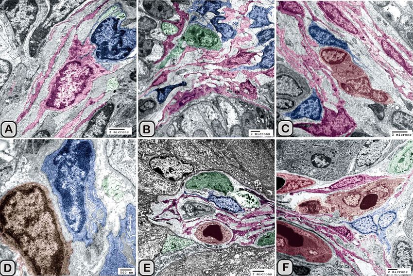
www.nature.com/scientificreports/
reside in many tissues of the body under the normal physiological conditions and function as immunological
sensors for many p athogens7. Dendritic cells are widely distributed in the lymphatic tissue. In addition, they are
found in the non-lymphoid organs such as the skin (Langerhans cells), heart, uterine tube, lungs, cornea and
the gastrointestinal tract (interstitial DC)8–11. Immature dendritic cells phagocytose antigens by endocytosis or
macropinocytosis then process these antigens into peptides to be presented to the major histocompatibility com-
plex class II molecule (MHC-II) and activate the naive T- lymphocyte c ells12–14. The DCs become mature after its
stimulation with the lipopolysaccharide (LPS) and c ytokines15–17. The macrophages are professional antigen-pre-
senting cells (APC); however, the DCs are much more potent at initiating and expanding the secondary immune
responses18. There are several surface markers used to characterize DCs in several tissues such as CD1, CD11c,
CD13, CD14, CD56, CD34, CD68, S100 and L angerin19,20. Unfortunately, there is no, to date, a specific marker
for the DCs subtypes. Therefore, the ultrastructure approach is considered the gold standard for identifying the
DCs21,22. The circulating melatonin is the principal hormone synthesized and secreted rhythmically by the pineal
gland23,24, and to a lesser extent by other tissues such as the gastrointestinal tract, skin, retina, bone marrow, ovary
and testis25,26. The melatonin secretion influenced by the environmental photoperiod. Melatonin (5-methoxy-
N-acetyltryptamine) regulates the circadian rhythm and the seasonal changes via its nocturnal synthesis by the
pineal gland. In addition, it is also involved in many pathways including antiaging, antioxidant, free radical
scavenger, anticarcinogenic and immunomodulatory e ffects27–29. The immunomodulatory effect of melatonin
on the immune cells is supported by the existence of G-protein-coupled membrane receptors (MT1 and MT2
receptors). Both membrane and nuclear melatonin receptors have been recognized in immune cells30. Mela-
tonin induces the proliferative response of stimulated T and B- lymphocytes through the membrane receptors.
However, melatonin induces the cytokine production in the mononuclear cells such as DCs and macrophages
through the nuclear r eceptors31. It was demonstrated that melatonin induces IL-12 (Interleukin-12) and IL-6
secretion in monocytes which will stimulate the secretion of IL-2 to differentiate towards T-helper p henotype32.
Melatonin enhances the immune response through stimulation of natural killer cells, antigen presentation and
phagocytosis. In our previous study we investigated a significant increase in the number of dendritic cells and
its phagocytic vesicles in the adrenal gland of melatonin treated groups of Soay r am33. Melatonin is the main
regulator of reproduction especially in the photoperiodic animals as it modulates the synthesis and secretion of
gonadotrophic hormones in the hypothalamus 34. The exogenous melatonin treatment during the non-breeding
season increases the testosterone level in the blood plasma35,36, the sperm quality, the testicular parameters, the
spermatogenesis and fertility of rams during the long day’s p eriods37,38.
The present study was an extension from our previous study which demonstrated the stimulatory effect of
melatonin on the seminal gland of Soay ram during the non-breeding season. It was proved that melatonin
administration resulted in cytological signs of increase in the secretory activity and height of the principal cells
lining the glandular epithelium of the gland. In addition, we investigated a significant increase in the number
of telocytes and its stromal synapses with immune cells such as macrophages and mast cells after melatonin
administration39,40. Therefore, the current study continues our work to understand the broader effect of melatonin
on the immunity and validate its use in practice as an enhancer for immunity and fertility. To our knowledge
there has been no study about the effect of melatonin on the dendritic cells biology. Therefore, the objective of
the present study was to characterize the morphology and the distribution of the dendritic cells in the seminal
vesicle of Soay ram after melatonin administration using the transmission electron microscopy and the immu-
nohistochemical study.
Materials and methods
Animals and experimental design. Experiments were conducted in accordance with the U.K. Animals
(Scientific Procedures) Act of 1986 in MRC Reproductive Biology Unit, Centre for reproductive biology, Edin-
burgh, Scotland, U.K. This study was performed in strict accordance with the relevant guidelines and ethical
regulations (experiment No. S/17353). The animals and the experimental design have been discussed before
by39,41. In brief, the animals of Soay Ram breed (Ovis aries) were obtained from specialist breeders in Scot-
land. The seminal glands of 15 adult Soay rams (aged 1.5 years) were used in this study. At the end of May,
eight animals were given a subcutaneous implant containing melatonin (treated group), while another group of
seven animals were given empty implants (control group). The melatonin implants were made of Silastic sheet-
ing (500-1DOw Corning sheeting, Midland, MI, USA) sealed into an envelope, containing 1.0 gm melatonin
(Sigma Chemical, Poole, Dorset, UK). The implants were placed subcutaneously above the rib cage using local
anesthesia and left throughout the time of experiment. Such implants have been previously shown to maintain a
constant supply of 200–500 pg melatonin/ml plasma for many months without interfering with the endogenous
supply of the secreted m elatonin42. Eleven weeks after the onset of the experiment, all rams were sacrificed by
intravenous barbiturate injection, the seminal glands were carefully excised, and small samples were processed
for light and electron microscopic examination. Both light and electron microscopic examinations were done at
Assiut University, Egypt.
Light microscopy. The samples were obtained from the seminal glands of both control and melatonin treated
groups, dissected and immediately fixed in 4% paraformaldehyde in 0.1 M phosphate buffered saline (PBS,
pH 7.4) over night (ON) at 4 °C. The fixed samples were dehydrated in an ascending series of ethanol, cleared
in methyl benzoate then embedded in paraffin wax. Samples were cut at 5–8 μm in thickness transversely and
longitudinally. The staining protocols and used procedures were carried out following the descriptions of the his-
tological techniques as reported by43. The sections were stained with Haematoxylin and Eosin for demonstrating
the general histological structure of the examined seminal gland and to identify the DCs.
Scientific Reports | (2021) 11:872 | https://doi.org/10.1038/s41598-020-79529-y 2
Vol:.(1234567890)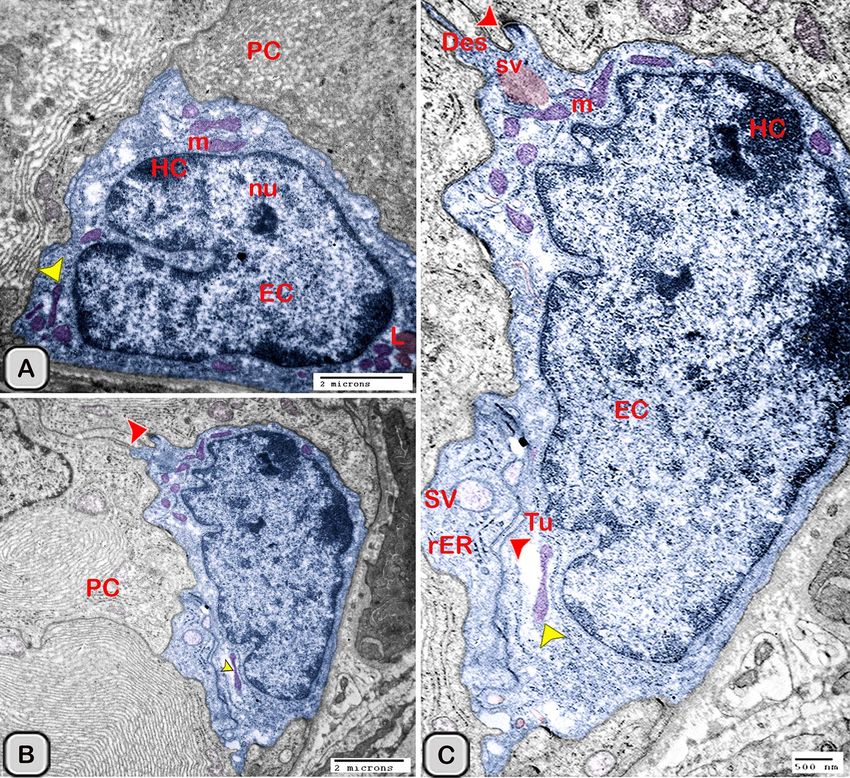
www.nature.com/scientificreports/
Primary antibody origin Primary antibody Biotinylated secondary
Target Primary antibody suppliers (catalog no.) Primary antibody dilution incubation period antibody
Anti-mouse IgG + anti-rabbit
S-100 protein Genemed Biotechnologies Rabbit (pc; 61-0061) 1:200 1 h at RT IgG (biotinylated goat anti-
polyvalent)b
Anti-mouse IgG + anti-rabbit
Estrogen receptor (SP1) Genemed Biotechnologies Rabbit (pc; RM_9101-S0 1:200 1 h at RT IgG (biotinylated goat anti-
polyvalent)b
Progesterone receptor Immunotech Mouse (mc; PR10A9) 1:50 Overnight at 4 °C (Rabbit anti-mouse IgG)b
Anti-mouse IgG + anti-rabbit
CD 34 (clone QBE d/10) ThermoFisher Scientific Mouse (mc; MS-363-R7) Ready to use 1 h at RT IgG (biotinylated goat anti-
polyvalent)a
Anti-mouse IgG + anti-rabbit
anti-c-kit (CD117) Genemed Biotechnologies Rabbit (pc) 1:50 1 h at RT IgG (biotinylated goat anti-
polyvalent)a
CD3 Novus Biologicals Rabbit (pc, NB 100-2000) 1: 100 Overnight at 4 °C (goat anti-rabbit IgG)b
CD56 Santa Cruz biotech Goat (pc, Sc 1507) 1:100 Overnight at 4 °C (Donkey anti-goat IgG)b
MHCII Abcam Mouse (mc, ab23990) 1:100 Overnight at 4 °C (Donkey anti-mouse IgG)b
Table 1. Identity, sources, and working dilutions of primary and secondary antibodies. pc polyclonal,
mc monoclonal, RT room temperature. a From ThermoFisher Scientific/Lab Vision, Fremont, CA, USA. b From
Dako, Hamburg, Germany.
Semi‑thin sections and transmission electron microscopic studies. The procedure of staining was done according
to44,45. The seminal glands were preserved by immersion in a mixture of 5% paraformaldehyde–glutaraldehyde
fixative overnight46. After fixation, the samples were washed in phosphate buffer solution and osmicated with 1%
osmium tetroxide in 0.1 mol/L Na-phosphate buffer at pH 7.3. After that, the samples were dehydrated in series
of ethanol followed by propylene oxide and embedded in Araldite. Semi-thin sections were cut at 1 µm thickness
with a Reichert Ultracut (Leica, Germany) and stained with toluidine blue for light microscopy.
Ultrathin sections were done with Ultrotom VRV (LKB Bromma, Germany). The sections (70 nm) were
stained with uranyl acetate and lead c itrate47 and examined in a JEOL 100CX II transmission electron microscope
(TEM) (JEOL, Tokyo, Japan) at the Electron Microscopy Unit of Assiut University.
Digitally colorization of TEM images. To increase the visual contrast between several structures on the same
electron micrograph, we have digitally colored specific elements [e.g., dendritic cells (DCs), telocytes (TCs),
nerve fibers and lymphocytes] to make them more visible. All the elements were carefully hand colored in Adobe
Photoshop software version 6. The methods were used by41,48–51.
CMEIAS color segmentation (for the supplementary images). Negative images were obtained using the CMEIAS
color segmentation software, which processes color images by segmenting the object of interest in the fore-
ground from the b ackground52. This process is conducted by the following steps: open image file with CMEIAS
color segmentation, then select “Process” from the menu items and subsequently choose “Negative image”53–55.
Immunohistochemistry technique. To demonstrate the dendritic cells in the seminal glands of Soay rams, six
different antibodies were used including, anti-estrogen receptor alpha (ER-α), anti-progesterone, CD3, CD117,
CD34, MHCII, CD56 and S-100 protein (Table 1). These antibodies have been validated in various studies in
sheep33,40,56. In addition, most of used antibodies in the current study their cross reactivity was proved in bovine
and goat (ruminants) according to manufacturer data sheet or not tested yet.
Immunohistochemistry staining technique had been performed on the paraffin fixed tissue. Sections were
deparaffinized with xylene and hydrated with a descending grade of ethanol then washed with 0.1 M PBS (3 ×
10 min). To inhibit the endogenous peroxidase activity the sections was incubated with 3% H 2O2 in H2O for
20 min at the room temperature (RT), followed by intense washing under running tap water for an additional
10 min. To decrease the masking of antigen epitopes, the antigen retrieval was carried out using 0.1 M sodium
citrate buffer solution (pH 6) for 15 min using a microwave (750 Watt). Then, sections were cooled to room
temperature for 20 min and washed with PBS (pH 7.4, for 10 min). The sections were covered with Ultra V Block
(ThermoFisher Scientific, TP-015-UB) for 5 min at room temperature to minimize the non-specific antibody
binding. Then, the sections were incubated with the primary antibodies diluted in the blocking solution [1.5%
normal donkey serum (NDS) + 0.2% Triton-X 100/PBS] (Table 1). The sections were rinsed in 0.2% Triton-X
100/PBS and followed by incubation with a biotinylated secondary antibody (Table 1) for 1 h at RT. Then, sections
were washed by PBS (pH 7.4, 3 times for 5 minutes) and, subsequently incubated with streptavidin-peroxidase
complex (ThermoFisher Scientific, TS-015-HR) for 15 min at room temperature. Visualization of the reaction
was carried out with a drop of DAB plus chromogen (ThermoFisher Scientific, TA-001-HCX) to 2 ml of DAB plus
substrate (ThermoFisher Scientific, TP-015-HSX) which applied on the sections for 5–10 min. The sections were
counterstained with Harris haematoxylin for 30 s. The sections were dehydrated in a graded series of ethanol,
Scientific Reports | (2021) 11:872 | https://doi.org/10.1038/s41598-020-79529-y 3
Vol.:(0123456789)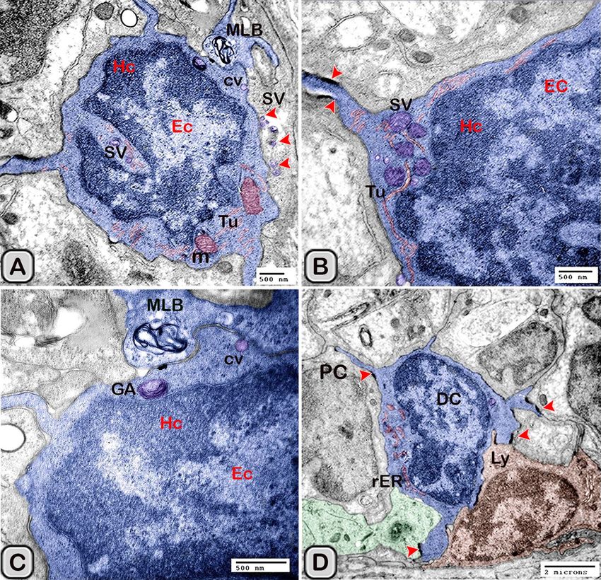
www.nature.com/scientificreports/
Figure 1. Paraffin sections stained with H&E showing the dendritic cells residing among the glandular
epithelium lining the seminal gland of the Soay ram. (A) DCs were small with a deeply stained nucleus,
acidophilic cytoplasm and a cytoplasmic process directed toward the lumen of the gland (arrowhead). (B) The
number of DCs was obviously increased in melatonin treated groups (arrowhead).
Measurements Control After melatonin treatment
Ratio of interstitial tissue/glandular tissue (Fig. 2 and other serial semithin sections) 0.494 ± 0.019 0.241 ± 0.021*
Cross sectional area of dendritic cells in the glandular epithelium (TEM images , Figs. 10,
20.97 ± 4.155 48.72 ± 7.528*
12, 13, 14)
Cross sectional area of dendritic cells in the interstitium (TEM images, Figs. 10, 16) 17.99 ± 3.972 15.78 ± 3.219 (ns)
Number of DCs in the glandular epithelium of the histological and semi-thin sections
3.667 ± 0.2562 8.917 ± 0.8569**
(Figs. 1, 2, 3)
Number of DCs in the glandular epithelium of estrogen alpha receptors (Fig. 6) 3.571 ± 0.4809 6.543 ± 0.5936***
Number of DCs in the glandular epithelium of progesterone receptors (Fig. 7) 3.000 ± 0.2582 6.500 ± 0.5627***
Number of DCs in the glandular epithelium of CD3 (Fig. 5) 4.750 ± 0.4787 8.500 ± 0.6455**
Table 2. The morphometric measurements of the control and melatonin treated groups. The measurements
are expressed as the mean ± SE. When P < 0.05 (*) is significant, P < 0.01 (**) and P < 0.01 (***) is highly
significant.
cleared with xylene and covered with DPX. Immunohistochemical staining was evaluated by LeitzDialux 20
Microscope and photos were photographed by cannon digital camera (Cannon Powershot A95). IHC staining
was quantified within control and melatonin treated groups for ER-α, CD3 and Progesterone.
Morphometrical and statistical analysis. The morphometric studies were performed on both light and elec-
tron- microscopic images of the seminal glands of both control and treated animals using Image-J software. The
measurements were carried out on 15 randomly selected sections of each gland per animal (5 different areas
were measured from each section) as follows: the interstitial connective tissue/glandular tissue ratio per 20 mm2
using 40× objective, the cross sectional area of dendritic cells in the glandular epithelium and the surrounding
interstitium per 20 μm2 in TEM images. The number of dendritic cells in the glandular epithelium of histological
sections, semi-thin sections and CD3, estrogen receptor alpha (ER-α) and progesterone immunostained sections
were performed per 20 mm2 using 20× objective. Per each animal the quantification were performed from each
fifth section (to avoid double counting of the same cells). All the data were expressed as mean± SE (standard
error) which was statistically analyzed using “T-Test Graphpad prism Software” (Version 6.05, International
Scientific Community) to compare between different measurements of both control and treated animals. Differ-
ences were considered significant if P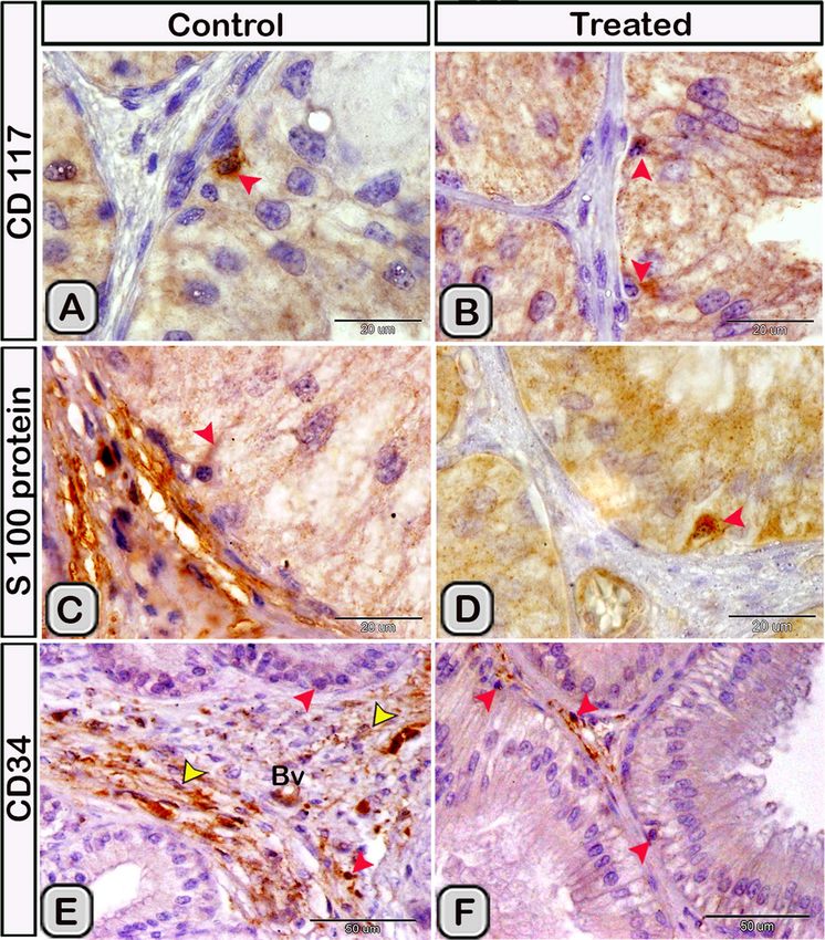
www.nature.com/scientificreports/

                                 Figure 2.  (A,B) Semi-thin sections stained with Toluidine blue showing the DCs (red arrowheads) in the
                                 control groups near the basement membrane lining the glandular epithelium and its close association to
                                 T- lymphocytes (black arrowhead) and telocytes (TCs, yellow arrowheads) in the interstitium (IT). (C) A
                                 triangular-shaped dendritic cell with a few vesicles in the cytoplasm and a long cytoplasmic process directed
                                 toward the lumen of the gland was observed. (D,E) In the melatonin treated groups, DCs (red arrowheads) were
                                 closely associated to T-lymphocytes (Ly, black arrowheads) and telocytes (yellow arrowheads) in the interstitium
                                 (IT). DCs were larger in size and their nucleus was euchromatic and more indented in comparison to control
                                 groups. Notice, the epithelium of the principal cells (PC) is higher in melatonin groups compared to control
                                 ones. (F) The cytoplasm of dendritic cells had large amount of the secretory vesicles in comparison to the
                                 control groups.

                                 Figure 3.  (A,B) The free movable DCs (red arrowheads) were observed within the glandular epithelium lining
                                 the seminal gland of the control groups. (C) Interstitial DCs (red arrowheads) were observed in a close contact
                                 to telocytes (TC, yellow arrowheads). (D–F) Semi-thin sections stained with Toluidine blue in the melatonin
                                 treated groups. (D,E) free movable dendritic cells were larger in size and more abundant in comparison to the
                                 control ones (red arrowheads). (F) Interstitial DCs (red arrowheads) were more abundant compared to the
                                 control group and in a close contact to telocytes (TC) and blood vessels (BVs).

Scientific Reports |   (2021) 11:872 |               https://doi.org/10.1038/s41598-020-79529-y                                                    5

                                                                                                                                          Vol.:(0123456789)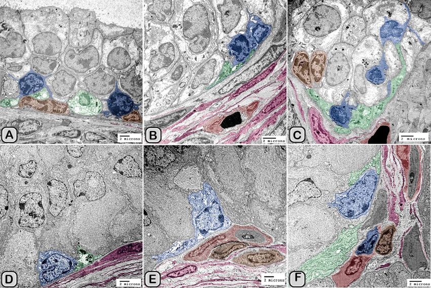
www.nature.com/scientificreports/

                                           Figure 4.  The effect of melatonin administration on the expression of CD117, S-100 protein and CD34 in
                                           dendritic cells (DCs). No obvious changes could be detected in the expression of CD117 (A,B) S-100 protein
                                           (C,D) and CD34 (E,F) (red arrowheads) in the melatonin treated group compared to the control. Notice: the
                                           expression of CD34 in telocytes (yellow arrowheads) endothelium of blood vessel (Bv).

                                           study was carried out in strict accordance with the national and ethical regulations and care of animals was in
                                           accordance with institutional guidelines. The protocol was approved by the Central Office for Research Ethics
                                           Committees (COREC) in Eastarbourne Terrace, United Kingdom.

                                           Results
                                           General morphology (characterization) of the dendritic cells.                 Histological analysis. In the paraf-
                                           fin sections stained with H&E of the control groups, the dendritic cells were demonstrated residing among the
                                           glandular epithelium lining the seminal vesicle. The cells were small with a deeply stained nucleus, acidophilic
                                           cytoplasm and a cytoplasmic process directed toward the lumen of the gland (Fig. 1A). However, in the mela-
                                           tonin treated groups, the number of dendritic cells in the epithelial lining the gland was obviously increased in
                                           comparison to the control ones (Fig. 1B, Table 2). In Toluidine blue stained semi-thin sections of the control

          Scientific Reports |   (2021) 11:872 |                https://doi.org/10.1038/s41598-020-79529-y                                                 6

Vol:.(1234567890)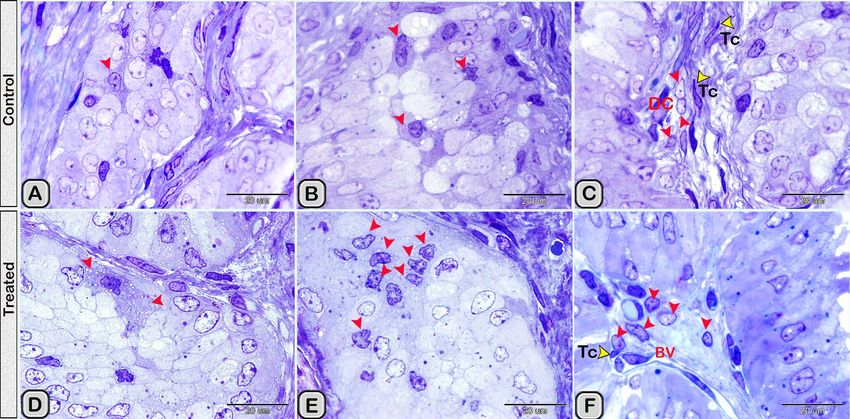
www.nature.com/scientificreports/

                                 Figure 5.  The effect of melatonin administration on the expression of CD3 in the dendritic cells (DC) and
                                 T-lymphocytes (Ly). (A,B) CD3 expressed in the DCs with its well defined processes (red arrowheads) and
                                 T-Lymphocytes (blue arrowheads) in the control groups. (C,D) The immunoreactivity for CD3 was more
                                 abundant in melatonin treated groups.

                                 groups, DCs were observed near the basement membrane lining the glandular epithelium and in a close asso-
                                 ciation to T- lymphocytes and telocytes (TCs) in the interstitium (Fig. 2A,B). Lymphocytes are usually smaller
                                 in size than DCs. Lymphocytes have a small spherical or oval nucleus and abundant darkly stained chromatin.
                                 A triangular-shaped dendritic cell with a few vesicles in the cytoplasm and a long cytoplasmic process directed
                                 toward the lumen of the gland was observed (Fig. 2C). Whereas, in the melatonin treated groups, DCs were
                                 larger in size and their nucleus was euchromatic and more indented in comparison to control groups. The as-
                                 sociated T-lymphocytes were active, larger in size and their nucleus was vesicular with chess like appearance of
                                 chromatin (Fig. 2D,E). The cytoplasm of dendritic cells had large amount of the secretory vesicles in comparison
                                 to the control groups (Fig. 2F). The free movable DCs were investigated within the glandular epithelium of the
                                 seminal gland (Fig. 3A,B). The interstitial DCs were investigated in the interstitium of the gland and were in
                                 close contact with telocytes (Fig. 3C). The movable DCs were larger in size and more abundant in the melatonin
                                 treated groups when compared to the control (Fig. 3D,E and Table 2). In addition, the interstitial DCs were more
                                 abundant and were in close contact with telocytes and blood vessels (Fig. 3F).

                                 Immunohistochemical analysis. Immunohistochemical staining was performed to characterize the dendritic
                                 cells in the seminal gland of the control and melatonin treated groups. The dendritic cells showed positive
                                 immunoreactivity for CD117/c-kit (Fig. 4A,B), S-100 proteins (Fig. 4C,D) and CD34 (Fig. 4E,F). CD3 immuno-
                                 reactivity were demonstrated in the T- lymphocytes which characterized by their small size and they were round
                                 to oval in shape. The immunoreactivity was also investigated in DCs with its well-defined processes (Fig. 5A,B).
                                 In the melatonin treated groups we demonstrated an obvious increase in the number of the positive immunore-
                                 active cells to CD3 (Fig. 5C,D and Table 2). The immunoreactivity for estrogen receptor (ER-α) and progesterone
                                 demonstrated in the lining epithelium and the interstitium of the seminal gland (Figs. 6A–C, 7A,B). However,
                                 in the melatonin treated groups DCs showed marked increase in the expression of estrogen receptor (ER-α) and
                                 progesterone (Figs. 6D–F; 7C,D and Table 2). Furthermore, we examined the expression of two specific mark-
                                 ers: CD56 and MHC-II (major histocompatability class II). DCs with its well-defined processes showed positive

Scientific Reports |   (2021) 11:872 |               https://doi.org/10.1038/s41598-020-79529-y                                                7

                                                                                                                                          Vol.:(0123456789)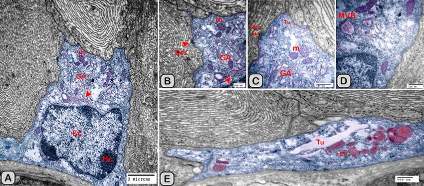
www.nature.com/scientificreports/

                                           Figure 6.  The effect of melatonin administration on the expression of estrogen alpha receptors in the dendritic
                                           cells (red arrowheads). (A,B) Expression of estrogen alpha receptor in the epithelium, (C) the expression in the
                                           interstitium of the control groups. (D,F) the expression of estrogen alpha receptor (ER-α) increased obviously in
                                           the melatonin treated groups.

                                           Figure 7.  The effect of melatonin administration on the expression of progesterone in the DCs (red
                                           arrowheads). (A,B) Expression of progesterone receptor in the DCs in the epithelium and the interstitium of
                                           the control groups respectively. (C,D) The expression of progesterone receptors increased obviously in the
                                           melatonin treated groups. Notice: the close relation between the DCs (red arrowheads) and TCs (telocytes).

          Scientific Reports |   (2021) 11:872 |               https://doi.org/10.1038/s41598-020-79529-y                                                  8

Vol:.(1234567890)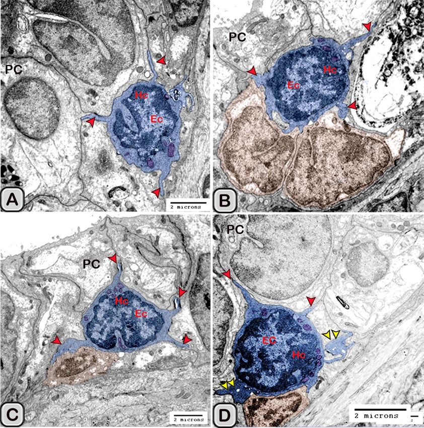
www.nature.com/scientificreports/

                                 Figure 8.  The effect of melatonin administration on the expression of CD56 in the DCs. (A) Expression of
                                 CD56 in the DCs lining the epithelium and the interstitium of the control groups. (B) the expression of CD56
                                 was more abundant in the melatonin treated groups. The expression of CD56 in the interstitial DCs was
                                 branched and anastomsing similar to a continuous network.

                                 Figure 9.  The effect of melatonin administration on the expression of MHC-II in the DCs. (A,B) The
                                 endosomal compartments of the dendritic cells showed positive immunoreactivity for MHC-II. (C,D)
                                 Expression of the MHCII in the endosomal compartments of the DCs was more abundant after melatonin
                                 administration (red arrowheads).

                                 immunoreactivity for CD56 in the lining epithelium and the interstitium of the seminal gland (Fig. 8A). How-
                                 ever, the expression of CD56 and MHC-II in the dendritic cells was abundant in the melatonin treated groups

Scientific Reports |   (2021) 11:872 |               https://doi.org/10.1038/s41598-020-79529-y                                                 9

                                                                                                                                         Vol.:(0123456789)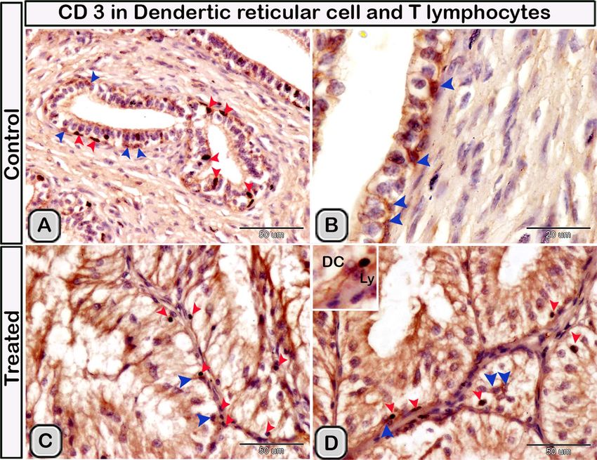
www.nature.com/scientificreports/

                                           Figure 10.  Digitally colored transmission electron microscope images of the dendritic cells in the seminal
                                           vesicle of the soay ram. (A–C) General morphology of the dendritic cells in the glandular epithelium lining
                                           the seminal vesicle of the control groups. The dendritic cells (blue) had a deeply indented nucleus and a
                                           great amount of the peripheral heterochromatin (HC). The DCs (blue) were observed in association to
                                           the T-lymphocytes (brown), nerve fibers (green) and telocytes in the interstitium (pink). (D–F) General
                                           morphology of the DCs in the glandular epithelium lining the seminal vesicle of the melatonin treated groups.
                                           The nucleus of the DCs (blue) was euchromatic (EC) (D–F). DCs varied in shape which could be large
                                           polyhedral (D,E) or triangular (F). DCs were closely associated with lymphoblast (brown) and telocytes (pink).

                                           compared to the control (Figs. 8B, 9A,B). Expression of the MHCII in the endosomal compartments of the DCs
                                           was more numerous compared to the control ones (Fig. 9C,D).

                                           Ultrastructure of the dendritic cell. In addition to the immunohistochemical examinations, transmission elec-
                                           tron microscopy (TEM) was performed to identify and characterize the dendritic cells in the seminal gland of
                                           Soay ram. TEM demonstrated the presence of dendritic cells (DCs) interdigitating between the epithelial cells
                                           lining the seminal gland through their long dendrites. The nucleus of the DCs in the untreated groups retained
                                           deeply indented nucleus with a peripheral distribution of heterochromatin. We observed a close association
                                           between the DCs, T-lymphocytes and nerve fibers (Fig. 10A–C). Dendritic cells located at the basement mem-
                                           brane (Fig. 10A,B) or free mobile among the glandular epithelium lining the seminal gland (Fig. 10C). In the
                                           melatonin treated groups the activated dendritic cells contain a large cell body and euchromatic nucleus and
                                           cells appeared variable in shapes including rounded, polyhedral and triangular. The DCs lining the seminal
                                           gland were observed closely related to telocytes (TCs) with its telopodes (Tp), nerve fibers, lymphoblast and the
                                           blood vessels in the interstitium of the gland (Fig. 10D–F). We observed that the cytoplasm of the dendritic cells
                                           contained relatively few membranous cell organelles including the mitochondria, Golgi apparatus and short cis-
                                           ternae of rough endoplasmic reticulum (rER) which arranged close to the nucleus (Fig. 11A–D). The endosomal
                                           tubulo-vesicular system observed in the cytoplasm of the dendritic cells, which including the coated vesicles,
                                           multilamellar bodies (MLB), numerous tubules and vesicles (Fig. 11A–C). Among the morphological features of
                                           the dendritic cells were the desmosomal intercellular junctions with the adjacent principal cells lining the semi-
                                           nal gland and the associated T-lymphocyte (Fig. 12A–C). The dendritic cells demonstrated in a close contact to

          Scientific Reports |   (2021) 11:872 |                https://doi.org/10.1038/s41598-020-79529-y                                                10

Vol:.(1234567890)
www.nature.com/scientificreports/
Figure 11. Digitally colored transmission electron microscope images of the DCs in the seminal vesicles
of control groups. (A–C) The cytoplasm of the DCs (blue) contained relatively few cell organelles including
the mitochondria (m), Golgi apparatus (GA), and short cisternae of the rER (arrowhead). The endosomal
tubulo-vesicular system including the cavolae (cv), secretory vesicles (SV, red arrowhead) multilamellar bodies
(MLB), numerous tubules (Tu) were observed. (D) The intercellular junctions such as the desmosomal junction
(arrowheads) with the adjacent cells including T-lymphocytes (brown), principal cell (PC) lining the seminal
vesicle and nerve fiber (green) were investigated. HC heterochromatin, EC euchromatin.
one or two T-lymphocytes (Fig. 12B–D). The cytoplasmic cell processes of dendritic cells were long and thick
bulbous like cell processes (Fig. 12C,D).
The size (cross sectional area) of the dendritic cells increased significantly when compared to the control
groups (Table 2). The nucleus of the DCs was euchromatic and contained a prominent nucleolus (active cell),
in addition it was larger and more indented compared to the control group (Figs. 13 and 14). We demonstrated
an increase in the secretory activity of the dendritic cells after the melatonin treatment. There was an obvious
increase in the number of mitochondria, cisternae of rER and a well-developed supranuclear Golgi apparatus with
its secretory vesicle (Fig. 13A–D). The endosomal- lysosomal system was more developed than the control groups
and composed of numerous tubules and vesicles, multivesicular bodies and phago- lysosomes (Fig. 13D,E). In
addition, a rod-shaped Birbeck granule (club-shaped) demonstrated in the cytoplasm nearby the nucleus of the
dendritic cells (Fig. 14A–C). The cell process was thinner and shorter than those observed in the control groups.
The desmosomal cell junctions were more obvious between the dendritic cells and the neighboring principal
cells lining the seminal vesicle (Fig. 14B,C).
The free movable dendritic cells showed different stages of maturity in the melatonin treated groups. The
nucleus of immature free movable dendritic cells was euchroamtic and deeply indented with a distinct nucleo-
lus. The cytoplasm consisted of well-developed rER, a great amount of the dense granules, secretory vesicles, a
Scientific Reports | (2021) 11:872 | https://doi.org/10.1038/s41598-020-79529-y 11
Vol.:(0123456789)www.nature.com/scientificreports/
Figure 12. Digitally colored transmission electron microscope images of the DCs in the seminal vesicles of
control groups. (A–D) DCs (blue) varied in shape which could be polyhedral, round or triangular. (A–C) The
DCs located at the basement membrane or near from the basement membrane and in close association to
T-lymphocytes (brown). (A–C) The cytoplasmic cell processes of dendritic cells were long (red arrowheads) and
thick bulbous like cell processes (D, yellow arrow heads). HC heterochromatin, EC euchromatin, PC principal
cell.
moderate number of mitochondria and an ill-developed tubule-vesicular system. The cell processes were short
and thin or thick (Fig. 15A,B).
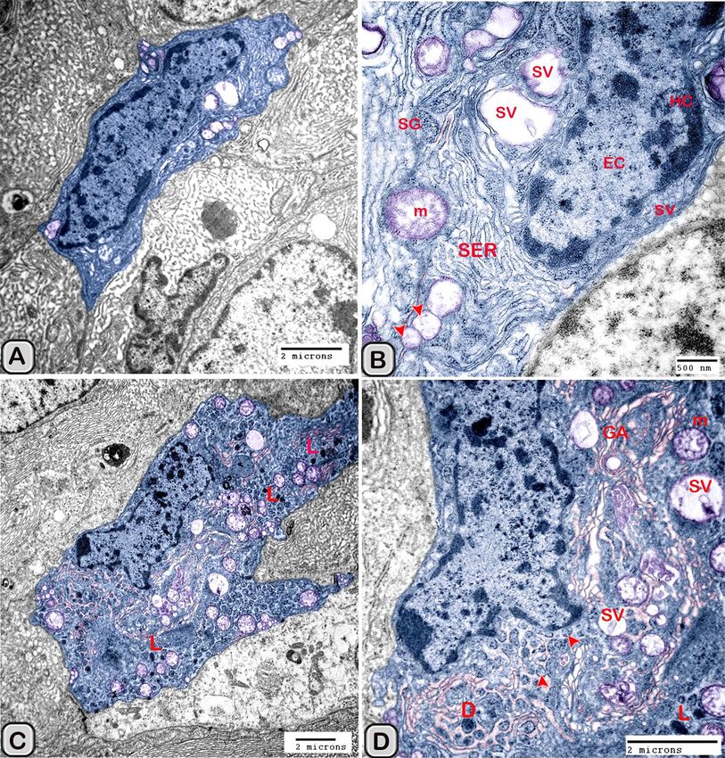
Interstitial dendritic cells were demonstrated in the interstitium of the control and melatonin treated groups.
Interstitial DCs were observed in close contact with telocytes and its telopodes (Tps), T-Lymphocytes, Schwann
cell and blood vessels (Fig. 16A–F). The cross sectional area of the interstitial DCs varied between the control
and melatonin treated groups, however they are not significantly different between the two groups (Table 2)
Macrophages were also demonstrated among the epithelium lining the seminal gland in the control groups
and nearby the dendritic cells (Fig. 17A,B). The macrophages nucleus was kidney shaped and the cytoplasm con-
tained lysosomes and ill-developed tubular endosomal system. In the melatonin treated groups, the macrophages
appeared activated (Fig. 18A–D). The cytoplasm contained a well-developed tubulo-vesicular endosomal system,
smooth endoplasmic reticulum (SER), secretory vesicles, mitochondria and a well-developed Golgi apparatus.
In addition, the dense bodies and lysosomes were more evident compared to the control ones.
Scientific Reports | (2021) 11:872 | https://doi.org/10.1038/s41598-020-79529-y 12
Vol:.(1234567890)www.nature.com/scientificreports/
Figure 13. Digitally colored transmission electron microscope images of the activated DCs in the melatonin
treated group. (A) General view of an active polyhedral dendritic cell with an abundant cell organelles and
euchromatic nucleus (Ec). (B–D) Higher magnification showing abundant mitochondria (m) and well
developed Golgi apparatus (GA). (B–E) Birbeck granules (red arrowheads), multivesicular bodies (MVB),
tubules (Tu) and phagolysosomes (L) were observed. Desmosomal cell junction with the neighboring cell (red
arrowhead, Des) was observed.
Discussion
In the current study, we identified and characterized the dendritic cells in the glandular epithelium and the
surrounding interstitial tissue of the Soay ram seminal glands. DCs have a unique morphological structure
and functional properties when they compared to other antigen presenting cells such as macrophages as they
are strong naïve T-lymphocytes stimulators via peptide-presenting MHC (major histocomptability) class II
molecules57–59. In the present work, we identified the dendritic cells morphologically by conventional stains,
immunohistochemistry and ultra-structure. They are characterized by their typical cytoplasmic processes (den-
drites), indented nuclei and by their close association to T- lymphocyte cells. Dendritic cells (DCs) and mac-
rophages are highly dynamic cells which show changes in their function, distribution and phenotype. Dendritic
cells are critically involved in the initiation and modulation of an appropriate immune response by linking innate
to adaptive immune responses 12. Interstitial dendritic cells were demonstrated in the interstitium of the seminal
gland in close association to the blood vessels, telocytes and nerve fibers. Telocytes had been identified in the
interstitium of many organs; to react with the interstitial cells and immune c ells60,61. We demonstrated a close
association between the dendritic cells and the nerve fibers. These areas of contact contain important neurome-
diators including substance P and calcitonin-gene related peptide (CGRP) which are critical for the crosstalk
between the nervous and immune s ystems62. The current study demonstrated a stimulatory effect exerted by the
melatonin on the dendritic cells, such as increasing the number and the secretory activity of DCs observed by
TEM. The endosomal- lysosomal system was more developed in the melatonin treated groups compared to the
control ones. The current result revealed that the endosoaml system consisted of numerous vesicular and tubular
compartments, cavolae, multivesicular bodies and lysosomes. This pathway is highly dynamic and involved in
segregating the components fated for degradation in lysosomes or recycling to the plasma membrane or Golgi
apparatus63,64. The endosomal system had a role in the endocytosis of the antigen for processing, then transport
the peptide/MHC class II to the plasma membrane. Therefore, the endosomal tubulo-vesicular system acts as
sites for the activation of T-lymphocyte cell. Moreover, it acts as a storage site for several immunoregulatory
factors which secreted form the D Cs65,66. We demonstrated Birbeck granules, which are club shaped structure
in the melatonin treated groups. Birbeck granules composed of Langerin protein (CD207), which found in the
endosomal recycling compartment67. Birbeck granules play an important role in antigen processing as they allow
internalization of antigens until their presentation to T-Lymphocytes68.
Melatonin exerts its action through M1 and M2 membrane receptors69. Melatonin receptors had been identi-
fied in the ram testis, epididymes, vas deferens and seminal vesicles70. Melatonin is an immunomodulator, which
produced by the immunocompetent cells and lymphocytes71. Moreover, melatonin enhances the proliferation
Scientific Reports | (2021) 11:872 | https://doi.org/10.1038/s41598-020-79529-y 13
Vol.:(0123456789)www.nature.com/scientificreports/
Figure 14. Digitally colored transmission electron microscope images of the activated DCs in the melatonin
treated group. (A,B) An active polyhedral and triangular shaped dendritic cell with abundant cell organelles and
euchromatic nucleus (Ec) with a distinct nucleolus (nu) were investigated. (C) Higher magnification showing
abundant mitochondria (m), rER, secretory vesicles (Sv), tubules (Tu), lysosomes (L) and Birbeck granules
(yellow arrowheads). Moreover, the cell processes were thin and short and desmosomal junctions (Des, red
arrowheads) were observed.
and maturation of all immune cells including T and B lymphocytes, granulocytes and m onocytes72. In addition,
melatonin increases testosterone level during the non-breeding season in the blood plasma, the sperm quality,
the testicular parameters, the spermatogenesis and fertility of rams. In our previous studies we investigated
the enhancement role of melatonin on the secretory activity of the seminal gland and its glandular epithelium.
Therefore, we concluded that the effect of melatonin on DCs could be directly through the melatonin receptor
or indirectly through the increase in testosterone level.
In the current work, we demonstrated that the dendritic cells showed positive immunoreactivity for CD117
and CD34. CD117/c-KIT is a tyrosine-kinase receptor expressed on the outer surfaces of hematopoietic cells
and stem cells (Martin et al., 1990). Moreover, CD117 and CD 34 are used as a specific marker for telocytes
Scientific Reports | (2021) 11:872 | https://doi.org/10.1038/s41598-020-79529-y 14
Vol:.(1234567890)www.nature.com/scientificreports/
Figure 15. Digitally colored transmission electron microscope images of the free movable immature dendritic
cells. (A,B) The nucleus was euchroamtic (Ec) and deeply indented with a distinct nucleolus (nu). (B) The
cytoplasm consisted of well-developed rER, a great amount of the dense granules (G), secretory vesicles (SV) a
moderate number of mitochondria (m) and ill-developed tubule-vesicular system (Tu). The cell processes were
short and thin or thick (red arrowheads). HC heterochromatin, EC euchromatin, PC principal cell.
Figure 16. Digitally colored transmission electron microscope images of the Interstitial DCs in the control
(A–C) and melatonin treated groups (D–F). DCs process (blue) form hetercellular junctions with telocytes and
its telopodes (pink). In addition, DCs were observed in a close contact to T-Lymphocytes (brown), Schwann
cell, nerve fibers (green) and blood vessels (reddish brown).
Scientific Reports | (2021) 11:872 | https://doi.org/10.1038/s41598-020-79529-y 15
Vol.:(0123456789)www.nature.com/scientificreports/
Figure 17. Digitally colored transmission electron microscope images of Macrophage (violet) among the
epithelium of Soay ram seminal gland in the control groups. (A,B) macrophages were observed nearby the
dendritic cells (blue), T-lymphocytes (brown) and nerve fibers (green). The nucleus was kidney shape and their
cytoplasm contains lysosomes (L) and ill-developed endosomal tubular system (Tu).
in the seminal vesicles of Soay ram39. CD117 expression in most mature hematopoietic cells is absent during
the final stages of differentiation. However, some subsets of dendritic cells express CD117 throughout their
development73. CD34 is a marker for blood progenitor cells and many undifferentiated cells74. It is well known
that CD3 is a marker of T- lymphocytes in all stages of m aturation75. We demonstrated that CD3 expressed in
the T-lymphocytes and the DCs. A unique feature of DCs was their ability to proliferate and activate MHC class
II dependent which primarily involves CD3 and CD4 T cells. Afterwards, the proliferating T-cells were able to
produce the effectors cytokines interleukin-4 and i nterferon76. S-100 is a calcium-binding protein which is widely
expressed in a variety of cell, such as glial and Schwann cells of the nervous system, epidermal langerhans cells,
melanocytes and telocytes in different o rgans9,77,78 mentioned that most of the immunoreactive cells to S-100
protein in the human uterine tube were related to the dendritic cells. In the current work, we observed an obvi-
ous increase in the number of DCs, which immunoreacted positively with estrogen receptor alpha (ER-α) and
progesterone receptors. It is well known that the function of dendritic cells is highly controlled by sex steroid
hormones. In addition, DCs express receptors for the steroid hormones which, acts as a primary target for their
actions during infection79. Estrogen receptors, particularly ER-α have been identified in many different immune
cell types including hematopoietic precursors, CD34+ human hematopoietic progenitor cells and DCs subsets80,81.
Estrogens act directly through their receptors, on progenitor cells to regulate various facets of convential and
plasmacytoid DCs82–84. CD56 (the neural cell adhesion molecule, NCAM) is expressed by many immune cell
subsets including natural killer cells, monocytes, gamma delta T cells and dendritic cells It has been investigated
that both plasmacytoid and myeloid DCs can adapt a C D56+ phenotype and acquire cytotoxic functions85. The
current study demonstrated that CD56 expression was more abundant in the melatonin treated group in the
epithelium and the interstitium. The expression of CD56 in the interstitial DCs was branched and anastomosing
similar to a continuous network. CD56 had a vital role in the formation of preferential synapses between the
CD56+ immune cells85. The MHCII molecule is expressed by all subtypes of DCs as well as macrophages and
certain populations of B-cells86. The current study demonstrated that endosomal compartments were more
numerous in the melatonin treated groups, indicating the activation of the DCs. We confirmed this observation
utilizing the transmission electron microscopy.
In conclusion, we observed melatonin administration elicits a stimulatory action on the dendritic cells and
macrophages of the Soay ram seminal gland. The current work supports that melatonin enhances the immune
response through increasing the size, the number and the endosomal compartments of DCs and macrophages
which may correlate to increased immunity. Dendritic cells have an emerging role in novel cancer t herapies87.
Therefore, Future research in this field should be done to ensure the effectiveness of melatonin on therapeutic
causes under clinical conditions.
Scientific Reports | (2021) 11:872 | https://doi.org/10.1038/s41598-020-79529-y 16
Vol:.(1234567890)www.nature.com/scientificreports/
Figure 18. Digitally colored transmission electron microscope images of Macrophage among the epithelium
of Soay ram seminal gland in the melatonin treated groups (A–D). (A,B)The cytoplasm contained a well-
developed tubulo-vesicular endosomal system (Tu), smooth endoplasmic reticulum (SER), secretory vesicles
(Sv) and a well-developed Golgi (GA). In addition, the dense bodies (DB) and lysosomes (L) were more obvious
compared to the control ones.
Data availability
All data generated or analyzed during this study are included in this published article and its Supplementary
Information files. The datasets used and/or analyzed during the current study are available from the correspond-
ing author on reasonable request.
Received: 17 March 2020; Accepted: 9 December 2020
References
1. Rahman, M. S., Islam, M. S., Rahman, M., Parvez, N. & Rahman, M. Morphometric analysis of vesicular glands of indigenous bull.
Int. J. Sustain. Crop Prod. 5, 11–14 (2010).
2. Clavert, A., Cranz, C. & Bollack, C. Functions of the seminal vesicle: Funktionen der Bläschendrüse. Andrologia 22, 185–192
(1990).
3. Gonzales, G. F. Function of seminal vesicles and their role on male fertility. Asian J. Androl. 3, 251–258 (2001).
4. Gordon, S. & Taylor, P. R. Monocyte and macrophage heterogeneity. Nat. Rev. Immunol. 5, 953 (2005).
5. Hume, D. A. The mononuclear phagocyte system. Curr. Opin. Immunol. 18, 49–53 (2006).
6. Wu, L. & Liu, Y.-J. Development of dendritic-cell lineages. Immunity 26, 741–750 (2007).
7. Steinman, R. M. & Hemmi, H. in From Innate Immunity to Immunological Memory 17–58 (Springer, New York, 2006).
8. Cella, M., Sallusto, F. & Lanzavecchia, A. Origin, maturation and antigen presenting function of dendritic cells. Curr. Opin. Immu‑
nol. 9, 10–16 (1997).
Scientific Reports | (2021) 11:872 | https://doi.org/10.1038/s41598-020-79529-y 17
Vol.:(0123456789)www.nature.com/scientificreports/
9. Rabi, S., Indrasingh, I. & Lionel, J. Ultrastructural demonstration of antigen presenting cells in human uterine tube. Eur. J. Anat
18, 253–260 (2014).
10. Steinman, R. M., Pack, M. & Inaba, K. Dendritic cells in the T-cell areas of lymphoid organs. Immunol. Rev. 156, 25–37 (1997).
11. Yamagami, S. et al. Distinct populations of dendritic cells in the normal human donor corneal epithelium. Invest. Ophthalmol. Vis.
Sci. 46, 4489–4494 (2005).
12. Banchereau, J. & Steinman, R. M. Dendritic cells and the control of immunity. Nature 392, 245 (1998).
13. Chung, Y. et al. Anatomic location defines antigen presentation by dendritic cells to T cells in response to intravenous soluble
antigens. Eur. J. Immunol. 37, 1453–1462 (2007).
14. Gordon, S. Alternative activation of macrophages. Nat. Rev. Immunol. 3, 23–35. https://doi.org/10.1038/nri978 (2003).
15. Sallusto, F. & Lanzavecchia, A. Efficient presentation of soluble antigen by cultured human dendritic cells is maintained by granu-
locyte/macrophage colony-stimulating factor plus interleukin 4 and downregulated by tumor necrosis factor alpha. J. Exp. Med.
179, 1109–1118 (1994).
16. MacDonald, K. P. et al. Characterization of human blood dendritic cell subsets. Blood 100, 4512–4520 (2002).
17. Sallusto, F. & Lanzavecchia, A. Mobilizing dendritic cells for tolerance, priming, and chronic inflammation. J. Exp. Med. 189,
611–614 (1999).
18. Pavli, P., Hume, D., Van De Pol, E. & Doe, W. Dendritic cells, the major antigen-presenting cells of the human colonic lamina
propria. Immunology 78, 132 (1993).
19. Liu, Y.-J. Dendritic cell subsets and lineages, and their functions in innate and adaptive immunity. Cell 106, 259–262 (2001).
20. Segerer, S. et al. Compartment specific expression of dendritic cell markers in human glomerulonephritis. Kidney Int. 74, 37–46
(2008).
21. Rogers, A. V., Ädelroth, E., Hattotuwa, K., Dewar, A. & Jeffery, P. K. Bronchial mucosal dendritic cells in smokers and ex-smokers
with COPD: An electron microscopic study. Thorax 63, 108–114 (2008).
22. Mokhtar, D. M. & Hussein, M. M. Morphological characteristic and functional dependencies of dendritic cell in developing rabbit
lung during fetal and neonatal life. Dev. Biol. 454, 29–43 (2019).
23. Calvo, J. R., Gonzalez-Yanes, C. & Maldonado, M. The role of melatonin in the cells of the innate immunity: A review. J. Pineal
Res. 55, 103–120 (2013).
24. Donmez, N., Karaca, F., Belge, F. & Ates, C. The effects of melatonin application on some haematological parameters and thyroid
hormones and testosterone in male goats’ non-breeding season. Veterinarski Arhiv 74, 281–288 (2004).
25. Cecon, E., Oishi, A. & Jockers, R. Melatonin receptors: molecular pharmacology and signalling in the context of system bias. Br.
J. Pharmacol. 175, 3263–3280 (2018).
26. Chen, C.-Q., Fichna, J., Bashashati, M., Li, Y.-Y. & Storr, M. Distribution, function and physiological role of melatonin in the lower
gut. World J. Gastroenterol. (WJG) 17, 3888 (2011).
27. Esposito, E. & Cuzzocrea, S. Antiinflammatory activity of melatonin in central nervous system. Curr. Neuropharmacol. 8, 228–242
(2010).
28. Hardeland, R., Tan, D. X. & Reiter, R. J. Kynuramines, metabolites of melatonin and other indoles: The resurrection of an almost
forgotten class of biogenic amines. J. Pineal Res. 47, 109–126 (2009).
29. Galano, A., Tan, D. X. & Reiter, R. J. On the free radical scavenging activities of melatonin’s metabolites, AFMK and AMK. J. Pineal
Res. 54, 245–257 (2013).
30. Carrillo-Vico, A. et al. Expression of membrane and nuclear melatonin receptor mRNA and protein in the mouse immune system.
J. Cell. Mol. Life Sci. (CMLS) 60, 2272–2278 (2003).
31. Maestroni, G. J. & Mazzola, P. J. J. O. N. Langerhans cells β2-adrenoceptors: role in migration, cytokine production, Th priming
and contact hypersensitivity. J. Neuroimmunol. 144, 91–99 (2003).
32. Lissoni, P. J. N. L. The pineal gland as a central regulator of cytokine network. J. Neuro Endocrinol. Lett. 20, 343–350 (1999).
33. Hussein, M. T., Mokhtar, D. M. & Hassan, A. S. Melatonin activates the vascular elements, telocytes, and neuroimmune com-
munication in the adrenal gland of Soay rams during the non-breeding season. Protoplasma 1–17 (2019).
34. Satake, H., Matsubara, S., Aoyama, M., Kawada, T. & Sakai, T. GPCR heterodimerization in the reproductive system: Functional
regulation and implication for biodiversity. Front. Endocrinol. 4, 100 (2013).
35. Lincoln, G. & Ebling, F. Effect of constant-release implants of melatonin on seasonal cycles in reproduction, prolactin secretion
and moulting in rams. Reproduction 73, 241–253 (1985).
36. Kokolis, N. et al. The effect of melatonin implants on blood testosterone and acrosin activity in spermatozoa of the ram. Andrologia
32, 107–114 (2000).
37. Casao, A. et al. Effects of melatonin implants during non-breeding season on sperm motility and reproductive parameters in Rasa
Aragonesa rams. Reprod. Domest. Anim. 45, 425–432 (2010).
38. Rosa, H., Silva, C. & Bryant, M. The effect of melatonin treatment in rams on seasonal variation of testicular size and semen
production parameters. Small Ruminant Res. 102, 197–201 (2012).
39. Abd-Elhafeez, H. H., Mokhtar, D. M. & Hassan, A. H. Effect of melatonin on telocytes in the seminal vesicle of the Soay ram: An
immunohistochemical, ultrastructural and morphometrical study. Cells Tissues Organs 203, 29–54 (2017).
40. Mokhtar, D. M., Abd-Elhafeez, H. H., Abou-Elmagd, A. & Hassan, A. H. Melatonin administration induced reactivation in the
seminal gland of the soay rams during non-breeding season: An ultrastructural and morphometrical study. J. Morphol. 277,
231–243 (2016).
41. Abd-Elhafeez, H. H. & Soliman, S. A. New description of telocyte sheaths in the bovine uterine tube: An immunohistochemical
and scanning microscopic study. Cells Tissues Organs 203, 295–315 (2017).
42. Lincoln, G. & Almeida, O. Melatonin and the seasonal photoperiodic response in sheep (reproductive physiology). in Colloques
de l’INRA (France) (1981).
43. Bancroft, J., Layton, C. & Suvarna, S. Bancroft’s theory and practice of histological techniques. in Churchill Livingstone, 7th edn
(2013).
44. Abd-Elhafeez, H. H., Abou-Elhamd, A. S., Abdo, W. & Soliman, S. A. Migratory activities and stemness properties of rodlet cells.
J. Microsc. Microanal. 26, 1–18 (2020).
45. Suvarna, S. & Layton, C. J. E. A. E., Frye FL. Comparative veterinary histology. Manson publishing. Bancroft’s theory and practice
of Histological techniques. Churchill Livingstone. 21, 173–186 (2013).
46. Morris, J. K. A formaldehyde glutaraldehyde fixative of high osmolality for use in electron microscopy. J. Cell Biol. 27, 1A-149A
(1965).
47. Reynolds, E. S. The use of lead citrate at high pH as an electron-opaque stain in electron microscopy. J. Cell Biol. 17, 208 (1963).
48. Abd-Elhafeez, H. & Soliman, S. Origin of rodlet cells and mapping their distribution in ruby-red-fin shark (rainbow shark)
Epalzeorhynchos frenatum (Teleostei: Cyprinidae): Light, immunohistochemistry and ultrastructure study. J. Cytol. Histol. 7, 435
(2016).
49. Abdel-Maksoud, F. M., Abd-Elhafeez, H. H. & Soliman, S. A. Morphological changes of telocytes in camel efferent ductules in
response to seasonal variations during the reproductive cycle. Sci. Rep. 9, 4507. https: //doi.org/10.1038/s41598 -019-41143- y (2019).
50. Abdel-Maksoud, F. M., Hussein, M. T. & Attaai, A. Seasonal variation of the intraepithelial gland in camel epididymis with special
reference to autophagosome. J. Microsc. Microanal. 25, 1–9 (2019).
Scientific Reports | (2021) 11:872 | https://doi.org/10.1038/s41598-020-79529-y 18
Vol:.(1234567890)www.nature.com/scientificreports/
51. Yousef, M. S., Abd-Elhafeez, H. H., Talukder, A. K. & Miyamoto, A. Ovulatory follicular fluid induces sperm phagocytosis by
neutrophils, but oviductal fluid around oestrus suppresses its inflammatory effect in the buffalo oviduct in vitro. Mol. Reprod. Dev.
86, 835–846 (2019).
52. Gross, C. A., Reddy, C. K. & Dazzo, F. B. CMEIAS color segmentation: An improved computing technology to process color images
for quantitative microbial ecology studies at single-cell resolution. Microb. Ecol. 59, 400–414 (2010).
53. Mustafa, F.E.-Z.A. Putative primo-vascular system in rabbit placenta. J. Acupuncture Meridian Stud. 12, 20–24 (2019).
54. Soliman, S. A., Kamal, B. M. & Abd-Elhafeez, H. H. Cellular invasion and matrix degradation, a different type of matrix-degrading
cells in the cartilage of catfish (Clarias gariepinus) and Japanese quail embryos (Coturnix coturnix japonica). Microsc. Microanal.
25, 1283–1292 (2019).
55. Abdel-Hakeem, S. S., Mahmoud, G.A.-E. & Abdel-Hafeez, H. H. Evaluation and microanalysis of parasitic and bacterial agents of
Egyptian fresh Sushi, Salmo salar. Microsc. Microanal. 25, 1498–1508 (2019).
56. Mustafa, F. E. A., Abdel-Maksoud, F. M., Hassan, A. H. S. & Mokhtar, D. M. Melatonin induces a stimulatory action on the scrotal
skin components of Soay ram in the non-breeding season. Sci. Rep. 10, 10154. https: //doi.org/10.1038/s41598 -020-67103- 5 (2020).
57. Amigorena, S. & Savina, A. Intracellular mechanisms of antigen cross presentation in dendritic cells. Curr. Opin. Immunol. 22,
109–117 (2010).
58. Durai, V. & Murphy, K. M. Functions of murine dendritic cells. Immunity 45, 719–736 (2016).
59. Satpathy, A. T., Wu, X., Albring, J. C. & Murphy, K. M. Re (de) fining the dendritic cell lineage. Nat. Immunol. 13, 1145 (2012).
60. Cretoiu, D., Cretoiu, S. M., Simionescu, A. A. & Popescu, L. Telocytes, a distinct type of cell among the stromal cells present in the
lamina propria of jejunum. J. Mol. Reprod. Dev. 86, 835–846 (2012).
61. Luesma, M. J., Gherghiceanu, M. & Popescu, L. M. Telocytes and stem cells in limbus and uvea of mouse eye. J. Cell Mol. Med. 17,
1016–1024 (2013).
62. Lambrecht, B. N. et al. Endogenously produced substance P contributes to lymphocyte proliferation induced by dendritic cells
and direct TCR ligation. Eur. J. Immunol. 29, 3815–3825 (1999).
63. Grant, B. D. & Donaldson, J. G. Pathways and mechanisms of endocytic recycling. Nat. Rev. Mol. Cell Biol. 10, 597 (2009).
64. Marks, M. S., Heijnen, H. F. & Raposo, G. Lysosome-related organelles: Unusual compartments become mainstream. Curr. Opin.
Cell Biol. 25, 495–505 (2013).
65. Turley, S. J. et al. Transport of peptide-MHC class II complexes in developing dendritic cells. Science 288, 522–527 (2000).
66. Compeer, E. B. & Boes, M. MICAL-L1-related and unrelated mechanisms underlying elongated tubular endosomal network
(ETEN) in human dendritic cells. Commun. Integr. Biol. 7, e994969 (2014).
67. Romani, N., Brunner, P. M. & Stingl, G. Changing views of the role of Langerhans cells. J. Invest. Dermatol. 132, 872–881 (2012).
68. Valladeau, J., Dezutter-Dambuyant, C. & Saeland, S. Langerin/CD207 sheds light on formation of birbeck granules and their pos-
sible function in Langerhans cells. Immunol. Res. 28, 93–107 (2003).
69. Jockers, R., Maurice, P., Boutin, J. & Delagrange, P. Melatonin receptors, heterodimerization, signal transduction and binding sites:
What’s new?. Br. J. Pharmacol. 154, 1182–1195 (2008).
70. González-Arto, M. et al. Melatonin MT1 and MT2 receptors in the ram reproductive tract. Int. J. Mol. Sci. 18, 662 (2017).
71. Carrillo-Vico, A., Guerrero, J. M., Lardone, P. J. & Reiter, R. J. A review of the multiple actions of melatonin on the immune system.
Endocrine 27, 189–200 (2005).
72. Miller, S. C., Pandi, P. S., Esquifino, A. I., Cardinali, D. P. & Maestroni, G. J. The role of melatonin in immuno-enhancement:
potential application in cancer. Int. J. Exp. Path 87, 81–87 (2006).
73. Ray, P., Krishnamoorthy, N., Oriss, T. B. & Ray, A. Signaling of c-kit in dendritic cells influences adaptive immunity. Ann. N. Y.
Acad. Sci. 1183, 104 (2010).
74. Sidney, L. E., Branch, M. J., Dunphy, S. E., Dua, H. S. & Hopkinson, A. Concise review: Evidence for CD34 as a common marker
for diverse progenitors. Stem Cells 32, 1380–1389 (2014).
75. Vernau, W. & Moore, P. F. An immunophenotypic study of canine leukemias and preliminary assessment of clonality by polymerase
chain reaction. Vet. Immunol. Immunopathol. 69, 145–164 (1999).
76. Cavatorta, D. J., Erb, H. N. & Felippe, M. J. Activation-induced FoxP3 expression regulates cytokine production in conventional
T cells stimulated with autologous dendritic cells. Clin. Vaccine Immunol. 19, 1583–1592 (2012).
77. Popescu, L., Gherghiceanu, M., Cretoiu, D. & Radu, E. The connective connection: interstitial cells of Cajal (ICC) and ICC‐like
cells establish synapses with immunoreactive cells: Electron microscope study in sity. J. Cell. Mol. Med. 9, 714–730 (2005).
78. Turusov, V. Protein S-100 in the histological diagnosis of tumors. Arkh. Patol. 52, 71–78 (1990).
79. Butts, C. et al. Progesterone regulation of uterine dendritic cell function in rodents is dependent on the stage of estrous cycle.
Mucosal Immunol. 3, 496 (2010).
80. Maret, A. et al. Estradiol enhances primary antigen‐specific CD4 T cell responses and Th1 development in vivo. Essential role of
estrogen receptor α expression in hematopoietic cells. Eur. J. Immunol. 33, 512–521 (2003).
81. Kovats, S. Estrogen receptors regulate innate immune cells and signaling pathways. Cell. Immunol. 294, 63–69 (2015).
82. Paharkova-Vatchkova, V., Maldonado, R. & Kovats, S. Estrogen preferentially promotes the differentiation of CD11c+ CD11binter-
mediate dendritic cells from bone marrow precursors. J. Immunol. 172, 1426–1436 (2004).
83. Medina, K. L. et al. Identification of very early lymphoid precursors in bone marrow and their regulation by estrogen. Nat. Immunol.
2, 718 (2001).
84. Carreras, E. et al. Estrogen receptor signaling promotes dendritic cell differentiation by increasing expression of the transcription
factor IRF4. Blood 115, 238–246 (2010).
85. Nieda, M., Terunuma, H., Eiraku, Y., Deng, X. & Nicol, A. J. Effective induction of melanoma-antigen-specific CD 8+ T cells via
Vγ9γδT cell expansion by CD 56high+ Interferon-α-induced dendritic cells. Exp. Dermatol. 24, 35–41 (2015).
86. Romero-Palomo, F. et al. Immunohistochemical detection of dendritic cell markers in cattle. Vet. Pathol. 50, 1099–1108 (2013).
87. Palucka, A. K., Ueno, H., Fay, J. & Banchereau, J. Dendritic cells: A critical player in cancer therapy? J. Immunother. (Hagerstown,
Md.: 1997) 31, 793 (2008).
Acknowledgements
Prof .Dr. A.H.S. Hassan was on sabbatical leave—from the department of Anatomy and Histology, Faculty of
Veterinary Medicine, Assiut university- at MRC, Reproductive Biology Unit, Edinburgh, UK, and supported by
British Council grant (1986). The authors are grateful to Prof. G.A. Lincoln, professor of physiology at MRC,
Edinburgh, for his great support in the collection of samples for this experiment. In addition, we thank the Elec-
tron Microscopy Unit technicians at Assiut University for their help in processing the imaging of the electron
microscopy samples. Finally, we are grateful for Dr. Ehab Misk, Research Scientist. Huntsman Marine Science
Center, Saint Andrew’s NB. Canada E5B 2L7 for language editing of the manuscript.
Scientific Reports | (2021) 11:872 | https://doi.org/10.1038/s41598-020-79529-y 19
Vol.:(0123456789)You can also read