Mesothelin blockage by Amatuximab suppresses cell invasiveness, enhances gemcitabine sensitivity and regulates cancer cell stemness in ...
←
→
Page content transcription
If your browser does not render page correctly, please read the page content below
Matsuzawa et al. BMC Cancer      (2021) 21:200
https://doi.org/10.1186/s12885-020-07722-3

 RESEARCH ARTICLE                                                                                                                                  Open Access

Mesothelin blockage by Amatuximab
suppresses cell invasiveness, enhances
gemcitabine sensitivity and regulates
cancer cell stemness in mesothelin-positive
pancreatic cancer cells
Fumihiko Matsuzawa1, Hirofumi Kamachi1*, Tatsuzo Mizukami1, Takahiro Einama2, Futoshi Kawamata1, Yuki Fujii1,
Moto Fukai1, Nozomi Kobayashi1, Yutaka Hatanaka3 and Akinobu Taketomi1

  Abstract
  Background: Mesothelin is a 40-kDa glycoprotein that is highly overexpressed in various types of cancers, however
  molecular mechanism of mesothelin has not been well-known. Amatuximab is a chimeric monoclonal IgG1/k antibody
  targeting mesothelin. We recently demonstrated that the combine therapy of Amatuximab and gemcitabine was
  effective for peritonitis of pancreatic cancer in mouse model.
  Methods: We discover the role and potential mechanism of mesothelin blockage by Amatuximab in human
  pancreatic cells both expressing high or low level of mesothelin in vitro experiment and peritonitis mouse model of
  pancreatic cancer.
  Results: Mesothelin blockage by Amatuximab lead to suppression of invasiveness and migration capacity in
  AsPC-1 and Capan-2 (high mesothelin expression) and reduce levels of pMET expression. The combination of
  Amatuximab and gemcitabine suppressed proliferation of AsPC-1 and Capan-2 more strongly than gemcitabine
  alone. These phenomena were not observed in Panc-1 and MIA Paca-2 (Mesothelin low expression). We
  previously demonstrated that Amatuximab reduced the peritoneal mass in mouse AsPC-1 peritonitis model and
  induced sherbet-like cancer cell aggregates, which were vanished by gemcitabine. In this study, we showed that
  the cancer stem cell related molecule such as ALDH1, CD44, c-MET, as well as proliferation related molecules,
  were suppressed in sherbet-like aggregates, but once sherbet-like aggregates attached to peritoneum, they
  expressed these molecules strongly without the morphological changes.
  Conclusions: Our work suggested that Amatuximab inhibits the adhesion of cancer cells to peritoneum and
  suppresses the stemness and viability of those, that lead to enhance the sensitivity for gemcitabine.
  Keywords: Amatuximab, Mesothelin, Peritoneal metastasis, Cancer stem cell, Pancreatic cancer, pMET, C-MET

* Correspondence: hkamachi@db3.so-net.ne.jp
1
 Department of Gastroenterological Surgery I, Hokkaido University Graduate
School of Medicine North 15, West 7, Kita-Ku, Sapporo, Hokkaido 060-8638,
Japan
Full list of author information is available at the end of the article

                                       © The Author(s). 2021 Open Access This article is licensed under a Creative Commons Attribution 4.0 International License,
                                       which permits use, sharing, adaptation, distribution and reproduction in any medium or format, as long as you give
                                       appropriate credit to the original author(s) and the source, provide a link to the Creative Commons licence, and indicate if
                                       changes were made. The images or other third party material in this article are included in the article's Creative Commons
                                       licence, unless indicated otherwise in a credit line to the material. If material is not included in the article's Creative Commons
                                       licence and your intended use is not permitted by statutory regulation or exceeds the permitted use, you will need to obtain
                                       permission directly from the copyright holder. To view a copy of this licence, visit http://creativecommons.org/licenses/by/4.0/.
                                       The Creative Commons Public Domain Dedication waiver (http://creativecommons.org/publicdomain/zero/1.0/) applies to the
                                       data made available in this article, unless otherwise stated in a credit line to the data.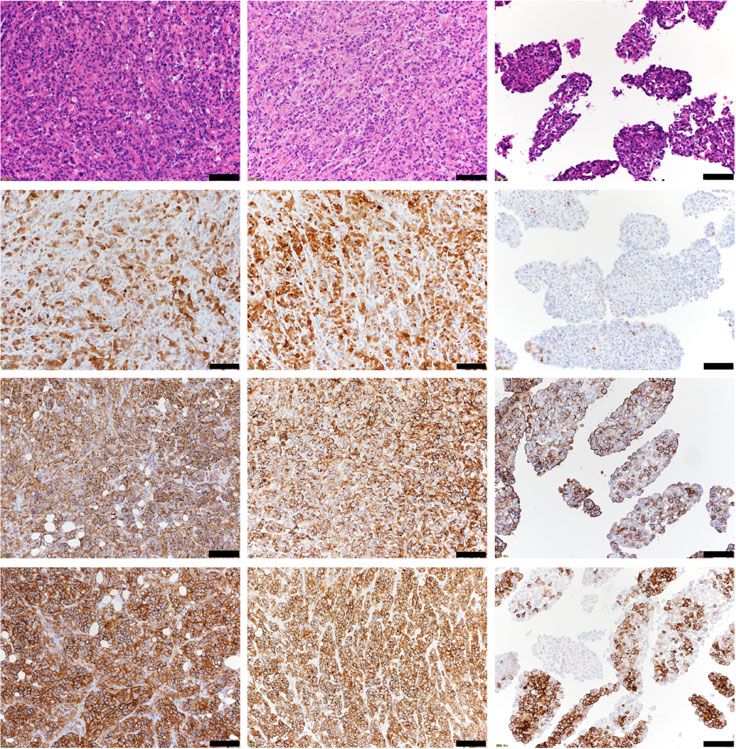
Matsuzawa et al. BMC Cancer   (2021) 21:200                                                                   Page 2 of 16

Background                                                     unresectable stage 3 or 4 pancreatic cancer. However, the
Pancreatic cancer shows rapid growth and metastasis            study did not demonstrate the efficacy of combined ther-
and is one of the most fatal human cancers. Over half of       apy compared with either Amatuximab or chemotherapy
patients are diagnosed at a stage where metastases have        alone. The trial included patients with unresectable bulky
developed, and the overall 5-year survival rate for the        pancreatic cancer, and we suspect that Amatuximab could
pancreatic patients with metastases is only 10% [1, 2].        not be delivered to pancreatic tumor cells in these
Only 15–20% of patients have resectable disease at the         patients.
time of diagnosis [3]. The majority of patients have local        We previously reported that Amatuximab monother-
and distant micrometastases at the time of surgery.            apy reduced the peritoneal mass volume in peritonitis
Therefore, disease recurrence following operation is very      model mouse of human pancreatic cancer cells and ob-
high. Hattangadi et al. reported that the peritoneum           served the appearance of sherbet-like aggregates in this
(55%) and the liver (53%) were the most common sites           model [21]. We demonstrated that the combined ther-
of recurrence [4].                                             apy using Amatuximab and gemcitabine reduced the
   Adjuvant therapies such as radiotherapy and chemo-          peritoneal mass and eliminated the sherbet-like aggre-
therapy (or a combination) help improve the prognosis          gates. However, the molecular mechanism of this
of pancreatic cancer. However, their effectiveness is lim-     phenomenon is not yet understood.
ited. Gemcitabine [5] or S-1 [6] is the most common ad-           The aim of this study is to clarify the role and poten-
juvant chemotherapy for pancreatic cancer after surgery.       tial mechanism of mesothelin receptor blockage by
Recent studies showed that in patients with advanced           Amatuximab in human pancreatic cell lines both in vitro
disease, combining gemcitabine with other systemic             and in vivo.
agents can improve patient outcome compared with
standard gemcitabine monotherapy [7]. For example, the         Methods
combined therapy with gemcitabine plus albumin-bound           Cell culture
paclitaxel reached 8.7 months of median overall survival       The human pancreatic adenocarcinoma AsPC-1 and
in phase III trial [2]. Although the prognosis of this dis-    Capan-2 cell lines were cultured in RPMI-1640 medium
ease has been improved, these results were not accept-         supplemented with 10% FBS, 100 units/ml penicillin and
able for the patients in clinical. Thus, the identification    100 μg/ml streptomycin. Panc-1 and MIA Paca-2 cell
of novel and effective strategies is critical to improve the   lines were cultured in DMEM medium supplemented
prognosis for pancreatic cancer patients.                      with 10% FBS, 100 units/ml penicillin and 100 μg/ml
   Mesothelin is a 40-kDa glycosylphosphatidyl inositol-       streptomycin. The cells were cultured and maintained as
linked protein that is expressed in mesothelial cells lin-     previously described [21]. We obtained AsPC-1 cells
ing the peritoneum among the human normal tissues              (ATCC® CRL-1682™) in 2010, MIA Paca-2 (ATCC® CRL-
[8]. Mesothelin is highly overexpressed in various types       1420™) cells and Panc-1 (ATCC® CRL-1469™) cells in
of cancers, such as mesothelioma [8], ovarian [8] and          2016 and Capan-2 cells (ATCC® HTB-80™) in 2019. All
pancreatic cancer [9]. Recent studies have demonstrated        cell lines were obtained from ATCC (Manassas, VA,
a role for mesothelin in cell survival, cell migration, cell   USA). After obtaining cells from ATCC, cells were fro-
invasion and tumor progression [10]. The expression of         zen at low passage (p3-p5), and then thawed and used at
mesothelin is also related to epithelial-mesenchymal           passage 10 or lower. These cell lines were tested for
transition (EMT) and cancer stem cells (CSCs) [11]. We         mycoplasma contamination at regular intervals. we did
previously reported the significance of mesothelin ex-         not authenticate these cell lines.
pression in the poor prognosis of gastrointestinal cancer
patients [12–17]. Because of the expression pattern,           Reagents
mesothelin has recently been targeted for immunother-          Amatuximab (also known as MORAb-009) was obtained
apy [18].                                                      from MORPHOTEK INC (Exton, PA, USA). Gemcita-
   Amatuximab is an investigational chimeric high-affinity     bine hydrochloride (Gemzar) was purchased from Eli
monoclonal IgG1/k antibody that targets mesothelin [19].       Lilly (Indianapolis, IN, USA).
In vitro, Amatuximab elicits antibody-dependent cellular
cytotoxicity against mesothelin-expressing tumor cell lines    Proliferation assay (cell counting method)
and inhibits heterotypic cell adhesion of mesothelin-          Pancreatic cancer cell lines were seeded at a density of
positive tumor cells to CA125-expressing tumor cells.          1.5 × 10⁵ cells per 2 mL medium in a 35 mm plate, and
Based on its safety in a Phase I study and preclinical stud-   Amatuximab was added at various concentrations (0,
ies showing synergy with chemotherapy [20], Amatuximab         0.01, 1 or 100 μg/mL). After 48 h, cells were harvested
was combined with gemcitabine in a Phase II study              by Trypsin/EDTA (0.05%) (Thermo Fisher Scientific
(NCT00570713) in patients with previously untreated            Inc., Waltham, MA, USA). Next, 50 μL of the cell
Matsuzawa et al. BMC Cancer    (2021) 21:200                                                                Page 3 of 16

suspension were mixed with 50 μL of 0.4% trypan blue        and cells were cultured for 24 or 48 h. After cells were
(#207–17,081, Wako, Tokyo, Japan) by gentle pipetting       harvested, western blot analysis was performed as previ-
and then 20 μL of the mix were loaded into a chamber        ously described [21]. The cells were homogenized at 4 °C
in a hemocytometer. Counts were performed in tripli-        in lysis buffer (0.1% SDS, 1% Igepal CA-630, 0.5% sodium
cate by one analyst under a 40× objective according to      deoxycholate) and a protease inhibitor cocktail (Sigma-Al-
standard methodology using BIOREVO (#BZ-9000,               drich, St. Louis, MO, USA). Cell lysates (20–50 μg) were
KEYENCE, Osaka, Japan).                                     resolved by electrophoresis on polyacrylamide gels and
                                                            transferred to PVDF membranes (Millipore, Billerica, MA,
Invasion assay                                              USA). After blocking the membranes in 5% non-fat dry
In vitro invasion assays were performed using the BD        milk or 3% bovine serum albumin in Tris-buffered saline
Bio-Coat Matrigel invasion assay system (BD Biosci-         for 1 h at room temp, the blots were hybridized overnight
ences, San Jose, CA, USA) according to the manufac-         at 4 °C with primary antibodies. After hybridization with
turer’s instructions. Briefly, cells were seeded into       secondary antibodies conjugated with HRP (Cell Signaling
Matrigel-precoated Transwell chambers consisting of         Technology, Danvers, MA, USA), immunocomplexes were
polycarbonate membranes with 8.0 μm pores. The              visualized using an enhanced chemiluminescence kit (GE
Transwell chambers were then placed into 24-well            Healthcare, Chalfont St. Giles, UK). Densitometric analysis
plates, into which basal medium only or basal medium        of western blots was performed using a ChemiDoc XRS
containing 100 μg/mL Amatuximab or control IgG was          Plus system with Image Lab Software (Bio-Rad, Hercules,
added. We decided the concentration of Amatuximab or        CA, USA). Primary antibodies are described in Supple-
control IgG from our data in proliferation assay and the    mental Table 1.
past literature, which showed the strong blockage of ad-
hesion between mesothelin and CA125 in this concen-         FACS analysis
tration of Amatuximab [19]. After incubating cells for      We harvested, washed and resuspended the cells in ice-
24 h, the upper surface of the Transwell chambers was       cold PBS. Primary antibody was added at 2 μg/ml and
wiped with a cotton swab and the invading cells were        incubated for 1 h on ice. Cells were then washed with
fixed and stained using the Diff-Quick cell staining kit    PBS, incubated with Alexa Fluor 488 goat-anti-mouse
(Dade Behring, Inc., Newark, DE, USA). We counted the       secondary antibody (Invitrogen, Carlsbad, CA, USA) di-
number of invading cells in five random microscopic         luted to 1 μg/ml, washed as above and analyzed on BD
fields (200× magnification) using BIOREVO (#BZ-9000,        FACS Canto™ II flow cytometer (BD Biosciences).
KEYENCE, Osaka, Japan).
                                                            Animals
Migration assay                                             Six-week-old female BALB/c Slc-nu/nu mice (body
In vitro migration assays were performed using the BD       weight 14-19 g) were purchased from CLEA Japan, INC.
Bio-Coat migration assay system (BD Biosciences) ac-        (Tokyo, Japan). Mice were maintained under specific
cording to the manufacturer’s instructions and the          pathogen-free conditions in laminar-flow benches and
methodology for the invasion assays.                        were allowed to adapt to the environment for 1 or 2
                                                            weeks before experiments. All procedures involving ani-
Cell growth inhibition assay                                mals and their care were approved by the Institutional
Pancreatic cancer cells were seeded at a density of 1.5 ×   Animal Care and Use Committee of National University
105 cells per 35 mm dish in culture medium containing       Corporation Hokkaido University and were conducted
10% FBS with 1 μM gemcitabine and 100 μg/ml Amatux-         under National University Corporation Hokkaido
imab or control IgG. After 48 h, the numbers of viable      University Regulations on Animal Experimentation.
cells were counted as described in the previous section.
The concentration of gemcitabine was determined based       Generation of peritoneal model mouse using AsPC-1 cells
on previous studies [22] and our preliminary experi-        We generated the peritoneal model mouse as described
ments. Counts were performed in triplicate by one ana-      previously [21]. Briefly, 5 × 10⁶ AsPC-1 cells were
lyst under a 40× objective according to standard            injected to the abdominal cavity of 7-week-old nude
methodology using BIOREVO (#BZ-9000, KEYENCE,               mice. The mice were divided to two groups randomly.
Osaka, Japan).                                              Amatuximab (200 mg/kg) (n = 6) or isotype control IgG
                                                            (200 mg/kg) (n = 4), so totally using 10 mice, was admin-
Western blot analysis                                       istered by injection into the abdominal cavity every 2
Cells were seeded at a density of 1.5 × 10⁶ cells in 2 ml   days for 22 days. Two groups were maintained in the
medium in a 100 mm dish. Amatuximab (100 μg/ml) or          same type of cage and the environment. The mice were
control IgG was added to medium at the time of seeding      euthanized by isoflurane inhalation followed by cervical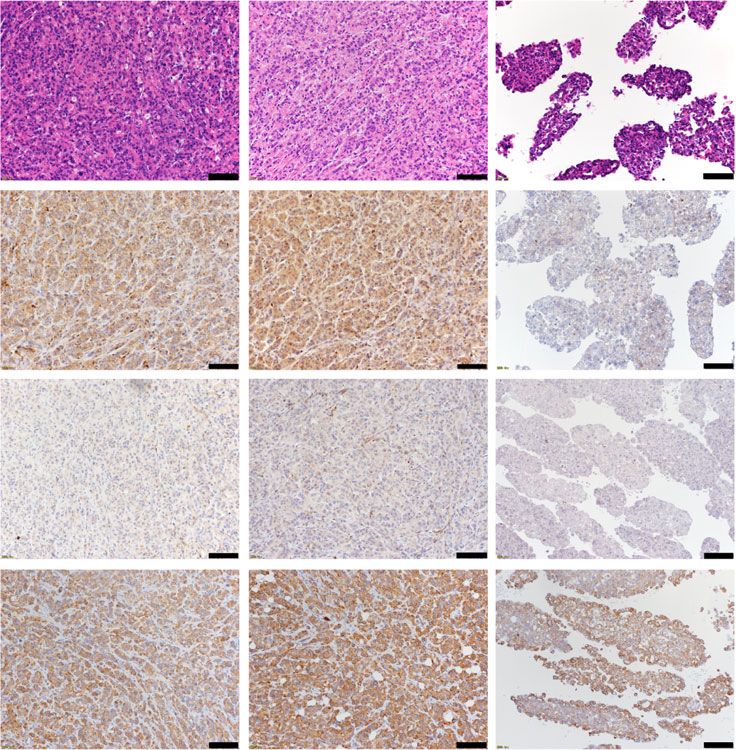
Matsuzawa et al. BMC Cancer   (2021) 21:200                                                                   Page 4 of 16

dislocation on day 22 and peritoneal masses and              Statistical analysis
sherbet-like aggregations were harvested. No expected        Data are shown as mean ± SD. Significance of differences
or unexpected adverse events happened. The resected          was determined by Student’s t-test, one-way ANOVA,
specimens were fixed with formalin for 6 h immediately       two-way ANOVA or Mann-Whitney U-test. P < 0.05 was
after harvest and embedded in paraffin, and then sliced      considered statistically significant. All statistical proce-
sequentially at a thickness of 3 μm. We established the      dures were performed using JMP Pro 10 (SAS Institute,
humane endpoints that when the model mouse got poor          Japan).
physical condition during the experiment, we planned to
euthanize by isoflurane inhalation followed by cervical      Results
dislocation. No animals were adapted to the criteria. We     Comparison of mesothelin expression in four pancreatic
set no specific criteria used for including and excluding    cancer cell lines
animals during the analysis. The first author understood     We first evaluated mesothelin expression in AsPC-1,
the group allocation totally through the experiment.         Capan-2, Panc-1 and MIA Paca-2 pancreatic cell
These protocols were prepared before the study.              lines. Western blotting analysis revealed that Capan-
                                                             2 cells showed the strongest expression of mesothe-
                                                             lin among the four cell lines. Although the expres-
Immunohistochemistry                                         sion of mesothelin in AsPC-1 cells was weaker than
We performed the immunohistochemistry using the En-          that in Capan-2, AsPC-1 showed the stronger ex-
Vision+ System-HRP (Dako Japan, Tokyo), in the same          pression than those in Panc-1 and MIA Paca-2 cells.
method as described previously [21]. Briefly, the sections   Although the expression of mesothelin in Panc-1
were mounted on glass slides, deparaffinized, and rehy-      was detected, that was weak compared with those in
drated through several graded ethanol. We retrieved the      Capan-2 and AsPC-1 (Fig. 1a). The result using an-
antigens in Dako EnVision FLEX Target Retrieval Solu-        other primary antibody for mesothelin showed that
tion low pH using Dako PT Link for 20 min at 97 °C ac-       the mesothelin expression in Panc-1 was low level
cording to the manufacturer’s instructions (Dako,            (Supplemental Figure 1), and the order of expres-
Japan). After we blocked the endogenous peroxidase ac-       sion level was same in both results. FACS (Fig. 1b)
tivity with 0.03% hydrogen peroxide, the tissue sections     and immunocytochemistry analysis (Supplemental
were incubated with primary antibodies at room temp          Figure 7) of mesothelin expression in the four pan-
for 30 min and then we reacted the samples with a dex-       creatic cell lines were consistent with the western
tran polymer reagent combined with secondary anti-           blot analysis. In consideration of these results to-
bodies and peroxidase for 30 min at room temperature.        tally, we thus selected AsPC-1 and Capan-2 cells as
Specific antigen-antibody reactions were visualized with     pancreatic cancer cell lines strongly expressing
diaminobenzidine chromogen applied for 10 min. After         mesothelin (mesothelin-high), and Panc-1 and MIA
that, these slides were stained with hematoxylin, dehy-      Paca-2 cells as cell lines expressing low levels of
drated and mounted. The primary antibodies are de-           mesothelin (mesothelin-low) for subsequent in vitro
scribed in Supplemental Table 1. The percentage of           experiments.
stain-positive cells and strength of staining were evalu-
ated in the analysis. The representative images were         Suppression of invasiveness and migration capacity in
taken using OLYMPUS equipment described below                mesothelin-high pancreatic cancer cell lines by
(microscope BX63–44-FLD-2, Camera DP80, software             Amatuximab treatment
cellSens, OLYMPUS, Tokyo, Japan).                            We next examined the effects of blockade of
                                                             mesothelin by Amatuximab on the proliferation of
                                                             pancreatic cancer cells. AsPC-1, Capan-2, Panc-1 and
Quantitative RT-PCR                                          MIA Paca-2 cells were treated with various concen-
Total RNA from tissues and cells was isolated using          trations of Amatuximab (0, 0.01, 1 or 100 μg/mL).
TRIzol reagent (Invitrogen). Relative levels of E-           Amatuximab treatment had no effect on the prolifera-
cadherin, ALDH1, CD44 and c-MET mRNA were exam-              tion of either of the cell lines (Fig. 2a). We examined
ined using SYBR Green Real-time Quantitative RT-PCR          the effects of Amatuximab on the invasion capacities
(qRT-PCR) (LightCycler 480 Roche, Switzerland) and           of AsPC-1, Capan-2, Panc-1 and MIA Paca-2 cells.
were normalized to levels of GAPDH mRNA. We de-              We showed the representative images of invasion
scribed the primers in Supplemental Table 2. Relative        assay upon AsPC-1 cells in Fig. 2b. The results
expression was calculated using the 2-ΔΔCT method. All       showed that the invasiveness was suppressed in
qRT-PCR analyses were carried out in triplicate, and the     AsPC-1 and Capanc-2 cells upon treatment with
data are presented as means±standard of the means.           Amatuximab compared with control treatments. In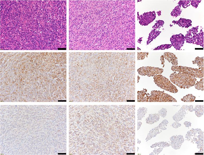
Matsuzawa et al. BMC Cancer       (2021) 21:200                                                                                        Page 5 of 16

 Fig. 1 Comparison of mesothelin expression in human pancreatic cancer cell lines. a Whole cell lysates of AsPC-1, Capan-2, Panc-1 and MIA Paca-
 2 cells were analyzed for mesothelin expression by western blotting. β-actin was used as an internal control. Full-length blots/gels are presented
 in Supplemental Figure 2. Densitometric analysis of western blots was performed using a ChemiDoc XRS Plus system with Image Lab Software
 (Bio-Rad, Hercules, CA, USA). b FACS analysis of mesothelin expression on the surface of the four human pancreatic cancer cell lines. Onecomp
 eBeads (#01–1111, eBioscience) is used for positive control and FITC Mouse IgG1κ Isotype Control (#555748, BD Biosciences) is used for negative
 control. The results of analysis of mesothelin expression in the four human pancreatic cancer cells by immunocytochemistry were shown in
 Supplementary Figure 2

contrast, Amatuximab had no impact on the invasion                          Reduced levels of pMET expression in mesothelin-high
of Panc-1 and MIA Paca-2 cells (Fig. 2c). We also ex-                       pancreatic cell lines treated with Amatuximab
amined the effects of Amatuximab on the migration                           We next investigated the mechanism underlying the ef-
capacities of AsPC-1 and MIA Paca-2 cells. The rep-                         fects of Amatuximab in pancreatic cancer cells by exam-
resentative images were shown in Fig. 2d. The results                       ining changes in molecular factors in response to
showed that the migration capacities were suppressed                        Amatuximab treatment using western blotting analysis.
in AsPC-1 cells upon treatment with Amatuximab                              We examined a panel of CSC-related molecules and
compared with control treatments. In contrast, Ama-                         found that the levels of p-MET were reduced in both
tuximab had no impact on the migration capacity of                          AsPC-1 and Capan-2 cells (mesothelin-high) treated
MIA Paca-2 cells (Fig. 2e).                                                 with Amatuximab compared with controls. In contrast,

Matsuzawa et al. BMC Cancer   (2021) 21:200                                                                     Page 6 of 16

                                               Fig. 2 Effect of Amatuximab on pancreatic cancer cell proliferation,
                                               invasion and migration. a AsPC-1, Capan-2, Panc-1 and MIA Paca-2 cells
                                               were incubated with the indicated concentrations of Amatuximab for 48 h.
                                               Viable cells were stained with trypan blue and counted. N.S., not significant.
                                               b, c Invasion assays were performed in AsPC-1, Capan-2, Panc-1 and MIA
                                               Paca-2 cells treated with Amatuximab (100 μg/mL) or control IgG (100 μg/
                                               mL) for 13 h. d, e Migration assays were performed in AsPC-1 cells and MIA
                                               Paca-2 cells treated with Amatuximab (100 μg/mL) or control IgG (100 μg/
                                               mL) for 13 h. *P < 0.05. 1HPF: one high power field. Scale bar, 100 μm

                                              no changes in p-MET levels were observed in Panc-1
                                              and MIA Paca-2 cells (mesothelin-low) treated with
                                              Amatuximab. The expression level of CD44 in AsPC-1
                                              cells treated with Amatuximab was decreased compared
                                              with controls, however this phenomenon was not ob-
                                              served in Capan-2 cells and other mesothelin-low cells
                                              (Fig. 3a).

                                              No significant changes in expression of ERK/MEK pathway
                                              nor NFκB/Stat3 pathway proteins in mesothelin-high
                                              pancreatic cancer cells treated with Amatuximab
                                              We also examined molecular factors related to prolifera-
                                              tion and chemoresistance in pancreatic cell lines treated
                                              with Amatuximab. We observed the slight decrease in
                                              the expressions of MEK1/2 and ERK1/2 in AsPC-1 cells
                                              upon treatment with Amatuximab, but not in significant.
                                              Using the same approach, we examined molecules re-
                                              lated to cell survival, invasion and migration in pancre-
                                              atic cell lines treated with Amatuximab. We observed
                                              the slight decrease in the expressions of p-NFκB and p-
                                              Stat3 in AsPC-1 cells upon treatment with Amatuximab,
                                              but not in significant (Fig. 3b).
                                                We did not detect any changes in EMT-related pro-
                                              teins or mesothelin expression in either cells upon Ama-
                                              tuximab treatment (Fig. 3c).

                                              Suppression on cell growth of combined Amatuximab
                                              and gemcitabine treatment in mesothelin-high pancreatic
                                              cancer cell lines
                                              We next examined the effect of combination treatment
                                              with Amatuximab and gemcitabine in pancreatic cancer
                                              cell lines. The results showed that the combined therapy
                                              suppressed the proliferation of AsPC-1 and Capan-2
                                              cells (mesothelin-high) more strongly than gemcitabine
                                              alone. Notably, these results were not observed in Panc-
                                              1 and MIA Paca-2 cells (Fig. 4).

                                              Suppression of E-cadherin in sherbet-like aggregates
                                              We generated the mouse peritoneal dissemination model
                                              of pancreatic cancer using the previously reported proto-
                                              col [21] and treated mice either with Amatuximab or
                                              isotype control IgG as described in methods. No animals
                                              were excluded during the experiment and the analysis.
                                              We examined mass lesions and sherbet like aggregates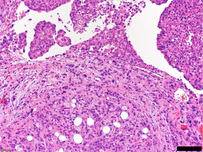
Matsuzawa et al. BMC Cancer          (2021) 21:200                                                                                                Page 7 of 16

 Fig. 3 Molecular changes in human pancreatic cancer cells exposed to Amatuximab. Western blot analyses in AsPC-1, Capan-2, Panc-1 and MIA Paca-2 cells
 treated with control IgG or Amatuximab for the indicated proteins related to a cancer stem cells, b survival and chemoresistance, and c epithelial-mesenchymal
 transition. GADPH was used as an internal control. Ful-length blots are presented in Supplemental Figures 3, 4 and 5. Densitometric analysis of western blots
 was performed using a ChemiDoc XRS Plus system with Image Lab Software (Bio-Rad, Hercules, CA, USA)
Matsuzawa et al. BMC Cancer (2021) 21:200 Page 8 of 16
Fig. 4 Effect of Amatuximab and gemcitabine combination treatment in human pancreatic cancer cell lines. AsPC-1, Capan-2, Panc-1 and MIA Paca-2
cells were treated with gemcitabine (1 μM) and/or Amatuximab (100 μg/mL) for 48 h. Viable cells were stained with trypan blue and counted
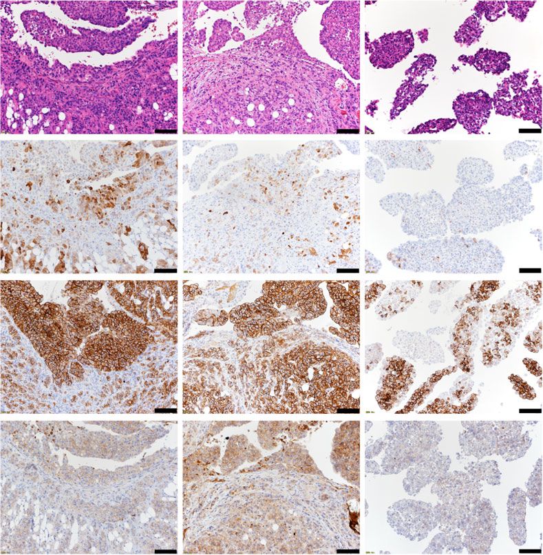
by immunohistochemistry. No changes were observed in immunohistochemistry results (Fig. 5g). These results
mesothelin expression between control and Amatuximab showed that the stemness of sherbet-like aggregates in
mice (Fig. 5a). The expression of p-ERK1/2 was the Amatuximab group was suppressed compared with
heterogenous in sherbet-like aggregates of Amatuximab- that of peritoneal metastasis tissues.
treated group in contrast to strong homogenous expres-
sions in mass lesions of both Amatuximab-treated group
and control group. Ki-67 expression was reduced in Cancer cell clusters adhesive to the surface of peritoneal
sherbet-like aggregates of Amatuximab-treated group metastasis are morphologically similar to sherbet-like
compared with mass lesions of both Amatuximab- aggregates
treated group and control group (Fig. 5a, b). Evaluation We found the cancer cell clusters adhesive to the surface
of EMT-related protein expression revealed no changes of peritoneal metastasis, those were morphologically
except for the suppression of E-Cadherin expression in similar to the sherbet-like aggregates (Fig. 6a). The fre-
Amatuximab groups (Fig. 5c). quencies of these clusters were not related to the treat-
ment or absence of Amatuximab (data not shown).
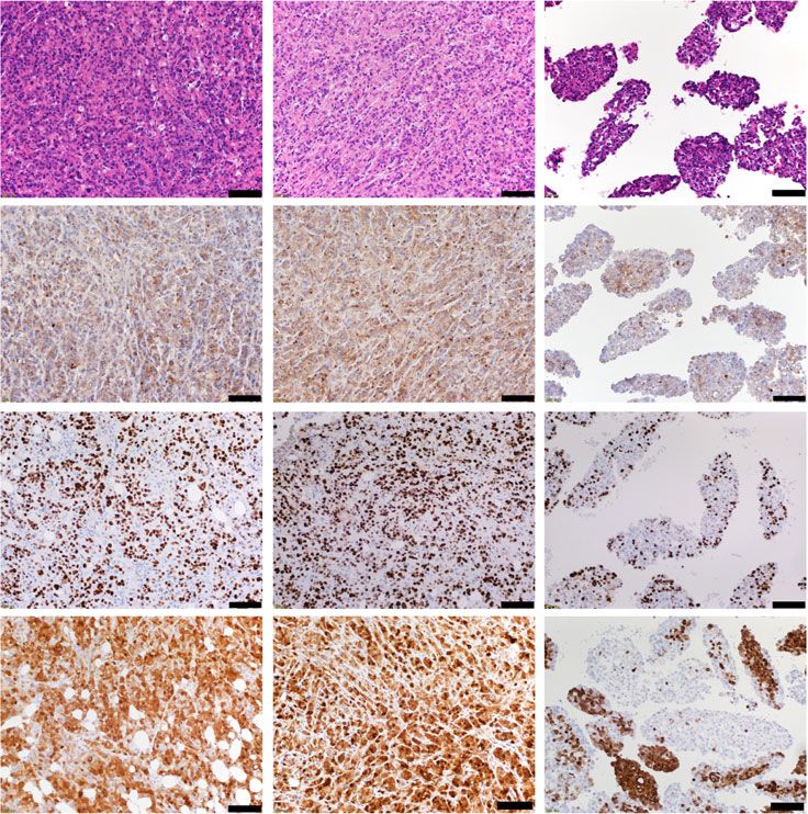
Suppression of CSC-related molecules in sherbet-like
aggregates
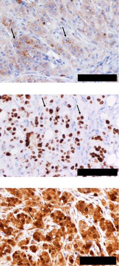
We next examined the expressions of CSC-related mole- Protein expressions in cancer cell clusters attached to the
cules. We observed the suppression of ALDH1/c-MET/ peritoneal metastasis tissue
CD44 expression in sherbet-like aggregates in Amatuxi- We next examined the protein expressions in the cancer
mab mice compared with those of mass lesions in both cell clusters by immunohistochemistry. The expressions
Amatuximab-treated group and control group (Fig. 5d, of ALDH1, c-MET and CD44 were enhanced in cancer
e). Expression of CXCR4 showed no change, while cell clusters attached to the metastases compared with
CD133 expression was enhanced in sherbet-like aggre- those in sherbet-like aggregates in Amatuximab mice
gates compared with those of mass lesions in both (Fig. 6b). The protein expressions in cancer cells adhered
Amatuximab-treated group and control group (Fig. 5f). to metastases were more similar to those of peritoneal
We also examined the mRNA expressions of these mole- metastasis tissue than those of the sherbet-like aggre-
cules, and the results were consistent with the gates (Fig. 6b, c).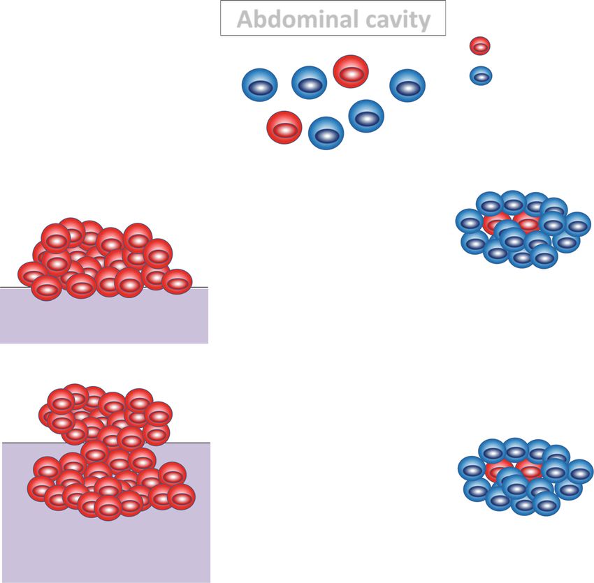
Matsuzawa et al. BMC Cancer        (2021) 21:200                                                                                                                            Page 9 of 16

   A                                                                                                                         C

   B                                                                     100
                                                                                             Mesothelin
                                                                                                 N.S.                       D
                                                                          80                            N.S.
                                         % Mesothelin positive cells

                                                                          60

                                                                          40

                                                                          20

                                                                           0
                                                                                   Control     Amatuximab      Amatuximab
                                                                                    mass         mass            sherbet

                                                                                                 Ki-67

                                                                         100
                                                % Ki-67 positive cells

                                                                          80

                                                                          60

                                                                          40

                                                                          20

                                                                           0
                                                                                   Control      Amatuximab     Amatuximab
                                                                                    mass          mass           sherbet

                                                                                                 p-ERK 1/2
                                          % p-ERK1/2 positive cells

                                                                          100

                                                                           80

                                                                           60

                                                                           40

                                                                           20

                                                                               0
                                                                                   Control     Amatuximab      Amatuximab
                                                                                    mass         mass            sherbet

 Fig. 5 Immunohistochemical analysis in the pancreatic peritoneal dissemination model treated by Amatuximab. Immunohistochemistry was
 performed on indicated sections from Amatuximab or control treated model mice for proteins related with a chemoresistance, c epithelial
 mesenchymal transition, adhesion, d, f stemness and metastasis. b, e Quantification of positively stained cells for the indicated proteins. Arrows
 indicate the representative image of the positively stained cells. The frequencies of positively stained cells were counted in six high power fields
 that were chosen at random. g Quantitative PCR of the indicated factors in RNA samples harvested from blocks. *P < 0.05. Scale bar, 100 μm

Amatuximab inhibits the adhesion of cancer cells to                                                                         adhesion and the molecular change by mesothelin, al-
peritoneum and suppresses the stemness and viability of                                                                     though a part of disseminated cancer cells makes peri-
those, that lead to enhanced sensitivity for gemcitabine                                                                    toneal metastasis alike those without Amatuximab,
A schematic for a model of our proposed hypothesis is                                                                       another part of cancer cells was inhibited to adhere to
shown in Fig. 7. Pancreatic cancer cells disseminated in                                                                    the peritoneum and keep differentiated floating in the
the abdominal cavity adhere to the peritoneum and de-                                                                       ascites. These floating cancer cell clusters have high sen-
differentiate, invade and undergo metastasis. The stable                                                                    sitivity for gemcitabine and can be eliminated by the
metastases eventually acquire resistance to gemcitabine.                                                                    combination chemotherapy of gemcitabine and
Upon treatment with Amatuximab that blocks the                                                                              Amatuximab.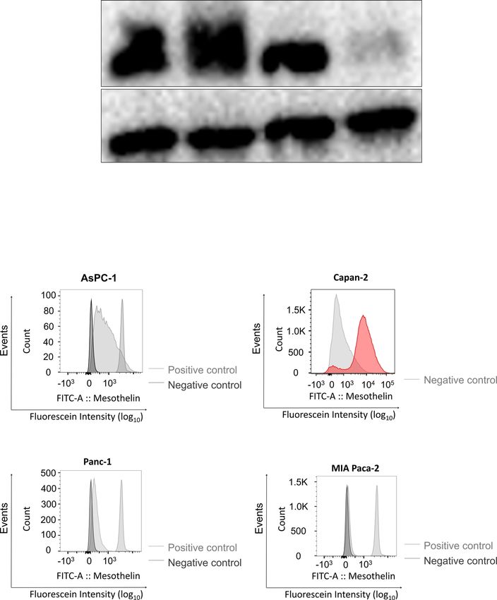
Matsuzawa et al. BMC Cancer (2021) 21:200 Page 10 of 16
Figure 5 continued
E ALDH1 F
100
% ALDH1 positive cells
80
60
40
20
0
Control Amatuximab Amatuximab
mass mass sherbet
CD44
% CD44 positive cells
100
80
60
40
20
0
Control Amatuximab Amatuximab
mass mass sherbet
c-MET
% c-MET positive cells
100
80
60
40
20
0
Control Amatuximab Amatuximab
mass mass sherbet
E-Cadherin ALDH1
Relative mRNA expression of E-Cadherin
G
Relative mRNA expression of ALDH1
1.4
1.2
1.2
1
1
0.8
0.8
0.6
0.6
0.4 0.4
0.2 0.2
0 0
Control Amatuximab Amatuximab Control Amatuximab Amatuximab
mass mass sherbet mass mass sherbet
CD44 c-MET
Relative mRNA expression of c-MET
Relative mRNA expression of CD44
1.2 1.2
1 1
0.8 0.8
0.6 0.6
0.4 0.4
0.2 0.2
0 0
Control Amatuximab Amatuximab Control Amatuximab Amatuximab
mass mass sherbet mass mass sherbet
Fig. 5 Immunohistochemical analysis in the pancreatic peritoneal dissemination model treated by Amatuximab. Immunohistochemistry was
performed on indicated sections from Amatuximab or control treated model mice for proteins related with a chemoresistance, c epithelial
mesenchymal transition, adhesion, d, f stemness and metastasis. b, e Quantification of positively stained cells for the indicated proteins. Arrows
indicate the representative image of the positively stained cells. The frequencies of positively stained cells were counted in six high power fields
that were chosen at random. g Quantitative PCR of the indicated factors in RNA samples harvested from blocks. *P < 0.05. Scale bar, 100 μm
Discussion cancer cells. Amatuximab also induced the downregula-
In the present study, we demonstrated significant effects tion of CSC-related proteins such as pMET in these cell
of Amatuximab by mesothelin blockage in pancreatic lines.
cancer cells. Our in vitro results showed that Amatuxi- A full understanding of the biological functions of
mab treatment suppressed the invasiveness and gemcita- mesothelin is lacking given that mesothelin knockout
bine sensitivity of AsPC-1 and Capan-2 pancreatic mice do not show any developmental phenotype [23].Matsuzawa et al. BMC Cancer (2021) 21:200 Page 11 of 16 Recent reports indicate that mesothelin may play an im- the other side that downregulates mesenchymal and portant role in cell adherence, cell survival/proliferation, CSC regulatory genes that relieves self-renewal, prolifer- tumor progression and chemoresistance [24]. In pancre- ation, dissemination and metastasis of cancer cells. atic cancer, Stat3 plays a pivotal role in oncogenic trans- Overexpression of mesothelin in normal cells stimulates formation [25, 26], cell survival, proliferation [25, 27] anchorage-independent growth, migration and invasion and resistance to apoptosis [28]. Stat3 is also aberrantly [11]. In our study, the mesothelin blockage by Amatuxi- activated in a subset of pancreatic tumor tissues and cell mab directly suppressed the expression level of pMET lines [27]. Bharadwaj et al. showed that MSLN upregula- and led to suppression of malignant features of AsPC-1 tion induces the activation of Stat3 in pancreatic cancer and Capan-2 pancreatic cancer cells. To the best of our cells [29]. Furthermore, the authors showed that knowledge, this is the first study to establish a relation- mesothelin induced an NFκB/Akt-dependent anti- ship between mesothelin blockage and CSC markers. apoptotic pathway that can protect pancreatic cancer MET (also known as c-MET) is a receptor of the tyro- cells from TNF-α-induced apoptosis, and this was a sine kinase family that acts as a proto-oncogene and is probable mechanism of pancreatic cancer cell survival in stimulated by hepatocyte growth factor to mediate mo- midst of inflammation and inflammatory mediators. tility, invasion, and metastasis [43]. The intracellular sig- Other groups found that mesothelin could confer resist- naling cascades activated by MET include the RAS- ance to cytotoxic drug-induced apoptosis via the ERK MAPK and PI3K-Akt pathways, NFκB and Wnt/GSK- signaling pathway [30, 31]. Some mesothelin monoclonal 3β/β-Catenin signaling [44]. The levels of c-MET are in- antibodies were reported as unable to inhibit cancer cell creased in pancreatic carcinoma where c-MET signaling proliferation because the majority of these antibodies induces growth and invasion and some authors have re- target N terminal region I rather than a key signaling ported c-MET as a stem cell marker in pancreatic tissue domain in mesothelin. Our results showed that Amatux- [44]. In our results, we concluded that MET was the im- imab did not inhibit cell proliferation, but the agent sup- portant factor in the effects of Amatuximab by mesothe- pressed the invasiveness and chemoresistance in AsPC-1 lin blockage in pancreatic cancer cells. and Capan-2 pancreatic cancer cells. We could observe Although the molecular changes of AsPC-1 show the the slight changes of the molecule expression level in limited effects of Amatuximab in vitro study, drastic the pathway described above in AsPC-1 only. These re- changes observed in vivo study. Using the peritoneal dis- sults might be caused by the difference of the expression semination model, we demonstrated that several pro- level of mesothelin, that of the expression pattern of teins related to proliferation, cancer stemness and MMP7, or that of the mutation pattern of p53 between chemoresistance were suppressed in the cancer cell clus- AsPC-1 cells and Capan-2 cells [32, 33]. These factors ters (which we named sherbet-like aggregates) that were might lead to the dissociated results. blocked to attach to peritoneum by mesothelin blockage. CSCs are a population of undifferentiated tumorigenic We previously demonstrated that the sherbet-like aggre- cells that are responsible for tumor initiation, tumor gates were sensitive to gemcitabine [21]. Marjanovic pre- maintenance and tumor cell spreading to distant organ sented a plastic CSC theory as a model of tumor sites [34]. These cells exhibit unlimited proliferation po- heterogeneity [45]. The classical CSC theory proposes tential, self-renewal ability and the capacity for the gen- that tumor heterogeneity would arise when cancer cells eration of a progeny of differentiated cells which within a given tumor reside in different states of stem- constitute the major tumor population. CSCs can be ness or differentiation. Critical to this theory is the no- characterized by chemoresistance [35], multipotency, tion that CSC-to-non-CSC conversion is a unidirectional tumorigenicity [36], stem gene expression [37] and alde- process. The plastic CSC theory describes an evolving hyde dehydrogenase activity [38]. Several studies showed model in which bidirectional conversions exist between that chemotherapy treatment in pancreatic cancer cell non-CSCs and CSCs that are controlled by extrinsic fea- lines led to an increased number of CSCs [39, 40]. CSCs tures, such as extracellular matrix or blood vessels, and are also thought to be related to the invasiveness of can- intrinsic features, such as genetic or epigenetic changes. cer cells [41]. Ortensi et al. reported that brain tumor This model implies that non-CSCs can continually cre- cells enriched for stem cell markers displayed greater ate CSC populations throughout tumorigenesis. In other migratory and invasive potential compared with stem words, this theory suggests that CSCs are kept in differ- cell marker-negative tumor cells. We can identify the entiated states by their surroundings, and their malig- CSCs by the expression of CSC-specific cell surface nancy is suppressed in this condition. Stankevicius et al. markers, such as CD133, CD44, c-MET and ALDH1 reported the importance of microenvironment, including [42]. He et al. showed that mesothelin regulates EMT scaffold, for the cancer cell stemness in human colorec- and CSC traits. Knockdown of mesothelin upregulates tal cancer cells [46]. The authors demonstrated that the epithelial and adhesion molecules on one side and on expressions of several CSC markers were increased in
Matsuzawa et al. BMC Cancer (2021) 21:200 Page 12 of 16
A B
C c-MET
ALDH1
100
% c-MET positive cells
% ALDH1 positive cells
80
100
60 80
60
40
40
20 20
0
0 Control Amatuximab Amatuximab
Control Amatuximab Amatuximab adhesive sherbet adhesive sherbet sherbet
adhesive sherbet adhesive sherbet sherbet
CD44
100
% CD44 positive cells
80
60
40
20
0
Control Amatuximab Amatuximab
adhesive sherbet adhesive sherbet sherbet
Fig. 6 Molecular transition of sherbet like aggregates attaching to mesothelium. a H&E image of peritoneal mass from Amatuximab treated mice.
b Immunohistochemistry of the indicated proteins. c Immunohistochemistry of the indicated proteins shown on the left; the frequencies of
positive stained cells in each treatment group were calculated and shown on the right. Arrows indicate the representative image of the positively
stained cells *P < 0.05. Scale bar, 100 μm
3D cultures considered as scaffold compared with those molecules using the cells those expression of
in 2D monolayer cultures. Our results suggested that mesothelin were genetically controlled. Second, we
not only the direct effects of Amatuximab but also the could not confirm the definite role of p-MET down-
environmental changes induced by Amatuximab could regulation in the antitumor activity of Amatuximab
control the stemness of pancreatic cancer cells express- directly in this study. To confirm that, we should
ing high mesothelin, and these resulted in an improve- carry out the experiment such as invasion assay, mi-
ment of sensitivity for gemcitabine in mesothelin- gration assay and gemcitabine sensitivity experiments
expressing pancreatic cancer cells. using AsPC-1 cells or Capan-2 cells those expression
There are several limitations in the present study. of p-MET is controlled genetically or using inhibitor
First, we did not demonstrate the relationship be- for p-MET. Third, we carried out our experiments
tween the expression of mesothelin and CSCs. We using whole pancreatic cancer cells, not dividing cells
need to examine the expression changes of CSC to CSCs or non-CSCs. We need to determine theMatsuzawa et al. BMC Cancer (2021) 21:200 Page 13 of 16 Fig. 7 Schematic model for the proposed mechanism by Amatuximab inhibits the chemoresistance of pancreatic cancer in the peritoneal dissemination model. Pancreatic cancer cells disseminated in the abdominal cavity adhere to the peritoneum and dedifferentiate, invade and undergo metastasis. The stable metastases eventually acquire resistance to gemcitabine. Upon treatment with Amatuximab that blocks the adhesion and the molecular change by mesothelin, although a part of disseminated cancer cells makes peritoneal metastasis alike those without Amatuximab, another part of cancer cells was inhibited to adhere to the peritoneum and keep differentiated floating in the ascites. These floating cancer cell clusters have high sensitivity for gemcitabine and can be eliminated by the combination chemotherapy of gemcitabine and Amatuximab specific effects of Amatuximab on CSC population showing that Amatuximab inhibited the adhesion of separated by sorting technique for further investiga- cancer cells to peritoneum, we could not confirm the tion in the future. Fourth, our in vitro data could not mechanism in this study. Additionally, we examined show the relationship between the inhibition of inva- the in vivo experiments using only AsPC-1 cell lines. sion/migration and enhancement of antitumor activity To validate the efficacy of Amatuximab for pancreatic of gemcitabine in mesothelin-high pancreatic cancer cancer cells and the relationship between effect of cell lines. The setting of the in vitro experiments Amatuximab and expression of mesothelin, we need could not mimic the condition of ex vivo study com- to perform the experiment using other several pancre- pletely. 3D culture might be suitable for the investiga- atic cancer cell lines. Our findings are limited to pan- tion of cancer stemness and of our ex vivo study. creatic cancer cell lines. Further research is needed in Shishido reported the relationship between the ovar- other types of cancer cells that highly express ian cancer cells and peritoneal cells in the co-culture mesothelin. Finally, we did not demonstrate the relation- experiments [47]. The ovarian cancer cells showed ship between the microenvironment and the phenomenon the stem like features more strongly in co-culture in the previous study. We need to reveal these mecha- condition than in monoculture. Co-culture study nisms by establishing the experimental system in those might be useful in our investigation of CSCs. Fifth, the microenvironment was controlled. our data could not reveal how Amatuximab caused the generations of sherbet like aggregates. Some pre- Conclusion vious reports showed that CA125 was expressed in In summary, we demonstrated that mesothelin block- the peritoneum in the mouse [48] and that Amatuxi- age by Amatuximab directly suppressed the expres- mab inhibited the interaction of mesothelin-CA125 sion of CSC-related molecules and cell invasiveness. [49]. Although we tried to reveal the direct evidence In addition, mesothelin blockage suppressed the
Matsuzawa et al. BMC Cancer           (2021) 21:200                                                                                            Page 14 of 16

adhesion of pancreatic cancer cells to mesothelium in                           Paca-2 cells. The image of Capan-2 was taken by deferent researcher in
a peritoneal dissemination mouse model. These ef-                               another time, so in a little bit deferent condition. Scale bar, 100 μm.
fects led to the enhancement of sensitivity for gemci-
tabine. These results suggest a new possibility for                            Abbreviations
Amatuximab as a therapeutic agent for mesothelin-                              IgG: Immunogloblin G; pMET: Phosphorylated Met; ALDH1: Aldehyde
                                                                               dehydrogenase 1; CD44: Cluster of differentiation 44; cMET: Hepatocyte
expressing cancers. Future studies will examine these                          growth factor receptor; EMT: Epithelial-Mesenchymal Transition; CSC: Cancer
findings through in vivo experiments and clinical                              stem cell; CA125: Carbohydrate antigen 125;
investigations.                                                                EDTA: Ethylenediaminetetraacetic acid; FBS: Fetal bovine serum;
                                                                               FACS: Fluorescence activated cell sorter; RT-PCR: Reverse transcriptase
                                                                               polymerase chain reaction; GAPDH: Glyceraldehyde 3-phosphate dehydro-
                                                                               genase; mRNA: Messenger ribonucleic acid; ERK: Extracellular signal-regulated
                                                                               Kinase; MEK: MAPK/ERK kinase; NFκB: Nuclear factor-kappa B; TNF-α: Tumor
Supplementary Information                                                      Necrosis Factor α; MMP7: Matrix metalloproteinase; CD133: Cluster of
The online version contains supplementary material available at https://doi.   differentiation 133; MAPK: Mitogen-activated Protein Kinase;
org/10.1186/s12885-020-07722-3.                                                PI3K: Phosphoinositide 3-kinase; GSK-3β: Glycogen synthase kinase 3β

 Additional file 1: Supplemental Table 1. First antibodies those were          Acknowledgments
 used for western blotting analysis and immunohistochemistry.                  We are grateful to Dr. Keiji Furuuchi (Morphotek Inc.) and for providing
                                                                               Amatuximab from Morphotek Inc. We also thank Edanz Group (www.
 Additional file 2: Supplemental Table 2. The primers list those were          edanzediting.com/ac) for editing a draft of this manuscript.
 used for Quantitative RT-PCR.
 Additional file 3: Supplemental Figure 1. Analysis for mesothelin             Authors’ contributions
 expression in the four human pancreatic cancer cells by western blotting      FM and HK designed the study conception. TM, YF and NK performed
 using another primary antibody. Ful-length blots are presented in Supple-     acquisition of the data. FM, TE and FK drafted the manuscript. YH, MF and TF
 mentary Figure 6. Densitometric analysis of western blots was performed       conducted analyses and interpretation of data. HK and AT performed critical
 using a ChemiDoc XRS Plus system with Image Lab Software (Bio-Rad,            revision of the manuscript. All authors read and approved the final
 Hercules, CA, USA). We cut the membranes according to the standard            manuscript.
 protein size markers and detected the blot using the images in those the
 blotting picture and marker were merged.                                      Authors’ information
 Additional file 4: Supplemental Figure2. Corresponding uncropped              Not applicable.
 full-length blot images for Fig. 1a. The cropped blots were marked with
 black frame. Densitometric analysis of western blots was performed using      Funding
 a ChemiDoc XRS Plus system with Image Lab Software (Bio-Rad, Hercules,        This study was funded by Morphotek Inc. (Exton, USA). The sponsor of the
 CA, USA). We cut the membranes according to the standard protein size         study had no role in the study design, conduct of the study, data collection,
 markers and detected the blot using the images in those the blotting          data interpretation or preparation of the report.
 picture and marker were merged.
 Additional file 5: Supplemental Figure 3. Corresponding uncropped             Availability of data and materials
 full-length blot images for Fig. 3a. The cropped blots were marked with       All data generated or analysed during this study are included in this
 black frame. Densitometric analysis of western blots was performed using      published article and its supplementary information files and available.
 a ChemiDoc XRS Plus system with Image Lab Software (Bio-Rad, Hercules,
 CA, USA). We cut the membranes according to the standard protein size         Ethics approval and consent to participate
 markers and detected the blot using the images in those the blotting          This study was approved by the Institutional Animal Care and Use Committee
 picture and marker were merged.                                               of National University Corporation Hokkaido University and were conducted
                                                                               under National University Corporation Hokkaido University Regulations on
 Additional file 6: Supplemental Figure 4. Corresponding uncropped             Animal Experimentation. The utilization of the cell lines in this study was also
 full-length blot images for Fig. 3b. The cropped blots were marked with       approved by the Ethics Committee of National University Corporation Hokkaido
 black frame. Densitometric analysis of western blots was performed using      University.
 a ChemiDoc XRS Plus system with Image Lab Software (Bio-Rad, Hercules,
 CA, USA). We cut the membranes according to the standard protein size
                                                                               Consent for publication
 markers and detected the blot using the images in those the blotting
                                                                               Not applicable.
 picture and marker were merged.
 Additional file 7: Supplemental Figure 5. Corresponding uncropped             Competing interests
 full-length blot images for Fig. 3c. The cropped blots were marked with       The authors declare that they have no competing interests.
 black frame. Densitometric analysis of western blots was performed using
 a ChemiDoc XRS Plus system with Image Lab Software (Bio-Rad, Hercules,        Author details
 CA, USA). We cut the membranes according to the standard protein size         1
                                                                                Department of Gastroenterological Surgery I, Hokkaido University Graduate
 markers and detected the blot using the images in those the blotting          School of Medicine North 15, West 7, Kita-Ku, Sapporo, Hokkaido 060-8638,
 picture and marker were merged.                                               Japan. 2Department of Surgery, National Defense Medical College, Namiki
 Additional file 8: Supplemental Figure 6. Corresponding uncropped             3-2, Tokorozawa, Saitama 359-8513, Japan. 3Research Division of Companion
 full-length blot images for supplemental Figure 1. The cropped blots          Diagnostics, Hokkaido University Hospital, Kita 14, Nishi 5, Kita-ku, Sapporo,
 were marked with black frame. Densitometric analysis of western blots         Hokkaido 060-8638, Japan.
 was performed using a ChemiDoc XRS Plus system with Image Lab Soft-
 ware (Bio-Rad, Hercules, CA, USA). We cut the membranes according to          Received: 2 October 2020 Accepted: 9 December 2020
 the standard protein size markers and detected the blot using the images
 in those the blotting picture and marker were merged.
 Additional file 9: Supplemental Figure 7. Analysis of mesothelin              References
                                                                               1. Kleeff J, Korc M, Apte M, La Vecchia C, Johnson CD, Biankin AV, Neale RE,
 expression in the four human pancreatic cancer cells by
 immunocytochemistry: (a) AsPC-1, (b) Capan-2, (c) Panc-1 and (d) MIA              Tempero M, Tuveson DA, Hruban RH, et al. Pancreatic cancer. Nat Rev Dis
                                                                                   Primers. 2016;2:16022.
Matsuzawa et al. BMC Cancer (2021) 21:200 Page 15 of 16
2. Tempero MA, Malafa MP, Al-Hawary M, Asbun H, Bain A, Behrman SW, against mesothelin-high expressing pancreatic cancer cells in a peritoneal
Benson AB 3rd, Binder E, Cardin DB, Cha C, et al. Pancreatic metastasis mouse model. Oncotarget. 2018;9(73):33844–52.
adenocarcinoma, version 2.2017, NCCN clinical practice guidelines in 22. Awasthi N, Monahan S, Stefaniak A, Schwarz MA, Schwarz RE. Inhibition of
oncology. J Natl Compr Canc Netw. 2017;15(8):1028–61. the MEK/ERK pathway augments nab-paclitaxel-based chemotherapy effects
3. Vauthey JN, Dixon E. AHPBA/SSO/SSAT consensus conference on Resectable in preclinical models of pancreatic cancer. Oncotarget. 2018;9(4):5274–86.
and borderline Resectable pancreatic Cancer: rationale and overview of the 23. Bera TK, Pastan I. Mesothelin is not required for normal mouse
conference. Ann Surg Oncol. 2009;16(7):1725–6. development or reproduction. Mol Cell Biol. 2000;20(8):2902–6.
4. Hattangadi JA, Hong TS, Yeap BY, Mamon HJ. Results and patterns of failure 24. Tang Z, Qian M, Ho M. The role of mesothelin in tumor progression and
in patients treated with adjuvant combined chemoradiation therapy for targeted therapy. Anti Cancer Agents Med Chem. 2013;13(2):276–80.
resected pancreatic adenocarcinoma. Cancer. 2009;115(16):3640–50. 25. Scholz A, Heinze S, Detjen KM, Peters M, Welzel M, Hauff P, Schirner M,
5. Neoptolemos JP, Stocken DD, Bassi C, Ghaneh P, Cunningham D, Goldstein Wiedenmann B, Rosewicz S. Activated signal transducer and activator of
D, Padbury R, Moore MJ, Gallinger S, Mariette C, et al. Adjuvant transcription 3 (STAT3) supports the malignant phenotype of human
chemotherapy with fluorouracil plus folinic acid vs gemcitabine following pancreatic cancer. Gastroenterology. 2003;125(3):891–905.
pancreatic cancer resection: a randomized controlled trial. JAMA. 2010; 26. DeArmond D, Brattain MG, Jessup JM, Kreisberg J, Malik S, Zhao S, Freeman
304(10):1073–81. JW. Autocrine-mediated ErbB-2 kinase activation of STAT3 is required for
6. Uesaka K, Boku N, Fukutomi A, Okamura Y, Konishi M, Matsumoto I, Kaneoka growth factor independence of pancreatic cancer cell lines. Oncogene.
Y, Shimizu Y, Nakamori S, Sakamoto H, et al. Adjuvant chemotherapy of S-1 2003;22(49):7781–95.
versus gemcitabine for resected pancreatic cancer: a phase 3, open-label, 27. Toyonaga T, Nakano K, Nagano M, Zhao G, Yamaguchi K, Kuroki S, Eguchi T,
randomised, non-inferiority trial (JASPAC 01). Lancet (London, England). Chijiiwa K, Tsuneyoshi M, Tanaka M. Blockade of constitutively activated Janus
2016;388(10041):248–57. kinase/signal transducer and activator of transcription-3 pathway inhibits
7. Ducreux M, Boige V, Malka D. Treatment of advanced pancreatic cancer. growth of human pancreatic cancer. Cancer Lett. 2003;201(1):107–16.
Semin Oncol. 2007;34(2 Suppl 1):S25–30. 28. Greten FR, Weber CK, Greten TF, Schneider G, Wagner M, Adler G, Schmid
8. Chang K, Pastan I. Molecular cloning of mesothelin, a differentiation antigen RM. Stat3 and NF-kappaB activation prevents apoptosis in pancreatic
present on mesothelium, mesotheliomas, and ovarian cancers. Proc Natl carcinogenesis. Gastroenterology. 2002;123(6):2052–63.
Acad Sci U S A. 1996;93(1):136–40. 29. Bharadwaj U, Li M, Chen C, Yao Q. Mesothelin-induced pancreatic cancer cell
9. Hassan R, Laszik ZG, Lerner M, Raffeld M, Postier R, Brackett D. Mesothelin is proliferation involves alteration of cyclin E via activation of signal transducer
overexpressed in pancreaticobiliary adenocarcinomas but not in normal and activator of transcription protein 3. Mol Cancer Res. 2008;6(11):1755–65.
pancreas and chronic pancreatitis. Am J Clin Pathol. 2005;124(6):838–45. 30. Uehara N, Matsuoka Y, Tsubura A. Mesothelin promotes anchorage-independent
10. Li M, Bharadwaj U, Zhang R, Zhang S, Mu H, Fisher WE, Brunicardi FC, Chen growth and prevents anoikis via extracellular signal-regulated kinase signaling
C, Yao Q. Mesothelin is a malignant factor and therapeutic vaccine target pathway in human breast cancer cells. Mol Cancer Res. 2008;6(2):186–93.
for pancreatic cancer. Mol Cancer Ther. 2008;7(2):286–96. 31. Chang MC, Chen CA, Hsieh CY, Lee CN, Su YN, Hu YH, Cheng WF.
11. He X, Wang L, Riedel H, Wang K, Yang Y, Dinu CZ, Rojanasakul Y. Mesothelin Mesothelin inhibits paclitaxel-induced apoptosis through the PI3K pathway.
promotes epithelial-to-mesenchymal transition and tumorigenicity of Biochem J. 2009;424(3):449–58.
human lung cancer and mesothelioma cells. Mol Cancer. 2017;16(1):63. 32. Tan X, Egami H, Abe M, Nozawa F, Hirota M, Ogawa M. Involvement of MMP-7
12. Einama T, Homma S, Kamachi H, Kawamata F, Takahashi K, Takahashi N, in invasion of pancreatic cancer cells through activation of the EGFR mediated
Taniguchi M, Kamiyama T, Furukawa H, Matsuno Y, et al. Luminal MEK-ERK signal transduction pathway. J Clin Pathol. 2005;58(12):1242–8.
membrane expression of mesothelin is a prominent poor prognostic factor 33. Zheng C, Jia W, Tang Y, Zhao H, Jiang Y, Sun S. Mesothelin regulates
for gastric cancer. Br J Cancer. 2012;107(1):137–42. growth and apoptosis in pancreatic cancer cells through p53-dependent
13. Einama T, Kamachi H, Nishihara H, Homma S, Kanno H, Ishikawa M, and -independent signal pathway. J Exp Clin Cancer Res. 2012;31(1):84.
Kawamata F, Konishi Y, Sato M, Tahara M, et al. Importance of luminal 34. Reya T, Morrison SJ, Clarke MF, Weissman IL. Stem cells, cancer, and cancer
membrane mesothelin expression in intraductal papillary mucinous stem cells. Nature. 2001;414(6859):105–11.
neoplasms. Oncol Lett. 2015;9(4):1583–9. 35. Dean M, Fojo T, Bates S. Tumour stem cells and drug resistance. Nat Rev
14. Einama T, Kamachi H, Nishihara H, Homma S, Kanno H, Takahashi K, Sasaki Cancer. 2005;5(4):275–84.
A, Tahara M, Okada K, Muraoka S, et al. Co-expression of mesothelin and 36. Rosen JM, Jordan CT. The increasing complexity of the cancer stem cell
CA125 correlates with unfavorable patient outcome in pancreatic ductal paradigm. Science. 2009;324(5935):1670–3.
adenocarcinoma. Pancreas. 2011;40(8):1276–82. 37. Ouyang G, Wang Z, Fang X, Liu J, Yang CJ. Molecular signaling of the
15. Einama T, Kawamata F, Kamachi H, Nishihara H, Homma S, Matsuzawa F, epithelial to mesenchymal transition in generating and maintaining cancer
Mizukami T, Konishi Y, Tahara M, Kamiyama T, et al. Clinical impacts of stem cells. Cell Mol Life Sci. 2010;67(15):2605–18.
mesothelin expression in gastrointestinal carcinomas. World J Gastrointest 38. Awad O, Yustein JT, Shah P, Gul N, Katuri V, O'Neill A, Kong Y, Brown ML,
Pathophysiol. 2016;7(2):218–22. Toretsky JA, Loeb DM. High ALDH activity identifies chemotherapy-resistant
16. Kawamata F, Homma S, Kamachi H, Einama T, Kato Y, Tsuda M, Tanaka Ewing's sarcoma stem cells that retain sensitivity to EWS-FLI1 inhibition.
S, Maeda M, Kajino K, Hino O, et al. C-ERC/mesothelin provokes PLoS One. 2010;5(11):e13r943.
lymphatic invasion of colorectal adenocarcinoma. J Gastroenterol. 2014; 39. Mueller MT, Hermann PC, Witthauer J, Rubio-Viqueira B, Leicht SF, Huber S,
49(1):81–92. Ellwart JW, Mustafa M, Bartenstein P, D'Haese JG, et al. Combined targeted
17. Kawamata F, Kamachi H, Einama T, Homma S, Tahara M, Miyazaki M, Tanaka treatment to eliminate tumorigenic cancer stem cells in human pancreatic
S, Kamiyama T, Nishihara H, Taketomi A, et al. Intracellular localization of cancer. Gastroenterology. 2009;137(3):1102–13.
mesothelin predicts patient prognosis of extrahepatic bile duct cancer. Int J 40. Dylla SJ, Beviglia L, Park IK, Chartier C, Raval J, Ngan L, Pickell K, Aguilar
Oncol. 2012;41(6):2109–18. J, Lazetic S, Smith-Berdan S, et al. Colorectal cancer stem cells are
18. Morello A, Sadelain M, Adusumilli PS. Mesothelin-targeted CARs: driving T enriched in xenogeneic tumors following chemotherapy. PLoS One.
cells to solid tumors. Cancer Discov. 2016;6(2):133–46. 2008;3(6):e2428.
19. Hassan R, Ebel W, Routhier EL, Patel R, Kline JB, Zhang J, Chao Q, Jacob S, 41. Ortensi B, Setti M, Osti D, Pelicci G. Cancer stem cell contribution to
Turchin H, Gibbs L, et al. Preclinical evaluation of MORAb-009, a chimeric glioblastoma invasiveness. Stem Cell Res Ther. 2013;4(1):18.
antibody targeting tumor-associated mesothelin. Cancer Immun. 2007;7:20. 42. Eramo A, Lotti F, Sette G, Pilozzi E, Biffoni M, Di Virgilio A, Conticello C, Ruco
20. Fujisaka Y, Kurata T, Tanaka K, Kudo T, Okamoto K, Tsurutani J, Kaneda H, L, Peschle C, De Maria R. Identification and expansion of the tumorigenic
Okamoto I, Namiki M, Kitamura C, et al. Phase I study of amatuximab, a lung cancer stem cell population. Cell Death Differ. 2008;15(3):504–14.
novel monoclonal antibody to mesothelin, in Japanese patients with 43. Michieli P, Mazzone M, Basilico C, Cavassa S, Sottile A, Naldini L, Comoglio
advanced solid tumors. Investig New Drugs. 2015;33(2):380–8. PM. Targeting the tumor and its microenvironment by a dual-function
21. Mizukami T, Kamachi H, Fujii Y, Matsuzawa F, Einama T, Kawamata F, decoy met receptor. Cancer Cell. 2004;6(1):61–73.
Kobayashi N, Hatanaka Y, Taketomi A. The anti-mesothelin monoclonal 44. Gherardi E, Birchmeier W, Birchmeier C, Vande Woude G. Targeting MET in
antibody amatuximab enhances the anti-tumor effect of gemcitabine cancer: rationale and progress. Nat Rev Cancer. 2012;12(2):89–103.You can also read