Cancer Association of South Africa (CANSA)
←
→
Page content transcription
If your browser does not render page correctly, please read the page content below
Cancer Association of South Africa (CANSA)
Fact Sheet
on
Cancer of the Mouth
Introduction
In human anatomy, the mouth is the first portion of
the alimentary canal (digestive system). In addition to
its primary role as the beginning of the digestive
system, in humans the mouth also plays a significant
role in communication. While primary aspects of the
voice are produced in the throat, the tongue, lips, and
jaw are also needed to produce the range of sounds
included in human language.
[Picture Credit: Mouth]
The mouth, consists of two regions, the vestibule and
the oral cavity proper. The vestibule is the area between the teeth, lips and cheeks. The oral cavity is
bounded at the sides and in front by the alveolar process (containing the teeth) and at the back by the
isthmus of the fauces. Its roof is formed by the hard palate and soft palate and the floor is formed by
the mylohyoid muscle and is occupied mainly by the tongue. Mucous membrane lines the sides and
under surface of the tongue to the gum lining the inner aspect of the jaw mandible. It receives the
secretions from the submaxillary and sublingual salivary glands.
Cancer of the Mouth
Mouth cancer, also known as oral cancer, is where a tumour
develops on the surface of the tongue, mouth, lips or gums.
Tumours can also occur in the salivary glands, tonsils and the
pharynx (the part of the throat from the mouth to the
windpipe) but these are less common.
(NHS Choices).
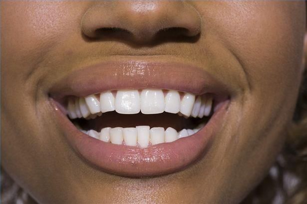
[Picture Credit: Mouth Cancer]
Mouth cancer occurs when cells on the lips or in the mouth
develop changes (mutations) in their DNA. These mutations
allow cancer cells to grow and divide when healthy cells would die. The accumulating mouth cancer
Researched and Authored by Prof Michael C Herbst
[D Litt et Phil (Health Studies); D N Ed; M Art et Scien; B A Cur; Dip Occupational Health; Dip Genetic Counselling; Diagnostic
Radiographer; Dip Audiometry and Noise Measurement; Medical Ethicist]
Approved by Ms Elize Joubert, Chief Executive Officer [BA Social Work (cum laude); MA Social Work]
January 2021 Page 1
cells can form a tumour. With time they may spread to other areas of the mouth and on to other areas
of the head and neck or other parts of the body.

Mouth cancers most commonly begin in the flat, thin cells (squamous cells) that line the lips and the
inside of the mouth. Most oral cancers are squamous cell carcinomas.

It is not always clear what causes the mutations in squamous cells that lead to mouth cancer, but
doctors have identified factors that may increase the risk of mouth cancer.

Inchingolo, F., Santacroce, L., Ballini, A., Topi, S., Dipalma, G., Haxhirexha, K., Bottalico, L. & Charitos,
I.A. 2020. Oral cancer: a historical review. Int J Environ Res Public Health. 2020 May 2;17(9):3168.
Aim: This historical medical literature review aims at understanding the evolution of the medical
existence of oral cancer over times, particularly better comprehending if the apparent lower
prevalence of this type of cancer in antiquity is a real value due to the absence of modern
environmental and lifestyle factors or it is linked to a misinterpretation of ancient foreign terms found
in ancient medical texts regarding oral neoplasms.
Methods: The databases MedLne, PubMed, Web of Science, Elsevier's EMBASE.com, Cochrane
Review, National Library of Greece (Stavros Niarchos Foundation, Athens) and the Library of the
School of Health Sciences of the National and Kapodistrian University of Athens (Greece) were
extensively searched for relevant studies published during the past century on the history of
oral cancer and its treatment from antiquity to modern times, in addition to the WHO website to
analyse the latest epidemiological data. In addition, we included historical books on the topic of
interest and original sources.
Results: Historical references reveal that the cradle of the oral oncology was in ancient Egypt, the
Asian continent and Greece and cancer management was confined to an approximate surgical
practice, in order to remove abnormal masses and avoid bleeding with cauterization. In the Medieval
Age, little progress occurred in medicine in general, oral cancers management included. It is only from
the Renaissance to modern times that knowledge about its pathophysiological mechanisms and
histopathology and its surgical and pharmacological treatment approaches became increasingly deep
all over the world, evolving to the actual integrated treatment. Despite the abundant literature
exploring oncology in past civilizations, the real prevalence of oral cancer in antiquity is much less
known; but a literature analysis cannot exclude a consistent prevalence of this cancer in past
populations, probably with a likely lower incidence than today, because many descriptions of its
aggressiveness were found in ancient medical texts, but it is still difficult to be sure that each single
description of oral masses could be associated to cancer, particularly for what concerns the period
before the Middle Ages.
Conclusions: Modern oncologists and oral surgeons must learn a lot from their historic counterparts
in order to avoid past unsuccessful efforts to treatment oral malignancies. Several descriptions of oral
cancers in the antiquity that we found let us think that this disease might be linked to mechanisms not
strictly dependent on environmental risk factors, and this might guide future research on oral cavity
treatments towards strategical cellular and molecular techniques.

Mummudi, N., Agarwal, J.P., Chatterjee, S., Mallick, I., Ghosh-Laskar, S. 2019.
“Oral cavity cancer (OCC) poses a global challenge that plagues both the Orient and the Occident,
accounting for an estimated 350 000 new cases and 177 000 deaths in 2018. OCC is a major public
health problem in the Indian subcontinent, where it ranks among the top three cancer types in both
incidence and mortality. Major risk factors are the use of tobacco, betel quid and alcohol consumption.
OCC is a heterogeneous group of multiple histologies that affects multiple subsites. The oral
cavity includes the lips, buccal mucosa, teeth, gingiva, anterior two-thirds of the tongue, floor of

Researched and Authored by Prof Michael C Herbst
[D Litt et Phil (Health Studies); D N Ed; M Art et Scien; B A Cur; Dip Occupational Health; Dip Genetic Counselling; Diagnostic
Radiographer; Dip Audiometry and Noise Measurement; Medical Ethicist]
Approved by Ms Elize Joubert, Chief Executive Officer [BA Social Work (cum laude); MA Social Work]
January 2021                                                                                                            Page 2
the mouth and hard palate. OCC is defined as cancer of lips, mouth and tongue as defined by the
International Classification of Diseases coding scheme. The epidemiology, aetio-pathogenesis and
treatment philosophy are similar within this group. Although salivary gland malignancies, sarcomas,
mucosal melanomas and lymphomas can also arise within the oral cavity, this review will focus on
squamous cell cancer, which is the predominant histology in OCC. We review and contrast data from
developing and developed countries. We also highlight the unique regional challenges that countries
in the East face; citing India as an example, we elaborate on the opportunities and scope for
improvement in the management of OCC.”

Incidence of Cancer of the Mouth in South Africa
According to the outdated National Cancer Registry (2017) the following number of cancer of the
mouth cases was histologically diagnosed in South Africa during 2017:

 Group - Males                         Actual                 Estimated           Percentage of
 2017                                No of Cases            Lifetime Risk          All Cancers
 All males                              337                     1:484                 0,84%
 Asian males                             11                     1:698                 1,13%
 Black males                            183                     1:571                 1,40%
 Coloured males                          56                     1:308                 1,15%
 White males                             87                     1:372                 0,41%

 Group - Females                       Actual                 Estimated           Percentage of
 2017                                No of Cases            Lifetime Risk          All Cancers
 All females                            189                    1:1 206                0,45%
 Asian females                           21                     1:391                 1,63%
 Black females                           84                    1:2 060                0,44%
 Coloured females                        29                     1:821                 0,64%
 White females                           55                     1:637                 0,31%

The frequency of histologically diagnosed cases of cancer of the mouth in South Africa for 2017 was
as follows (National Cancer Registry, 2017):

 Group - Males                    0 – 19         20 – 29         30 – 39       40 – 49       50 – 59      60 – 69       70 – 79         80+
 2017                             Years           Years           Years         Years         Years        Years         Years         Years
 All males                           1              2              10            38            123          109           35             19
 Asian males                         0              0               1             3              3           2             1             1
 Black males                         0              2               7            17             73           65           14             5
 Coloured males                      0              0               1             8             21           16            9             1
 White males                         1              0               1            10             26           26           11             12

 Group - Females                  0 – 19         20 – 29         30 – 39       40 – 49       50 – 59      60 – 69       70 – 79         80+
 2017                             Years           Years           Years         Years         Years        Years         Years         Years
 All females                         3              5              11            20            54           55            25             16
 Asian females                       0              1               0             4             3            6             5             2
 Black females                       3              3               9             9            23           22             9             6
 Coloured females                    0              1               0             2            12            7             5             2
 White females                       0              0               2             5            16           20             6             6
N.B. In the event that the totals in any of the above tables do not tally, this may be the result of uncertainties as to the age, race or sex of
the individual. The totals for ‘all males’ and ‘all females’, however, always reflect the correct totals.

Researched and Authored by Prof Michael C Herbst
[D Litt et Phil (Health Studies); D N Ed; M Art et Scien; B A Cur; Dip Occupational Health; Dip Genetic Counselling; Diagnostic
Radiographer; Dip Audiometry and Noise Measurement; Medical Ethicist]
Approved by Ms Elize Joubert, Chief Executive Officer [BA Social Work (cum laude); MA Social Work]
January 2021                                                                                                            Page 3
Risks and Causes of Cancer of the Mouth
The following risks and causes of mouth cancer has been identified.

Alcohol Consumption – There is no safe level of alcohol consumption. Drinking alcohol increases the
risk of oropharyngeal cancer and may increase mouth cancer risk when combined with smoking. A
large Cancer Research UK study looking at lifestyle factors that cause cancer found that around a third
of cancers of the mouth and throat (30%) were caused by drinking alcohol.

Alcohol contain nitrosamines that are known to cause cancer. The nitrosamines in alcohol pass over
the mouth, throat and top of the epiglottis as one swallows.

Tobacco use - smoking tobacco (cigarettes, cigars and pipes) form part of the main risk factors for
mouth and oropharyngeal cancers in the western world. If a person smokes, he/she is at a higher than
average risk of developing oropharyngeal cancers. People exposed to secondhand smoke at home or
in the environment have a small increase in their risk of mouth or oropharyngeal cancer.

Cigarettes contain nitrosamines and other chemicals that are known to cause cancer. When smoking,
the smoke passes through the mouth, throat and the larynx on its way to the lungs. The risk increases
the longer one smokes.

Individuals who smoke are more likely to develop cancer of the mouth or oropharynx. If one smokes
and regularly drinks alcohol, the risk is especially high. Cancers of the mouth or oropharynx do
sometimes occur in people who do not smoke or drink, but this is less common.

Al Feghali, K.A., Ghanem, A.I., Burmeister, C., Chang, S.S., Ghanem, T., Keller, C. & Siddiqui, F. 2019.
OBJECTIVES: We sought to determine whether smokers with oral cavity squamous cell carcinoma
(OCSCC) have tumors with more adverse pathological features than in nonsmokers and whether or
not these are predictive of outcomes.
MATERIALS AND METHODS: We retrospectively identified 163 patients with American Joint
Committee on Cancer stages I-IVa OCSCC diagnosed between 2005 and 2015 and treated with
curative intent. A pathological risk score (PRS) was calculated using the National
Comprehensive Cancer Network adverse risk factors: positive margin, extracapsular extension of
lymph node metastases, pT3 or pT4 primary, N2 or N3 nodal disease, perineural invasion, and
lymphovascular space invasion. Multivariable models were constructed to determine the
independent predictors of overall survival (OS), recurrence-free survival (RFS), and PRS.
RESULTS: A total of 108 (66.26%) were smokers and 55 nonsmokers. Three-year actuarial OS and RFS
were 62% and 68% in smokers and 81% and 69% in nonsmokers, respectively (P = 0.06 and P = 0.63).
Smokers were more likely to have advanced disease stage and tumors with aggressive pathological
features than nonsmokers. Smokers had significantly worse PRS (mean ± standard deviation; 2.38 ±
2.19, median; 2.00) than nonsmokers (0.89 ± 1.21, 0.00) (P < 0.001). Older age, higher PRS, and
smoking status were independent predictors of OS. Smoking or PRS did not predict for worse RFS. On
multivariate analysis, independent predictors of PRS were smoking status and grade (P < 0.001).
CONCLUSION: In patients with OCSCC, smokers have more aggressive disease as evidenced by more
adverse pathological features than nonsmokers. Moreover, smoking is an independent predictor of
OS but not RFS. The PRS is a significant predictor of OS and needs validation in the future studies.

Researched and Authored by Prof Michael C Herbst
[D Litt et Phil (Health Studies); D N Ed; M Art et Scien; B A Cur; Dip Occupational Health; Dip Genetic Counselling; Diagnostic
Radiographer; Dip Audiometry and Noise Measurement; Medical Ethicist]
Approved by Ms Elize Joubert, Chief Executive Officer [BA Social Work (cum laude); MA Social Work]
January 2021                                                                                                            Page 4
Chewing tobacco or betel quid - chewing tobacco (smokeless tobacco) or betel quid (gutkha; areca
nut) is known to cause mouth cancer and oropharyngeal cancer. It is not a safe alternative to
cigarettes. It is very common in parts of Asia. It is also popular in some immigrant groups in Europe,
North America and Australia.

The term ‘quid’ means a substance or mixture of substances put in the mouth and chewed, usually for
long periods. It usually contains tobacco, either on its own or mixed with areca nut (from the Areca
catechu tree) and slaked lime and sometimes spices. The mixture is wrapped in a leaf called a betel
leaf, which is where the name betel quid (also called ‘paan’) comes from. The harmful substances in
tobacco and betel quid can cause cancer if they are in contact with the gums and tongue over long
periods.

Mouth cancer is more common in parts of the world where people chew betel quid. Of the estimated
400 000 cases of oral cancer worldwide each year, around two thirds occur in developing countries.
In some parts of India, it is the most common type of cancer.

Diet - a poor diet may increase one’s risk of certain types of mouth and oropharyngeal cancer. This
may be due to a lack of vitamins and minerals, such as iron or folic acid. Poor diet can lead to a
breakdown in the oral mucosa and this can make it more prone to developing cancer. If one eats a
well balanced diet with plenty of protein, one is unlikely to be short of vitamins and minerals.

A diet high in fresh fruit (in season) and vegetables seems to reduce the risk of developing cancer of
the mouth. This may be because these foods contain a lot of antioxidant vitamins and other
substances that help prevent damage to body cells.

Human papilloma virus (HPV) - viruses can help to cause some cancers. But this does not mean that
one can catch these cancers like an infection. The virus can cause genetic changes in cells that make
them more likely to become cancerous in the future.

Mouth and oropharyngeal cancers have been linked to the human papilloma virus (HPV), especially
type 16. There are more than 100 different types of (HPV). Some types are called the wart virus,
because they cause warts on the genital area or skin. Other types of HPV are known to increase the
risk of some types of cancer. These include cancer of the cervix, vaginal cancer, vulval cancer and anal
cancer.

HPV can be passed on during sexual contact. Most sexually active adults will be infected with at least
one type of HPV at some time during their life. For many people, the virus causes no harm and goes
away without treatment. Only a very small percentage of people with HPV develop oropharyngeal
cancer. The risk of HPV infection in the mouth and throat is linked to certain sexual behaviours, such
as oral sex. The risk increases with the number of sexual partners a person has. Smoking also increases
the risk of HPV infection in the mouth.

Li, H., Park, H.S., Osborn, H.A. & Judson, B.L. 2018.
BACKGROUND: Human papilloma virus (HPV)-associated head and neck cancer is now recognized as
a distinct clinical entity from HPV-negative tumors, which are primarily associated with tobacco and
alcohol exposure.Little is known, however, about the behavior of HPV-associated
oropharynx (OP) and oral cavity (OC) SCCs as two distinct cancers and how sex affects the overall

Researched and Authored by Prof Michael C Herbst
[D Litt et Phil (Health Studies); D N Ed; M Art et Scien; B A Cur; Dip Occupational Health; Dip Genetic Counselling; Diagnostic
Radiographer; Dip Audiometry and Noise Measurement; Medical Ethicist]
Approved by Ms Elize Joubert, Chief Executive Officer [BA Social Work (cum laude); MA Social Work]
January 2021                                                                                                            Page 5
survival (OS) in these two cancers. The objective of our study is to determine if sex is associated with
overall survival (OS) in patients with high-risk human papillomavirus (HPV)-positive and HPV-negative
squamous cell carcinomas (SCC) in the oropharynx and oral cavity sites.
METHODS: This is a retrospective cohort study using a national database. Data were extracted from
the National Cancer Database (NCDB) of patients diagnosed with OP or OC SCC from 2010 to 2014.
Univariate and multivariate survival analyses were conducted with chi-square tests, Kaplan-Meier
estimates, log-rank tests, and Cox proportional hazards multivariable modeling.
RESULTS: A total of 30,707 patients (13,694 OP HPV-associated, 7933 OP HPV-, 1220 OC HPV-
associated, 7860 OC HPV-) were identified. In all four groups, women tended to be older and have
lower T and N clinical classification than men. Though there were no significant differences in OS
between the sexes in OP HPV-associated cancers, female sex was associated with worse OS in OP
HPV- cancers (HR: 1.15; 95% CI 1.04-1.28, p = 0.004), whereas it was associated with improved OS in
OC HPV-associated and HPV- cancers (HPV-associated: HR: 0.71; 95% CI 0.50-0.99, p = 0.048; HPV-:
HR: 0.87; 95% CI 0.78-0.95, p = 0.004).
CONCLUSION: The effect of sex on OS in OC and OP SCC appears to vary based on tumor location and
HPV status. While the source of this difference in prognostic association is unclear, it may be related
to an emerging difference in the biology of HPV carcinogenesis in these locations.

Low immunity - research has found that people have an increased risk of mouth cancer if they have a
reduced immunity due to HIV or AIDS. Taking medicines to suppress immunity after organ transplants
also gives a higher risk of mouth cancer than in the general population.

Sunlight and sunbeds - skin cancers are relatively common on the face and neck, as these areas are
often exposed to ultraviolet light (UV). Both the sun and tanning beds give off UV rays. These rays can
cause skin cancers in unprotected skin. Some studies have shown an increase of skin cancer in people
who regularly use sunbeds. Melanoma is the most serious type of skin cancer and can occur on the
lip.

Previous cancer - people who have had mouth or oropharyngeal cancer have an increased risk of
getting a second one. People who have had some other types of cancer also have an increased risk of
mouth cancer. These include
•       Cancer of the food pipe (oesophagus)
•       Squamous cell skin cancer
•       Cervical, anal or genital cancer in women
•       Cancer of the anus or penis in men

Family history - people often worry that they are at a higher risk of cancer because someone in their
family has it. There does seem to be a slightly higher risk of getting mouth cancer if one has a close
relative (a parent, brother, sister or child) who has had mouth cancer. The reason for this is no known.

Mouth conditions - sometimes changes can happen in the cells of the lining of the mouth and they
cause red or white patches to appear. Doctors call these red patches erythroplakia and white patches
leukoplakia.

Researched and Authored by Prof Michael C Herbst
[D Litt et Phil (Health Studies); D N Ed; M Art et Scien; B A Cur; Dip Occupational Health; Dip Genetic Counselling; Diagnostic
Radiographer; Dip Audiometry and Noise Measurement; Medical Ethicist]
Approved by Ms Elize Joubert, Chief Executive Officer [BA Social Work (cum laude); MA Social Work]
January 2021                                                                                                            Page 6
In some people these changes may develop into cancer over some years. Dentists can see these
patches during dental checks so it is important to have regular dental appointments to find these
changes early.

Genetic conditions - people with certain syndromes caused by inherited changes (mutations) in
particular genes have a high risk of mouth and throat cancer.

These include:

•    Fanconi anaemia – a genetic disorder that can affect children and adults from any ethnic
     background. It is also called Fanconi's syndrome. People with Fanconi anaemia are short, have
     bone changes, and are at risk of developing cancers, leukaemia, and bone marrow failure (aplastic
     anaemia)
•    Dyskeratosis congenita – a genetic syndrome that can cause aplastic anaemia, skin rashes, and
     abnormally shaped fingernails and toenails. People with this syndrome have a high risk of
     developing cancer of the mouth and throat when they are young.

Hydrochlorothiazide - hydrochlorothiazide is a drug used to treat high blood pressure (hypertension).
One of its possible side effects is photosensitivity (increased sensitivity to sunlight). A small study
showed hydrochlorothiazide may increase the risk of developing lip cancer.

Types of Mouth Cancers
The following types of mouth cancer have been identified:

•         Squamous cell carcinoma - more than 90% of cancers that occur in the oral cavity and
          oropharynx are squamous cell carcinoma. Normally, the throat and mouth are lined with so-
          called squamous cells, which are flat and arranged in a scale-like way. Squamous cell
          carcinoma means that some squamous cells are abnormal.

•         Verrucous carcinoma - about 5% of all oral cavity tumours are verrucous carcinoma, which is
          a type of very slow-growing cancer made up of squamous cells. This type of oral cancer rarely
          spreads to other parts of the body, but can invade the tissue surrounding the site of origin.

•         Minor salivary gland carcinomas - this category includes several kinds of oral cancer that can
          develop on the minor salivary glands, which are found throughout the lining of the mouth and
          throat. These types include adenoid cystic carcinoma, mucoepidermoid carcinoma, and
          polymorphous low-grade adenocarcinoma.

•         Lymphomas - oral cancers that develop in lymph tissue, which is part of the immune system,
          are known as lymphomas. The tonsils and base of the tongue both contain lymphoid tissue.

•         Benign oral cavity and oropharyngeal tumours - several types of non-cancerous tumours and
          tumour-like conditions can arise in the oral cavity and oropharynx. Sometimes, these
          conditions may develop into cancer. For this reason, benign tumours, which usually do not
          recur, are often surgically removed.
Researched and Authored by Prof Michael C Herbst
[D Litt et Phil (Health Studies); D N Ed; M Art et Scien; B A Cur; Dip Occupational Health; Dip Genetic Counselling; Diagnostic
Radiographer; Dip Audiometry and Noise Measurement; Medical Ethicist]
Approved by Ms Elize Joubert, Chief Executive Officer [BA Social Work (cum laude); MA Social Work]
January 2021                                                                                                            Page 7
The types of benign (non-cancerous) lesions include:
▪ Eosinophilic granuloma
▪ Fibroma
▪ Granular cell tumour
▪ Karatoacanthoma
▪ Leiomyoma
▪ Osteochondroma
▪ Lipoma
▪ Schwannoma
▪ Neurofibroma
▪ Papilloma
▪ Condyloma acuminatum
▪ Verruciform xanthoma
▪ Pyogenic granuloma
▪ Rhabdomyoma
▪ Odontogenic tumors (lesions that begin in tooth-forming tissues)
• Leukoplakia and erythroplakia - these non-cancerous conditions mean that there are certain types
of abnormal cells in the mouth or throat. With leukoplakia, a white area can be seen, and with
erythroplakia, there is a red area, flat or slightly raised, that often bleeds when scraped. Both
conditions may be precancerous; that is, they can develop into different types of cancer. When
these conditions occur, a biopsy or other test is done to determine whether the cells are
cancerous.
Most leukoplakia is benign. About 25% of cases of leukoplakia are either cancerous when first
discovered or become precancerous. Erythroplakia is usually more serious, with about 70% of
cases cancerous either at the time of diagnosis or later.
Diagnosis of Mouth Cancers
Any discussion of diagnosis must be prefaced with the issue of discovery. While an annual screening
for oral cancer is important, it is possible that you will notice some change in your mouth or throat
that needs examination between your annual screenings. You are the most important factor in an
early diagnosis. You should always contact your doctor or dentist immediately if you notice the
following symptoms in yourself or a loved one:
• A sore or lesion in the mouth that does not heal within two weeks.
• A lump or thickening in the cheek.
• A white or red patch on the gums, tongue, tonsil, or lining of the mouth.
• A sore throat or a feeling that something is caught in the throat.
• Difficulty chewing or swallowing.
• Difficulty moving the jaw or tongue.
• Numbness of the tongue or other area of the mouth.
• Swelling of the jaw that causes dentures to fit poorly or become uncomfortable.
• Chronic hoarseness.
Researched and Authored by Prof Michael C Herbst
[D Litt et Phil (Health Studies); D N Ed; M Art et Scien; B A Cur; Dip Occupational Health; Dip Genetic Counselling; Diagnostic
Radiographer; Dip Audiometry and Noise Measurement; Medical Ethicist]
Approved by Ms Elize Joubert, Chief Executive Officer [BA Social Work (cum laude); MA Social Work]
January 2021 Page 8These symptoms may be caused by other, less serious problems, but they also indicate the possible presence of oral cancer. Only a professional will be able to tell. Some think that a visit to their medical doctor is the appropriate course of action. But remember that dentists are trained in this simple, quick screening, which involves the examination of the oral cavity as a whole and not just the teeth. Besides a visual examination of all the tissues in one’s mouth, a doctor will feel the floor of the mouth and portions of the back of the throat with his/her fingers, in the search for abnormalities. A thorough oral screening also includes indirect examination of the nasopharynx and larynx, and involves manually feeling the neck for swollen lymph nodes, and other abnormalities such as hardened masses. The doctor will also check the mouth for white patches, red patches, ulcerations, lumps, loose teeth, and review dental X-rays for abnormalities. Be sure to tell the doctor if you have been a tobacco user in any form. Tobacco use is implicated in many cases of oral cancer. After the physical examination of the mouth, if the doctor finds any areas that are suspicious, he/she may recommend a biopsy. This is simply taking a small portion of the suspicious tissue for examination under a microscope. The most traditional type of biopsy is incisional. It may be done by the doctor who examines the patient, or the patient may be referred to another doctor for the procedure. In an incisional biopsy, the doctor will remove part or all of the lesion depending on its size and his/her ability to define the extent of it at this early stage. The sample of tissue is then sent to a pathologist who examines the tissue under a microscope to check for abnormal, or malignant cells. When dealing with an area of significant mass, such as an enlarged lymph node, fine needle aspiration cytology (fine needle biopsy or FNB) has found an increasing role in diagnosis. The technique is reliable and relatively inexpensive. In it, a small needle attached to a syringe is inserted into the questionable mass, and cells are aspirated, or pulled out into the syringe as the doctor draws back the piston of the syringe. The success of this method depends on how accurately the needle is placed, and, as with all biopsies, on the skill and experience of the tissue pathologist who will be examining the cells. It is likely that the doctor will insert the needle and draw out cellular material from several different locations in the mass to ensure that a thorough and representative sample has been taken. Another form of incisional biopsy is referred to as a punch biopsy. In this case, a very small circular blade is pressed down into the suspect area cutting a round border. The doctor then pulls on the centre of this area, and with a scalpel or a pair of small tissue scissors snips it free of the surrounding tissue, removing a perfect plug of cells from the sampled area. As before this is sent to a pathologist for examination. The area where the plug was removed will not bleed much, and heals normally without the need for any stitches since it is so small. Some dental offices are doing a "brush biopsy" where a sampling of cells is collected by aggressively rubbing a brush against the suspect area. While this has some usefulness in preliminary evaluation of a suspect area, it is not a stand-alone procedure, and if a positive find returns, this must be confirmed by a conventional incisional biopsy. The entire point of course, is that no treatment decisions should be made before there is confirmation of malignancy. Even in the case of what would seem to be an obvious malignancy, appearances can occasionally be misleading, hence the need for a proper biopsy. Also, the degree of differentiation between healthy and malignant tissues, along with the stage of the disease will influence treatment strategy and prognosis. Other ways to determine the presence or extent of oral cancer exist. For instance, radiographs, also referred to as X-rays, can assist in determining the potential growth of a tumour into bone. While oral Researched and Authored by Prof Michael C Herbst [D Litt et Phil (Health Studies); D N Ed; M Art et Scien; B A Cur; Dip Occupational Health; Dip Genetic Counselling; Diagnostic Radiographer; Dip Audiometry and Noise Measurement; Medical Ethicist] Approved by Ms Elize Joubert, Chief Executive Officer [BA Social Work (cum laude); MA Social Work] January 2021 Page 9
cancers unlike many other malignancies can usually be seen with the naked eye, some cancers are
located internally in the body, making their detection difficult. Different scanning options, some of
which assist in determining the presence of tumours or growths, and some of which can even detect
malignancy, are necessary in these instances.
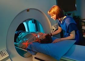
CT, or CAT (co-axial tomography) scan
technology has developed rapidly over the last
few decades, and these scans can provide
images of great diagnostic quality and
usefulness. A CT scan could be described as a
series of X-rays, each one a view of a 3mm
section of the area being scanned, which are
then manipulated by a computer, allowing
doctors a dynamic view of the affected soft
tissue areas of the body with much greater
detail than a simple X-ray. However, CT is only
able to detect the actual presence of masses,
and only a biopsy can verify that the mass is
malignant. Another recent technology, Magnetic Resonance Imaging (MRI), is helpful in providing
accurate views of the affected area. MRI is a procedure in which pictures are created using magnets
and radio frequencies linked to a computer imaging system. The hydrogen atoms in the patient's body
react to the magnetic field and emit signals that are analysed by a computer to produce detailed
images of organs and structures in the body. Occasionally a dye is injected into the bloodstream during
scanning to bring greater detail to the soft tissue areas of the scan. Again, this procedure is only able
to detect the actual presence of masses, and it still requires a biopsy for confirmation.
PET, or Positron Emission Tomography, provides another kind of image of the body's interior. Instead
of taking a picture of the bones, like an X-ray, or the internal organs and soft tissue, like a MRI, PET
scanning lets doctors display the body's actual metabolism. Since cells use a simple sugar, glucose, as
a source of energy, PET can track down how much glucose is being metabolized in different areas of
the body.
Because cancer cells are dividing rapidly, they break down glucose much faster than normal cells. The
increased activity will show up on a PET scan, and can indicate both primary and metastatic tumours.
Although less frequently used for oral cancer detection, ultrasonography is another way to produce
pictures of areas in the body. In it, high-frequency sound waves (ultrasound) are bounced off organs
and tissue. The pattern of echoes produced by these waves creates a picture called a sonogram. It is
useful in finding masses with in an area, if palpation discloses something of questionable nature.
Radionuclide scanning can show whether cancer has spread to other organs elsewhere in the body. In
it, the patient swallows or receives an injection of a mildly radioactive substance, and a scanner
measures and records the level of radioactivity in certain organs to reveal abnormal areas.
All these types of scans are still used largely for confirmation or measuring extent. The best indicator
of tumour involvement is still the clinical assessment, relying on both direct examination of the area
as well as biopsy. The ability to detect cancer at the earliest stages, as well as its precise location in
the body, can improve the survival rate of this disease, and allow for less disfiguring ways to address
the tumours and lesions associated with oral cancer.
Researched and Authored by Prof Michael C Herbst
[D Litt et Phil (Health Studies); D N Ed; M Art et Scien; B A Cur; Dip Occupational Health; Dip Genetic Counselling; Diagnostic
Radiographer; Dip Audiometry and Noise Measurement; Medical Ethicist]
Approved by Ms Elize Joubert, Chief Executive Officer [BA Social Work (cum laude); MA Social Work]
January 2021 Page 10Ilhan, B., Gunerik P. & Wilder-Smith, P. 2020. “Early diagnosis is the most important determinant of oral and oropharyngeal squamous cell carcinoma (OPSCC) outcomes, yet most of these cancers are detected late, when outcomes are poor. Typically, nonspecialists such as dentists screen for oral cancer risk, and then they refer high-risk patients to specialists for biopsy-based diagnosis. Because the clinical appearance of oral mucosal lesions is not an adequate indicator of their diagnosis, status, or risk level, this initial triage process is inaccurate, with poor sensitivity and specificity. The objective of this study is to provide an overview of emerging optical imaging modalities and novel artificial intelligence-based approaches, as well as to evaluate their individual and combined utility and implications for improving oral cancer detection and outcomes. The principles of image-based approaches to detecting oral cancer are placed within the context of clinical needs and parameters. A brief overview of artificial intelligence approaches and algorithms is presented, and studies that use these 2 approaches singly and together are cited and evaluated. In recent years, a range of novel imaging modalities has been investigated for their applicability to improving oral cancer outcomes, yet none of them have found widespread adoption or significantly affected clinical practice or outcomes. Artificial intelligence approaches are beginning to have considerable impact in improving diagnostic accuracy in some fields of medicine, but to date, only limited studies apply to oral cancer. These studies demonstrate that artificial intelligence approaches combined with imaging can have considerable impact on oral cancer outcomes, with applications ranging from low-cost screening with smartphone-based probes to algorithm-guided detection of oral lesion heterogeneity and margins using optical coherence tomography. Combined imaging and artificial intelligence approaches can improve oral cancer outcomes through improved detection and diagnosis.” Loree, J.T., Popat, S.R., Burke, M.S., Frustino, J., Grewal, J.S. & Loree, T.R. 2019. BACKGROUND AND OBJECTIVES: The management of the clinically N0 (cN0) neck is controversial for early stage squamous cell carcinoma of the oral cavity (OSCC). This paper represents a single institution series analyzing the efficacy of sentinel lymph node biopsy (SNB) for early stage oral cavity cancers. METHODS: From 2005 to 2017, 108 patients with cN0 OSCC were treated with primary resection and SNB. Patients with positive biopsy results proceeded to neck dissection with or without adjuvant chemoradiotherapy. Mean follow-up for the entire cohort was 50.8 months (range: 8-147 months). Clinically, 56 patients were T1N0, 49 patients were T2N0, and three patients were T3N0 or greater. RESULTS: Disease-specific survival was 93% within the entire cohort. Sentinel lymph nodes were identified in 95.4% of patients. Twenty one patients had a positive biopsy. There were seven false- negative biopsies. The overall rate of nodal disease was 26%. Accuracy of node biopsy was 93%, with sensitivity of 75%, and negative predictive value of 91%. Recurrence rate was 19% (20/108), with an overall survival of 60% in this subgroup. CONCLUSION: SNB is a safe, effective, and well tolerated method for staging cN0 OSCC. Treatment of Mouth Cancer Oral cancer treatment may include surgery, radiation therapy, or chemotherapy. Some patients have a combination of treatments. At any stage of disease, people with mouth (oral) cancer may have treatment to control pain and other symptoms, to relieve the side effects of therapy, and to ease emotional and practical problems. This kind of treatment is called supportive care, symptom management, or palliative care. Researched and Authored by Prof Michael C Herbst [D Litt et Phil (Health Studies); D N Ed; M Art et Scien; B A Cur; Dip Occupational Health; Dip Genetic Counselling; Diagnostic Radiographer; Dip Audiometry and Noise Measurement; Medical Ethicist] Approved by Ms Elize Joubert, Chief Executive Officer [BA Social Work (cum laude); MA Social Work] January 2021 Page 11
A patient may want to talk to the doctor about taking part in a clinical trial, a research study of new
treatment methods.

Surgery - surgery to remove the tumour in the mouth or throat is a common treatment for oral cancer.
Sometimes the surgeon also removes lymph nodes in the neck. Other tissues in the mouth and neck may
be removed as well. Patients may have surgery alone or in combination with radiation therapy.

Tahmasebi, E., Alikhani, M., Yazdanian, A., Yazdanian, M., Tebyanian, H. & Seifalian, A. 2020.
“Head and neck cancer (HNC) constitute 5% of all reported cancers. Among all, the oral cavity cancer
is the most frequent type of HNC which accounts for over half of HNC cases. Mouth cancer ranks the
sixth leading cause of cancer-related mortality. Generally, conventional chemotherapy has shown
success at decreasing relapse and metastasis rates and improves the overall prognosis. Recently,
target therapy and targeted drug delivery systems have been introduced as promising treatments. The
elimination of efficiency of current therapeutic strategies due to the spared cancer stem cells that
cause chemotherapy resistance, relapse and metastasis. Inefficiency methodologies in the elimination
of all cancer cells in the body are a major problem that remained to be resolved before to confront
the new cancer therapies. Many studies imply to cancer stem cell markers as important agents for
targeted anti-cancer as well as improving chemotherapy efficiencies. The potentials of targeted cancer
therapy led us to search for novel markers in the mouth cancer stem cells especially in rare cancers.
The aimed of this research was, first a comprehensive critical review of the previous studies on the
markers of cancer stem cells in oral cancers including oral squamous cell carcinoma, salivary gland
cancers, and to highlight the most common cancer stem cell markers which have potential to be
exploited as indicators for the preneoplastic lesion malignancy, oral cancer progression, and/or
treatment prognosis.”

Kamat, M., Rai, B.D., Puranik, R.S. & Datar, U.V. 2019.
“Oral squamous cell carcinoma (OSCC) is the most common malignancy of the oral cavity, and surgery
is the most accepted line of treatment. The surgical margins (SMs) or resection margins are boundaries
of resection specimen excised by the surgeon. The status of these resected SMs is an important and
valuable tool to predict the treatment outcome. It is necessary to attain optimal SM to avoid local
recurrence and improve overall survival. However, the controversies exist regarding the concept of
optimal SM. There are various factors that influence the assessment of the SMs. In addition, apart
from routine histopathology, the molecular assessment of resected margins has recently gained value
which has a promising role for margin surveillance. Furthermore, the histological and molecular
appraisal of tumor-free margins is also necessary to standardize the treatment modalities. Hence, this
review aims to summarize the above issues that influence the evaluation of SMs of OSCC along with
recent updates. Furthermore, an attempt has been made to give an overview about future possible
approaches for the tumor-free margins. An electronic search was performed for items related to the
evaluation of SMs in OSCC, and the obtained articles were critically assessed and the relevant
information was extracted and summarized.”

Radiation therapy - radiation therapy (also called radiotherapy) is a type of local therapy. It affects cells
only in the treated area. Radiation therapy is used alone for small tumours or for patients who cannot have
surgery. It may be used before surgery to kill cancer cells and shrink the tumour. It also may be used after
surgery to destroy cancer cells that may remain in the area.
Radiation therapy uses high-energy rays to kill cancer cells. Doctors use two types of radiation therapy to
treat oral cancer:

Researched and Authored by Prof Michael C Herbst
[D Litt et Phil (Health Studies); D N Ed; M Art et Scien; B A Cur; Dip Occupational Health; Dip Genetic Counselling; Diagnostic
Radiographer; Dip Audiometry and Noise Measurement; Medical Ethicist]
Approved by Ms Elize Joubert, Chief Executive Officer [BA Social Work (cum laude); MA Social Work]
January 2021                                                                                                           Page 12
•    External radiation: The radiation comes from a machine. Patients go to the hospital or clinic once or
     twice a day, generally 5 days a week for several weeks.
•    Internal radiation (implant radiation): The radiation comes from radioactive material placed in seeds,
     needles, or thin plastic tubes put directly in the tissue. The patient stays in the hospital. The implants
     remain in place for several days. Usually they are removed before the patient goes home.

Some people with oral cancer have both kinds of radiation therapy.

Chemotherapy - chemotherapy uses anticancer drugs to kill cancer cells. It is called systemic therapy
because it enters the bloodstream and can affect cancer cells throughout the body. A new targeted therapy
called cetuximab, which blocks a growth factor upon which cancer cells may depend, is being used today,
either alone or in combination with radiation and older chemotherapy drugs.

Chemotherapy is usually given by injection. It may be given in an outpatient part of the hospital, at the
doctor's office, or at home. Rarely, a hospital stay may be needed.

Szturz, P. & Vermorken, J.B. 2020.
“In recurrent and/or metastatic squamous cell carcinoma of the head and neck (R/M-SCCHN), the
armamentarium of systemic anti-cancer modalities continues to grow in parallel with innovations in
and better integration of local approaches. The backbone of cytotoxic chemotherapy remains cisplatin
with 5-fluorouracil or a taxane. In contrast to cisplatin, the tumoricidal activity of carboplatin
monotherapy is debatable. Adding the epidermal growth factor receptor (EGFR) inhibitor cetuximab
to a platinum/5-fluorouracil doublet (the so-called EXTREME regimen) produced a statistically but also
clinically significant improvement of survival and became thus the standard first-line palliative
treatment in adequately fit patients. Interestingly, three large randomized trials (EXTREME,
SPECTRUM, and ZALUTE) evaluating different anti-EGFR monoclonal antibodies (cetuximab,
panitumumab, and zalutumumab, respectively) demonstrated preferential anti-tumour efficacy in
patients with primary cancer in the oral cavity. Modern immunotherapy with immunomodulating
antibodies, dubbed immune checkpoint inhibitors, such as anti-programmed cell death protein-1
(anti-PD-1) inhibitors nivolumab and pembrolizumab, showed unprecedented activity in one first-line
(KEYNOTE-048) and several second-line trials (CheckMate-141, KEYNOTE-012, KEYNOTE-055, and
KEYNOTE-040). In a minority of also heavily-pretreated patients, these agents generate long-lasting
responses without the typical chemotherapy-related toxicity, however, at a price of a low overall
response rate, rare but potentially life-threatening immune-related adverse events, the risk of
hyperprogression, and high costs. In oligometastatic disease, emerging data indicate long-term
benefit with locally ablative techniques including metastasectomy and stereotactic radiotherapy of
pulmonary but also hepatic and other distant lesions. In the frame of highly-individualized cancer care,
a particularly intriguing approach is a combination of systemic and local therapies.”

About Clinical Trials
Clinical trials are research studies that involve people. They are conducted under controlled
conditions. Only about 10% of all drugs started in human clinical trials become an approved drug.

Clinical trials include:
     • Trials to test effectiveness of new treatments
     • Trials to test new ways of using current treatments

Researched and Authored by Prof Michael C Herbst
[D Litt et Phil (Health Studies); D N Ed; M Art et Scien; B A Cur; Dip Occupational Health; Dip Genetic Counselling; Diagnostic
Radiographer; Dip Audiometry and Noise Measurement; Medical Ethicist]
Approved by Ms Elize Joubert, Chief Executive Officer [BA Social Work (cum laude); MA Social Work]
January 2021                                                                                                           Page 13
• Tests new interventions that may lower the risk of developing certain types of cancers
• Tests to find new ways of screening for cancer
The South African National Clinical Trials Register provides the public with updated information on
clinical trials on human participants being conducted in South Africa. The Register provides
information on the purpose of the clinical trial; who can participate, where the trial is located, and
contact details.
For additional information, please visit: www.sanctr.gov.za/
Medical Disclaimer
This Fact Sheet is intended to provide general information only and, as such, should not be considered
as a substitute for advice, medically or otherwise, covering any specific situation. Users should seek
appropriate advice before taking or refraining from taking any action in reliance on any information
contained in this Fact Sheet. So far as permissible by law, the Cancer Association of South Africa
(CANSA) does not accept any liability to any person (or his/her dependants/estate/heirs) relating to
the use of any information contained in this Fact Sheet.
Whilst CANSA has taken every precaution in compiling this Fact Sheet, neither it, nor any
contributor(s) to this Fact Sheet can be held responsible for any action (or the lack thereof) taken by
any person or organisation wherever they shall be based, as a result, direct or otherwise, of
information contained in, or accessed through, this Fact Sheet.
Sources and References Consulted or Utilised
Al Feghali, K.A., Ghanem, A.I., Burmeister, C., Chang, S.S., Ghanem, T., Keller, C. & Siddiqui, F. 2019. Impact of smoking on
pathological features in oral caviaty squamous cell carcinoma. J Cancer Res Ther. 2019 Jul-Sep;15(3):582-588. doi:
10.4103/jcrt.JCRT_641_16.
Cancer.Net
http://www.cancer.net/cancer-types/oral-and-oropharyngeal-cancer/stages-and-grades
Cancer Research UK
http://www.cancerresearchuk.org/about-cancer/type/mouth-cancer/about/risks/definite-risks-for-mouth-and-
oropharyngeal-cancer
Cancer Treatment Centers of America
http://www.cancercenter.com/oral-cancer/types/
Ilhan, B., Gunerik P. & Wilder-Smith, P. 2020. Improving oral cancer outcomes with imaging and artificial intelligence. J Dent
Res. 2020 Mar;99(3):241-248.
Inchingolo, F., Santacroce, L., Ballini, A., Topi, S., Dipalma, G., Haxhirexha, K., Bottalico, L. & Charitos, I.A. 2020. Oral cancer:
a historical review. Int J Environ Res Public Health. 2020 May 2;17(9):3168.
Kamat, M., Rai, B.D., Puranik, R.S. & Datar, U.V. 2019. A comprehensive review of surgical margin in oral squamous cell
carcinoma highlighting the significance of tumor-free surgical margins. J Cancer Res Ther. 2019 Jul-Sep;15(3):449-454. doi:
10.4103/jcrt.JCRT_273_17.
Researched and Authored by Prof Michael C Herbst
[D Litt et Phil (Health Studies); D N Ed; M Art et Scien; B A Cur; Dip Occupational Health; Dip Genetic Counselling; Diagnostic
Radiographer; Dip Audiometry and Noise Measurement; Medical Ethicist]
Approved by Ms Elize Joubert, Chief Executive Officer [BA Social Work (cum laude); MA Social Work]
January 2021 Page 14Li, H., Park, H.S., Osborn, H.A. & Judson, B.L. 2018. Sex differences in patients with high risk HPV-associated negative oropharyngeal and oral cavity squamous cell carcinomas. Cancers Head Neck. 2018 Jun 20;3:4. doi: 10.1186/s41199-018- 0031-y. eCollection 2018. Loree, J.T., Popat, S.R., Burke, M.S., Frustino, J., Grewal, J.S. & Loree, T.R. 2019. Sentinel lymph node biopsy for management of the N0 neck in oral cavity squamous cell carcinoma. J Surg Oncol. 2019 May 16. doi: 10.1002/jso.25494. [Epub ahead of print] Mayo Clinic http://www.mayoclinic.org/diseases-conditions/mouth-cancer/basics/causes/con-20026516 Medicine.Net http://www.medicinenet.com/oral_cancer/page6.htm Mouth http://www.yes30.com/2015/01/25/need-a-little-mouth-to-mouth/ Mummudi, N., Agarwal, J.P., Chatterjee, S., Mallick, I., Ghosh-Laskar, S. 2019. Oral cavity cancer in the Indian subcontinent – challenges and opportunities. Clin Oncol (R Coll Radiol). 2019 Jun 4. pii: S0936-6555(19)30205-5. doi: 10.1016/j.clon.2019.05.013. [Epub ahead of print] National Cancer Institute http://www.cancer.gov/about-cancer/treatment/clinical-trials/what-are-trials NHS Choices http://www.nhs.uk/conditions/cancer-of-the-mouth/pages/introduction.aspx Szturz, P. & Vermorken, J.B. 2020. Management of recurrent and metastataic oral cavity cancer: raising the bar a step higher. Oral Oncol. 2020 Feb;101:104492. Tahmasebi, E., Alikhani, M., Yazdanian, A., Yazdanian, M., Tebyanian, H. & Seifalian, A. 2020. The current markers of cancer stem cell in oral cancers. Life Sci. 2020 May 15;249:117483. The Oral Cancer Foundation http://www.oralcancerfoundation.org/discovery-diagnosis/diagnosis.php Researched and Authored by Prof Michael C Herbst [D Litt et Phil (Health Studies); D N Ed; M Art et Scien; B A Cur; Dip Occupational Health; Dip Genetic Counselling; Diagnostic Radiographer; Dip Audiometry and Noise Measurement; Medical Ethicist] Approved by Ms Elize Joubert, Chief Executive Officer [BA Social Work (cum laude); MA Social Work] January 2021 Page 15
You can also read