The clinical and functional significance of c-Met in breast cancer: a review
←
→
Page content transcription
If your browser does not render page correctly, please read the page content below
Ho-Yen et al. Breast Cancer Research (2015) 17:52
DOI 10.1186/s13058-015-0547-6

 REVIEW                                                                                                                                         Open Access

The clinical and functional significance of c-Met
in breast cancer: a review
Colan M Ho-Yen1*, J Louise Jones2 and Stephanie Kermorgant2

  Abstract
  c-Met is a receptor tyrosine kinase that upon binding of its ligand, hepatocyte growth factor (HGF), activates
  downstream pathways with diverse cellular functions that are important in organ development and cancer
  progression. Anomalous c-Met signalling has been described in a variety of cancer types, and the receptor is
  regarded as a novel therapeutic target. In breast cancer there is a need to develop new treatments, particularly for
  the aggressive subtypes such as triple-negative and basal-like cancer, which currently lack targeted therapy. Over
  the last two decades, much has been learnt about the functional role of c-Met signalling in different models of
  breast development and cancer. This work has been complemented by clinical studies, establishing the prognostic
  significance of c-Met in tissue samples of breast cancer. While the clinical trials of anti-c-Met therapy in advanced
  breast cancer progress, there is a need to review the existing evidence so that the potential of these treatments
  can be better appreciated. The aim of this article is to examine the role of HGF/c-Met signalling in in vitro and
  in vivo models of breast cancer, to describe the mechanisms of aberrant c-Met signalling in human tissues, and to
  give a brief overview of the anti-c-Met therapies currently being evaluated in breast cancer patients. We will show
  that the HGF/c-Met pathway is associated with breast cancer progression and suggest that there is a firm basis for
  continued development of anti-c-Met treatment, particularly for patients with basal-like and triple-negative breast
  cancer.

Introduction                                                                          cancer. Although distinct, BL tumours can be considered
The receptor tyrosine kinase (RTK) c-Met was originally                               an aggressive subgroup of TN cancers, and both are
identified as the product of a transforming gene generated                            characterised by a lack of oestrogen receptor and c-
from a chemically transformed osteosarcoma cell line [1].                             erbB2 (Her2) expression, limiting systemic treatment op-
In 1991, c-Met was discovered to be the receptor for hep-                             tions [12,13]. Since their discovery, the literature regard-
atocyte growth factor (HGF), a protein that had previously                            ing c-Met and HGF in the breast has grown rapidly, and
been shown to promote hepatocyte growth in culture                                    there is now a need to consolidate the findings from
[2,3]. Mutations in the MET gene were subsequently                                    these studies to better understand the relevance of anti-
described in hereditary and sporadic papillary renal cell                             c-Met therapy in breast cancer.
carcinomas [4]. Since then, dysregulation of c-Met signal-                              The aim of this review is to explore the roles of HGF/c-
ling has been identified in a variety of malignant and pre-                           Met signalling in breast development, different in vitro
malignant lesions, including those arising in the breast,                             and in vivo models of breast cancer, and the various mech-
lung, stomach, pharynx, colorectum and cervix [5-10]. Ac-                             anisms of aberrant c-Met signalling identified in breast
cordingly, the utility of targeting c-Met in different cancer                         cancer tissue. We will also outline the anti-c-Met com-
types is now being evaluated in clinical trials [11].                                 pounds currently being investigated as possible breast can-
   New therapeutic targets are needed in breast cancer,                               cer treatments.
particularly in patients with triple-negative (TN) breast
cancer and the related basal-like (BL) subgroup of breast                             Structure and function
                                                                                      c-Met is first produced as a 170 kDa precursor that then
* Correspondence: Colan.HoYen@stgeorges.nhs.uk
1
                                                                                      undergoes proteolytic cleavage, generating a 50 kDa α-
 Department of Cellular Pathology, St George’s Healthcare NHS Trust,
Blackshaw Road, Tooting, London SW17 0QT, UK                                          subunit and a 145 kDa β-subunit [3,14]. The extracellular
Full list of author information is available at the end of the article                α-subunit is attached to the transmembrane β-subunit by a
                                         © 2015 Ho-Yen et al.; licensee BioMed Central. This is an Open Access article distributed under the terms of the Creative
                                         Commons Attribution License (http://creativecommons.org/licenses/by/4.0), which permits unrestricted use, distribution, and
                                         reproduction in any medium, provided the original work is properly credited. The Creative Commons Public Domain
                                         Dedication waiver (http://creativecommons.org/publicdomain/zero/1.0/) applies to the data made available in this article,
                                         unless otherwise stated.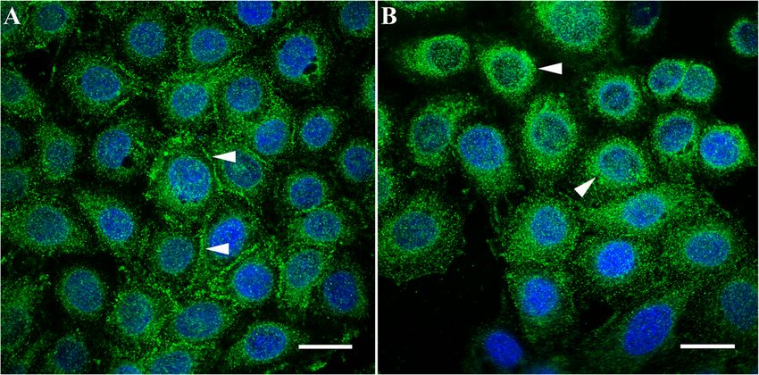
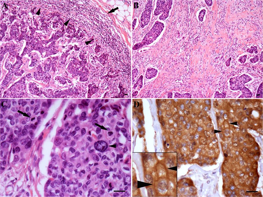
Ho-Yen et al. Breast Cancer Research (2015) 17:52                                                                                              Page 2 of 11

disulphide bond (reviewed in [15]). A Sema domain, a PSI                        intracellular degradation of the receptor, resulting in sup-
domain (so-called because it is present in plexins, sema-                       pression of c-Met-mediated cell migration and growth [16].
phorins and integrins) and four IPT domains (immuno-                              In common with other RTKs, c-Met is regulated by the
globulin-like fold shared by plexins and transcription                          ubiquitin ligase, Cbl [19,20]. Following c-Met activation,
factors) make up the extracellular portion of c-Met. The                        phosphorylation of the Y1003 residue in the juxtamem-
intracellular aspect contains three further domains: the                        brane region recruits Cbl to c-Met, permitting polyubiqui-
juxtamembrane region, which is important in downgrading                         tination and degradation of the receptor [19,20]. Although
kinase activity following Ser 975 phosphorylation; the cata-                    c-Met internalisation is part of the process of signal at-
lytic domain that harbours the Y1234 and Y1235 residues;                        tenuation, trafficking of the receptor within endosomes,
and the multifunctional carboxy-terminal docking site [15].                     under the control of protein kinase C, results in sustained
   The only known mammalian agonistic ligand for c-Met                          signalling and is necessary for HGF-mediated migration
is HGF (also known as scatter factor) [16]. As is the case                      [21-23] (Figure 1).
with c-Met, HGF is secreted first as a precursor, which
must then be activated by proteases, resulting in the for-                      Hepatocyte growth factor/c-Met signalling in
mation of a mature heterodimer composed of an α-chain                           breast development
and a β-chain [17].                                                             The relationship between HGF and c-Met during develop-
   When HGF binds to c-Met, the receptor undergoes au-                          ment is complex and dynamic. In a study of mouse em-
tophosphorylation of the Y1234 and Y1235 residues in the                        bryos, Andermarcher and colleagues described a shift in
kinase domain [14]. Subsequently, tyrosine residues in the                      expression of HGF and c-Met from gastrulation to early
docking site (Y1349 and Y1356) become phosphorylated,                           organogenesis [24]. It was noted that while HGF and c-
permitting binding of adaptor molecules including growth                        Met were coexpressed in endodermal and mesodermal
factor receptor-bound protein 2, growth factor receptor-                        cells during gastrulation, the onset of organogenesis coin-
bound protein 2-associated binder 1 and Shc [14,15].                            cided with HGF being localised to the mesenchyme and c-
These molecules facilitate downstream signalling through                        Met to the epithelium and endothelium [24]. The authors
several pathways, such as the Rac1/Cdc42 pathway, the                           suggested that this change from an autocrine to a para-
phosphoinositide 3-kinase/Akt pathway, signal transducer                        crine relationship may reflect the different roles of the
and activator of transcription 3 and the Erk/mitogen-acti-                      pathway at different stages of development [24].
vated protein kinase cascade [15,18]. Together, these path-                       The HGF/c-Met pathway may also have distinct roles in
ways regulate cellular proliferation, motility, migration,                      the different compartments of the mammary epithelium.
invasion and tubulogenesis [18].                                                Exposure of luminal epithelial and myoepithelial cells to
   The only other ligand known to bind c-Met in mammals                         HGF evokes contrasting effects in the two cell types [25].
is decorin, a leucine-rich proteoglycan [16]. Decorin has                       The growth rate of luminal cells increased fivefold to
been shown to antagonise c-Met signalling by promoting                          ninefold with HGF compared with controls, and no

 Figure 1 Trafficking of c-Met in MDA-MB-468 cells. (A) In resting cells there is prominent membrane expression of the receptor (white arrowheads).
 (B) Following hepatocyte growth factor stimulation there is internalisation of the receptor and a predominantly perinuclear, granular pattern of staining
 (white arrowheads), consistent with the presence of c-Met within endosomes. Immunofluorescence (green, c-Met; blue, nuclei); ×63 objective under oil
 immersion. Scale bars represent 20 μm.
Ho-Yen et al. Breast Cancer Research (2015) 17:52                                                                                                   Page 3 of 11

morphological changes were seen. In contrast, HGF had                              protein overexpression of c-Met and the relationship be-
no effect on myoepithelial cell growth but did induce ex-                          tween levels of the receptor and prognostic factors/
tensive branch formation [25]. RT-PCR analysis showed c-                           survival.
Met expression was higher in luminal cells than in myoe-
pithelial cells, which may explain these differences. The au-                      Gene mutation
thors of this study considered the developmental relevance                         Following the discovery of mutations in the tyrosine kin-
of this arrangement, and hypothesised that the myoepithe-                          ase domain of MET in hereditary and sporadic papillary
lial cells lay down the ductal framework that the luminal                          renal cell carcinomas [4], MET mutations have also been
cells proliferate and migrate along, thus populating and ex-                       found in up to 30% of cancers of unknown primary ori-
tending the ductal system [25].                                                    gin [29]. These mutations include those in the SEMA
   Other studies utilising primary murine mammary epithe-                          domain and the juxtamembrane domain and an activat-
lial cells in in vivo models have further emphasised the                           ing mutation in the tyrosine kinase domain [29]. How-
importance of the HGF/c-Met pathway in mammary devel-                              ever, few studies have assessed the frequency of MET
opment [26,27]. Overexpression of HGF in primary murine                            mutations in primary breast cancer. In a small study
mammary epithelial cells and subsequent transplantation of                         comprised of 11 patients with breast cancer (including
these cells into the cleared mammary fat pads of mice re-                          six patients that showed loss of heterozygosity in the re-
sulted in a marked increase in ductal branches/bifurcations,                       gion of the MET gene), no mutations in the tyrosine kin-
along with an increase in the size and number of ductal end                        ase domain of the MET were identified [30], suggesting
buds [26,27]. Importantly, immunohistochemistry revealed                           that this is not a common event in breast cancer. In con-
an increase in basal/myoepithelial marker expression                               trast, a mutation in the HGF promoter region referred
(smooth muscle actin, cytokeratin 14 and p63) and a reduc-                         to as the deoxyadenosine tract element appears to be a
tion in luminal marker expression (cytokeratin 18 and                              frequent event, having been identified in 15% of European
oestrogen receptor) compared with control mice [27]. This                          breast cancer patients and over 50% of African Americans
finding led the authors to suggest that c-Met signalling di-                       with breast cancer [31]. A truncation mutation in the
rects progenitor cells towards a basal phenotype over lu-                          deoxyadenosine tract element activates the HGF promoter
minal differentiation [27], which is reflected in the pattern of                   in breast cancer cells, leading to the formation of an HGF/
c-Met staining seen in different breast cancer subtypes (as                        c-Met autocrine loop [31].
discussed later).
                                                                                   Gene amplification
Aberrant c-Met signalling in breast cancer                                         Amplification of the MET gene (located on chromosome 7),
A broad range of mechanisms may result in aberrant c-                              like mutation, is unusual in invasive breast cancer: in a study
Met signalling, including activating gene mutations, gene                          of 155 patients, Carracedo and colleagues did not identify
amplification, protein overexpression, increased ligand-                           MET amplification at all (although 22% of tumours showed
dependent paracrine stimulation and the acquisition of                             low-grade polysomy) [32]. Elsewhere, in a much larger
autocrine signalling [28] (Table 1). In breast cancer, the                         study, Gonzalez-Angulo and colleagues found increased
majority of studies have looked at the significance of                             copy numbers of MET in a minority of cases (82 out of 971

Table 1 Mechanisms of aberrant c-Met signalling in invasive breast cancer
Mechanism                  Frequency/prognostic significance in breast cancer                                                                          Reference
Gene mutation              MET mutations are uncommon; HGF promoter region mutations occur in 15 to 51% of breast cancers                              [30,31]
Gene amplification         MET amplification is uncommon, occurring in 0 to 8% of breast cancers; MET copy number is positively                        [32,33]
                           correlated with TN tumours
                           Patients with trastuzumab-treated Her2-positive metastatic breast cancer show MET amplification in 27.7%                    [34]
                           of cases and HGF amplification in 39.3% of cases; patients with MET-amplified Her2-positive tumours have
                           a shorter time to progression
Autocrine signalling       HGF and MET mRNA detected in tumour cells in all breast cancers analysed, with strongest positivity at the                  [35]
                           advancing edge of the tumour
                           On IHC, autocrine pattern of staining seen in 46.6% of tumours                                                              [37]
Paracrine signalling       On IHC, paracrine pattern seen in 59.1% of tumours; paracrine signalling is associated with a worse outcome                 [68]
                           when c-Met staining is more intense at the tumour front
C-Met activity             Using RPPA, 47.9% of tumours showed high phospho-c-Met expression; inconsistent relationship with                           [43,44]
(phosphorylation)          molecular subtype; high phospho-c-Met associated with an increased risk of tumour recurrence
Frequency and prognostic significance of the different mechanisms of aberrant c-Met signalling in invasive breast cancer, identified in studies using human tissue
samples. HGF, hepatocyte growth factor, IHC, immunohistochemistry, RPPA, reverse-phase protein arrays; TN, triple negative.
Ho-Yen et al. Breast Cancer Research (2015) 17:52 Page 4 of 11
tumours studied) [33]. Although a high copy number of samples is complicated by the poor stability of phospho-
MET was not an independent predictor of recurrence-free epitopes in general [39,40] and by the limited sensitivity
survival, these workers did note lower recurrence-free sur- and specificity of phospho-specific antibodies [41,42]. It
vival rates in the MET-amplified group on univariate analysis is therefore perhaps not surprising that few studies have
[33]. Moreover, there was a positive correlation between investigated the prognostic significance of c-Met phos-
MET copy number and TN status [33]. phorylation in invasive breast cancer. Two studies that
Amplification of MET may also be important in other have managed to identify phospho-c-Met in breast can-
molecular subtypes of breast cancer: in a study of 130 cer did so using reverse-phase protein analysis, with
Her2-positive breast cancers, both MET and HGF ampli- contrasting results [43,44]. In a study of 107 primary
fication were associated with trastuzumab failure and pa- breast cancer patients, Hochgräfe and colleagues found
tients with MET amplified tumours had a shorter time higher phospho-c-Met expression (pY1234/5) in TN tu-
to progression [34]. mours [43], whereas Raghav and colleagues found no
difference in phospho-c-Met expression (pY1235) be-
Autocrine/paracrine signalling and c-Met activation tween different molecular subtypes but did find higher
Several lines of evidence suggest that HGF-dependent c- recurrence rates in patients whose tumours showed high
Met signalling (both paracrine and autocrine) is an import- levels of pY1235 [44].
ant mediator of breast cancer progression [35-38]. HGF and
c-Met are frequently coexpressed in invasive breast cancers: Protein overexpression
c-Met in epithelial cells and HGF in epithelial cells (auto- c-Met protein overexpression, as assessed by immunohis-
crine pattern) and/or stromal cells (paracrine pattern) tochemistry (IHC)/immunofluorescence, is now generally
[35-37]. HGF/c-Met coexpression is often strongest at the accepted to be a poor prognostic factor in invasive
infiltrative margins of tumours [35,37]. Moreover, when tu- breast cancer [6,38,45-51] (Table 2). Exactly what con-
mours demonstrate this strong coexpression at the advan- stitutes overexpression is less clear, and several different
cing edge, there is a significant correlation with high tumour scoring methods and cutoff points were utilised in
grade, an increased proliferation index and reduced survival, these studies, resulting in a variable proportion of cases
compared with cancers that are negative for coexpression being classified as c-Met-positive (15 to 63%)
[37]. Expression of matriptase (an activator of HGF) is posi- [6,38,45-49,51]. Of course, the characteristics of the
tively correlated with both c-Met and HGF in invasive breast study population and the choice of antibody for the
cancer, and high levels of c-Met and matriptase are associ- IHC assay are additional variables that may influence
ated with reduced 30-year survival at univariate analysis the proportion of c-Met-positive cases. Two particular
[38]. issues related to c-Met IHC that deserve further com-
While analysis of HGF/c-Met coexpression provides ment are the reproducibility of staining from commer-
useful insight into the role of ligand-dependent c-Met cially available antibodies and the domain of the
activation, measuring c-Met phosphorylation would also receptor targeted by the antibody.
take into account ligand-independent c-Met activation, In an analysis of six different commercial c-Met anti-
theoretically giving a more global readout of c-Met sig- bodies (five of which recognised the protein at western
nalling. Unfortunately, the detection of c-Met phosphor- blot), Pozner-Moulis and colleagues found a low correl-
ylation in human formalin-fixed, paraffin-embedded ation between c-Met expression on different sections of
Table 2 Relationship between c-Met expression and prognostic factors
Prognostic parameter Relationship Reference
Age at presentation No established relationship [6,46,49,50]
Tumour size Most studies have found no relationship [36,46,49,53]
We found inverse correlation between c-Met expression and tumour size [50]
Lymph node status Most studies show no relationship [6,49,53]
We found higher c-Met expression in node-negative tumours [50]
Tumour grade Mixed; some studies show no association [6,36,48]
Some studies show increased c-Met expression in high-grade tumours [46,49]
One study showed increased c-Met in low-grade tumours [53]
Histological subtype Increased c-Met in tubular carcinoma, decreased in lobular carcinoma [50]
Molecular subtype Increased c-Met in basal-like breast cancer [50,51,55,82]
Survival Increased c-Met associated with reduced survival [6,38,45-51]Ho-Yen et al. Breast Cancer Research (2015) 17:52 Page 5 of 11
the same tissue microarrays stained with the same anti- tumour subtype characterised by angulated tubules [54].
body [52]. Moreover, when different lots of the same These observations are reminiscent of findings from the
monoclonal antibody were applied to the same tumour, aforementioned studies on mammary development, where
marked differences were seen in the staining pattern. HGF stimulated tubule formation in murine mammary
These findings suggest that many c-Met antibodies may epithelial cells [26,27]. We also demonstrated, for the first
not be providing a reproducible evaluation of c-Met ex- time, that c-Met protein expression was independently as-
pression [52]. Several studies have also commented on sociated with BL breast cancer (Figure 3), a finding sup-
the importance of selecting c-Met antibodies that target ported by the results from the most recent IHC analyses
the intracellular domain, since expression of this part of [50,51]. Together these findings indicate that patients with
the receptor appears to have more prognostic relevance BL cancer should be included in clinical trials of anti-c-
than those directed against the extracellular region Met therapy.
[38,47,52]. Thus, there is now a need to develop standar-
dised guidelines for the methodology (such as the use of
Hepatocyte growth factor/c-Met signalling in
validated anti-c-Met antibodies) and interpretation of c-
breast cancer cells
Met IHC.
A variety of breast cancer cell lines (BCLs) have been
Most studies have found no association between c-
used to study the role of HGF and c-Met in a range of
Met expression and established prognostic factors, such
cellular processes implicated in the progression of breast
as age at presentation, tumour size and lymph node sta-
cancer. Although these BCLs include those representa-
tus [6,36,46,49,53], perhaps explaining why c-Met ex-
tive of the luminal, Her2-overexpressing and BL sub-
pression retains prognostic power after correcting for
types, MET overexpression at the RNA level and c-Met
these factors on multivariate analysis [38,45-48,50].
protein overexpression are more often seen in the BL
Interestingly, a large recent study utilising breast cancers
BCLs [55,56].
from 924 patients did find a positive correlation between
c-Met expression and both increasing tumour size and
nodal involvement; c-Met-overexpressing tumours were Tubulogenesis
associated with worse survival, but not on multivariate The extent of tubule formation is a key component of the
analysis [51]. With regard to tumour grade there is no grading system in invasive breast cancer [57]. A lack of
consensus, with some studies finding no association tubular differentiation is a feature of high-grade tumours,
[6,36,48], other studies finding increased c-Met expression which are associated with a poorer outlook than their low-
in high-grade tumours [46,49] and one study identifying grade counterparts [57]. Tubule formation has been
more frequent immunoreactivity in grade 1 tumours com- observed in T47D and MCF7 cells in response to HGF
pared with grade 3 cancers (75% versus 43.8%, respect- treatment and c-Met expression has been identified at
ively) [53]. confocal microscopy in T47D cells that bordered luminal
In our own analysis we identified significantly different structures [58]. However, the relationship seems complex.
levels of c-Met expression in two special histological When similar experiments were performed on colon car-
subtypes of breast cancer: levels were lower in the E- cinoma cell lines, low doses of HGF (1 to 10 ng/ml) stimu-
Cadherin-negative invasive lobular carcinomas and higher lated tubule formation, but higher doses (up to 100 ng/ml)
in tubular carcinomas [50] (Figure 2), a well-differentiated appeared to abrogate this phenomenon [58].
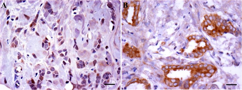
Figure 2 c-Met expression varies between histological subtypes of breast cancer. (A) Invasive lobular carcinoma characterised by
discohesive tumour cells with low c-Met expression. (B) Tubular carcinoma with cohesive tumour cells arranged in angulated tubules with strong
expression of c-Met. Immunohistochemistry, ×40 objective. Scale bars represent 20 μm.Ho-Yen et al. Breast Cancer Research (2015) 17:52 Page 6 of 11 Figure 3 c-Met expression in basal-like breast cancer. Characteristic features of basal-like breast cancer (images are from the same tumour). (A) Circumscribed tumour front (arrowheads) and associated chronic inflammatory cell infiltrate (arrow). (B) Tumour fibrosis. (C) High-grade cytology, with nuclear enlargement and pleomorphism (arrowheads), along with prominent mitotic figures (arrows). Haematoxylin and eosin; (A) and (B) × 10 objective, (C) × 40 objective. Scale bars represent 20 μm. (D) High cytoplasmic and membranous (arrowheads) expression of c-Met. Immunohistochemistry, ×40 objective. Scale bar represents 20 μm. Inset image is at 200% magnification. Migration and invasion Numerous studies have sought to uncover the mecha- The promigratory and proinvasive effects of HGF have nisms through which HGF/c-Met signalling contributes to been shown in several BCLs, including MCF7, MCF10. the migratory and invasive phenotype in breast cancer, DCIS and MDA-MB-231 [59-62]. Administration of particularly focusing on pathways associated with epithe- HGF, either in the form of recombinant HGF, condi- lial adhesion [65-67]. E-Cadherin is a key component of tioned media from HGF-secreting fibroblasts or by way adherens junctions (specialised intraepithelial junctions) of co-culture with HGF-secreting fibroblasts, has been [68,69], and is regarded by some as a tumour suppressor shown to significantly increase migration and invasion important in the prevention of cell migration, invasion in wound closure and transwell invasion assays [59-62]. and metastasis [69]. In MCF7 cells, E-Cadherin and c-Met In addition, adipose-derived mesenchymal cells isolated are co-localised at the cell membrane in regions of cell– from lipoaspirates express variable levels of HGF and cell contact [65,70]. Following treatment with HGF, lysates co-culture of these cells with MDA-MB-231 cells re- from these cells show a reduction in E-Cadherin expres- sulted in increased migration [63]. These studies are sion, and immunofluorescent studies demonstrate asym- supported by work demonstrating that NK4, a variant metric accumulation of c-Met and E-Cadherin in the form of HGF that competitively inhibits HGF binding cytosol, ultimately leading to complete internalisation of [64], reduces HGF-mediated c-Met phosphorylation and both proteins after 2 hours [61,70]. inhibits HGF-induced scattering and invasion of MCF7 In addition to favouring epithelial dissociation via E- and MDA-MB-231 cells [59]. Cadherin downregulation/internalisation, there is some
Ho-Yen et al. Breast Cancer Research (2015) 17:52                                                                   Page 7 of 11

evidence that HGF/c-Met signalling contributes to breast         the c-erbB family in particular has received considerable
cancer progression by promoting cancer cell adhesion to          interest. HGF has been shown to trans-activate epidermal
components of the extracellular matrix [66]. HGF                 growth factor receptor (EGFR) in PyVmT mouse mammary
treatment increased adhesion of MtLn3 rat mammary                carcinoma cells [76]. Moreover, the EGFR inhibitor gefitinib
adenocarcinoma cells to laminin, type 1 collagen and fi-         blocked HGF-mediated proliferation in PyVmT cells, mi-
bronectin, compared with control cells [66]. Furthermore,        gration in PyVmT cells and NMuMG cells, and invasion in
treatment with HGF was associated with lamellipodia for-         PyVmT cells, NMuMG cells and MDA-MB-231 cells [76].
mation, focal adhesion kinase phosphorylation and focal          The authors went on to show that gefitinib effects c-Met
adhesion kinase expression at focal contacts, suggesting         activation in an EGFR-dependent process (as opposed to
that c-Met and focal adhesion kinase cooperate to pro-           directly targeting c-Met) by finding no effect on c-Met acti-
mote cancer cell/substrate adhesion [66].                        vation when EGFR-null/c-Met expressing haematopoietic
   Proteolytic pathway regulation is another mechanism in-       32D cells were treated with the inhibitor [76].
fluenced by HGF/c-Met signalling in the in vitro setting            Similarly, another member of the c-erbB family – Her2 –
[60]. Conditioned media from HGF-secreting fibroblasts           has been noted to cross-talk with c-Met in HCC1954 breast
and recombinant HGF treatment resulted in increased se-          cancer cells, which overexpress c-Met and Her2 [77]. In
cretion of both urokinase-type plasminogen activator and         HCC1954 cells, knockdown of MET resulted in increased
its receptor (urokinase-type plasminogen activator recep-        Her2 phosphorylation and, conversely, knockdown of Her2
tor) by different ductal carcinoma in situ cell lines            was associated with an increase in c-Met activity [77].
(MCF10.DCIS cells and SUM102 cells). Increased collagen             The relationship between c-Met and other RTKs has im-
IV degradation was also demonstrated, along with in-             portant implications for the development of resistance to
creased numbers of invasive outgrowths in three-                 anti-RTK therapies already in clinical use – now a significant
dimensional cultures of ductal carcinoma in situ cells when      problem in breast cancer treatment [77]. Indeed, in the
in the presence of HGF [60]. Together, these findings im-        EGFR tyrosine kinase inhibitor-resistant cell line SUM229,
plicate HGF-secreting fibroblasts in the progression of          c-Met is phosphorylated and thought to stimulate EGFR
ductal carcinoma in situ to invasive cancer [60].                phosphorylation in the presence of EGFR inhibitors in a
                                                                 Src-mediated process [78]. Likewise, treatment of the Her2-
                                                                 overexpressing BT-474 and SKBR3 cells with trastuzumab
Cell survival
                                                                 upregulated c-Met protein expression in just 48 hours [79].
HGF/c-Met signalling has been associated with both pro-
                                                                 HGF-mediated c-Met phosphorylation in these cells op-
apoptotic and anti-apoptotic effects [71]. Using the murine
                                                                 posed trastuzumab-mediated growth inhibition by abrogat-
hepatocellular carcinoma cell line Hepa1-6, Wang and col-
                                                                 ing p27 induction [79]. c-Met therefore plays an important
leagues established that c-Met and the death receptor FAS
                                                                 role in breast cancer cell function and signalling by virtue of
formed a complex; they proposed a model in which c-Met
                                                                 its ability to interact with other RTKs.
sequesters FAS, thus preventing ligand-independent activa-
tion (due to clustering) and FAS ligand/FAS binding, result-
ing in cell survival [71]. In this model, high levels of HGF
                                                                 In vivo models of HGF/c-Met-mediated tumour
(or FAS ligand) would cause dissociation of the c-Met/FAS
                                                                 formation
                                                                 The in vivo effects of aberrant HGF/c-Met signalling have
complex, leaving the cells vulnerable to FAS-mediated apop-
                                                                 been explored in different mouse models [80-83]. Mice
tosis. Such a model would explain the paradoxical effects of
                                                                 harbouring the whey acidic protein WAP-HGF transgenic
HGF/c-Met on cell survival [71].
                                                                 construct show elevated HGF expression in mammary
   A similar mechanism may also exist in breast cancer cells,
                                                                 epithelium, compared with wild-type mice, and go on to
where treatment of preneoplastic MCF-10AT breast epithe-
                                                                 develop mammary tumours characterised by a high Ki67
lial cells with anti-FAS (an activator of FAS signalling) in-
                                                                 proliferation index, a reduced progesterone receptor im-
duced c-Met/FAS complex dissociation and apoptosis [72].
                                                                 munoreactivity and areas of squamous differentiation (a
It has also been shown that HGF protects MDA-MB-453
                                                                 feature of BL breast cancers) [80,84].
breast cancer cells from adriamycin-induced apoptosis [73].
                                                                    Squamous metaplasia was also detected in a high propor-
Preincubation of these cells with HGF blocked adriamycin-
                                                                 tion (65%) of mammary tumours that developed in mice
mediated FAS ligand upregulation and inhibited the reduc-
                                                                 with mutationally activated MET [82]. The majority of these
tion in levels of the anti-apoptotic protein Bcl-XL [73,74].
                                                                 tumours also expressed the basal cytokeratin, cytokeratin 5
                                                                 [82]. Elsewhere, Ponzo and colleagues studied transgenic
Cross-talk with other receptor tyrosine kinases                  mice that express oncogenic MET in the mammary epithe-
It is widely appreciated that c-Met can cross-talk with a        lium, under the control of the murine mammary tumour
variety of other cell surface receptors (reviewed in [75]). In   virus promoter [81]. About one-half of the tumours that de-
breast cancer, cross-talk between c-Met and members of           veloped in these mice showed variable histological patterns
Ho-Yen et al. Breast Cancer Research (2015) 17:52 Page 8 of 11
Table 3 Anti-c-Met therapies currently under investigation in clinical trials for breast cancer [11]
Compound Target/mechanism of action ClinicalTrials.gov identifier
Tivantinib (ARQ197) c-Met/non-ATP kinase inhibitor NCT 01575522
Cabozantinib (XL184) c-Met and VEGFR, along with RET, KIT, AXL/kinase inhibitor NCT 01738438
Foretinib (XL880) c-Met and VEGFR, along with KIT, Flt-3, PDGFR, Tie-2/kinase inhibitor NCT 01147484
MetMab (onartuzumab) c-Met/anti-c-Met antibody NCT 01186991
PDGFR, platelet-derived growth factor receptor; VEGFR, vascular endothelial growth factor receptor.
which included BL features [85] such as squamous/spindle RET, KIT and AXL, but particularly c-Met and VEGFR2
cell differentiation, high nuclear grade, necrosis and lympho- [91]. Cabozantinib not only inhibits c-Met phosphorylation
cytic infiltration [81]. These tumours also expressed cytoker- in vivo, but also promotes tumour hypoxia and cell death
atin 5/6 and cytokeratin 14 (another basal cytokeratin) on and inhibits the growth of MDA-MB-231 tumours in a
IHC [81]. Interestingly, in a subsequent study these workers dose-dependent manner [91]. It has been suggested that the
found that loss of TP53, in addition to oncogenic MET ex- ability of cabozantinib to inhibit both c-Met and vascular
pression, was associated with the formation of tumours with endothelial growth factor receptor 2 may actually counter
a claudin-low profile, a recently described subgroup of TN the c-Met-dependent resistance noted when only the vascu-
tumours that is distinct from the BL subtype [83,86]. lar endothelial growth factor pathway is targeted [91].
Foretinib is a small molecule kinase inhibitor that princi-
Anti-c-Met therapy in invasive breast cancer pally targets c-Met and vascular endothelial growth factor
There are various strategies for antagonising HGF/c-Met receptor [92]. Foretinib inhibits HGF-induced c-Met phos-
signalling: antibodies can be directed against c-Met; HGF phorylation, inhibits tumour cell growth in hypoxic and nor-
itself can be targeted with antibodies; and the catalytic moxic conditions, and has been shown to reduce tumour
function of c-Met can be opposed with tyrosine kinase in- cell burden in an in vivo model of lung cancer [92]. A phase
hibitors, which account for the majority of anti-c-Met I study in patients with a wide variety of solid organ cancers
compounds under investigation [87]. Four therapies cur- (including one breast cancer patient) found the inhibitor to
rently in phase II clinical trials for the treatment of ad- be safe and noted a partial response in 7.5% of patients and
vanced TN breast cancer are tivantinib (also known as stable disease in a further 55% of patients [93].
ARQ197) [ClinicalTrials.gov:NCT 01575522], cabozanti-
nib (alternatively known as XL184) [ClinicalTrials.gov: Conclusion
NCT 01738438], MetMab (onartuzumab) [ClinicalTrials. Much progress has been made in our understanding
gov:NCT 01186991] and foretinib (XL880) [ClinicalTrials. of c-Met/HGF signalling in recent years, and there is
gov:NCT 01147484] [11] (Table 3). now convincing in vitro and in vivo evidence that this
As well as breast cancer, the anti-c-Met monoclonal is an important pathway in mammary development
antibody MetMab is also being trialled in lung cancer and cancer progression. Clinical studies have con-
and colon cancer [88]. Unlike many other anti-c-Met firmed the prognostic significance of c-Met expression
antibodies, MetMab is monovalent – therefore it does in breast cancer and highlight the potential of c-Met
not promote dimerisation when it binds to c-Met, thus inhibitors as a novel form of targeted therapy. The
avoiding the agonistic effects associated with similar possible role of c-Met signalling in promoting BL
therapies [88]. breast cancer is noteworthy, and merits further inves-
Tivantinib belongs to the c-Met tyrosine kinase inhibi- tigation in the experimental and clinical trial settings.
tor class of c-Met antagonists, and is a non-ATP com- The outcomes of ongoing and future clinical trials of
petitive inhibitor of the receptor [89,90]. A phase I trial anti-c-Met therapy will be eagerly anticipated, but is-
of tivantinib in 51 patients with solid tumours (including sues such as receptor cross-talk and resistance may
two patients with breast cancer) found the inhibitor to need to be addressed if treatment efficacy is to be
be well tolerated, with fatigue, nausea and vomiting be- maximised. It is also important to stratify patients ap-
ing the most common adverse effects [90]. Furthermore, propriately, and the development of standardised
when pretreatment and on-treatment tumour biopsies prognostic/predictive assays will be crucial in identify-
were compared, there was a reduction in total and phos- ing those subgroups of patients most likely to benefit
phorylated c-Met expression in the on-treatment sam- from anti-c-Met therapy.
ples, suggesting that tivantinib inhibited intra-tumoural
c-Met signalling [90]. Abbreviations
BCL: Breast cancer cell line; BL: Basal-like; EGFR: Epidermal growth factor
Cabozantinib is another small molecule inhibitor of c- receptor; HGF: Hepatocyte growth factor; IHC: Immunohistochemistry;
Met, which targets a range of tyrosine kinases, including RTK: Receptor tyrosine kinase; TN: Triple negative.Ho-Yen et al. Breast Cancer Research (2015) 17:52 Page 9 of 11
Competing interests 19. Abella JV, Peschard P, Naujokas MA, Lin T, Saucier C, Urbé S, et al. Met/
The authors declare that they have no competing interests. hepatocyte growth factor receptor ubiquitination suppresses transformation
and is required for Hrs phosphorylation. Mol Cell Biol. 2005;25:9632–45.
20. Peschard P, Park M. From Tpr-Met to Met, tumorigenesis and tubes.
Acknowledgements Oncogene. 2007;26:1276–85.
The authors apologise that they have been unable to cite all relevant 21. Kermorgant S, Zicha D, Parker PJ. PKC controls HGF-dependent c-Met traffic,
studies, due to space constraints. CMH-Y is funded by a Cancer Research UK signalling and cell migration. EMBO J. 2004;23:3721–34.
Clinical Research Fellowship. JLJ is funded by the Breast Cancer Campaign 22. Kermorgant S, Parker PJ. Receptor trafficking controls weak signal delivery: a
Tissue Bank. strategy used by c-Met for STAT3 nuclear accumulation. J Cell Biol.
2008;182:855–63.
Author details 23. Barrow-McGee R, Kermorgant S. Met endosomal signalling: in the right
1
Department of Cellular Pathology, St George’s Healthcare NHS Trust, place, at the right time. Int J Biochem Cell Biol. 2014;49:69–74.
Blackshaw Road, Tooting, London SW17 0QT, UK. 2Centre for Tumour 24. Andermarcher E, Surani MA, Gherardi E. Co-expression of the HGF/SF and c-
Biology, Barts Cancer Institute, Charterhouse Square, London EC1M 6BQ, UK. met genes during early mouse embryogenesis precedes reciprocal expression
in adjacent tissues during organogenesis. Dev Genet. 1996;18:254–66.
Received: 25 July 2014 Accepted: 5 March 2015 25. Niranjan B, Buluwela L, Yant J, Perusinghe N, Atherton A, Phippard D, et al.
HGF/SF: a potent cytokine for mammary growth, morphogenesis and
development. Development. 1995;121:2897–908.
26. Yant J, Buluwela L, Niranjan B, Gusterson B, Kamalati T. In vivo effects of
References
hepatocyte growth factor/scatter factor on mouse mammary gland
1. Cooper CS, Park M, Blair DG, Tainsky MA, Huebner K, Croce CM, et al.
development. Exp Cell Res. 1998;241:476–81.
Molecular cloning of a new transforming gene from a chemically
27. Gastaldi S, Sassi F, Accornero P, Torti D, Galimi F, Migliardi G, et al. Met signaling
transformed human cell line. Nature. 1984;311:29–33.
regulates growth, repopulating potential and basal cell-fate commitment of
2. Nakamura T, Nawa K, Ichihara A. Partial purification and characterization of
mammary luminal progenitors: implications for basal-like breast cancer.
hepatocyte growth factor from serum of hepatectomized rats.
Oncogene. 2013;32:1428–40.
Biochem Biophys Res Commun. 1984;122:1450–9.
3. Bottaro DP, Rubin JS, Faletto DL, Chan AM, Kmiecik TE, Vande Woude GF, 28. Sierra JR, Tsao MS. c-MET as a potential therapeutic target and biomarker in
et al. Identification of the hepatocyte growth factor receptor as the c-met cancer. Ther Adv Med Oncol. 2011;3:S21–35.
proto-oncogene product. Science. 1991;251:802–4. 29. Stella GM, Benvenuti S, Gramaglia D, Scarpa A, Tomezzoli A, Cassoni P, et al.
4. Schmidt L, Duh FM, Chen F, Kishida T, Glenn G, Choyke P, et al. Germline MET mutations in cancers of unknown primary origin (CUPs). Hum Mutat.
and somatic mutations in the tyrosine kinase domain of the MET proto- 2011;32:44–50.
oncogene in papillary renal carcinomas. Nat Genet. 1997;16:68–73. 30. Bièche I, Champème MH, Lidereau R. Infrequent mutations of the MET gene
5. Walker F, Kermorgant S, Daraï E, Madelenat P, Cremieux AC, Hénin D, et al. in sporadic breast tumours. Int J Cancer. 1999;82:908–10.
Hepatocyte growth factor and c-Met in cervical intraepithelial neoplasia: 31. Ma J, DeFrances MC, Zou C, Johnson C, Ferrell R, Zarnegar R. Somatic
overexpression of proteins associated with oncogenic human papillomavirus mutation and functional polymorphism of a novel regulatory element in
and human immunodeficiency virus. Clin Cancer Res. 2003;9:273–84. the HGF gene promoter causes its aberrant expression in human breast
6. Lengyel E, Prechtel D, Resau JH, Gauger K, Welk A, Lindemann K, et al. cancer. J Clin Invest. 2009;119:478–91.
C-Met overexpression in node-positive breast cancer identifies patients with 32. Carracedo A, Egervari K, Salido M, Rojo F, Corominas JM, Arumi M, et al. FISH
poor clinical outcome independent of Her2/neu. Int J Cancer. and immunohistochemical status of the hepatocyte growth factor receptor
2005;113:678–82. (c-Met) in 184 invasive breast tumors. Breast Cancer Res. 2009;11:402.
7. Kim CH, Kim J, Kahng H, Choi EC. Change of E-cadherin by hepatocyte 33. Gonzalez-Angulo AM, Chen H, Karuturi MS, Chavez-MacGregor M, Tsavachidis
growth factor and effects on the prognosis of hypopharyngeal carcinoma. S, Meric-Bernstam F, et al. Frequency of mesenchymal–epithelial transition
Ann Surg Oncol. 2007;14:1565–74. factor gene (MET) and the catalytic subunit of phosphoinositide-3-kinase
8. Engelman JA, Zejnullahu K, Mitsudomi T, Song Y, Hyland C, Park JO, et al. (PIK3CA) copy number elevation and correlation with outcome in patients with
MET amplification leads to gefitinib resistance in lung cancer by activating early stage breast cancer. Cancer. 2013;119:7–15.
ERBB3 signaling. Science. 2007;316:1039–43. 34. Minuti G, Cappuzzo F, Duchnowska R, Jassem J, Fabi A, O’Brien T, et al.
9. De Oliveira AT, Matos D, Logullo AF, DA Silva SR, Neto RA, Filho AL, et al. Increased MET and HGF gene copy numbers are associated with trastuzumab
MET Is highly expressed in advanced stages of colorectal cancer and failure in HER2-positive metastatic breast cancer. Br J Cancer. 2012;107:793–9.
indicates worse prognosis and mortality. Anticancer Res. 2009;29:4807–11. 35. Tuck AB, Park M, Sterns EE, Boag A, Elliott BE. Coexpression of hepatocyte
10. Li Y, Chen CQ, He YL, Cai SR, Yang DJ, He WL, et al. Abnormal expression of growth factor and receptor (Met) in human breast carcinoma. Am J Pathol.
E-cadherin in tumor cells is associated with poor prognosis of gastric 1996;148:225–32.
carcinoma. J Surg Oncol. 2012;106:304–10. 36. Jin L, Fuchs A, Schnitt SJ, Yao Y, Joseph A, Lamszus K, et al. Expression of
11. ClinicalTrials.gov. US National Institutes of Health, Bethesda, MD. scatter factor and c-met receptor in benign and malignant breast tissue.
http://www.clinicaltrials.gov. Accessed Jun 2014. Cancer. 1997;79:749–60.
12. Cheang MC, Voduc D, Bajdik C, Leung S, McKinney S, Chia SK, et al. Basal- 37. Edakuni G, Sasatomi E, Satoh T, Tokunaga O, Miyazaki K. Expression of the
like breast cancer defined by five biomarkers has superior prognostic value hepatocyte growth factor/c-Met pathway is increased at the cancer front in
than triple-negative phenotype. Clin Cancer Res. 2008;14:1368–76. breast carcinoma. Pathol Int. 2001;51:172–8.
13. Ho-Yen C, Bowen RL, Jones JL. Characterization of basal-like breast cancer: 38. Kang JY, Dolled-Filhart M, Ocal IT, Singh B, Lin CY, Dickson RB, et al. Tissue
an update. Diagn Histopathol. 2012;18:104–11. microarray analysis of hepatocyte growth factor/Met pathway components
14. Hanna JA, Bordeaux J, Rimm DL, Agarwal S. The function, proteolytic reveals a role for Met, matriptase, and hepatocyte growth factor activator
processing, and histopathology of Met in cancer. Adv Cancer Res. inhibitor 1 in the progression of node-negative breast cancer. Cancer Res.
2009;103:1–23. 2003;63:1101–5.
15. Trusolino L, Bertotti A, Comoglio PM. MET signalling: principles and 39. Baker AF, Dragovich T, Ihle NT, Williams R, Fenoglio-Preiser C, Powis G.
functions in development, organ regeneration and cancer. Nat Rev Mol Cell Stability of phosphoprotein as a biological marker of tumor signaling.
Biol. 2010;11:834–48. Clin Cancer Res. 2005;11:4338–40.
16. Goldoni S, Humphries A, Nyström A, Sattar S, Owens RT, McQuillan DJ, et al. 40. Dua R, Zhang J, Parry G, Penuel E. Detection of hepatocyte growth factor
Decorin is a novel antagonistic ligand of the Met receptor. J Cell Biol. (HGF) ligand-c-MET receptor activation in formalin-fixed paraffin embedded
2009;185:743–54. specimens by a novel proximity assay. PLoS One. 2011;6:e15932.
17. Birchmeier C, Birchmeier W, Gherardi E, Vande Woude GF. Met, metastasis, 41. Jarvius M, Paulsson J, Weibrecht I, Leuchowius KJ, Andersson AC, Wählby C,
motility and more. Nat Rev Mol Cell Biol. 2003;4:915–25. et al. In situ detection of phosphorylated platelet-derived growth factor
18. Gherardi E, Birchmeier W, Birchmeier C, Vande WG. Targeting MET in cancer: receptor beta using a generalized proximity ligation method. Mol Cell
rationale and progress. Nat Rev Cancer. 2012;12:89–103. Proteomics. 2007;6:1500–9.Ho-Yen et al. Breast Cancer Research (2015) 17:52 Page 10 of 11
42. Blokzijl A, Friedman M, Pontén F, Landegren U. Profiling protein expression 65. Hiscox S, Jiang WG. Association of the HGF/SF receptor, c-met, with the
and interactions: proximity ligation as a tool for personalized medicine. cell-surface adhesion molecule, E-cadherin, and catenins in human tumor
J Intern Med. 2010;268:232–45. cells. Biochem Biophys Res Commun. 1999;261:406–11.
43. Hochgräfe F, Zhang L, O’Toole SA, Browne BC, Pinese M, Porta Cubas A, 66. Beviglia L, Kramer RH. HGF induces FAK activation and integrin-mediated
et al. Tyrosine phosphorylation profiling reveals the signaling network adhesion in MTLn3 breast carcinoma cells. Int J Cancer. 1999;83:640–9.
characteristics of Basal breast cancer cells. Cancer Res. 2010;70:9391–401. 67. Reshetnikova G. Met receptor subcellular localization depends on E-
44. Raghav KP, Wang W, Liu S, Chavez-MacGregor M, Meng X, Hortobagyi GN, cadherin function. ScientificWorldJournal. 2007;7:2009–11.
et al. cMET and phospho-cMET protein levels in breast cancers and survival 68. Thiery JP. Epithelial–mesenchymal transitions in tumour progression.
outcomes. Clin Cancer Res. 2012;18:2269–77. Nat Rev Cancer. 2002;2:442–54.
45. Ghoussoub RA, Dillon DA, D’Aquila T, Rimm EB, Fearon ER, Rimm DL. 69. Rodriguez FJ, Lewis-Tuffin LJ, Anastasiadis PZ. E-cadherin’s dark side:
Expression of c-met is a strong independent prognostic factor in breast possible role in tumor progression. Biochim Biophys Acta.
carcinoma. Cancer. 1998;82:1513–20. 1826;2012:23–31.
46. Camp RL, Rimm EB, Rimm DL. Met expression is associated with poor 70. Matteucci E, Ridolfi E, Desiderio MA. Hepatocyte growth factor differently
outcome in patients with axillary lymph node negative breast carcinoma. influences Met-E-cadherin phosphorylation and downstream signaling
Cancer. 1999;86:2259–65. pathway in two models of breast cells. Cell Mol Life Sci. 2006;63:2016–26.
47. Tolgay Ocal I, Dolled-Filhart M, D’Aquila TG, Camp RL, Rimm DL. Tissue microarray- 71. Wang X, DeFrances MC, Dai Y, Pediaditakis P, Johnson C, Bell A, et al. A
based studies of patients with lymph node negative breast carcinoma show that mechanism of cell survival: sequestration of Fas by the HGF receptor Met.
met expression is associated with worse outcome but is not correlated with Mol Cell. 2002;9:411–21.
epidermal growth factor family receptors. Cancer. 2003;97:1841–8. 72. Shen K, Novak RF. Fas-signaling and effects on receptor tyrosine kinase
48. Chen HH, Su WC, Lin PW, Guo HR, Lee WY. Hypoxia-inducible factor-1alpha signal transduction in human breast epithelial cells. Biochem Biophys Res
correlates with MET and metastasis in node-negative breast cancer. Breast Commun. 1997;230:89–93.
Cancer Res Treat. 2007;103:167–75. 73. Fan S, Wang JA, Yuan RQ, Rockwell S, Andres J, Zlatapolskiy A, et al. Scatter
49. Zagouri F, Bago-Horvath Z, Rössler F, Brandstetter A, Bartsch R, Papadimitriou factor protects epithelial and carcinoma cells against apoptosis induced by
CA, et al. High MET expression is an adverse prognostic factor in patients with DNA-damaging agents. Oncogene. 1998;17:131–41.
triple-negative breast cancer. Br J Cancer. 2013;108:1100–5. 74. Gao M, Fan S, Goldberg ID, Laterra J, Kitsis RN, Rosen EM. Hepatocyte
50. Ho-Yen CM, Green AR, Rakha EA, Brentnall AR, Ellis IO, Kermorgant S, et al. growth factor/scatter factor blocks the mitochondrial pathway of apoptosis
C-Met in invasive breast cancer: is there a relationship with the basal-like signaling in breast cancer cells. J Biol Chem. 2001;276:47257–65.
subtype? Cancer. 2014;120:163–71. 75. Lai AZ, Abella JV, Park M. Crosstalk in Met receptor oncogenesis. Trends Cell
51. Kim YJ, Choi JS, Seo J, Song JY, Lee SE, Kwon MJ, et al. MET is a potential Biol. 2009;19:542–51.
target for use in combination therapy with EGFR inhibition in triple- 76. Bonine-Summers AR, Aakre ME, Brown KA, Arteaga CL, Pietenpol JA, Moses
negative/basal-like breast cancer. Int J Cancer. 2014;134:2424–36. HL, et al. Epidermal growth factor receptor plays a significant role in
52. Pozner-Moulis S, Cregger M, Camp RL, Rimm DL. Antibody validation by hepatocyte growth factor mediated biological responses in mammary
quantitative analysis of protein expression using expression of Met in breast epithelial cells. Cancer Biol Ther. 2007;6:561–70.
cancer as a model. Lab Invest. 2007;87:251–60. 77. Paulson AK, Linklater ES, Berghuis BD, App CA, Oostendorp LD, Paulson JE,
53. Nakopoulou L, Gakiopoulou H, Keramopoulos A, Giannopoulou I, Athanassiadou et al. MET and ERBB2 are coexpressed in ERBB2+ breast cancer and
P, Mavrommatis J, et al. c-met tyrosine kinase receptor expression is associated contribute to innate resistance. Mol Cancer Res. 2013;11:1112–21.
with abnormal beta-catenin expression and favourable prognostic factors in 78. Mueller KL, Hunter LA, Ethier SP, Boerner JL. Met and c-Src cooperate to
invasive breast carcinoma. Histopathology. 2000;36:313–25. compensate for loss of epidermal growth factor receptor kinase activity in
54. Harris G, Pinder SE, O’Malley FP. Invasive carcinoma: special types. In: O’Malley breast cancer cells. Cancer Res. 2008;68:3314–22.
FP, Pinder SE, editors. Breast pathology. Philadelphia: Elsevier; 2006. p. 201–23. 79. Shattuck DL, Miller JK, Carraway 3rd KL, Sweeney C. Met receptor
55. Charafe-Jauffret E, Ginestier C, Monville F, Finetti P, Adélaïde J, Cervera N, contributes to trastuzumab resistance of Her2-overexpressing breast cancer
et al. Gene expression profiling of breast cell lines identifies potential new cells. Cancer Res. 2008;68:1471–7.
basal markers. Oncogene. 2006;25:2273–84. 80. Gallego MI, Bierie B, Hennighausen L. Targeted expression of HGF/SF in
56. Lehmann BD, Bauer JA, Chen X, Sanders ME, Chakravarthy AB, Shyr Y, et al. mouse mammary epithelium leads to metastatic adenosquamous
Identification of human triple-negative breast cancer subtypes and preclinical carcinomas through the activation of multiple signal transduction pathways.
models for selection of targeted therapies. J Clin Invest. 2011;121:2750–67. Oncogene. 2003;22:8498–508.
57. Elston CW, Ellis IO. Pathological prognostic factors in breast cancer. I. The 81. Ponzo MG, Lesurf R, Petkiewicz S, O’Malley FP, Pinnaduwage D, Andrulis IL,
value of histological grade in breast cancer: experience from a large study et al. Met induces mammary tumors with diverse histologies and is
with long-term follow-up. Histopathology. 1991;19:403–10. associated with poor outcome and human basal breast cancer. Proc Natl
58. Tsarfaty I, Resau JH, Rulong S, Keydar I, Faletto DL, Vande Woude GF. The Acad Sci U S A. 2009;106:12903–8.
met proto-oncogene receptor and lumen formation. Science. 82. Graveel CR, DeGroot JD, Su Y, Koeman J, Dykema K, Leung S, et al. Met
1992;257:1258–61. induces diverse mammary carcinomas in mice and is associated with
59. Hiscox S, Parr C, Nakamura T, Matsumoto K, Mansel RE, Jiang WG. Inhibition human basal breast cancer. Proc Natl Acad Sci U S A. 2009;106:12909–14.
of HGF/SF-induced breast cancer cell motility and invasion by the HGF/SF 83. Knight JF, Lesurf R, Zhao H, Pinnaduwage D, Davis RR, Saleh SM, et al. Met
variant, NK4. Breast Cancer Res Treat. 2000;59:245–54. synergizes with p53 loss to induce mammary tumors that possess features
60. Jedeszko C, Victor BC, Podgorski I, Sloane BF. Fibroblast hepatocyte growth of claudin-low breast cancer. Proc Natl Acad Sci U S A. 2013;110:E1301–10.
factor promotes invasion of human mammary ductal carcinoma in situ. 84. Fulford LG, Easton DF, Reis-Filho JS, Sofronis A, Gillett CE, Lakhani SR, et al.
Cancer Res. 2009;69:9148–55. Specific morphological features predictive for the basal phenotype in grade
61. Hung CM, Kuo DH, Chou CH, Su YC, Ho CT, Way TD. Osthole suppresses 3 invasive ductal carcinoma of breast. Histopathology. 2006;49:22–34.
hepatocyte growth factor (HGF)-induced epithelial–mesenchymal transition 85. Livasy CA, Karaca G, Nanda R, Tretiakova MS, Olopade OI, Moore DT, et al.
via repression of the c-Met/Akt/mTOR pathway in human breast cancer Phenotypic evaluation of the basal-like subtype of invasive breast
cells. J Agric Food Chem. 2011;59:9683–90. carcinoma. Mod Pathol. 2006;19:264–71.
62. Ayoub NM, Akl MR, Sylvester PW. Combined γ-tocotrienol and Met inhibitor 86. Herschkowitz JI, Simin K, Weigman VJ, Mikaelian I, Usary J, Hu Z, et al.
treatment suppresses mammary cancer cell proliferation, epithelial-to- Identification of conserved gene expression features between murine mammary
mesenchymal transition and migration. Cell Prolif. 2013;46:538–53. carcinoma models and human breast tumors. Genome Biol. 2007;8:R76.
63. Eterno V, Zambelli A, Pavesi L, Villani L, Zanini V, Petrolo G, et al. Adipose- 87. Gentile A, Trusolino L, Comoglio PM. The Met tyrosine kinase receptor in
derived mesenchymal stem cells (ASCs) may favour breast cancer development and cancer. Cancer Metastasis Rev. 2008;27:85–94.
recurrence via HGF/c-Met signaling. Oncotarget. 2014;5:613–33. 88. Yano S, Nakagawa T. The current state of molecularly targeted drugs
64. Date K, Matsumoto K, Shimura H, Tanaka M, Nakamura T. HGF/NK4 is a targeting HGF/Met. Jpn J Clin Oncol. 2014;44:9–12.
specific antagonist for pleiotrophic actions of hepatocyte growth factor. 89. Cecchi F, Rabe DC, Bottaro DP. Targeting the HGF/Met signalling pathway
FEBS Lett. 1997;420:1–6. in cancer. Eur J Cancer. 2010;46:1260–70.Ho-Yen et al. Breast Cancer Research (2015) 17:52 Page 11 of 11
90. Yap TA, Olmos D, Brunetto AT, Tunariu N, Barriuso J, Riisnaes R, et al. Phase I
trial of a selective c-MET inhibitor ARQ 197 incorporating proof of
mechanism pharmacodynamic studies. J Clin Oncol. 2011;29:1271–9.
91. Yakes FM, Chen J, Tan J, Yamaguchi K, Shi Y, Yu P, et al. Cabozantinib
(XL184), a novel MET and VEGFR2 inhibitor, simultaneously suppresses
metastasis, angiogenesis, and tumor growth. Mol Cancer Ther.
2011;10:2298–308.
92. Qian F, Engst S, Yamaguchi K, Yu P, Won KA, Mock L, et al. Inhibition of
tumor cell growth, invasion, and metastasis by EXEL-2880 (XL880,
GSK1363089), a novel inhibitor of HGF and VEGF receptor tyrosine kinases.
Cancer Res. 2009;69:8009–16.
93. Eder JP, Shapiro GI, Appleman LJ, Zhu AX, Miles D, Keer H, et al. A phase I
study of foretinib, a multi-targeted inhibitor of c-Met and vascular
endothelial growth factor receptor 2. Clin Cancer Res. 2010;16:3507–16.You can also read