Insulin Receptor Substrate-2 Deficiency Impairs Brain Growth and Promotes Tau Phosphorylation
←
→
Page content transcription
If your browser does not render page correctly, please read the page content below
7084 • The Journal of Neuroscience, August 6, 2003 • 23(18):7084 –7092
Cellular/Molecular
Insulin Receptor Substrate-2 Deficiency Impairs Brain
Growth and Promotes Tau Phosphorylation
Markus Schubert,1* Derek P. Brazil,1* Deborah J. Burks,1* Jake A. Kushner,1 Jing Ye,1 Carrie L. Flint,1
Janet Farhang-Fallah,1 Pieter Dikkes,2 Xavier M. Warot,2 Carlos Rio,2 Gabriel Corfas,2 and Morris F. White1
1Howard Hughes Medical Institute, Joslin Diabetes Center, Harvard Medical School, Boston, Massachusetts 02215, and 2Division of Neuroscience,
Department of Neurology, Harvard Medical School, Boston, Massachusetts 02115
Insulin resistance and diabetes might promote neurodegenerative disease, but a molecular link between these disorders is unknown.
Many factors are responsible for brain growth, patterning, and survival, including the insulin–insulin-like growth factor (IGF)-signaling
cascades that are mediated by tyrosine phosphorylation of insulin receptor substrate (IRS) proteins. Irs2 signaling mediates peripheral
insulin action and pancreatic -cell function, and its failure causes diabetes in mice. In this study, we reveal two important roles for Irs2
signaling in the mouse brain. First, disruption of the Irs2 gene reduced neuronal proliferation during development by 50%, which
dissociated brain growth from Irs1-dependent body growth. Second, neurofibrillary tangles containing phosphorylated tau accumulated
in the hippocampus of old Irs2 knock-out mice, suggesting that Irs2 signaling is neuroprotective. Thus, dysregulation of the Irs2 branch
of the insulin–Igf-signaling cascade reveals a molecular link between diabetes and neurodegenerative disease.
Key words: diabetes; insulin receptor substrates; Irs2; growth factors; tau phosphorylation; brain size; neuron survival; Alzheimer’s
disease
Introduction standing species-specific expansion of the brain and body size
Among mammals including mice, the size of the brain and its during development and neonatal growth (Ludwig et al., 1996;
components are related to body size (Finlay and Darlington, Goldowitz and Hamre, 1998).
1995). Patterning and size of the brain are determined by the The receptors for insulin and IGF1 are tyrosine kinases that
coordinated proliferation, death, and migration of neuronal pre- mediate phosphorylation of the insulin receptor substrates
cursors in the cerebellum, hippocampus, retinal ganglion, and (IRSs), including Irs1, Irs2, Irs3, and Irs4 (Yamada et al., 1997;
other regions (Strom and Williams, 1998; Airey et al., 2001; Lu et Yenush and White, 1997). Irs1 and Irs2 are widely expressed and
al., 2001; Chenn and Walsh, 2002). Various factors regulate these mediate insulin action in most tissues (Sun et al., 1992, 1995;
processes, including extracellular matrix proteins, immunoglo- Bernal et al., 1998), Irs3 is restricted to rodent adipose tissue but
bins, peptide growth factors, inflammatory cytokines, and neu- might be missing in people (Lavan et al., 1997; Bjornholm et al.,
rotransmitters (Horner and Gage, 2000). Developmental signals 2002), and Irs4 is primarily detected in the brain, including the
are especially important, including the sonic hedgehog pathway thymus, pituitary, and hypothalamus (Fantin et al., 1999; Uchida
that balances proliferation or differentiation of neuronal precur- et al., 2000). Irs1 mediates the effects of Igf1 on somatic growth,
sors, and the wnt3-catenin pathway that controls cell cycle exit because Irs1 ⫺/⫺ mice are ⬃50% smaller than normal. In con-
(Goldowitz and Hamre, 1998; Chenn and Walsh, 2002). At least trast, Irs2 ⫺ / ⫺ mice reach nearly normal size (Withers et al.,
50% of brain and body growth is mediated by the insulin–Igf- 1999). Although both Irs1 ⫺ / ⫺ and Irs2 ⫺ / ⫺ mice are insulin re-
signaling system (Liu et al., 1993). Various heterologous signals sistant, only Irs2 ⫺ / ⫺ mice develop diabetes resulting from im-
integrate the effect of the insulin-like growth factors (IGFs) with paired pancreatic -cell survival and function (Bruning et al.,
other systems. Cytoskeletal signals might modulate insulin–IGF 1998; Withers et al., 1998, 1999; Kulkarni et al., 1999; Michael et
signaling through the activity of GTPases (Sordella et al., 2002), al., 2000; Previs et al., 2000). Female Irs2 knock-out mice are
and growth hormone upregulates peripheral but not central IGF1 infertile and display increased food intake and mild obesity, sug-
levels, so the body can grow faster than the brain (Lupu et al., gesting that Irs2 signaling also promotes hypothalamic control of
2001). These relationships provide a molecular basis for under- appetite and reproduction (Burks et al., 2000).
Phosphorylated IRS proteins activate multiple signaling path-
Received March 17, 2003; revised April 23, 2003; accepted May 8, 2003. ways, including the phosphatidylinositol 3 (PI3)-kinase and ex-
This work was supported by National Institutes of Health Grants DK43808 and DK38712. We are grateful to C. tracellular signal-regulated kinase (ERK) cascades that directly
Cahill for preparation of ultrathin sections and procedural help and K. Copps for discussion and editorial help. We regulate various physiological processes (Saltiel, 2001). Activa-
thank M. Montminy for the Pdx1 transgenic mouse. tion of the PI3-kinase3 Akt cascade promotes neuronal growth
*M.S., D.P.B., and D.J.B. contributed equally to this work.
Correspondence should be addressed to Dr. Morris F. White, Howard Hughes Medical Institute, Joslin Diabetes
and survival by phosphorylating Foxo-transcription factors to
Center, 1 Joslin Place, Boston, MA 02215. E-mail: morris.white@joslin.harvard.edu. regulate gene transcription and phosphorylating Bcl-2-
Copyright © 2003 Society for Neuroscience 0270-6474/03/237084-09$15.00/0 associated death (BAD) protein to inhibit apoptosis (Brunet etSchubert et al. • Insulin Receptor Substrate-2 Deficiency J. Neurosci., August 6, 2003 • 23(18):7084 –7092 • 7085
al., 1999; Rodgers and Theibert, 2002). Akt inactivates glycogen biotinylated UTP nick end labeling (TUNEL) assays were performed
synthase kinase-3 (GSK3), which promotes cellular metabo- using the DNA FragEL Kit (Oncogen Research Products, San Diego, CA).
lism, proliferation, and survival (Cross et al., 1995). Activated Nissl and hematoxylin-eosin (HE) staining as well as immunostaining
PI3-kinase also promotes GABAergic transmission in the pre- using anti-glial fibrillary acidic protein (GFAP) (GP-52; Progen, Darra,
Queensland, Australia) or anti-bromodeoxyuridine (BrdU) (Boehringer
frontal cortical pyramidal neurons that play a key role in learning
Mannheim, Indianapolis, IN) as primary antibody were performed as
and memory (Ma et al., 2003). Akt also blocks the accumulation described previously. For quantitation, sections were viewed using a
of -amyloid plaques and neurofibrillary tangles that are associ- Ziess Axiovert S100 microscope and video camera. Counts and analysis
ated with Alzheimer’s disease (Kaytor and Orr, 2002). were done using Openlab software (Improvision, Lexington, MA).
Alzheimer’s disease is characterized by progressive memory BrdU incorporation. BrdU (100 g/gm body weight; Boehringer
loss, impaired language, and behavior that culminate in death. Mannheim) was injected intraperitoneally into pregnant mice [embry-
Epidemiological studies suggest that insulin resistance and type 2 onic day (E) 14] or postnatal day (P) 6 pups. Tissues were harvested after
diabetes increase the risk for age-related cognitive decline (Finch 48 hr and fixed in 4% PFA.
and Cohen, 1997; Carantoni et al., 2000). Although the long-term Isolation and culture of cerebellar granule cells. Cerebellar granule neurons
sequelae of hyperglycemia, including vascular complications, are were isolated from 5-d-old mouse litters. All manipulations were performed
at 4°C unless indicated otherwise. Individual cerebella were isolated, the
frequently implicated as the culprit, defects in the insulin–IGF-
meninges were removed using a dissecting microscope, and the cerebella
signaling system might contribute directly to memory loss and were washed three times in HHGN solution (1⫻ HBSS, 2.5 mM HEPES, pH
dementia (Stolk et al., 1997; Lovestone, 1999; Ott et al., 1999). 7.4, 35 mM glucose, 4 mM sodium bicarbonate). Cerebella were then incu-
Like diabetes, Alzheimer’s disease occurs in many forms, in bated in trypsin solution (10 mg/ml of trypsin, 100 g/ml of DNase, in
which most instances are late onset and sporadic, whereas well HHGN, pH 7.0, with 0.1N NaOH) for 15 min at room temperature (RT).
defined autosomal mutations cause early onset familial Alzhei- Cerebella were placed on ice, washed three times in HHGN, and then tritu-
mer’s disease (Hoyer, 2002). The disorder is ordinarily character- rated ⬃25 times with 1 ml of DNase solution [10 g/ml of DNase in basal
ized by the accumulation of extracellular deposits of amyloid medium eagle (BME)]. The cells were allowed to settle for 5 min at room
-peptide that promotes inflammation and the intracellular ac- temperature, the supernatant was transferred into fresh tubes, and the re-
cumulation of neurofibrillary tangles composed of paired helical maining pellet was triturated with an additional 1 ml of DNase solution for
another 25 times. After settling, the supernatants were combined and the
filaments assembled from hyperphosphorylated forms of the
cells were centrifuged for 5 min at 1000 ⫻ g. Cell pellets were suspended in
microtubule-associated protein tau (Lucas et al., 2001; Selkoe, BME containing 10% fetal bovine serum, 100 U of penicillin–streptomycin,
2001; Weggen et al., 2001; Hardy and Selkoe, 2002). However, tau 2 mM glutamine, and 25 mM KCl (culture medium), counted, and plated on
neurofibrillary tangles cause frontotemporal dementia without poly-D-lysine-coated 96-well plates. After 24 hr, 10 M cytosine arabinoside
detectable -amyloid plaques, suggesting that -amyloid toxicity (araC) was added to the cultures to inhibit proliferation of non-neuronal
might be tau dependent (Selkoe and Podlisny, 2002). Insulin– cells.
IGF-signaling pathways might protect neurons from inflamma- Neuronal apoptosis. Apoptosis assays were performed after 96 hr (4 d in
tion caused by -amyloid by decreasing intracellular concentra- vitro). Cerebellar granule cells were washed with serum-free BME containing
tions of -amyloid (Solano et al., 2000; Gasparini et al., 2001). 0.2% BSA. Cells were then incubated with the indicated factors (IGF1 or
Neurons in transgenic mice overexpressing a mutant form of BDNF) in BME containing 100 U of penicillin–streptomycin, 2 mM glu-
tamine, 0.2% BSA, and 10 M araC for 24 hr at 37°C. For experiments using
human amyloid precursor protein are resistant to apoptosis and
LY294002 (LY; 25 M) and PD98059 (PD; 40 M), neurons were washed as
develop -amyloid deposits slowly, resulting from, at least in described above and preincubated with either LY or PD for 30 min at 37°C.
part, upregulation of insulin-like growth factor-2 and activation The cells were fixed directly in the wells using 10% (w/v) paraformaldehyde
of the insulin–IGF-signaling cascade (Stein and Johnson, 2002). [4% (w/v) final], washed three times with PBS, permeabilized with 0.2%
Tau phosphorylation is mediated by several kinases, including (v/v) Triton X-100, washed twice with PBS, and then incubated with
GSK3 (Hong and Lee, 1997; Lovestone and Reynolds, 1997). Hoechst dye 33342 (10 g/ml) in PBS for 10 min at RT.
Thus, tau phosphorylation might be a direct consequence of re- Immunoblotting. For immunoblotting, brains were homogenized in a
duced insulin–IGF signaling that occurs during aging. polytron containing 50 mM Tris-HCl, pH 7.4, 150 mM NaCl, 1% (v/v)
Here, we reveal two important roles for the Irs2 branch of the NP40, 5 mM EDTA, 5% (v/v) glycerol, 10 g/ml leupeptin, 10 g/ml
insulin–IGF-signaling cascade in the brain. First, Irs2 signaling aprotinin, 1 mM phenylmethylsulfonyl fluoride, and 1 mM Na3VO4. Pro-
tein expression was determined from whole-brain lysates (50 g) dis-
promotes neuronal proliferation that increases brain size during
solved in Laemmli buffer and resolved on 7.5 or 10% SDS-PAGE gels.
development. Second, Irs2 signaling promotes the dephosphor- Proteins were transferred to nitrocellulose; the membrane was blocked
ylation of tau. Thus, failure of the Irs2 branch of the insulin–IGF- with 3% dry milk solution and incubated with the appropriate antisera.
signaling pathway might link neurodegeneration with dysregu- Primary antibodies against GFAP (GP-52; Serotec, Indianapolis, IN),
lated nutrient homeostasis caused by insulin resistance and -cell protein phosphatase (PP) 2A (multiple subunits; Santa Cruz Biotechnol-
failure. ogy, Santa Cruz, CA), PP2B catalytic subunit (Santa Cruz Biotechnolo-
gy), tau 1/2 (Santa Cruz Biotechnology), AT8, and AT180 (Innogenetics,
Materials and Methods Gent, Belgium) were used. After incubation with protein A-HRP or
Mice. The generation of Irs1 and Irs2 knock-out mice has been described HRP-labeled secondary antibodies (Santa Cruz Biotechnology), signals
previously (Withers et al., 1998, 1999). Intercrosses with mice lacking one were detected using ECL reagents and exposure to x-ray film.
copy of the Igf1 receptor were created, and genotyping of the animals was
performed by Southern blotting as described previously (Withers et al., Results
1999). Mice were maintained on a normal light/dark cycle and handled in Brain growth in Irs1- and Irs2-deficient mice
accordance with Joslin Diabetes Center Care and Use Committee protocols.
The size of nonolfactory brain components varies predictably
Histology and immunostaining. For immunostaining, animals were
perfused for 60 sec with PBS and fixed for 30 min by transcardiac perfu- among vertebrates and is closely related to body size (Finlay and
sion with 4% paraformaldehyde (PFA). The brains were dissected from Darlington, 1995). For 230 different species of mice, the brain–
perfused animals and postfixed in 4% PFA for 4 hr. Brains were either body ratio is 2.1%, and our population of wild-type C57BL/6
paraffin embedded or cryoprotected in 30% sucrose, and 5–30 m sec- mice was indistinguishable from this average value (Table 1).
tions were prepared. Terminal deoxynucleotidyl transferase-mediated However, before diabetes developed between 6 and 8 weeks of7086 • J. Neurosci., August 6, 2003 • 23(18):7084 –7092 Schubert et al. • Insulin Receptor Substrate-2 Deficiency
Table 1. Body and brain weights from mice 3– 4 weeks of age stained serial brain sections from Irs2 ⫺ / ⫺ or Irs1 ⫺ / ⫺ mice at 4
⫺/⫺ ⫺/⫺ weeks of age did not reveal gross structural malformations (Fig. 1c).
Wild-type Irs1 Irs2
The cytoarchitecture of the cortical layers was unchanged in Irs2 ⫺ / ⫺
Body weight (gm) 21.2 ⫾ 1.4 8.9 ⫾ 2.1 22.4 ⫾ 3.7
Brain weight (gm) 0.45 ⫾ 0.02 0.36 ⫾ 0.02 0.28 ⫾ 0.02
and Irs1⫺/⫺ mice compared with wild-type controls (data not
Brain/body (%) 2.1 ⫾ 0.2 4.2 ⫾ 0.9 1.3 ⫾ 0.2 shown). Moreover, the cell surface area of motor neurons in the
(p ⫽ 0.94) (p ⬍ 0.001) (p ⬍ 0.001) hypoglossal nucleus and large cell bodies in layer V of the motor
Forebrain weight (gm) 0.25 ⫾ 0.02 0.23 ⫾ 0.02 0.17 ⫾ 0.03 cortex of Irs2 ⫺ / ⫺ and Irs1⫺/⫺ mice was identical to wild-type con-
Cerebellum weight (gm) 0.061 ⫾ 0.004 0.05 ⫾ 0.01 0.046 ⫾ 0.007 trols (Table 1). Immunostaining using antibodies against GFAP at
Cell surface (m2) (motor neurons P5, P7, P9, 3 weeks, 6 weeks, and 8 weeks of age revealed no patho-
of the hypoglossal nucleus) 349 ⫾ 33.9 350 ⫾ 29 353 ⫾ 48 logical astrocyte activation in Irs2 ⫺ / ⫺ mice (data not shown). These
Forebrains and cerebella were dissected carefully and weighed separately. Cell surface areas of the motor neurons data excluded changes in cell size or brain inflammation as the cause
(150 cells per group) of the hypoglossal nucleus were measured using OpenLab software (Improvision). All mea- of the small brains in Irs2 ⫺ / ⫺ mice. Because cell density was normal
surements represent the average ⫾SD of six animals per group; p values for the brain/body ratio are relative to the
mean brain/body ratio (2.1 ⫾ 0.29) reported for 230 different mouse strains (available at in the different cortical regions of Irs2 ⫺ / ⫺ mice, we hypothesized
http://www.nervenet.org/main/databases.html). that the small Irs2 ⫺ / ⫺ brains contained fewer cells and confirmed
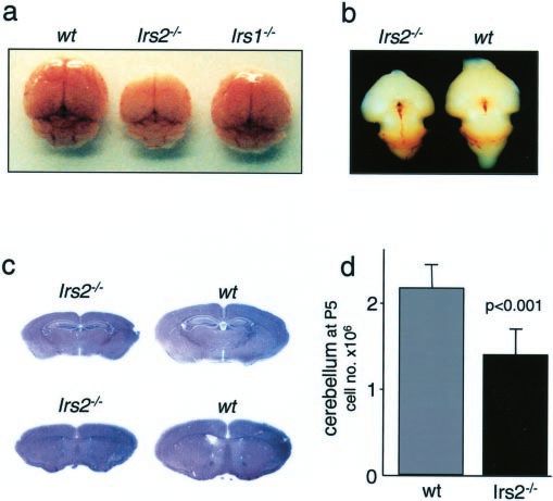
this hypothesis by counting the cells in dispersed preparations of P5
cerebella (Fig. 1d).
Apoptosis in IRS protein-deficient neurons
We postulated that the decreased number of cells in the Irs2 ⫺ / ⫺
brain might be attributable to increased apoptosis, because pre-
vious studies reveal that IGF1 signaling strongly inhibits neuro-
nal apoptosis (Kaytor and Orr, 2002). Surprisingly, the number
of apoptotic cells detected by TUNEL assays was not increased in
Irs2 ⫺ / ⫺ cerebella during prenatal (E12, E14, and E16) or postna-
tal (P5, P6, P7, P9, 3 weeks, 6 weeks, and 8 weeks) development
(Fig. 2a) (data not shown). Apoptosis in situ can be difficult to
quantify, so we investigated cultured granule cells obtained from
the cerebella to determine whether the deletion of Irs1 or Irs2
accelerates apoptosis. Previous results suggest that activation of
the PI3-kinase3 Akt cascade promotes neuronal survival by pro-
moting phosphorylation of GSK3, Foxo1, and BAD (Brunet et
al., 1999; Kaytor and Orr, 2002). As shown previously, with-
drawal of serum and the reduction of KCl to 5 mM stimulated
apoptosis of cultured granule cells (Fig. 2b). However, apoptosis
increased similarly in wild type, Irs1 ⫺ / ⫺, or Irs2 ⫺ / ⫺ granule
cells, suggesting that Irs2 expression was not especially important
Figure 1. Brains of Irs2 ⫺ / ⫺ mice are reduced in size. a, Brains of Irs2 ⫺ / ⫺, Irs1⫺/⫺, and (Fig. 2b). IGF1 and BDNF added separately restored normal sur-
wild-type (wt) mice at 4 weeks of age. b, Brains of Irs2 ⫺ / ⫺ and wild-type mice at E16. Embryos vival, whereas IGF1 and BDNF added together were even more
were collected from timed pregnant mothers and fixed in 4% paraformaldehyde. All visible effective (Fig. 2b). Moreover, IGF1 was equipotent in supporting
brain regions of the Irs2 ⫺ / ⫺ appear small but normal. c, HE-stained sections of Irs2 ⫺ / ⫺ and the survival of granule cells from wild-type, hemizygous (Irs1⫹/⫺
wild-type brains showing a global reduction in all regions. d, Total cell number of the cerebella or Irs2 ⫹/ ⫺), or homozygous (Irs1⫺/⫺ or Irs2 ⫺ / ⫺) mice (Fig.
of Irs2 ⫺ / ⫺ and wild-type mice at P5. Cerebella were dispersed and the total cell number was 2c,d), and dose–response studies revealed no differences in IGF1
determined (n ⫽ 6 per group). sensitivity (data not shown). The protective effect of IGF1 was
abolished in all genotypes by the PI3-kinase inhibitor LY294002
age, male Irs2⫺/⫺ mice displayed a nearly normal body size with but not by the MAP kinase kinase inhibitor PD98059 (data not
unexpectedly small brains (Fig. 1, Table 1). The small brains were shown). Because apoptosis was not enhanced in Irs2 ⫺ / ⫺ neu-
detected in Irs2 ⫺ / ⫺ embryos at E16 (Irs2 ⫺ / ⫺, 51 ⫾ 6 mg; wild rons, these data exclude cell death as the molecular basis of the
types, 74 ⫾ 1 mg; p ⬍ 0.05) (Fig. 1b), and 3– 4 weeks after birth, reduced cell number and brain size and suggest that either Irs1 or
the brain/body ratio was only 1.4% (Table 1). Deletion of Irs2 had Irs2 signaling is sufficient or another signaling pathway might
a global effect on brain size because the forebrain, cerebellum, contribute.
and brainstem decreased proportionally (Table 1). In contrast,
the brains in Irs1 ⫺ / ⫺ mice were only moderately smaller, and the IRS2 promotes neuronal proliferation
brain/body ratio was 4.2% (Fig. 1, Table 1), similar to the effect of Proliferation of neuronal precursors is critical during brain develop-
growth hormone receptor knock-out (Lupu et al., 2001). In each ment, and defects in this process will alter brain size and function. To
case, the smaller skull might limit brain growth. Although male compare neuronal proliferation within the frontal cortex of wild-
Irs2 ⫺ / ⫺ mice die between 10 and 15 weeks of age because of type and Irs2 ⫺ / ⫺ mice, BrdU was injected into pregnant females at
extreme hyperglycemia, female Irs2 ⫺ / ⫺ mice develop diabetes E14, and the embryonic tissue was harvested 48 hr later. Under these
more slowly and survive for ⱖ6 months (Burks et al., 2000). experimental conditions, the total number of BrdU-labeled neurons
However, longitudinal studies with female mice revealed that in Irs2 ⫺ / ⫺ mice decreased 33% compared with wild-type controls
brain size remained proportionately small until the experiment (Irs2 ⫺ / ⫺, 281 ⫾ 67 cells per 0.1 mm 2; wild type, 419 ⫾ 72 cells per
was terminated after 5 months (data not shown). 0.1 mm 2; p ⫽ 0.002); experiments conducted at E12 showed similar
We investigated various aspects of CNS development and struc- results (data not shown). Although reduced, the distribution of
ture but failed to reveal specific brain defects. Hematoxylin-eosin- BrdU-labeled neurons within the developing cortex was normal,Schubert et al. • Insulin Receptor Substrate-2 Deficiency J. Neurosci., August 6, 2003 • 23(18):7084 –7092 • 7087
Figure 2. Apoptosis in brains and cultured neurons from mice lacking IRS proteins. a, TUNEL
assay of the cerebellum at P6 reveals no differences between Irs2 ⫺ / ⫺, Irs1 ⫺ / ⫺, and wild-
type (wt) mice. Quantification of apoptotic nuclei showed the same number per area in all
genotypes. b, IGF1 and BDNF protect against neuronal apoptosis in the absence of Irs2. Cerebel- Figure 3. Neuronal proliferation is impaired in Irs2 ⫺ / ⫺ brains. a, Four pregnant mice were
lar granule cells (P5) were cultured for 4 d in medium containing normal serum. Neurons were injected intraperitoneally with BrdU at E14, and the embryos were harvested after 48 hr. Serial sec-
then incubated for an additional 24 hr in serum-free media containing 5 mM KCl, 5 mM KCl plus tionsofthefrontalcortexwereobtained,andthetotalnumberofBrdU-labeledneuronsperareawas
10 nM IGF1, 5 mM KCl plus 50 ng/ml BDNF, or 25 mM KCL. Cells were stained with Hoechst 33342 quantified using the Openlab software (Improvision). b, Mice (P6) were injected with BrdU, brains
and counted. Percentage apoptosis was calculated (n ⫽ 3 per genotype and experiment; 3 were fixed after 24 hr, and cryostat sections of the cerebellum were obtained (n ⫽ 4 per group).
independent experiments showed similar results). c, Cerebellar granule cells from Irs2⫹/⫺ ⫻ Sections were stained using an anti-BrdU antibody (Boehringer Mannheim). Labeled neurons were
Irs2 ⫹/ ⫺ litters (Irs2 ⫹/ ⫺ or Irs2 ⫺ / ⫺ cells) were cultured for 4 d, followed by incubation for counted using the Openlab software (Improvision). c, Enlargement of b, demonstrating labeled neu-
24 hr in serum-free media containing 25 mM KCl plus 10 nM IGF1 (⫹KCl, ⫹IGF1), 10 nM IGF1 ronsintheouterandinnergranulelayer.d,e,QuantificationofBrdU-labeledcellsinthefrontalcortex
(⫺KCl, ⫹IGF1), 25 mM KCl (⫹KCl, ⫺IGF1), or minimal medium (⫺KCl, ⫺IGF1) (n ⫽ 2 per (d) and cerebellum (e) from wild-type (wt) or Irs2⫺/⫺ sections.
genotype and experiment; 3 independent experiments showed similar results). d, Cerebellar
granule cells from Irs1⫹/⫺ ⫻ Irs1 ⫹/ ⫺ litters (Irs1 ⫹/ ⫺ or Irs1 ⫺ / ⫺ cells) were cultured for with reduced neurogenesis (Fig. 3b,c). Similar experiments with
4 d, followed by incubation for 24 hr as described in c. Irs1 ⫺ / ⫺ mice revealed a highly variable reduction in proliferation
of granule cells (13.7 ⫾ 6.2%) that was disproportionate to the
50% reduced body size.
suggesting that Irs2 was not required for the migration of newly
formed neurons (Fig. 3a,d). Igf1 receptor3Irs2 signaling promotes brain growth
To study the proliferation of granule cells in neonates, BrdU Many studies show that Igf1 promotes brain growth and survival
was injected at P6, and cerebella were harvested 2 d later. The (Recio-Pinto et al., 1984; Dudek et al., 1997). To determine the rela-
number of BrdU-labeled granule cells in Irs2 ⫺ / ⫺ brains was re- tionship between Igf1 receptor (Igf1r) and Irs2 signaling during neu-
duced 23% compared with wild-type mice (Irs2 ⫺ / ⫺, 224 ⫾ 25 rogenesis, we crossed Igf1r ⫹/ ⫺::Irs2 ⫹/ ⫺ compound heterozygous
cells per 0.1 mm 2; wild type, 291 ⫾ 24 cells per 0.1 mm 2; p ⬍ mice to generate the following five viable genotypes: Igf1r ⫹/ ⫺,
0.001) (Fig. 3e). Interestingly, the relative decrease in BrdU label- Irs2 ⫹/ ⫺, Irs2 ⫹/ ⫺::Igf1r⫹/⫺, Irs2 ⫺ / ⫺, and Irs2 ⫺ / ⫺::Igf1r ⫹/ ⫺;
ing (⬃37% reduction in the frontal cortex) paralleled the reduc- Igf1r⫺/⫺ mice die at birth and were not studied (Liu et al., 1993).
tion in total brain weight, consistent with reduced proliferation Heterozygosity for either Irs2 or Igf1r did not influence brain size,
as the cause for decreased Irs2 ⫺ / ⫺ brain size. Depth of the folia whereas brain size was reduced slightly in Irs2 ⫹/ ⫺::Igf1r⫹/⫺ mice
was also reduced in the Irs2 ⫺ / ⫺ cerebella sections, consistent (Fig. 4a). The Irs2 ⫺ / ⫺::Igf1r ⫹/ ⫺ brains were 15% smaller than the7088 • J. Neurosci., August 6, 2003 • 23(18):7084 –7092 Schubert et al. • Insulin Receptor Substrate-2 Deficiency
Figure 5. Immunoblotting with various antibodies conducted with whole-brain lysates. a,
Lysates from 6-week-old mice were immunoblotted with antibodies against tau1/2 protein,
the AT8 antibody against tau phosphorylated at Ser 202, and the AT180 antibody against tau
phosphorylated at Thr 231. b, Lysates from 4.5- and 6-week-old mice were immunoblotted with
AT8 antibody against tau phosphorylated at Ser 202. c, Lysates from 6-week-old mice were
immunoblotted with antibodies against phospho-GSK3 (Ser 9) and GSK3 protein. d, Lysates
from 6-week-old mice were immunoblotted with antibodies against PP2A scaffold subunit
(PR65) and PP2A catalytic subunits. wt, Wild type.
Figure 4. Igf1r heterozygosity causes a further reduction in brain size in Irs2 ⫺ / ⫺ mice. a, Brain
size of 6-week-old wild-type (wt), Igf1r ⫹/ ⫺, Irs2 ⫹/ ⫺, Irs2 ⫹/ ⫺ Igf1r ⫹/ ⫺, Irs2 ⫺ / ⫺, and rini et al., 2002; Hardy and Selkoe, 2002; Hoyer, 2002). Phos-
Igf1r ⫹/ ⫺::Irs2 ⫺ / ⫺ mice.b,Brain– bodyratio(%)of6-week-oldmiceofthedifferentgenotypes.c, phorylated tau accumulates in the hippocampus of patients with
BrdUincorporationincerebellagranulecellsatP6(48hrsurvival)fromwildtype,Irs2 ⫺ / ⫺,orIrs2 ⫺ / Alzheimer’s disease and is thought to contribute to the neuronal
⫺
Igf1r ⫹/ ⫺. Cell counts and average cell size were calculated with NIH ImageJ. d, Apoptosis in the degeneration (Hardy and Selkoe, 2002). Recent evidence suggests
cerebella granule cells from Igf1r ⫹/ ⫺, Irs2 ⫹/ ⫺ Igf1r ⫹/ ⫺, and Irs2 ⫺ / ⫺ Igf1r ⫹/ ⫺ mice (P5). that dysfunction of the insulin–Igf-signaling cascade might con-
Granule cells were cultured for 4 d and then incubated for an additional 24 hr in serum-free medium tribute to this disorder (Gasparini et al., 2002; Liolitsa et al.,
containing5mM KCl,5mM KClplus10nM IGF1,5mM KClplus50ng/mlBDNF.pvalueswerecalculated 2002). Because activation of the insulin–IGF-signaling cascade
using Sigmaplot (Sigma, St. Louis, MO) version 8.1.
promotes dephosphorylation of tau, we determined whether tau
phosphorylation was increased in Irs2 ⫺ / ⫺ brains. Tau was de-
Irs2 ⫺ / ⫺ brains (Fig. 4a); however, the corresponding decrease in tected by immunoblotting in both wild-type and Irs2 ⫺ / ⫺ brain
brain/body ratio was not significant (Fig. 4b). Consistent with these extracts at 6 weeks of age. Tau isolated from the Irs2 ⫺ / ⫺ brain
results, ⬃46% less Irs2 ⫺ / ⫺ cerebella granule cells were labeled by migrated more slowly during SDS-PAGE, suggesting that it
BrdU in P6 brains and further reduced in Irs2 ⫺ / ⫺::Igf1r ⫹/ ⫺ mice might be highly phosphorylated (Fig. 5a). Tau is phosphorylated
(Fig. 4c). at multiple sites including Ser 202 and Thr 231, but phosphoryla-
Apoptosis, assessed by TUNEL assays at various stages of de- tion of Ser 202, detectable with phosphospecific antibody AT8,
velopment (E14, E16, P5, P8, 3 weeks, and 6 weeks of age), was correlates closely with neurodegeneration (Kaytor and Orr,
not altered in any of the viable genotypes. Moreover, the ability of 2002). The phosphorylation of Ser 202 was significantly increased
Igf1 and BDNF to prevent apoptosis was unchanged in cultured in Irs2 ⫺ / ⫺ extracts, whereas phosphorylation of Thr 231 was not
cerebella granule cells from these mice (Fig. 4d). Viable embryos changed (Fig. 5a). Ser 202 phosphorylation was not increased at 4
(E16) deficient for both Igf1r and Irs2 did not show a further weeks of age but was readily detected at 6 weeks of age (Fig. 5b).
reduction in brain size compared with Igf1r ⫺ / ⫺ embryos Tau is phosphorylated on Ser 202 by GSK3, and insulin–Igf
(Igf1r ⫺ / ⫺, 42 ⫾ 4 mg; Irs2 ⫺ / ⫺Igf1r ⫺ / ⫺, 42 ⫾ 5 mg), suggesting inhibits tau phosphorylation by inhibiting GSK3 activity
that Igf1r signaling stimulates neuronal proliferation through the through Akt-mediated phosphorylation on Ser 9 (Kaytor and
Irs2 branch of the insulin–IGF-signaling cascade. Moreover, the Orr, 2002). However, GSK3 is probably not directly phosphor-
average size of the labeled cells was indistinguishable among ylating tau in Irs2 ⫺ / ⫺ mice, because its expression was normal at 6
wild-type, Irs2 ⫺ / ⫺, and Igf1r ⫹/ ⫺Irs2 ⫺ / ⫺ mice, suggesting that weeks of age, and the inhibitory Ser 9 phosphorylation was increased
the Igf1r3Irs2 signal did not control cell size (Fig. 4c). (Fig. 5c). Other kinases that might be involved, including CDK5
(cyclin-dependent kinase 5), MAPK (mitogen-activated protein ki-
Tau is phosphorylated in the Irs2 ⴚ / ⴚ brain nase), or casein kinase-1, were not investigated. Protein phosphatase
Alzheimer’s disease is characterized by dysregulated metabolism 2A dephosphorylates Ser 202 on tau, and its activity might be essential
that includes the accumulation of amyloid -peptide and neuro- to block the accumulation of phospho-tau (Sontag et al., 1996,
fibrillary tangles containing phosphorylated tau protein (Gaspa- 1999). PP2A is composed of a regulatory and catalytic subunit asso-Schubert et al. • Insulin Receptor Substrate-2 Deficiency J. Neurosci., August 6, 2003 • 23(18):7084 –7092 • 7089
Figure 7. AT8 immunostaining from 12- or 16-month-old brains of wild-type (wt), Pdx1 tg,
and Irs2 ⫺ / ⫺::Pdx1 tg mice. Pyramidal cells of the CA1 region or coronal sections through the
corpus striatum (Striatum) were stained with the AT8 antibody (CA1; original magnification,
200⫻).
small brains (Fig. 6b). Moreover, whole-brain lysates from 16-
month-old Irs2 ⫺ / ⫺::Pdx1 tg mice contain elevated levels of phos-
phorylated tau, compared with age-matched wild-type and Pdx1
transgenic mice (Fig. 6c). Immunostaining, using the AT8 anti-
body, revealed increased axonal staining and cytoplasmic depos-
its of phosphorylated tau in Irs2 ⫺ / ⫺::Pdx1 tg hippocampus sec-
tions compared with wild type and Pdx1 tg between 12 and 16
Figure 6. Glucose tolerance, brain size, and tau phosphorylation in wild-type (wt), Pdx1 tg, months of age (Fig. 7). Although tau was hyperphosphorylated in
and Irs2 ⫺ / ⫺::Pdx1 tg mice. a, Glucose tolerance was measured at 14 months of age. b, Brain young Irs2 ⫺ / ⫺::Pdx1 tg mice, cytoplasmic deposits were only de-
weight at ⬃14 months of age. c, Western blot analysis using phospho-tau Ser 202 antibody tected after 12 months and almost entirely in the hippocampus
(AT8) of whole-brain lysates at 16 months of age. (Fig. 7). Thus, Irs2 deficiency was associated with increased tau
phosphorylation that progressed to cytoplasmic deposits during
aging, even when diabetes was prevented. However, up to 16
ciated with a scaffold subunit (PR65) that stabilizes the catalytic months of age, TUNEL assays and glial fibrillary acidic protein
complex. Consistent with increased phosphorylation of Ser 202, the staining did not reveal increased apoptosis, suggesting that the
level of PP2A catalytic subunit was significantly reduced in Irs2 ⫺ / ⫺ accumulation of hyperphosphorylated tau in the Irs2 ⫺ / ⫺ brain
brain extracts, and PR65 was barely detected (Fig. 5d). Thus, disrup- did not reach cytotoxic levels in our experiments.
tion of the PP2A complex might contribute to tau phosphorylation
in Irs2 ⫺ / ⫺ brains. Discussion
The insulin–IGF1-signaling system has been recognized for a
Phosphorylated tau accumulates in old Irs2 ⴚ / ⴚ brains long time to play a critical role in the determination of body size
It is difficult to validate whether hyperphosphorylated tau forms (Birnbaum, 2002). Insulin and IGF1 signals are coordinated by
neurofibrillary tangles in old Irs2 ⫺ / ⫺ brains because Irs2 ⫺ / ⫺ tyrosine phosphorylation of homologous insulin receptor sub-
mice die from diabetes between 10 and 15 weeks of age (Withers strates Irs1 and Irs2 (White, 2002). Although both substrates have
et al., 1998). To study the effect of Irs2 deficiency in the brains of a similar composition, the Igf1r3 Irs2 branch of the pathway is
older mice, we restored -cell function by crossing Irs2 ⫺ / ⫺ mice entirely responsible for the effects of IGF1r on brain size. More-
with transgenic mice expressing Pdx1 (Dutta et al., 2001). Pdx1 is over, Irs2 protects the aging brain from accumulation of phos-
a transcription factor that is critical for -cell function because it phorylated tau that might form neurofibrillary tangles. These
regulates pancreas development and promotes -cell function in results are consistent with the role of Igf1 for neuronal prolifera-
adults (Kushner et al., 2002). Pdx1 expression is significantly tion in vivo and in vitro (DiCicco-Bloom and Black, 1988; Zack-
reduced in Irs2 ⫺ / ⫺ -cells, but transgenic expression of Pdx1 in enfels et al., 1995; Ye et al., 1996; Anlar et al., 1999; Dentremont et
Irs2 ⫺ / ⫺ pancreas restores -cell function and glucose- al., 1999; Aberg et al., 2000; Pixley et al., 2000) and the ability of
stimulated insulin secretion (Kushner et al., 2002). Thus, glucose insulin or Igf1 to reduce tau phosphorylation in cultured neurons
tolerance of Irs2 ⫺ / ⫺::Pdx1 tg improves significantly, and the mice through a PI3-kinase-dependent mechanism (Hong and Lee,
survive for nearly 2 years (Fig. 6a). 1997). Because Irs2 signaling also mediates peripheral insulin
Similar to young Irs2 ⫺ / ⫺ mice, Irs2 ⫺ / ⫺::Pdx1 tg mice have action and promotes pancreatic -cell function, dysregulation of7090 • J. Neurosci., August 6, 2003 • 23(18):7084 –7092 Schubert et al. • Insulin Receptor Substrate-2 Deficiency
Irs2 signaling provides a molecular basis for understanding the White, 1997). Cell-based experimental systems such as 32D myeloid
relationship between neurodegeneration and peripheral insulin progenitor cells fail to show clear differences in the regulation of
resistance and diabetes (Gasparini et al., 2002). downstream signaling pathways, including the PI3-kinase or the
We were surprised to find that brain and body growth diverge ERK cascade (Uchida et al., 2000). In vitro cultured neurons used in
at the IRS proteins. Thus, upregulation of Irs2 might increase this study also failed to reveal the specificity for Irs1 or Irs2 during
brain size with a minimal effect on the body, providing a mech- IGF1-stimulated anti-apoptosis. However, regulatory differences
anism for adaptive increase in brain size without a proportional are likely to exist at the level of gene regulation, message stability and
increase in body size. This mechanism contrasts the growth hor- translation, protein stability, and interactions with upstream and
mone (GH)/GH receptor 3 Igf1/Igf1r3 Irs1 pathway that pro- downstream elements. Controlling these processes might provide
motes body growth. Because Irs2 is also critical for pancreatic ways to treat certain diseases.
-cell growth, survival, and function, the increased demand for Cognitive impairment is common in diabetes, and cerebral
nutrients caused by a large brain might be compensated by in- atrophy is common among young, otherwise healthy patients
creased -cell capacity. However, this relationship might be with type 1 diabetes (Sharma et al., 2003). Moreover, diabetes is
problematic because the failure of Irs2 signaling, resulting from a associated with an increased incidence of Alzheimer’s disease,
predetermined genetic program, chronic inflammation, or aging, suggesting that abnormal insulin signaling might contribute to
might link insulin resistance syndromes and diabetes to dementia (Frolich et al., 1999; Blass et al., 2002; Gasparini et al.,
neurodegeneration. 2002; Hoyer, 2002). Activation of the insulin signaling machinery
In cell-based experiments, insulin and IGF1 promote prolif- might be important for many aspects of neuronal function. In
eration, increase neurite growth, and stimulate protein synthesis prefrontal cortical pyramidal neurons, insulin signaling pro-
(Recio-Pinto et al., 1984; Mill et al., 1985; Fernyhough et al., 1989; motes GABAergic transmission that facilitates learning and
Heidenreich and Toledo, 1989; Dudek et al., 1997). Our results memory (Ma et al., 2003). An important marker of Alzheimer’s
show that Irs2 plays a unique role in the proliferative branch of disease and other brain dysfunctions is the accumulation of neu-
the Igf1 signal in brain. Embryonic brain size is 55% of normal rofibrillary lesions composed of hyperphosphorylated tau (Spill-
(N) in mice lacking Igf1r, and Irs2 mediates most or all of this antini and Goedert, 1998). Tau is a neuronal microtubule-
effect. Hemizygous disruption of the Igf1r has no effect on brain associated protein found predominantly in axons, where it
size, but without Irs2, the Igf1r⫹/⫺ brain was also 55%N. Al- promotes tubulin polymerization and stabilizes microtubules
though complete disruption of Irs2 in Igf1r ⫺ / ⫺ mice is usually (Recio-Pinto et al., 1984; Binder et al., 1985; Drechsel et al.,
embryonic lethal, it does not further reduce brain size at E16, 1992). Because the disruption of Irs2 signaling promotes tau
suggesting that the effect of Igf1 on embryonic brain growth is phosphorylation in the mouse hippocampus, dysregulated Irs2
mediated entirely by the Irs2 branch of the pathway. signaling might be a common link between this marker of neu-
Insulin and IGF1 strongly promote neuronal survival in vitro rodegeneration and peripheral insulin resistance or diabetes.
(Russell et al., 1998; Anlar et al., 1999). IGF1 activates Akt to A direct role for dysregulated signal transduction in the back-
promote BAD phosphorylation and its association with 14 –3-3 ground of diabetes is difficult to establish because of multiple meta-
to release and activate Bcl-2 (Datta et al., 1997; Brunet et al., bolic changes during peripheral insulin resistance and hyperglyce-
1999). However, in our experience, the lack of either Irs2 or Irs1 mia. Even male Irs2 ⫺ / ⫺ mice cannot be studied in old age because
does not reduce survival of cultured cerebella granule cells, and they die between 12 and 15 weeks of age. Normalization of -cell
Igf1 or Bdnf prevents apoptosis normally in cerebella granule function by transgenic expression of Pdx1 in the pancreas of Irs2⫺/⫺
cells lacking Irs1 or Irs2. Thus, Irs1 and Irs2 might be exactly mice prevents the onset of diabetes (Kushner et al., 2002). However,
redundant regarding Igf1-mediated anti-apoptosis, or other Igf- as the euglycemic Irs2 ⫺ / ⫺::Pdx1 tg mice age, neurofibrillary tangles
mediated signals might be involved. containing phosphorylated tau accumulate in the hippocampus
Similar to mice, IGF1 deficiency in people causes mental re- (Kushner et al., 2002). Thus, failure of Irs2 signaling in peripheral
tardation and microcephaly, suggesting that IGF1 has a critical tissues and the brain reveals a potential mechanism to link insulin
role in human brain development (Woods et al., 1996). Direct resistance and neurodegeneration without diabetes (Hoyer, 2002).
evidence that Irs2 plays a role in human brain growth is not Several kinases are reported to phosphorylate tau, including
available. However, breaks at the distal end of human chromo- Gsk3, Cdk5, and probably others (Lee et al., 2000; Kaytor and
some 13 (13q) near the IRS2 gene between microsatellite Orr, 2002). Gsk3 is thought to play an important role in tau
D13S285 and D13S1295 are frequently associated with micro- phosphorylation, but its activity is not elevated in Irs2 ⫺ / ⫺ brains.
cephaly (Towfighi et al., 1987; Bektas et al., 1999). Interestingly, Gsk3 transcript level [estimated from Affymetrix (Santa Clara,
very distal deletions between D13S274 and D13S1311 are associ- CA) MGU74v2 arrays; data not shown] and protein level were
ated with microcephaly and neural tube defects, suggesting that normal, and its specific activity is probably reduced because of
partial Irs2 deficiency might contribute at least in part to the elevated phosphorylation of Ser 9 (we did not assess a possible role
observed microcephaly (Luo et al., 2000). Because the elimina- for Cdk5). However, tau phosphorylation might be increased by
tion of the Igf1r3 Irs2 signaling pathway in people or mice does relative inactivation of PP2A in Irs2 ⫺ / ⫺ brains. PP2A associates
not cause anencephaly, neurogenesis needed to establish the basic with tau and dephosphorylates Ser 202 (Garcia et al., 2000), and in
brain pattern, including fibroblast growth factors, sonic hedge- Irs2 ⫺ / ⫺ brains, its catalytic and scaffold subunits are significantly
hog, and wnt3-catenin signaling is independent of Igf3 Irs2 reduced. However, direct experiments are required to establish
signaling cascade (Vaccarino et al., 1999). the biochemical mechanism of tau hyperphosphorylation in
The molecular basis for the distinct functions of Irs1 and Irs2 Irs2 ⫺ / ⫺ brains.
during neuronal proliferation and survival in the brain is not clear. The accumulation of -amyloid deposits is also an important
Both proteins have similar composition, including a pleckstrin ho- characteristic of Alzheimer’s disease, and several reports suggest that
mology domain and a phosphotyrosine-binding domain that couple amyloid -peptide plaques are associated with the accumulation of
to activated receptors, and a tail of tyrosine phosphorylation sites phosphorylated tau (Hardy and Selkoe, 2002). Amyloid -peptide
that engage SH2 domains in various signaling proteins (Yenush and plaques activate microglial and astrocytes that cause inflammationSchubert et al. • Insulin Receptor Substrate-2 Deficiency J. Neurosci., August 6, 2003 • 23(18):7084 –7092 • 7091
in the brain (Selkoe and Podlisny, 2002). Many proinflammatory Datta SR, Dudek H, Tao X, Masters S, Fu H, Gotoh Y, Greenberg ME (1997)
signaling pathways inhibit the activity of Irs proteins by promoting Akt phosphorylation of BAD couples survival signals to the cell-intrinsic
death machinery. Cell 91:231–241.
Jnk-mediated serine phosphorylation or Socs-mediated ubiquitina-
Dentremont KD, Ye P, D’Ercole AJ, O’Kusky JR (1999) Increased insulin-
tion (Aguirre et al., 2002; Rui et al., 2002). The inhibition of Irs2 like growth factor-I (IGF-I) expression during early postnatal develop-
signaling by -amyloid-induced inflammation might provide a mo- ment differentially increases neuron number and growth in medullary
lecular link between these hallmarks of Alzheimer’s disease. Because nuclei of the mouse. Brain Res Dev Brain Res 114:135–141.
the murine ortholog of human -amyloid fails to form deposits, we DiCicco-Bloom E, Black IB (1998) Insulin growth factors regulate the mi-
are unable to comment on the possibility that Irs2 ⫺ / ⫺ brains are totic cycle in cultured rat sympathetic neuroblasts. Proc Natl Acad Sci
prone to -amyloid deposition. USA 85:4066 – 4070.
Drechsel DN, Hyman AA, Cobb MH, Kirschner MW (1992) Modulation of
In summary, our results show that Igf1r3 Irs2 signaling is
the dynamic instability of tubulin assembly by the microtubule-associated
important for brain size by promoting proliferation during de- protein tau. Mol Biol Cell 3:1141–1154.
velopment. Moreover, during aging, defects in Irs2 signaling Dudek H, Datta SR, Franke TF, Birnbaum MJ, Yao R, Cooper GM, Segal RA,
might contribute to the pathobiology of neurodegenerative dis- Kaplan DR, Greenberg ME (1997) Regulation of neuronal survival by
orders such as Alzheimer’s disease. Because Irs2 signaling is also the serine-threonine protein kinase Akt. Science 275:661– 665.
required for pancreatic -cell growth and function, failure of Irs2 Dutta S, Gannon M, Peers B, Wright C, Bonner-Weir S, Montminy M (2001)
might be a common link between dysregulated peripheral nutri- PDX:PBX complexes are required for normal proliferation of pancreatic
cells during development. Proc Natl Acad Sci USA 98:1065–1070.
ent homeostasis and neurodegeneration. Lifestyle changes or
Fantin VR, Lavan BE, Wang Q, Jenkins NA, Gilbert DJ, Copeland NG, Keller
drugs that promote Irs2 signaling might provide a rational ap- SR, Lienhard GE (1999) Cloning, tissue expression, and chromosomal
proach to treat the complications of insulin resistance, including location of the mouse insulin receptor substrate 4 gene. Endocrinology
diabetes and neurodegeneration. 140:1329 –1337.
Fernyhough P, Mill JF, Roberts JL, Ishii DN (1989) Stabilization of tubulin
mRNAs by insulin and insulin-like growth factor I during neurite forma-
References tion. Brain Res Mol Brain Res 6:109 –120.
Aberg M, Aberg N, Hedbacker H, Oscarsson J, Eriksson P (2000) Peripheral Finch CE, Cohen DM (1997) Aging, metabolism, and Alzheimer disease:
infusion of IGF-1 selectively induces neurogenesis in the adult rat hip- review and hypotheses. Exp Neurol 143:82–102.
pocampus. J Neurosci 20:2896 –2903.
Finlay BL, Darlington RB (1995) Linked regularities in the development and
Aguirre V, Werner ED, Giraud J, Lee YH, Shoelson SE, White MF (2002)
evolution of mammalian brains. Science 268:1578 –1584.
Phosphorylation of ser307 in insulin receptor substrate-1 blocks interac-
Frolich L, Blum-Degen D, Riederer P, Hoyer S (1999) A disturbance in the
tions with the insulin receptor and inhibits insulin action. J Biol Chem
neuronal insulin receptor signal transduction in sporadic Alzheimer’s
277:1531–1537.
disease. Ann NY Acad Sci 893:290 –293.
Airey DC, Lu L, Williams RW (2001) Genetic control of the mouse cerebel-
Garcia A, Cereghini S, Sontag E (2000) Protein phosphatase 2A and phos-
lum: identification of quantitative trait loci modulating size and architec-
phatidylinositol 3-kinase regulate the activity of Sp1-responsive promot-
ture. J Neurosci 21:5099 –5109.
ers. J Biol Chem 275:9385–9389.
Anlar B, Sullivan KA, Feldman EL (1999) Insulin-like growth factor-I and
Gasparini L, Gouras GK, Wang R, Gross RS, Beal MF, Greengard P, Xu H
central nervous system development. Horm Metab Res 31:120 –125.
(2001) Stimulation of beta-amyloid precursor protein trafficking by in-
Bektas A, Warram JH, White MF, Krolewski AS, Doria A (1999) Exclusion
sulin reduces intraneuronal beta-amyloid and requires mitogen-activated
of insulin receptor substrate 2 (IRS-2) as a major locus for early-onset
protein kinase signaling. J Neurosci 21:2561–2570.
autosomal dominant type 2 diabetes. Diabetes 48:640 – 642.
Bernal D, Almind K, Yenush L, Ayoub M, Zhang Y, Rosshani L, Larsson C, Gasparini L, Netzer WJ, Greengard P, Xu H (2002) Does insulin dysfunc-
Pedersen O, White MF (1998) IRS-2 amino acid polymorphisms are not tion play a role in Alzheimer’s disease? Trends Pharmacol Sci 23:288 –293.
associated with random type 2 diabetes among Caucasians. Diabetes Goldowitz D, Hamre K (1998) The cells and molecules that make a cerebel-
47:976 –979. lum. Trends Neurosci 21:375–382.
Binder LI, Frankfurter A, Rebhun LI (1985) The distribution of tau in the Hardy J, Selkoe DJ (2002) The amyloid hypothesis of Alzheimer’s disease:
mammalian central nervous system. J Cell Biol 101:1371–1378. progress and problems on the road to therapeutics. Science 297:353–356.
Birnbaum MJ (2002) RhoGAP: the next big thing for small mice? Dev Cell Heidenreich KA, Toledo SP (1989) Insulin receptors mediate growth effects
2:521–523. in cultured fetal neurons. I. Rapid stimulation of protein synthesis. En-
Bjornholm M, He AR, Attersand A, Lake S, Liu SC, Lienhard GE, Taylor S, docrinology 125:1451–1457.
Arner P, Zierath JR (2002) Absence of functional insulin receptor Hong M, Lee VM (1997) Insulin and insulin-like growth factor-1 regulate
substrate-3 (IRS-3) gene in humans. Diabetologia 45:1697–1702. tau phosphorylation in cultured human neurons. J Biol Chem
Blass JP, Gibson GE, Hoyer S (2002) The role of the metabolic lesion in 272:19547–19553.
Alzheimer’s disease. J Alzheimers Dis 4:225–232. Horner PJ, Gage FH (2000) Regenerating the damaged central nervous sys-
Brunet A, Bonni A, Zigmond MJ, Lin MZ, Juo P, Hu LS, Anderson MJ, Arden tem. Nature 407:963–969.
KC, Blenis J, Greenberg ME (1999) Akt promotes cell survival by phos- Hoyer S (2002) The aging brain. Changes in the neuronal insulin/insulin
phorylating and inhibiting a Forkhead transcription factor. Cell receptor signal transduction cascade trigger late-onset sporadic Alzhei-
96:857– 868. mer disease (SAD). A mini-review. J Neural Transm 109:991–1002.
Bruning JC, Michael MD, Winnay JN, Hayashi T, Horsch D, Accili D, Good- Kaytor MD, Orr HT (2002) The GSK3 beta signaling cascade and neurode-
year LJ, Kahn CR (1998) A muscle-specific insulin receptor knockout generative disease. Curr Opin Neurobiol 12:275–278.
exhibits features of the metabolic syndrome of NIDDM without altering Kulkarni RN, Bruning JC, Winnay JN, Postic C, Magnuson MA, Kahn CR
glucose tolerance. Mol Cell 2:559 –569. (1999) Tissue-specific knockout of the insulin receptor in pancreatic 
Burks DJ, de Mora JF, Schubert M, Withers DJ, Myers MG, Towery HH, cells creates an insulin secretory defect similar to that in type 2 diabetes.
Altamuro SL, Flint CL, White MF (2000) IRS-2 pathways integrate fe- Cell 96:329 –339.
male reproduction and energy homeostasis. Nature 407:377–382. Kushner JA, Ye J, Schubert M, Burks DJ, Dow MA, Flint CL, Dutta S, Wright
Carantoni M, Zuliani G, Munari MR, D’Elia K, Palmieri E, Fellin R (2000) CV, Montminy MR, White MF (2002) Pdx1 restores beta cell function
Alzheimer disease and vascular dementia: relationships with fasting glu- in Irs2 knockout mice. J Clin Invest 109:1193–1201.
cose and insulin levels. Dement Geriatr Cogn Disord 11:176 –180. Lavan BE, Lane WS, Lienhard GE (1997) The 60-kDa phosphotyrosine pro-
Chenn A, Walsh CA (2002) Regulation of cerebral cortical size by control of tein in insulin-treated adipocytes is a new member of the insulin receptor
cell cycle exit in neural precursors. Science 297:365–369. substrate family. J Biol Chem 272:11439 –11443.
Cross DA, Alessi DR, Cohen P, Andjelkovich M, Hemmings BA (1995) In- Lee MS, Kwon YT, Li M, Peng J, Friedlander RM, Tsai LH (2000) Neuro-
hibition of glycogen synthase kinase-3 by insulin mediated by protein toxicity induces cleavage of p35 to p25 by calpain. Nature 405:360 –364.
kinase B. Nature 378:785–789. Liolitsa D, Powell J, Lovestone S (2002) Genetic variability in the insulin7092 • J. Neurosci., August 6, 2003 • 23(18):7084 –7092 Schubert et al. • Insulin Receptor Substrate-2 Deficiency
signalling pathway may contribute to the risk of late onset Alzheimer’s Sontag E, Nunbhakdi-Craig V, Lee G, Bloom GS, Mumby MC (1996) Reg-
disease. J Neurol Neurosurg Psychiatry 73:261–266. ulation of the phosphorylation state and microtubule-binding activity of
Liu JP, Baker J, Perkins JA, Robertson EJ, Efstratiadis A (1993) Mice carry- tau by protein phosphatase 2A. Neuron 17:1201–1207.
ing null mutations of the genes encoding insulin-like growth factor I Sontag E, Nunbhakdi-Craig V, Lee G, Brandt R, Kamibayashi C, Kuret J,
(Igf-1) and type 1 IGF receptor (Igf1r). Cell 75:59 –72. White III CL, Mumby MC, Bloom GS (1999) Molecular interactions
Lovestone S (1999) Diabetes and dementia: is the brain another site of end- among protein phosphatase 2A, tau, and microtubules. Implications for
organ damage? Neurology 53:1907–1909. the regulation of tau phosphorylation and the development of tauopa-
Lovestone S, Reynolds CH (1997) The phosphorylation of tau: a critical thies. J Biol Chem 274:25490 –25498.
stage in neurodevelopment and neurodegenerative processes. Neuro- Sordella R, Classon M, Hu KQ, Matheson SF, Brouns MR, Fine B, Zhang L,
science 78:309 –324. Takami H, Yamada Y, Settleman J (2002) Modulation of CREB activity
Lu L, Airey DC, Williams RW (2001) Complex trait analysis of the hip- by the Rho GTPase regulates cell and organism size during mouse embry-
pocampus: mapping and biometric analysis of two novel gene loci with onic development. Dev Cell 2:553–565.
specific effects on hippocampal structure in mice. J Neurosci Spillantini MG, Goedert M (1998) Tau protein pathology in neurodegen-
21:3503–3514. erative diseases. Trends Neurosci 21:428 – 433.
Lucas JJ, Hernandez F, Gomez-Ramos P, Moran MA, Hen R, Avila J (2001) Stein TD, Johnson JA (2002) Lack of neurodegeneration in transgenic mice
Decreased nuclear beta-catenin, tau hyperphosphorylation and neurode- overexpressing mutant amyloid precursor protein is associated with in-
generation in GSK-3beta conditional transgenic mice. EMBO J 20:27–39. creased levels of transthyretin and the activation of cell survival pathways.
Ludwig T, Eggenschwiler J, Fisher P, D’Ercole AJ, Davenport ML, Efstratiadis J Neurosci 22:7380 –7388.
A (1996) Mouse mutants lacking the type 2 IGF receptor (IGF2R) are Stolk RP, Breteler MM, Ott A, Pols HA, Lamberts SW, Grobbee DE, Hofman
rescued from perinatal lethality in Igf2 and Igf1r null backgrounds. Dev A (1997) Insulin and cognitive function in an elderly population. The
Biol 177:517–535. Rotterdam Study. Diabetes Care 20:792–795.
Luo J, Balkin N, Stewart J, Sarwark J, Charrow J, Nye J (2000) Neural tube Strom RC, Williams RW (1998) Cell production and cell death in the gen-
defects and the 13q deletion syndrome: evidence for a critical region in eration of variation in neuron number. J Neurosci 18:9948 –9953.
13q33–34. Am J Med Genet 91:227–230. Sun XJ, Miralpeix M, Myers Jr MG, Glasheen EM, Backer JM, Kahn CR,
Lupu F, Terwilliger JD, Lee K, Segre GV, Efstratiadis A (2001) Roles of White MF (1992) The expression and function of IRS-1 in insulin signal
growth hormone and insulin-like growth factor 1 in mouse postnatal transmission. J Biol Chem 267:22662–22672.
growth. Dev Biol 229:141–162. Sun XJ, Wang LM, Zhang Y, Yenush L, Myers Jr MG, Glasheen EM, Lane WS,
Ma XH, Zhong P, Gu Z, Feng J, Yan Z (2003) Muscarinic potentiation of Pierce JH, White MF (1995) Role of IRS-2 in insulin and cytokine sig-
GABA(A) receptor currents is gated by insulin signaling in the prefrontal
nalling. Nature 377:173–177.
cortex. J Neurosci 23:1159 –1168.
Towfighi J, Ladda RL, Sharkey FE (1987) Purkinje cell inclusions and “ate-
Michael MD, Kulkarni RN, Postic C, Previs S, Shulman GI, Magnuson MA,
lencephaly” in 13q-chromosomal syndrome. Arch Pathol Lab Med
Kahn CR (2000) Loss of insulin signaling in hepatocyte leads to severe
111:146 –150.
insulin resistance and progressive hepatic dysfunction. Mol Cell 6:87–97.
Uchida T, Myers Jr MG, White MF (2000) IRS-4 mediates activation of
Mill JF, Chao MV, Ishii DN (1985) Insulin, insulin-like growth factor II, and
PKB/Akt during insulin stimulation without inhibition of apoptosis. Mol
nerve growth factor effects on tubulin mRNA levels and neurite forma-
Cell Biol 20:126 –138.
tion. Proc Natl Acad Sci USA 82:7126 –7130.
Vaccarino F, Schwartz ML, Raballo R, Rhee L, Rhee J, Zhou M, Doetschman
Ott A, Stolk RP, van Harskamp F, Pols HA, Hofman A, Breteler MM (1999)
TC, Coffin J, Wyland J, Hung Y (1999) Changes in cerebral cortex size
Diabetes mellitus and the risk of dementia: the Rotterdam Study. Neurol-
are governed by fibroblast growth factor during embryogenesis. Nat Neu-
ogy 53:1937–1942.
Pixley SK, Danogoria NS, Odoms KK, Hastings L (2000) Effects of insulin- rosci 2:246 –253.
like growth factor 1 on olfactory neurogensis in vivo and in vitro. Ann NY Weggen S, Eriksen JL, Das P, Sagi SA, Wang R, Pietrzik CU, Findlay KA,
Acad Sci 855:244 –247. Smith TE, Murphy MP, Bulter T, Kang DE, Marquez-Sterling N, Golde
Previs SF, Withers DJ, Ren JM, White MF, Shulman GI (2000) Contrasting TE, Koo EH (2001) A subset of NSAIDs lower amyloidogenic Abeta42
effects of IRS-1 vs IRS-2 gene disruption on carbohydrate and lipid me- independently of cyclooxygenase activity. Nature 414:212–216.
tabolism in vivo. J Biol Chem 275:38990 –38994. White MF (2002) IRS proteins and the common path to diabetes. Am J
Recio-Pinto E, Lang FF, Ishii DN (1984) Insulin and insulin-like growth Physiol Endocrinol Metab 283:E413–E422.
factor II permit nerve growth factor binding and the neurite formation Withers DJ, Gutierrez JS, Towery H, Burks DJ, Ren JM, Previs S, Zhang Y,
response in cultured human neuroblastoma cells. Proc Natl Acad Sci USA Bernal D, Pons S, Shulman GI, Bonner-Weir S, White MF (1998) Dis-
81:2562–2566. ruption of IRS-2 causes type 2 diabetes in mice. Nature 391:900 –904.
Rodgers EE, Theibert AB (2002) Functions of PI 3-kinase in development of Withers DJ, Burks DJ, Towery HH, Altamuro SL, Flint CL, White MF (1999)
the nervous system. Int J Dev Neurosci 20:187–197. Irs-2 coordinates Igf-1 receptor-mediated beta-cell development and pe-
Rui L, Yuan M, Frantz D, Shoelson S, White MF (2002) SOCS-1 and ripheral insulin signalling. Nat Genet 23:32– 40.
SOCS-3 block insulin signaling by ubiquitin-mediated degradation of Woods KA, Camacho-Hubner C, Savage MO, Clark AJL (1996) Intrauter-
IRS1 and IRS2. J Biol Chem 277:42394 – 42398. ine growth retardation and postnatal growth failure associated with dele-
Russell JW, Windebank AJ, Schenone A, Feldman EL (1998) Insulin-like tion of the insulin-like growth factor I gene. N Engl J Med 335:1363–1367.
growth factor-I prevents apopstis in neurons after nerve growth factor Yamada M, Ohnishi O, Sano S, Nakatani A., Ikeuchi T, Hatanaka H (1997)
withdrawal. J Neurobiol 36:455– 467. Insulin receptor substrate (IRS)-1 and IRS-2 are tyrosine-phosphorylated
Saltiel AR (2001) New perspectives into the molecular pathogenesis and and associated with phosphatidylinositol 3-kinase in response to brain-
treatment of type 2 diabetes. Cell 104:517–529. derived neurotrophic factor cultured cerebral cortical neurons. J Biol
Selkoe DJ (2001) Alzheimer’s disease: genes, proteins, and therapy. Physiol Chem 272:30334 –30339.
Rev 81:741–766. Ye P, Xing Y, Dai Z, D’Ercole AJ (1996) In vivo actions of insulin-growth
Selkoe DJ, Podlisny MB (2002) Deciphering the genetic basis of Alzheimer’s factor-I (IGF-I) on cerebellum development in transgenic mice: evidence
disease. Annu Rev Genomics Hum Genet 3:67–99. that IGF-1 increases proliferation of granule cell progenitors. Brain Res
Sharma J, Bakshi R, Lee D, Hachinski V, Chan RKT (2003) Cerebral atrophy Dev Brain Res 95:44 –54.
in young, otherwise healthy patients with type 1 diabetes mellitus. Neu- Yenush L, White MF (1997) The IRS-signaling system during insulin and
rology [Suppl] 35:3–29. cytokine action. BioEssays 19:491–500.
Solano DC, Sironi M, Bonfini C, Solerte SB, Govoni S, Racchi M (2000) Zackenfels K, Oppenheim RW, Rohrer H (1995) Evidence for an important
Insulin regulates soluble amyloid precursor protein release via phosphati- role of IGF-I and IGF-II for the early development of chick sympathetic
dyl inositol 3 kinase-dependent pathway. FASEB J 14:1015–1022. neurons. Neuron 14:731–741.You can also read