Targeting USP47 overcomes tyrosine kinase inhibitor resistance and eradicates leukemia stem/ progenitor cells in chronic myelogenous leukemia - Nature
←
→
Page content transcription
If your browser does not render page correctly, please read the page content below
ARTICLE
                  https://doi.org/10.1038/s41467-020-20259-0                 OPEN

                  Targeting USP47 overcomes tyrosine kinase
                  inhibitor resistance and eradicates leukemia stem/
                  progenitor cells in chronic myelogenous leukemia
                  Hu Lei 1,8, Han-Zhang Xu1,8, Hui-Zhuang Shan1,8, Meng Liu1, Ying Lu1, Zhi-Xiao Fang1, Jin Jin1, Bo Jing1,
                  Xin-Hua Xiao1, Shen-Meng Gao2, Feng-Hou Gao3, Li Xia1, Li Yang1, Li-Gen Liu1, Wei-Wei Wang1, Chuan-Xu Liu4,
                  Yin Tong5, Yun-Zhao Wu1, Jun-Ke Zheng1, Guo-Qiang Chen 6 ✉, Li Zhou 7 ✉ & Ying-Li Wu 1 ✉
1234567890():,;

                  Identifying novel drug targets to overcome resistance to tyrosine kinase inhibitors (TKIs) and
                  eradicating leukemia stem/progenitor cells are required for the treatment of chronic mye-
                  logenous leukemia (CML). Here, we show that ubiquitin-specific peptidase 47 (USP47) is a
                  potential target to overcome TKI resistance. Functional analysis shows that USP47 knock-
                  down represses proliferation of CML cells sensitive or resistant to imatinib in vitro and
                  in vivo. The knockout of Usp47 significantly inhibits BCR-ABL and BCR-ABLT315I-induced CML
                  in mice with the reduction of Lin−Sca1+c-Kit+ CML stem/progenitor cells. Mechanistic
                  studies show that stabilizing Y-box binding protein 1 contributes to USP47-mediated DNA
                  damage repair in CML cells. Inhibiting USP47 by P22077 exerts cytotoxicity to CML cells
                  with or without TKI resistance in vitro and in vivo. Moreover, P22077 eliminates leukemia
                  stem/progenitor cells in CML mice. Together, targeting USP47 is a promising strategy to
                  overcome TKI resistance and eradicate leukemia stem/progenitor cells in CML.

                  1 Hongqiao  International Institute of Medicine, Shanghai Tongren Hospital/Faculty of Basic Medicine, Department of Pathophysiology, Key Laboratory of Cell
                  Differentiation and Apoptosis of the Chinese Ministry of Education, Shanghai Jiao Tong University School of Medicine, 200025 Shanghai, China. 2 Laboratory
                  of Internal Medicine, The First Affiliated Hospital of Wenzhou Medical University, 325000 Wenzhou, China. 3 Department of Oncology, Shanghai 9th
                  People’s Hospital, Shanghai Jiao Tong University School of Medicine, 639 Zhi Zao Ju Road, 200011 Shanghai, China. 4 Department of Hematology, Xinhua
                  Hospital, Shanghai Jiao Tong University School of Medicine, 200092 Shanghai, China. 5 Department of Hematology, Shanghai First People’s Hospital,
                  Shanghai Jiao Tong University School of Medicine, 200081 Shanghai, China. 6 Department of Pathophysiology, Key Laboratory of Cell Differentiation and
                  Apoptosis of the Chinese Ministry of Education and Chinese Academy of Medical Sciences Research Unit (NO.2019RU043), Shanghai Jiao Tong University
                  School of Medicine, 200025 Shanghai, China. 7 Shanghai Institute of Hematology, State Key Laboratory of Medical Genomics, National Research Center for
                  Translational Medicine at Shanghai, Ruijin Hospital affiliated to Shanghai Jiao Tong University School of Medicine, 200025 Shanghai, China. 8These authors
                  contributed equally: Hu Lei, Han-Zhang Xu, Hui-Zhuang Shan. ✉email: chengq@shsmu.edu.cn; zl10677@rjh.com.cn; wuyingli@shsmu.edu.cn

                  NATURE COMMUNICATIONS | (2021)12:51 | https://doi.org/10.1038/s41467-020-20259-0 | www.nature.com/naturecommunications                                   1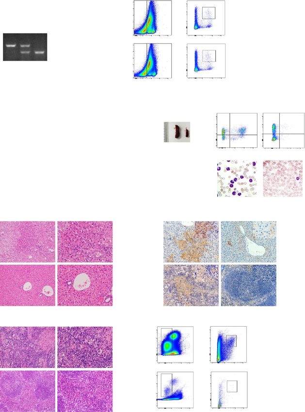
ARTICLE                                                                 NATURE COMMUNICATIONS | https://doi.org/10.1038/s41467-020-20259-0

C
        hronic myelogenous leukemia (CML) is a hematopoietic                  Results
        stem cell malignancy characterized by the t(9;22)(q34;q11)            BCR-ABL regulates USP47 through RAS/ERK and STAT5
        balanced reciprocal translocation of the Philadelphia (Ph)            pathway in CML. To screen the potential DUBs involved in the
chromosome, which leads to the generation of BCR-ABL onco-                    pathogenesis of CML, we compared the expression of DUBs in
genic fusion gene that encodes the chimeric BCR-ABL protein                   primary CML cells (chronic phase, n = 5) and normal bone
with constitutive kinase activity1,2. The introduction of imatinib            marrow (BM) CD34+ cells (n = 3). Compared with normal BM
(IM) in 2001, a tyrosine kinase inhibitor (TKI) that targets BCR-             CD34+ cells, some of the DUBs including USP47, USP9X,
ABL, revolutionized the prognosis of CML3. However, TKI                       USP14, OTUB2, etc. are expressed at higher levels in CML cells,
resistance, including BCR-ABL-dependent and -independent                      with USP47 being the most upregulated one (Fig. 1a). Con-
resistance, is a major problem in IM-based CML treatment. The                 sistent with this result, the USP47 protein level is significantly
BCR-ABL-dependent mechanism is mainly mediated through the                    upregulated in primary CML cells at different stages of disease
mutation of the ABL kinase domain, BCR-ABL overexpression,                    progression (Fig. 1b). Next, we determined whether USP47 is
or MDR1 upregulation4–7. To overcome IM resistance, the sec-                  regulated by BCR-ABL. Compared with the cells in the control
ond generation of TKIs, such as dasatinib and nilotinib, has been             group, the expression of USP47 at the mRNA (Supplementary
developed8. For the “gatekeeper” mutation T315I, which confers                Fig. 1a) and protein (Fig. 1c) levels are significantly upregu-
resistance to all first- and second-generation TKIs, the third-                lated in BCR-ABL-transfected myeloid progenitor 32D cells
generation TKI ponatinib was developed9. Nevertheless, the                    (32DBCR−ABL). In contrast, BCR-ABL knockdown or IM
toxicity of ponatinib limits its use in some patients10. On the               treatment attenuates the expression of USP47 at the mRNA
other hand, the underlying mechanisms of BCR-ABL-                             and protein levels (Fig. 1d–f). Furthermore, the expression of
independent TKI resistance are still not well understood. It has              USP47 is attenuated after RAS or ERK inhibition and
been reported that the leukemia stem cells (LSCs) in CML are                  STAT5 silence (Fig. 1g, h and Supplementary Fig. 1b, c). These
insensitive to TKI in a BCR-ABL-independent manner, thereby                   results suggest that BCR-ABL-induced upregulation of USP47
leading to relapse and minimal residual disease (MRD)11. Addi-                is mediated by the activation of RAS/ERK and STAT5 signaling
tionally, the aberrant activation of the PI3K and RAS/MAPK                    pathways.
signaling pathways also contributes to BCR-ABL-independent
TKI resistance12–15. Hence, identifying promising drug targets
                                                                              USP47 knockdown inhibits proliferation of CML cells. To
that overcome TKI resistance via both mechanisms is urgently
                                                                              investigate the role of USP47 in CML, USP47 was silenced in IM-
required to provide new possibilities for CML treatment.
                                                                              sensitive K562, IM-resistant K562R, and KBM5T315I cells. As
   To date, ~100 kinds of deubiquitinating enzymes (DUBs) have
                                                                              depicted in Fig. 2a, the proliferation of these three cell lines is
been identified16. DUBs remove ubiquitin conjugates from their
                                                                              compromised. Moreover, the numbers of sub-G1 and G2/M
substrates, thereby altering their stabilities, localizations, or
                                                                              phase cells increase after USP47 knockdown (Fig. 2b). The
activities17. Accumulating evidence shows that DUBs are pro-
                                                                              caspase-3 activity is also increased in USP47-knockdown cells
mising targets for cancer treatment, including hematopoietic
                                                                              (Fig. 2c), indicating the activation of apoptosis in CML cells.
malignancies. For instance, USP10 is involved in the pathogenesis
                                                                              Interestingly, USP47 knockdown in human normal BM CD34+
of FLT3-ITD-positive leukemia18; targeting USP1 and USP7 is
                                                                              cells does not inhibit cell viability or colony formation ability
effective in multiple myeloma cells19,20; and USP37 can stabilize
                                                                              (Fig. 2d–f). To investigate the function of USP47 in vivo, USP47-
PLZF/RARA in acute promyelocytic leukemia21. Although
                                                                              silenced and control K562 cells were subcutaneously injected into
USP9X has been demonstrated to be involved in the survival of
                                                                              nude mice. Compared with the control group, knockdown of
CML, how DUBs are related to CML pathogenesis is largely
                                                                              USP47 remarkably reduces the tumor volumes (Supplementary
unexplored22,23.
                                                                              Fig. 2a–c). Immunohistochemical staining shows that the per-
   Ubiquitin-specific peptidase 47 (USP47) is a member of the
                                                                              centages of PCNA-positive cells are significantly reduced, whereas
USP subfamily of DUBs24. Similar to other USPs, USP47 reg-
                                                                              the percentages of γH2AX (a well-recognized DNA damage
ulates cellular activities by removing ubiquitin conjugates from
                                                                              marker) positive cells are markedly increased compared with the
diverse substrates and, thereby, altering their stabilities, localiza-
                                                                              control group (Supplementary Fig. 2d). These data suggest that
tions, or activities. Specifically, USP47 deubiquitinates and sta-
                                                                              USP47 is involved in promoting CML cell proliferation both
bilizes MAPK25, DNA polymerase β (Polβ)26, E-cadherin27, β-
                                                                              in vitro and in vivo.
catenin28, SNAIL29, YAP30, β-Trcp31, and katanin-p6032. Hence,
USP47 is involved in cell proliferation33, cell survival31, DNA
damage repair26, NLRP3 inflammasome activation34, and                          Usp47 knockout suppresses BCR-ABL-induced CML in mice.
epithelial-mesenchymal transition29. USP47 plays an important                 To further explore the role of USP47 in CML, the Usp47
role in cancers such as gastric cancer, medulloblastoma, and                  knockout mouse model (Usp47−/−) was established (Fig. 3a).
colorectal cancer35–37; however, its role in CML remains                      Compared with Usp47+/+ mice, the numbers of total BM cells,
unexplored.                                                                   total white blood cells, red blood cells, granulocytes, lymphocytes,
   In this study, we reveal the critical role of USP47 in the                 and platelets are comparable with those in Usp47−/− mice
pathogenesis of CML. Specifically, we demonstrate that USP47                   (Fig. 3b and Supplementary Fig. 3a). The hematopoietic systems
is highly expressed in primary CML cells and promotes cell                    of Usp47−/− and Usp47+/+ mice are also similar (Supplementary
proliferation, while Usp47 knockout significantly prolongs the                 Fig. 3b, c). Moreover, the percentages of the bone marrow
survival of BCR-ABL and BCR-ABLT315I-induced CML mice                         Lin−Sca1+c-kit+ (LSK) cells, which are the target cells for BCR-
by reducing leukemia stem/progenitor cells. We further                        ABL, are also comparable between Usp47+/+ and Usp47−/− mice
demonstrate that USP47 facilitates DNA damage repair by                       (Fig. 3c). However, the mice received BCR-ABL-transformed
regulating a novel substrate, Y-box binding protein 1 (YB-1).                 Usp47−/− BM cells survived significantly longer than those
Moreover, we find that P22077, a USP47 inhibitor38, sub-                       receiving BCR-ABL-transformed Usp47+/+ cells (Fig. 3d).
stantially eliminates TKI-sensitive cells, TKI-resistant cells,               Moreover, the size and weight of the spleens in Usp47+/+ mice
leukemia stem/progenitor cells, and MRD in CML. We propose                    are significantly increased than those of the Usp47−/− mice
that USP47 is a promising target to overcome TKI resistance in                (Fig. 3e). Flow cytometry analysis showed that the percentage of
CML treatment.                                                                GFP+Gr-1+ cells in BM is significantly lower in the Usp47−/−

2                NATURE COMMUNICATIONS | (2021)12:51 | https://doi.org/10.1038/s41467-020-20259-0 | www.nature.com/naturecommunications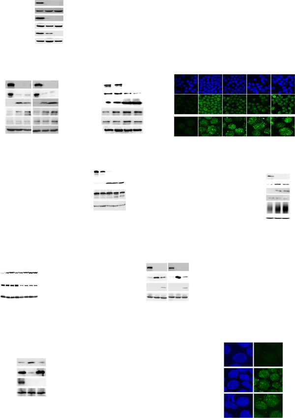
NATURE COMMUNICATIONS | https://doi.org/10.1038/s41467-020-20259-0                                                                      ARTICLE

Fig. 1 BCR-ABL regulates USP47 through RAS/ERK and STAT5 pathway in CML. a mRNA levels of DUBs in bone marrow (BM) mononuclear cells from
primary CML patients compared with normal BM CD34+ cells (CML n = 5, normal n = 3 biologically independent samples). Data are presented as mean ±
s.d. *p < 0.05; **p < 0.01; ***p < 0.001, using two-sided Student’s t-test. b USP47 and BCR-ABL protein expression were measured by western blot in
primary CML cells at different clinical stages compared with normal BM CD34+ cells. NM normal BM, CP chronic phase, AP accelerated phase, BC blast
crisis. c 32DMIGIR and 32DBCR−ABL cells were collected, and the indicated proteins were examined by western blot. d, e Protein (d) and mRNA (e) levels of
USP47 in BCR-ABL stably knocked down K562 cells (Sh BCR-ABL) and vector-transfected cells (Ctrl ShRNA) (n = 3 biologically independent samples per
group). Data are presented as mean ± s.d. ***p < 0.001, using two-sided Student’s t-test. f K562 cells were treated with IM for 24 h, and the indicated
proteins were examined by western blot. g STAT5 knockdown decreased USP47 expression in the K562 cells. h Pan-RAS-IN-1 (RAS inhibitor) and U0126
(ERK inhibitor) decreased USP47 expression in the K562 cells after 24 h of treatment. Source data are provided as a Source Data file.

NATURE COMMUNICATIONS | (2021)12:51 | https://doi.org/10.1038/s41467-020-20259-0 | www.nature.com/naturecommunications                                 3
ARTICLE                                                                  NATURE COMMUNICATIONS | https://doi.org/10.1038/s41467-020-20259-0

Fig. 2 Knockdown of USP47 inhibits the proliferation of CML cells. a USP47 was silenced in K562, K562R, and KBM5T315I cells using a retroviral
transduction system as described in Methods. Viable cells were counted in the transfected cell lines at different times (n = 3 biologically independent
samples per group). b Cell cycle was measured in the transfected cell lines on day 3 (n = 3 biologically independent samples per group). c Caspase-3
activity was measured after 3 days of retroviral transfection (n = 3 biologically independent samples per group). d Knockdown efficiency of USP47 in
normal human BM CD34+ cells was determined by real-time PCR (n = 3 biologically independent samples per group). e, f The number of colonies (e) and
cell viability (f) were measured. Data are mean ± s.d. p-values were analyzed by two-way analysis of variance (ANOVA; a, b) or one-way ANOVA (c–f). *p
< 0.05, **p < 0.01; ***p < 0.001; ****p < 0.0001; ns, no significant. Source data are provided as a Source Data file.

4                 NATURE COMMUNICATIONS | (2021)12:51 | https://doi.org/10.1038/s41467-020-20259-0 | www.nature.com/naturecommunications
NATURE COMMUNICATIONS | https://doi.org/10.1038/s41467-020-20259-0                                                              ARTICLE

group compared with that in the Usp47+/+ group (Fig. 3f, upper                  was detected in the liver and spleen of the Usp47+/+ mice
panel). Consistently, fewer CML leukemia cells were detected in                 (Fig. 3h). Furthermore, the number of CML leukemia stem/pro-
the PB of Usp47−/− group (Fig. 3f, lower panel). Also, the liver                genitor cells (GFP+ LSK cells) in the Usp47−/− group is sig-
and spleen tissues were more seriously damaged in the Usp47+/+                  nificantly decreased compared with the Usp47+/+ group (Fig. 3i).
group than in the Usp47−/− group (Fig. 3g). In addition, sub-                   These data demonstrate that USP47 plays a vital role in the
stantial infiltration of CML leukemia cells (GFP-positive cells)                 pathogenesis of BCR-ABL-induced CML.

NATURE COMMUNICATIONS | (2021)12:51 | https://doi.org/10.1038/s41467-020-20259-0 | www.nature.com/naturecommunications                       5
ARTICLE                                                                  NATURE COMMUNICATIONS | https://doi.org/10.1038/s41467-020-20259-0

Fig. 3 Usp47 knockout suppresses the development of BCR-ABL-induced CML in mice. a Genotyping of Usp47 knockout (Usp47−/−) mice by PCR. b The
total BM cells from 8-week-old Usp47+/+ and Usp47−/− mice were counted (n = 6 biologically independent samples per group). Data are mean ± s.d.
p-values were analyzed by two-sided Student’s t-test. ns, no significant. c Lin−Sca1+ c-kit+ (LSK) cells from 8-week Usp47+/+ and Usp47−/− mice (n = 3
biologically independent samples per group) were measured by FACS. Data are mean ± s.d. p-values were analyzed by two-sided Student’s t-test. ns no
significant. d Survival of mice after receiving BCR-ABL-transduced Usp47+/+ or Usp47−/− BM cells (n = 8 biologically independent samples per group).
The experiment was repeated three times. ****p < 0.0001, p-value was analyzed by Mantel-Cox-log-rank test. e The size and weight of the spleen in the
two groups of mice were shown on day 45 (n = 6 biologically independent samples per group). center lines of the box and whisker plot represent median
values, whereas the box edges indicate the 25th and 75th centiles, and the whiskers indicate the minimum and maximum values. f 35 days after
transplantation, the percentages of GFP+ and Gr-1+ cells in BM were examined by FACS analysis (upper panel). The morphology of the cells from PB was
examined by Wright-Giemsa staining (lower panel). Scale bar, 20 μm. g H&E staining of liver and spleen from the two groups of mice 45 days after
transplantation. Scale bars, 200 × , 100 μm; 400 × , 50 μm. h Immunohistochemical staining of liver and spleen with GFP antibody 45 days after
transplantation. Scale bars, 200 × , 100 μm; 400 × , 50 μm. i FACS analysis of the GFP+ LSK cells in BM 35 days after transplantation (n = 3 biologically
independent samples per group). Data are mean ± s.d. p-values were analyzed by two-sided Student’s t-test. **p < 0.01. Source data are provided as a
Source Data file.

Silencing Usp47 inhibits BCR-ABLT315I-induced CML and                          ubiquitination of full-length YB-1 and YB-1-S3, but not that of the
proliferation of KBMT315I in a xenograft mouse model. Next,                    truncated YB-1-S2, which cannot bind USP47. Consistent with the
we examined the role of Usp47 in a BCR-ABLT315I-driven CML                     deubiquitinating effect of USP47 on YB-1, less ubiquitinated YB-1
mouse model. Consistent with the findings above, Usp47 deple-                   is observed in primary CML cells than that in normal BM cells
tion substantially prolongs the survival of CML mice compared                  (Fig. 5e). Interestingly, the interaction between YB-1 and USP47 is
with the Usp47+/+ group (Fig. 4a). Compared with Usp47+/+                      not affected by the ubiquitination status of YB-1, as the mutation
mice, the spleen size and weight in Usp47−/− mice are sig-                     of lysine residues (Lys137, −164, and −170) abrogates the ubi-
nificantly decreased (Fig. 4b, c). Besides, there is a remarkable               quitination of YB-1, but not its interaction with USP47 (Fig. 5f, g).
decrease in the number of CML leukemia cells in the PB and BM                  We then investigated whether USP47 influences the stability of
of the Usp47−/− mice (Fig. 4d, e). Flow cytometry analysis shows               YB-1 and found that transient transfection of USP47 prolonged
that the number of GFP+Gr-1+ cells is comparable between the                   the half-life of YB-1 in K562 cells (Fig. 5h). In contrast, USP47
Usp47+/+ and Usp47−/− mice at 14 days after CML model                          knockdown or P22077 treatment reduces the half-life of YB-1
establishment, while the number of GFP+Gr-1+ cells decreases in                (Fig. 5i and Supplementary Fig. 5b). The half-life of YB-1 is also
Usp47−/− mice as time goes by (Supplementary Fig. 4a). The liver               reduced in Usp47 knockout MEF cells (Fig. 5j). The USP47
and spleen tissues are severely damaged in the Usp47+/+ group                  knockdown-induced downregulation of the YB-1 protein can be
compared with the Usp47−/− mice (Fig. 4f). Additionally, Usp47                 rescued by the pre-treatment of MG132, a proteasome inhibitor
knockout does not affect the homing efficacy of CML LSK cells                   (Fig. 5k). Meanwhile, knockdown of USP47 has no effect on YB-1
(Supplementary Fig. 4b). However, the number of CML GFP+                       or POLB at mRNA level and YB-1 mRNA expression is similar in
LSK cells in the Usp47−/− group is significantly lower than that in             normal and CML cells (Supplementary Fig. 5c, d), indicating the
the Usp47+/+ group (Supplementary Fig. 4c). To further validate                increased YB-1 protein levels in primary CML cells mainly attri-
this finding in human CML cells, we inoculated the USP47-                       butes to the post-transcriptional regulation of YB-1. Moreover,
knockdown KBM5T315I (ShUSP47-2) and the control KBM5T315I                      YB-1 expression is restored in Usp47−/− MEFs after the reintro-
(Ctrl shRNA) cells into B-NDG mice. Compared with the control                  duction of Usp47 (Fig. 5l). These findings suggest that YB-1 serves
group, the mice in the ShUSP47-2 group show more prolonged                     as a novel substrate for USP47.
survival with smaller spleen size and weight (Fig. 4g, h). More-
over, leukemic infiltration in the livers of the control mice is more           YB-1 contributes to USP47-mediated DNA damage repair in
obvious than that of the ShUSP47-2 mice (Fig. 4i). Also, the                   CML cells. We examined whether YB-1, a well-known multi-
hematoxylin and eosin (H&E) staining results show that the                     functional transcription factor and an oncoprotein in cancers39–42,
extent of leukemic infiltration in the liver and spleen in the                  contributes to the proliferation suppression of CML cells induced
ShUSP47-2 group is lower than the control group (Fig. 4j). These               by USP47 inhibition. As expected, YB-1 knockdown significantly
observations are further confirmed by CD45 antibody staining                    inhibits the proliferation of K562, K562R, and KBM5T315I cells
(Fig. 4k). These data further reveal the vital role of USP47 in the            (Fig. 6a). In agreement with previous studies that USP47 is
pathogenesis of CML with T315I mutation.                                       involved in the DNA damage repair pathway26,43, USP47 knock-
                                                                               down significantly increases γH2AX and ATR phosphorylation
USP47 interacts with and stabilizes YB-1 protein. We explored                  (Ser428) in CML cells (Fig. 6b). Furthermore, Usp47 knockout
the mechanism through which USP47 functions in CML.                            significantly enhances the expression of γH2AX in GFP+ BM cells
Although it has been reported that MAPK or β-catenin interacts                 from BCR-ABLT315I-induced CML mice (Fig. 6c). These results
with USP47 in Drosophila or tumors25,28, USP47 knockdown does                  were further confirmed by γH2AX foci and the number of AP sites
not affect the expression of MAPK or β-catenin in CML cells                    (DNA damage quantification analysis) (Fig. 6d, e). Time-course
(Supplementary Fig. 5a). We then immunoprecipitated, separated,                analysis of USP47 knockdown-induced DNA damage response
and analyzed the USP47-interacting proteins using mass spec-                   shows that, with the decrease of USP47, γH2AX increases, fol-
trometry (Fig. 5a). It was identified that YB-1 can interact with               lowed by the cleavage of PARP1, indicating that DNA damage
USP47 in K562 and primary CML cells (Fig. 5b). To map the                      response is not a secondary effect of cells already committed to
interaction domain of YB-1 with USP47, truncated forms of YB-1                 apoptosis (Fig. 6f and Supplementary Fig. 6a). Consistent with
(GFP tag) were constructed and co-transfected with USP47 (Flag                 previous findings that YB-1 regulates PCNA, epidermal growth
tag) into HEK293T cells. The results show that USP47 interacts                 factor (EGF), and DNA topoisomerase II α (TOPO IIα)44–46, YB-1
with the C-terminal domain (CTD) domain of YB-1 (Fig. 5c). We                  knockdown reduces the mRNA expression of PCNA and TOPO
further examined if USP47 regulates the ubiquitination of YB-1.                IIα (Fig. 6g and Supplementary Fig. 6b), indicating YB-1 is
As shown in Fig. 5d, USP47 overexpression significantly inhibits                involved in DNA damage response in CML cells. The role of YB-1

6                 NATURE COMMUNICATIONS | (2021)12:51 | https://doi.org/10.1038/s41467-020-20259-0 | www.nature.com/naturecommunications
NATURE COMMUNICATIONS | https://doi.org/10.1038/s41467-020-20259-0 ARTICLE in DNA damage repair was further validated as we found that YB- YB-1 and Polβ to USP47-mediated DNA damage repair in CML 1 knockdown induces the increase of ATR phosphorylation cells, we overexpressed USP47 in YB-1 and/or POLB knockdown (Ser428), PAR, and γH2AX in CML cells (Fig. 6h and Supple- K562 and KBM5T315I cells. As shown in Supplementary Fig. 6e, mentary Fig. 6c). YB-1 knockdown also causes γH2AX foci in knockdown of POLB cannot increase γH2AX expression. USP47 CML cells (Fig. 6d and Supplementary Fig. 6d). γH2AX clearance overexpression in YB-1 and/or Polβ depletion cells cannot abro- is also significantly slower in irradiation-induced YB-1 knockdown gate γH2AX expression, indicating that YB-1 contributes more to cells than in control cells (Fig. 6i). To assess the contribution of USP47-mediated DNA damage repair than Polβ in CML cells NATURE COMMUNICATIONS | (2021)12:51 | https://doi.org/10.1038/s41467-020-20259-0 | www.nature.com/naturecommunications 7
ARTICLE NATURE COMMUNICATIONS | https://doi.org/10.1038/s41467-020-20259-0
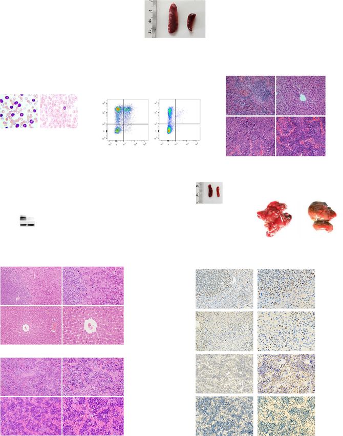
Fig. 4 Silence of Usp47 inhibits BCR-ABLT315I-induced CML and inhibits proliferation of KBMT315I in a xenograft model. a Survival of mice after the
transplantation of BCR-ABLT315I-transduced Usp47+/+ or Usp47−/− BM cells (n = 6 biologically independent samples per group). ****p < 0.0001, p-value
was analyzed by Mantel-Cox-log-rank test. The experiment was repeated three times. b, c Size (b) and weight (c) of the spleen in the two groups of mice
(n = 5 biologically independent samples per group) at 28 days after transplantation. Center lines of the box and whisker plot represent median values,
whereas the box edges indicate the 25th and 75th centiles, and the whiskers indicate the minimum and maximum values. d, e At 28 days after
transplantation, the morphology of cells from PB (d) was examined by Wright-Giemsa staining; the percentages of GFP+ and Gr-1+ cells (e) in BM were
examined by FACS analysis. f H&E staining of the liver and spleen at 28 days after transplantation. Scale bars, 50 μm. g Ctrl shRNA or ShUSP47-2
transfected KBM5T315I cells (n = 8 biologically independent samples per group) were injected into B-NDG mice through the tail vein. ****p < 0.0001, p-
value was analyzed by Mantel-Cox-log-rank test. h Size and weight of spleen at day 21 (n = 6 biologically independent samples per group). Center lines of
the box and whisker plot represent median values, whereas the box edges indicate the 25th and 75th centiles, and the whiskers indicate the minimum and
maximum values. i Infiltration of leukemia cells in the livers of the mice from Fig. 4h. Green arrows indicate infiltrated leukemic cells. Scale bars, 1 cm. j H&E
staining of liver and spleen from mice bearing leukemia. Scale bars, 200 × , 100 μm; 400 × , 50 μm. k Immunohistochemical staining with human CD45
antibody in the liver and spleen shown in Fig. 4j. Scale bars, 200 × , 100 μm; 400 × , 50 μm. Data are mean ± s.d. p-values were analyzed by two-sided
Student’s t-test (c, h) or Mantel-Cox-log-rank test (a, g). **p < 0.01; ****p < 0.0001. Source data are provided as a Source Data file.
(Supplementary Fig. 6f). As expected, overexpressing YB-1 P22077 exerts no cytotoxicity on normal CD34+ cells, but sig-
attenuates DNA damage response induced by USP47 knock- nificantly inhibits the colony-forming activity of CD34+ cells
down, as evidenced by the decrease of γH2AX expression and the from de novo and IM-resistant CML patients (Fig. 7j). Moreover,
number of AP sites (Fig. 6j, k). Also, YB-1 overexpression in P22077 significantly decreases the number of CML stem/pro-
Usp47−/− MEFs attenuates the upregulation of γH2AX (Fig. 6l). genitor cells in a BCR-ABL-induced CML mouse model (Fig. 7k).
Additionally, the utilization of AZD6738 (50 nM), an ATR path- P22077 treatment significantly eliminates the number of CML
way inhibitor, partially inhibits USP47 knockdown-induced sup- GFP+LSKs in the MRD mouse model (Fig. 7l). These results
pression of cell viability in CML cells (Supplementary Fig. 6g). As indicate that P22077 exerts cytotoxicity markedly against CML
DNA damage repair plays an essential role in the maintenance or cells, including CML stem/progenitor cells.
generation of CML stem/progenitor cells47,48, we hypothesized
that DNA damage induced by USP47 knockdown might be vital to Discussion
CML stem/progenitor cells. In agreement with our hypothesis, TKI resistance and CML leukemia stem cells remain the major
USP47 knockdown significantly decreases the colony formation challenges in the treatment of BCR-ABL+ CML. In this study, we
ability of primary CML CD34+ cells (Fig. 6m). The number of demonstrate that USP47 plays a critical role in the proliferation of
γH2AX foci also increases after USP47 knockdown in primary TKI-sensitive, TKI-resistant, and CML stem/progenitor cells both
CD34+ cells (Fig. 6n). The results indicate that YB-1 is involved in in vitro and in vivo. As a substrate of USP47, YB-1 contributes to
USP47-mediated DNA damage repair in CML cells. USP47-mediated DNA damage repair in CML cells. Hence, tar-
geting USP47 is a promising strategy to overcome TKI resistance
P22077 exerts remarkable toxicity on CML cells and CML LSCs and eradicate CML stem/progenitor cells.
in vitro and in vivo. P22077, a dual inhibitor of USP7/USP47, The role of USP47 in cancer is cancer type-dependent. It can
was used to assess the function of USP47 in CML cell lines and function as an oncoprotein in gastric cancer49, colorectal29,30,
primary CML cells. As depicted in Fig. 7a, P22077 exhibits low ovarian cancer50, and a tumor suppressor in medulloblastoma36.
toxicity to mononuclear cells from normal PB, while in contrast, Our study reveals that USP47 is essential in the pathogenesis of
P22077 inhibits the proliferation of both IM-sensitive and IM- CML. We show that knockout of Usp47 significantly prevents
resistant CML cell lines and primary CML cells in a dose- BCR-ABL or BCR-ABLT315I-induced leukemogenesis and redu-
dependent manner (Fig. 7b–d). Notably, P22077 shows evident ces the number of GFP+LSK cells in CML mice, indicating that
cytotoxicity to BM mononuclear cells derived from CML patients Usp47 is critical in the development of BCR-ABL-induced CML.
resistant to second-generation TKIs (Fig. 7e). In both primary Moreover, targeting USP47 is effective in CML cells regardless of
IM-sensitive and IM-resistant CML cells, P22077 treatment the presence of BCR-ABL with or without kinase domain
decreases YB-1 but increases the expression of γH2AX and the mutation. CML cells derived from all analyzed CML patients
cleavage of caspase-3 and PARP1, indicating the activation of (irrelevant to BCR-ABL mutation or TKI resistance) exhibit
apoptosis (Fig. 7f). In addition, USP47 is positively correlated similar sensitivity to USP47 inhibition-induced cytotoxicity.
with the expression of YB-1 in primary CML cells, and CML cells Furthermore, USP47 inhibition subdues the proliferation of
with higher levels of USP47 are more sensitive to P22077 treat- K562, K562R (without BCR-ABL mutation and insensitive to
ment than the cells with lower levels of USP47 (Supplementary IM), and KBM5T315I (with BCR-ABL T315I mutation and
Fig. 7a). We then evaluated the effect of P22077 on KBM5T315I insensitive to IM) CML cell lines both in vitro and in vivo.
cells using a B-NDG mouse CML model. The B-NDG mice Therefore, our study suggests that USP47 is a promising ther-
injected with KBM5T315I cells by tail vein were divided into four apeutic target in CML treatment, especially for CML patients with
groups and treated with the vehicle, IM, P22077, and P22077 plus TKI resistance.
imatinib, respectively. P22077 significantly prolongs the survival Consistent with previous reports26,43, we found that USP47 is
of CML mice, while P22077 combined with IM has no significant involved in the DNA damage repair in CML cells. We further
enhancement effect on P22077 (Fig. 7g). More importantly, using demonstrated that YB-1, a novel substrate of USP47, contributes
CML patient-derived xenograft model established from a TKI- to USP47-mediated DNA damage repair. In agreement with our
resistant CML patient, we found that P22077 treatment could findings, it has been reported that YB-1 is associated with cancer
significantly prolong the survival of mice (Supplementary Fig. 7b) progression and drug resistance in multiple cancers by stimulating
and reduce the percentages of CD34+CD38− cells in the sec- cell proliferation and promoting replicative immortality, genomic
ondary bone marrow transplantation (Fig. 7h, I). Given the instability, and metastasis39–42,51–53. Several studies reported that
important role of USP47 in CML stem/progenitor cells, we YB-1 is also involved in base excision and mismatch repair
investigated the effect of P22077 on primary CD34+ CML cells. pathways by interacting with multiple DNA repair proteins, such
8 NATURE COMMUNICATIONS | (2021)12:51 | https://doi.org/10.1038/s41467-020-20259-0 | www.nature.com/naturecommunicationsNATURE COMMUNICATIONS | https://doi.org/10.1038/s41467-020-20259-0 ARTICLE as DNA ligase IIIα54, APE155, MSH256, and PCNA57. In CML, Another important finding is that P22077 is effective in TKI- BCR-ABL compromises the fidelity of nucleotide excision repair sensitive and TKI-resistant CML cells, regardless of the presence and homologous recombination repair, thus promoting survival of T315I mutation. Interestingly, in addition to inhibiting the and resulting in the genomic instability of CML cells, especially activity of USP47, P22077 considerably reduces the protein level LSCs11,58–60. Therefore, it is reasonable that in CML cells, BCR- of USP47 in CML cells. P22077 may promote the degradation of ABL induces DNA damage, while USP47 and YB-1 are required to USP47 by inhibiting the auto-deubiquitinating process of repair DNA damage to ensure cell survival. USP4728, which is supported by the finding that MG132 reverses NATURE COMMUNICATIONS | (2021)12:51 | https://doi.org/10.1038/s41467-020-20259-0 | www.nature.com/naturecommunications 9
ARTICLE                                                                       NATURE COMMUNICATIONS | https://doi.org/10.1038/s41467-020-20259-0

Fig. 5 USP47 interacts with YB-1 and protects it from proteasomal degradation. a Immunoprecipitation with USP47 antibody in K562 cells, the proteins
were separated by SDS-PAGE and visualized by Coomassie brilliant blue (G250). b The interaction between endogenous USP47 and YB-1 was analyzed by
western blot in K562 and primary CML cells. c Full-length or several deletion constructs of GFP-YB-1 was co-transfected with Flag-USP47 in
HEK293T cells. Their interactions were examined by co-IP with anti-GFP antibody and by western blot with anti-Flag antibody. d GFP-YB-1-FL/GFP-YB-1-
S2/GFP-YB-1-S3 and HA-ubiquitin plasmids were co-transfected with or without Flag-USP47 into HEK293T cells. YB-1 was co-IP with anti-GFP antibody,
and its ubiquitination was measured with Ub antibody by western blot. e Normal and CML BM mononuclear cells were lysed and co-IP with YB-1 antibody,
YB-1 ubiquitination was detected with Ub antibody. f Flag-USP47, GFP-YB-1, and GFP-YB-1-mu (K137A, K164A, and K170A) were transfected in
HEK293T cells. Their interactions were examined by co-IP with anti-Flag antibody and measured by western blot with anti-GFP antibody. g GFP-YB-1 or
GFP-YB-1-mu was transfected with HA-Ub in HEK293T cells. YB-1 ubiquitination in transiently transfected cells was analyzed by co-IP with anti-GFP
antibody and western blot with Ub antibody. h USP47 plasmids and empty vectors were transfected into K562 cells. The transfected cells were treated
with cycloheximide (CHX, 10 μM) at different times. The indicated proteins were determined by western blot. i, j USP47 knockdown cells or Usp47−/−
MEFs were treated with cycloheximide (CHX, 10 μM) at different times together with the control cells, and the indicated proteins were determined by
western blot. k Control and USP47 stably knockdown K562 cells were treated with vehicle and MG132 (10 μM) for 4 h. The proteins were then extracted
and subjected to western blot. l YB-1 expression in Usp47−/− MEFs was measured by western blot after reintroduction of USP47. Source data are provided
as a Source Data file.

P22077-induced USP47 degradation (Supplementary Fig. 7c). It                        from Santa Cruz Biotechnology (Santa Cruz, CA, USA). Antibodies for Polβ
has been reported that P22077 also inhibits the activity of USP7                    (1:1000, #18003-1-AP, Lot 00009472), YB-1 (1:1000, #20339-1-AP, Lot 00046294),
                                                                                    Flag tag (1:1000, #66008−2-Ig), USP10 (1:1000, #19374-1-AP, Lot 00015015),
and USP10. To identify the specific USPs targeted by P22077 in                       TOPOIIα (1:1000, #20233-1-AP), SIRT6 (1:1000, #13572-1-AP, Lot 00004562), β-
CML cells, we measured the expression of substrates of USP7 and                     actin (1:1000, #HRP-66009) were purchased from Proteintech Group, Inc.
USP10, including p53, a substrate of both USP7 and USP1061,62,                      (Wuhan, CHN). The USP7 (1:1000, #A300-033A) antibody was purchased from
and SIRT6, a substrate of USP1063. We found that P22077                             Bethyl Laboratories Inc. (Montgomery, TX, USA). The USP16 (1:1000, GTX16439,
increases p53 expression (but not phosphorylated p53, ser15) in                     Lot 39631) antibody was purchased from GeneTex Inc. (Irvine, CA, USA). The
                                                                                    above antibodies were used in western blot. Lineage Antibody Cocktail (1:200, #88-
primary CML cells but does not affect SIRT6 expression. How-                        7772-72), sca-1 (1:200, #25-5981-82), c-kit (1:200, #17-1171-82), CD34 (1:200, #11-
ever, both p53 and SIRT6 expressions are not increased by                           0349-42), CD38 (1:200, #12-0389-42) and CD45 (1:200, #17-0459-42) for FACS
P22077 treatment in KBM5T315I cells (Fig. 7f). Also, USP10 or                       analysis were purchased from ebioscience Inc. (San Diego, CA, USA). Gr-1 anti-
USP7 knockdown does not affect the expression of YB-1 (Sup-                         body (1:200, #130-119-794) was purchased from Miltenyi Biotec (Auburn, CA,
                                                                                    USA). CD45 antibody (1:1000, #GB14038) for IHC was purchased from ServiceBio
plementary Fig. 7d). Moreover, P22077 still strongly inhibits the                   (Wuhan, CHN). Flag-bead (M20018) was purchased from Abmart (Shanghai,
viability of USP7 or USP10 stably knockdown K562 cells (Sup-                        CHN). EasySep Release Human CD45 Positive Selection Kit for Humanized Mice
plementary Fig. 7e). Therefore, we deem that USP47 is the main                      was purchased from STEMCELL (100-0107, Vancouver, BC, Canada). Imatinib
effector for P22077 to combat CML. In addition, it should be                        (S2475), P22077 (S7133), and MG132 (S2619) were purchased from Selleck Che-
                                                                                    micals (Houston, TX, USA). AZD6738 (T3338) was purchased from Target
noted that, mice treated with P22077 (30 mg/kg) for 2 weeks did                     Molecule Corp. (Shanghai, CHN). Pan-RAS-IN-1 (HY-101295) was purchased
not show health problems such as weight loss (Supplementary                         from MedChemExpress LLC. (Monmouth Junction, NJ, USA). The Cell Cycle
Fig. 7f), which was consistent with the previous reports64. These                   Detection Kit (340242) was purchased from BD Biosciences (San Jose, CA, USA).
data indicate P22077 is a promising candidate compound for the                      The Caspase-3 Activity Assay Kit (C1115) was purchased from Beyotime Bio-
                                                                                    technology (Jiangsu, CHN).
treatment of TKI-sensitive and TKI-resistant CML. Nevertheless,
further investigations are still needed to develop USP47-specific
                                                                                    Cell culture. The murine IL-3-dependent myeloid cell line 32D obtained from
inhibitors for CML treatment.                                                       ATCC (CRL-11346) was maintained in RPMI-1640 (21875109; Gibco. Grand
   Based on the fact that TKI reduces the expression of USP47 in                    Island, New York, USA) supplemented with 10% fetal bovine serum (FBS) (900-
CML cells and overexpression of BCR-ABL upregulates the                             108; Gemini Bio-Products Inc. Sacramento, USA) and 10% WEHI-3B conditional
expression of USP47 in 32D cells, we demonstrate that BCR-ABL                       medium containing IL-3; their counterparts transformed with p210BCR−ABL were
                                                                                    maintained in RPMI-1640 (11875; Gibco) supplemented with 10% FBS. The IM-
upregulates the expression of USP47. However, BCR-ABL may
                                                                                    sensitive K562 cells obtained from ATCC (CRL-243) and IM-resistant cell lines
not be the only regulator of USP47. First, the activation of RAS/                   K562R cells obtained from ATCC (CRL-3344), respectively, were cultured in
ERK, which is independent of BCR-ABL, can regulate the                              RPMI-1640 (11875; Gibco) supplemented with 10% FBS. K562R cells were cultured
expression of USP4765,66. Second, USP47 is highly expressed in                      in 1 μM IM to maintain their drug-resistant status. KBM5T315I cells67 were kindly
several types of BCR-ABL-negative cell lines, including colorectal                  provided by Jingxuan Pan in Sun Yat-sen University and were cultured in IMDM
                                                                                    (12440061; Gibco) supplemented with 10% heat-inactivated FBS, penicillin (50 U/
cancer cells and gastric cancer cells30,49. Therefore, USP47 may be                 ml)/ streptomycin (50 μg/ml) (E607011; Sangon Biotech Co, Shanghai, CHN). All
regulated through both BCR-ABL-dependent and -independent                           cells were cultured at 37 °C in a humidified atmosphere of 5% CO2. HEK293T cells
mechanisms.                                                                         were obtained from ATCC (CRL-3216) and cultured in DMEM (11971025, Gibco)
   In summary, we demonstrate that USP47 is a target for CML                        with 10% FBS. All cell stocks were routinely tested for mycoplasma contamination
                                                                                    and showed negative.
treatment and targeting USP47 is a promising strategy for over-
coming TKI resistance and eradicating leukemia stem/progenitor
                                                                                    Primary cells. BM samples were obtained from healthy donors and patients with
cells in CML.                                                                       CML (from 2011 to 2019) at different clinical stages or at drug resistance admitted
                                                                                    to Shanghai Jiao Tong University School of Medicine affiliated Ruijin Hospital,
Methods                                                                             Xinhua Hospital, Shanghai First People’s Hospital, Shanghai 9th People’s Hospital
                                                                                    and Shanghai Tongren Hospital. The details of patients were provided in Sup-
Reagents and antibodies. The p-Bcr (WB, 1:1000, #3901, Lot 1), c-Abl (1:1000,
                                                                                    plementary Table 1. Mononuclear cells were isolated by Ficoll-Paque Plus density
#2862, Lot 13), p-CrkL (1:1000, #3181, Lot 7), CrkL (1:1000, #3182, Lot 5), p-ERK
                                                                                    gradient media (GE Life Sciences, Chicago, IL, USA). Informed consent was
(1:1000, #9101, Lot 26), ERK (1:1000, #9102, Lot 25), p-P38 MAPK (1:1000, #9215,
                                                                                    obtained from all patients in accordance with the Declaration of Helsinki. All
Lot 19), p-JNK MAPK (1:1000, #9251, Lot 10), p-AKT (1:1000, #4060, Lot 30),
                                                                                    manipulations were approved by the Medical Ethic Committee of Shanghai Jiao
caspase-3 (1:1000, #9661, Lot 45), γH2AX (1:1000, #9718, Lot 1), p-p53 (1:1000,
                                                                                    Tong University School of Medicine.
#9284), p-ATR (1:1000, #2853, Lot 9), p-ATM (1:1000, #4526, Lot 14), β-catenin
(1:1000, #9587, Lot 2) and PAR (1:1000, #83732) antibodies were purchased from
Cell Signaling Technology (Danvers, MA, USA). The USP47 (1:500, sc-100633, Lot      RNA interference. ShRNA oligonucleotides targeting USP47, BCR-ABL, YB-1,
J1314), STAT5 (1:500, sc-74442), USP14 (1:500, sc-100630, Lot J3013), Ubiquitin     STAT5, USP7, and USP10 were designed and synthesized (Supplementary Table 2).
(1:500, sc-8017, Lot H0409), GFP (1:500, sc-9996, Lot J0813), PARP1 (1:500, sc-     For gene silencing, USP47, BCR-ABL, YB-1, STAT5, POLB, USP7, and USP10-
56197), PCNA (1:500, sc-53407), p53 (1:500, sc-126) antibodies were purchased       specific ShRNA and control ShRNA were cloned into the pSIREN-RetroQ vector.

10                  NATURE COMMUNICATIONS | (2021)12:51 | https://doi.org/10.1038/s41467-020-20259-0 | www.nature.com/naturecommunications
NATURE COMMUNICATIONS | https://doi.org/10.1038/s41467-020-20259-0 ARTICLE
The retrovirus supernatant was packaged in HEK293T cells by cotransfecting with ZsGreen-IRES-USP47 plasmid, envelope plasmid pMD2.G, and packaging plasmid
pSIREN-RetroQ, VSVG and gag-pol. The viral supernatant was collected after psPAX2. CML cells were infected twice by spinoculation (1500 × g, 60 min, room
transfection for 48 h. The cells were treated with viral supernatant and polybrene temperature) with virus-containing supernatants.
(4 μg/ml) for 48 h.
Cell cycle analysis. USP47-specific knockdown and control cells were collected
separately. Then, the cells were rinsed with cold PBS and fixed in 75% ethanol at
Lentiviral transduction in CML cells. Lentiviruses were produced by transient −20 °C for at least 24 h. Subsequently, the cells were rinsed again with cold PBS for
transfection in HEK293T cells using pLVX-ZsGreen-IRES-YB-1 or pLVX- two times, incubated with RNAase (10 μg/mL) at 37 °C for 30 min and stained with
NATURE COMMUNICATIONS | (2021)12:51 | https://doi.org/10.1038/s41467-020-20259-0 | www.nature.com/naturecommunications 11ARTICLE NATURE COMMUNICATIONS | https://doi.org/10.1038/s41467-020-20259-0
Fig. 6 YB-1 contributes to USP47-mediated DNA damage repair in CML cells. a YB-1 was silenced in K562, K562R, and KBM5T315I cells with a retroviral
transduction system, and viable cells were counted in the transfected cell lines at different times (n = 3 biologically independent samples per group). Data
are mean ± s.d. p-values were analyzed by two-way analysis of variance (ANOVA). ****p < 0.0001. b DNA damage-related protein expression was
measured by western blot after USP47 knockdown in the K562 and KBM5T315I cells. c GFP+ BM mononuclear cells were collected from Usp47+/+ and
Usp47−/− CML mice, and the indicated proteins were examined by western blot. d Immunofluorescence staining of γH2AX foci in the USP47 or YB-1
knockdown K562 cells. Scale bars, 20 μm. e The number of AP sites (DNA damage quantification) was detected in genomic DNA after USP47 knockdown
in the K562 and KBM5T315I cells (n = 3 biologically independent samples per group). Data are mean ± s.d. p-values were analyzed by one-way analysis of
variance (ANOVA). ***p < 0.001. f Time-course analysis of the USP47 knockdown-induced DNA damage response in K562 cells and cleaved-PARP1 by
western blot. g YB-1, PCNA, and TOPO IIα mRNA levels after YB-1 knockdown at day 7 in K562 cells (n = 3 biologically independent samples per group).
Data are mean ± s.d. p-values were analyzed by one-way analysis of variance (ANOVA). *p < 0.05; **p < 0.01; ****p < 0.0001. h DNA damage-related
protein expression was measured by western blot after YB-1 knockdown in the K562 cells. i The time-course of DNA damage is shown by γH2AX
expression after irradiation (IR, 3 Gy) in the control and YB-1 stably knockdown KBM5T315I cells. Band intensity (γH2AX relative to β-actin) is shown by the
histogram (n = 3 biologically independent samples per group). Data are mean ± s.d. p-values were analyzed by two-way analysis of variance (ANOVA).
***p < 0.001; ****p < 0.0001. j Exogenous YB-1 protein was expressed in the cell line with stably knocked down USP47, the expression of γH2AX and
exogenous YB-1 (GFP tag) was detected by western blot. k The number of AP sites was measured as described above (n = 3 biologically independent
samples per group). Data are mean ± s.d. p-values were analyzed by one-way analysis of variance (ANOVA). ***p < 0.001. l γH2AX expression in
Usp47−/− MEFs was measured by western blot after the reintroduction of YB-1. m, n USP47 was knocked down in primary CML CD34+ cells, then the cells
were cultured in a stem cell colony formation medium. The colonies (m) were counted on day 14 (n = 3 biologically independent samples per group). Data
are mean ± s.d. p-values were analyzed by one-way analysis of variance (ANOVA). ***p < 0.001. Immunofluorescence staining of γH2AX foci (n) was
performed (n = 3 biologically independent samples per group). Scale bars, 20 μm. Source data are provided as a Source Data file.
propidium iodide (PI; 50 μg/mL). Flow cytometry (BD FACS Calibur) was used to BCR-ABL retrovirus in the presence of interleukin-3 (IL-3), interleukin-6 (IL6) and
examine the percentages of cells at different cell cycle stages. stem cell factor (SCF). Wild-type recipient mice were subjected to 7 Gy γ-
irradiation and followed by injection with infected BM cells (1 × 10⁶ per mouse) via
tail vein injection. After transplantation, the incidence of CML in the recipient
Immunoprecipitation and immunoblot analysis. Whole-cell extracts were pre-
mice was observed and recorded.
pared with lysis buffer (P0013; Beyotime, Jiangsu, CHN), then they were incubated
The mice were bred in an equipped animal facility with the temperature at
with the appropriate antibody overnight at 4 °C. Protein A&G beads (sc-2003;
20–25 °C and humidity at 30–70%, with a 12 h light–dark cycle and ad libitum
Santa Cruz Biotechnology, Santa Cruz, CA, USA) were added, and the incubation
access to regular chow diet and water. All animal procedures were approved by the
was continued for 4 h at 4 °C. Beads were washed three times with PBS buffer. The
committee for the humane treatment of animals at Shanghai Jiao Tong University
bound proteins were then separated by sodium dodecyl sulphate–polyacrylamide
School of Medicine. The study was compliant with all of the relevant ethical
gel electrophoresis (SDS-PAGE), transferred to a nitrocellulose membrane, and
regulations regarding animal research.
probed with the appropriate antibodies. The image was visualized using a che-
miluminescence imaging analyzer (GE, ImageQuant LAS 4000).
B-NDG mouse model. Six-week-old NOD-SCID IL-2 receptor gamma (B-NDG)
Mass spectrometry analysis. Immunoprecipitation samples were subjected to null female mice were obtained from Jiangsu Biocytogen Co., Ltd. (Nantong,
SDS-PAGE and visualized with colloidal Coomassie blue. Two obvious bands were CHN). KBM5T315I cells (4 × 10⁶) transduced with control ShRNA or USP47
cut out and digested by sequencing-grade modified trypsin (Promega). The tryptic ShRNA were injected into the tail vein of mice (n = 8, each). The survival of mice
peptides were analyzed by liquid chromatography with tandem mass spectrometry from the tail vein model was analyzed and is shown with a Kaplan-Meier survival
((LC-MS/MS)) (liquid chromatography coupled with mass spectrometry). The MS plot. The spleen from the sacrificed mice was measured to determine whether the
experiments were performed on an LTQ orbitrap “XL” mass spectrometer mice had splenic enlargement. For drug administration experiments, KBM5T315I
assembled by an Easy-nLC 1000 via an Easy Spray (Thermo Fisher Scientific). The cells (2 × 10⁶) were transplanted via tail vein injection into 6-week-old B-NDG
peptide mixture was separated in a 50-cm-column (inner diameter = 0.075 mm) mice. Between 15 and 30 days, the mice were treated through tail vein injection of
packed with C18 2-mm Reversed Phase resins (PepMap RSLC) using 300-min vehicle (10% DMSO + 10% BASF-ELP + 10% 1,2-Propanediol + 70% physiologi-
linear gradient elution, from 95% solvent A (0.1% formic acid and 2% acetonitrile cal saline) (n = 6), P22077 (dissolved in the vehicle) (intraperitoneally, 30 mg/kg,
within 98% water) to 35% solvent B (0.1% formic acid and 2% water within 98% n = 6), IM (intraperitoneally, 50 mg/kg, n = 6) or P22077 + IM (intraperitoneally,
acetonitrile) at a flow rate of 200 nl/min. All MS/MS spectra were searched against n = 6). The survival time of the mice was determined. Primary CML-R cells (5 ×
the uniprot human database by Mascot Searching engine (Matrix Science, London, 106) were transplanted via tail vein injection into 6-week-old B-NDG mice.
UK). Finally, the validated peptides and protein identification were exported by Between 30 and 45 days, the mice were treated every day through tail vein injection
Scaffold (Proteome Software Inc., Portland, OR) using the Scaffold Local FDR of vehicle (n = 6), P22077 (dissolved in the vehicle) (intraperitoneally, 30 mg/kg,
algorithm. n = 6). For the secondary transplantation, human CD45 cells (2 × 10⁶) from BM
were transplanted into two groups (n = 7) of B-NDG mice. The survival times of
the mice were determined, and the percentages of CD34+CD38− cells in human
Quantitative real-time PCR (RT-PCR). Total RNA was extracted using Trizol CD45 cells from BM were measured. The graphical account for FACS (BD FACS
reagent (Invitrogen Corp. Waltham, MA, USA), and cDNA was prepared using a Calibur) sequential gating/sorting strategies was provided in Supplementary Fig. 8.
Reverse Transcriptase kit (Takara Bio Inc. Shiga, Japan). SYBR Master Mix (Roche
Diagnostics Corporation, Indianapolis, IN, USA) and the Applied Biosystems Step
One PlusTM detection system (ABI 7900) were used for real-time quantitative Immunohistochemical and H&E staining. The samples were fixed with 4% par-
PCR. The DUBs primer sequences (Supplementary Table 3) and specific primer aformaldehyde for 2 days, then dehydrated through a graded series of ethanol and
sequences (Supplementary Table 4) were used to analyze gene expression. embedded in paraffin. Sections were cut and stained with haematoxylin-eosin
(H&E). For immunohistochemistry, the samples were incubated overnight at 4 °C
with primary antibodies after antigen retrieval in citrate buffer. The samples were
CML mouse model. Usp47 knockout (Usp47−/−) mice (C57BL/6 J background) incubated for 30 min with biotinylated second antibody IgG and then for 20 min
were obtained from MMRRC (Mutant Mouse Resource & Research Centers, USA). with Streptavidin-HRP peroxidase. The reaction products were visualized with
To generate the Usp47−/− mouse with BALB/c background, we backcrossed these diaminobenzidine (DAB)-H2O2 as a substrate for peroxidase. All sections were
mice to the BALB/c mice for over 10 generations. All subsequent Usp47-deficient counterstained with hematoxylin. The image was visualized using a microscope
(−/−) or wild-type (WT, + / + ) mice used in this study were generated from (Olympus, CKX31).
mating with littermates. The Usp47 genotype was determined by PCR analysis. The
primers used were the following: β-Geo (580 bp) 5′-CAA ATG GCG ATT ACC
GTT GA−3′, 5′-TGC CCA GTC ATA GCC GAA TA-3′; Usp47 (907 bp) 5′-CTT Immunofluorescence staining. The cultured cells were fixed to a slide by a
CAC CTG TTC AAA TCC TCC G-3′, 5′-GTT CCT TTC TGT TCA TAC CCG cytospin and fixed with 0.3% Triton X-100. After permeabilization with 100% cold
ATG-3′. The retroviral plasmid MIGR1 carrying the p210BCR−ABL or T315I methanol and blocking with 2% (w/v) bovine serum albumin, the cells were
mutation p210BCR−ABL was prepared by transient transfection with the Ecopack incubated γH2AX antibody (1:100 dilution) overnight at 4 °C. Then, the slides were
construct in HEK293T cells. BM cells from 5-fluorouracil-treated (200 mg/kg) stained with the secondary antibody and DAPI. The fluorescence signal of the cells
Usp47+/+ or Usp47−/− BALB/c donor mice (8-week) were transduced twice with was detected by confocal microscopy (Nikon, A1R-si).
12 NATURE COMMUNICATIONS | (2021)12:51 | https://doi.org/10.1038/s41467-020-20259-0 | www.nature.com/naturecommunicationsNATURE COMMUNICATIONS | https://doi.org/10.1038/s41467-020-20259-0 ARTICLE
DNA damage quantification. Genomic DNA was isolated and purified using the using a Cell Counting Kit-8 (CK04, DOJINDO Laboratories, Kumamoto, Japan)
QIAamp DNA Mini Kit (51304; QIAGEN, Hilden, Germany). The number of according to the manufacturer’s instructions.
apurinic/apyrimidinic sites (AP sites) was determined using the DNA Damage
Quantification Kit (DK02, DOJINDO Laboratories, Kumamoto, Japan) according
Colony-forming assay. CD34+ cells from BM mononuclear cells of CML or
to the manufacturer’s instruction.
normal healthy volunteers were obtained using human CD34+ selection cocktail
(14756, STEMCELL, Vancouver, BC, Canada). CD34+ cells were infected with
Cell viability assay. CML cell lines and primary CML cells were incubated with USP47 knockdown lentivirus for 24 h, and then the cells were seeded at 1000 cells
different concentrations of P22077 and/or IM for 48 h. Cell viability was assayed per well of a 12-well plate. Different concentrations of P22077 and 1000 CD34+
NATURE COMMUNICATIONS | (2021)12:51 | https://doi.org/10.1038/s41467-020-20259-0 | www.nature.com/naturecommunications 13ARTICLE NATURE COMMUNICATIONS | https://doi.org/10.1038/s41467-020-20259-0
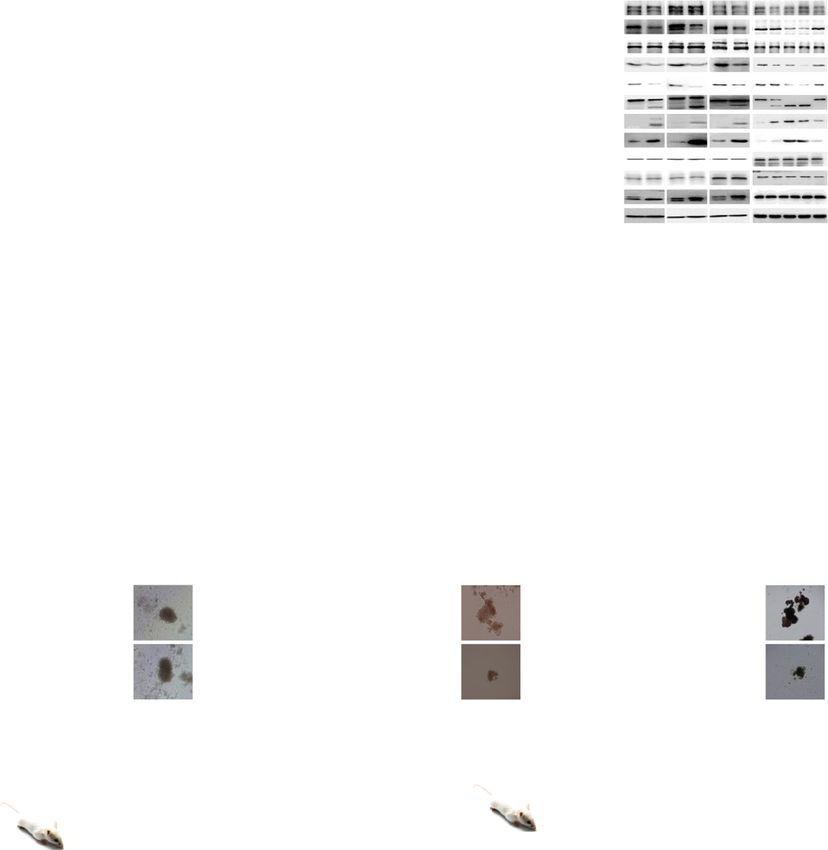
Fig. 7 P22077 shows remarkable toxicity on CML cells and CML leukemia stem/progenitor cells in vitro and in vivo. a–e Peripheral blood mononuclear
cells (PBMC) from normal donors (a) (n = 3 samples examined over three independent experiments), CML cell lines (b) (n = 3 cells examined over three
independent experiments), and BM mononuclear cells from IM-sensitive (c) (n = 5 samples examined over three independent experiments) and IM-
resistant (d) (n = 7 samples examined over three independent experiments) CML patients were treated with different concentrations of P22077 or IM for
48 h. Cell viability was measured by CCK8 assay. e BM mononuclear cells resistance to the second-generation TKIs (dasatinib, nilotinib, bosutinib) from
CML patients were treated with P22077 and IM for 48 h (n = 3 samples examined over three independent experiments). Cell viability was measured by
CCK8 assay. Data are presented as mean ± s.d. p-values were analyzed by one-way analysis of variance (ANOVA), *p < 0.05; **p < 0.01; ***p < 0.001,
****p < 0.0001. f Primary CML (IM-sensitive), CML-R (IM-resistant) BM mononuclear cells and KBM5T315I cells were treated with P22077 for 48 h. Cell
lysates from the indicated cells were extracted and subjected to immunoblotting with indicated antibodies. g P22077 (30 mg/kg/day), IM (50 mg/kg/
day), P22077 (30 mg/kg/day) in combination with IM (50 mg/kg/day) or vehicle were given to mice transplanted with KBM5T315I cells by intraperitoneal
injection (n = 6 biologically independent samples per group) from day 15 to day 30. **p < 0.01, by Mantel-Cox-log-rank test. h, i Human CD45 cells from
CML BM (patient-derived xenograft model) were transplanted into two groups of B-NDG mice. The survival time of the mice (h) (n = 7 biologically
independent samples per group) was determined, and the percentages of CD34+CD38− cells in human CD45 cells (i) (n = 3 biologically independent
samples per group) from BM were measured. Data are mean ± s.d. p-values were analyzed by Mantel-Cox-log-rank test (h) and two-sided Student’s t-test
(i). **p < 0.01; ***p < 0.001. j CD34+ cells derived from normal (n = 3 samples examined over three independent experiments), primary IM-sensitive CML
(n = 3 samples examined over three independent experiments), and primary IM-resistant CML BM mononuclear cells (n = 3 samples examined over three
independent experiments) were treated with different concentrations of P22077. The colonies were counted on day 14. Scale bars, 100 μm. Data are mean
± s.d. p-values were analyzed by one-way analysis of variance (ANOVA). *p < 0.05; **p < 0.01; ***p < 0.001; ****p < 0.0001; ns no significant. k Wild-type
mice received BCR-ABL retroviral transplantation for 21 days, and the mice were treated with control or P22077 by intraperitoneal injection for 14 days.
GFP+LSK cells from the BM of the two groups (n = 6 biologically independent samples per group) were evaluated by FACS. Data are mean ± s.d. p-values
were analyzed by two-sided Student’s t-test. ****p < 0.0001. l Wild-type mice received BCR-ABL retroviral transplantation for 21 days, and the mice were
treated with IM for 40 days. Mice were divided into two groups (n = 6 biologically independent samples per group). One group received P22077, and the
other group received the vehicle and used as the control. After 12 days, the number of GFP+LSK cells in the BM of the mice was examined. Data are mean
± s.d. p-values were analyzed by two-sided Student’s t-test. ****p < 0·0001. Source data are provided as a Source Data file.
cells were mixed with the stem cell medium (H4434, STEMCELL), and then data was generated in the Core Facility of Basic Medical Sciences of our school. Derived
continuously incubated at 37 °C in 5% humidified CO2. After incubation for data supporting the findings of this study are available from the corresponding author
14 days, the colonies were counted. upon reasonable request. Source data are provided with this paper.
MRD mouse model. BCR-ABL retrovirus-infected mouse BM cells were trans- Received: 15 January 2017; Accepted: 23 November 2020;
planted into 6-week-old female wild-type mice, and the GFP+Gr-1+ cells were
detected by FACS on day 21. The CML mice were treated with IM (50 mg/kg,
intraperitoneally) for 40 days, and then the mice were randomly divided into two
groups. One group was treated with P22077 (30 mg/kg, intraperitoneally), the other
group was given vehicle as control. The effect of P22077 on GFP+LSKs in BM was
examined by FACS after 12 days.
References
1. Radich, J. P. et al. Gene expression changes associated with progression and
Mouse xenograft model. K562 control and USP47 stably knockdown cells (8 × response in chronic myeloid leukemia. Proc. Natl Acad. Sci. USA 103,
106) were implanted subcutaneously into the right flanks of 6-week-old female 2794–2799 (2006).
Balb/c (nu/nu) mice (Slac Laboratory Animal Co., Ltd., China). Tumor sizes were 2. Hantschel, O. & Superti-Furga, G. Regulation of the c-Abl and Bcr-Abl
measured using calipers, and tumor volumes were calculated using a standard tyrosine kinases. Nat. Rev. Mol. cell Biol. 5, 33–44 (2004).
formula (width2 × length/2). Tumor cell proliferation and apoptosis were detected 3. O’Brien, S. G. et al. Imatinib compared with interferon and low-dose
by hematoxylin and eosin (H&E) staining, PCNA immunohistochemical (IHC) cytarabine for newly diagnosed chronic-phase chronic myeloid leukemia. N.
staining, and TUNEL assays were performed. Engl. J. Med. 348, 994–1004 (2003).
4. O’Hare, T., Eide, C. A. & Deininger, M. W. Bcr-Abl kinase domain mutations,
Homing analysis. We transduced BM from Usp47−/− and Usp47+/+ mice (n = 3, drug resistance, and the road to a cure for chronic myeloid leukemia. Blood
each group, 8-week-old) with BCR-ABL retrovirus for 48 h. GFP+LSK cells in 110, 2242–2249 (2007).
infected BM cells were measured by FACS. The infected BM cells (before trans- 5. Mathisen, M. S., Kantarjian, H. M., Cortes, J. & Jabbour, E. Mutant
plantation) were then injected into lethally irradiated 7.5 Gy Usp47+/+ mice. The BCR-ABL clones in chronic myeloid leukemia. Haematologica 96, 347–349
BM cells were collected 18 h after injection (after transplantation). The GFP+LSK (2011).
cells were monitored by FACS. The homing efficacy was calculated by the ratio of 6. Hochhaus, A. et al. Molecular and chromosomal mechanisms of resistance to
the percentage of GFP+LSK cells: [GFP+LSK] after transplantation 18 h/ [GFP imatinib (STI571) therapy. Leukemia 16, 2190–2196 (2002).
+LSK] before transplantation. 7. Mahon, F. X. et al. MDR1 gene overexpression confers resistance to imatinib
mesylate in leukemia cell line models. Blood 101, 2368–2373 (2003).
Statistics and reproducibility. All statistical tests were performed using GraphPad 8. Rosti, G., Castagnetti, F., Gugliotta, G. & Baccarani, M. Tyrosine kinase
Prism 6 (GraphPad Software Inc.). All data are presented as the mean ± sd. Student’s inhibitors in chronic myeloid leukaemia: which, when, for whom? Nat. Rev.
t-test was used to determine significant differences between two groups. One-way or Clin. Oncol. 14, 141–154 (2017).
two-way analysis of variance (ANOVA) was used to analyze the significant differences 9. Cortes, J. E. et al. A phase 2 trial of ponatinib in Philadelphia chromosome-
among multiple groups. For all statistical tests, p-values < 0.05 were considered to be positive leukemias. N. Engl. J. Med. 369, 1783–1796 (2013).
statistically significant. The number of independent experiments and replicates (n) is 10. Goodrich, A. D. Ponatinib in the leukemia world: why a reevaluation is
indicated in the figure legends. At least three biologically independent replicates were necessary for Philadelphia chromosome-positive patients with T315I
performed for each experiment and similar results were obtained. mutation. Expert Rev. Hematol. 7, 513–515 (2014).
11. Holyoake, T. L. & Vetrie, D. The chronic myeloid leukemia stem cell:
stemming the tide of persistence. Blood 129, 1595–1606 (2017).
Reporting summary. Further information on research design is available in the Nature
12. Quentmeier, H., Eberth, S., Romani, J., Zaborski, M. & Drexler, H. G. BCR-
Research Reporting Summary linked to this article.
ABL1-independent PI3Kinase activation causing imatinib-resistance. J.
Hematol. Oncol. 4, 6 (2011).
Data availability 13. Ma, L. et al. A therapeutically targetable mechanism of BCR-ABL-
The data supporting the findings of this study are available within the article and its independent imatinib resistance in chronic myeloid leukemia. Sci. Transl.
supplementary materials, which are provided as a Source Data file. In addition, some raw Med. 6, 252ra121 (2014).
14 NATURE COMMUNICATIONS | (2021)12:51 | https://doi.org/10.1038/s41467-020-20259-0 | www.nature.com/naturecommunicationsYou can also read