RING domains act as both substrate and enzyme in a catalytic arrangement to drive self-anchored ubiquitination - Nature
←
→
Page content transcription
If your browser does not render page correctly, please read the page content below
ARTICLE
                  https://doi.org/10.1038/s41467-021-21443-6                 OPEN

                  RING domains act as both substrate and enzyme
                  in a catalytic arrangement to drive self-anchored
                  ubiquitination
                  Leo Kiss        1 ✉,   Dean Clift      1,   Nadine Renner1, David Neuhaus                   1   & Leo C. James             1✉
1234567890():,;

                  Attachment of ubiquitin (Ub) to proteins is one of the most abundant and versatile
                  of all posttranslational modifications and affects outcomes in essentially all physiological
                  processes. RING E3 ligases target E2 Ub-conjugating enzymes to the substrate, resulting in its
                  ubiquitination. However, the mechanism by which a ubiquitin chain is formed on the sub-
                  strate remains elusive. Here we demonstrate how substrate binding can induce a specific
                  RING topology that enables self-ubiquitination. By analyzing a catalytically trapped structure
                  showing the initiation of TRIM21 RING-anchored ubiquitin chain elongation, and in combi-
                  nation with a kinetic study, we illuminate the chemical mechanism of ubiquitin conjugation.
                  Moreover, biochemical and cellular experiments show that the topology found in the
                  structure can be induced by substrate binding. Our results provide insights into ubiquitin
                  chain formation on a structural, biochemical and cellular level with broad implications for
                  targeted protein degradation.

                  1 MRC   Laboratory of Molecular Biology, Cambridge, UK. ✉email: lkiss@mrc-lmb.cam.ac.uk; lcj@mrc-lmb.cam.ac.uk

                  NATURE COMMUNICATIONS | (2021)12:1220 | https://doi.org/10.1038/s41467-021-21443-6 | www.nature.com/naturecommunications        1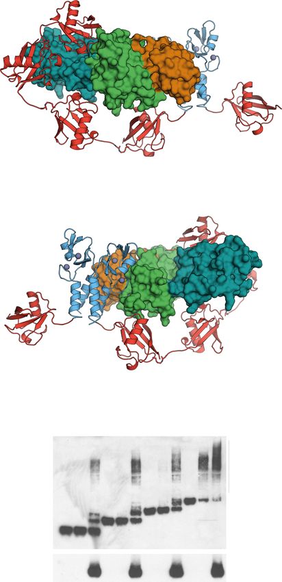
ARTICLE                                                                 NATURE COMMUNICATIONS | https://doi.org/10.1038/s41467-021-21443-6

U
         biquitin conjugation modifies cellular proteins with a               interaction with the E2 enzyme Ube2W12,13,20. Therefore, we
         highly versatile label and is used in virtually all physio-         attempted to address substrate-bound ubiquitination with
         logical pathways. The huge variety of possible ubiquiti-            TRIM21 RING and its chain forming E2 heterodimer Ube2N/
nation patterns includes modification with single ubiquitin                   Ube2V2. In crystallization trials, we used N-terminally mono-
molecules and/or up to eight different ubiquitin chain types1.               ubiquitinated TRIM21 RING domain (UbG75/76A-TRIM211–85 or
Furthermore, such modifications can be combined resulting in                  Ub-R), an isopeptide-linked, non-hydrolyzable ubiquitin-charged
highly specific signals that trigger dedicated processes. These               Ube2N conjugate (Ube2N~Ub) and Ube2V2. We solved the
modifications are achieved by a three-enzyme cascade. Ubiquitin               atomic structure of this complex at 2.2 Å resolution, with one
is first activated by an E1 enzyme and charged onto the active site           copy each of Ub-R, Ube2N~Ub and Ube2V2 in the asymmetric
cysteine of an E2 Ub-conjugating enzyme. Then the E2~Ub and a                unit (Supplementary Fig. 1 and Supplementary Table 1). The
third enzyme, an E3 ligase, ubiquitinate the substrate. Structural           naturally occurring TRIM21 RING homodimer21 was generated
insights into the process of substrate ubiquitination are limited to         in our model by invoking crystal symmetry (Fig. 1a). The RINGs
the transfer of one ubiquitin2 or ubiquitin-like protein3,4. How             engage Ube2N~Ub in the closed conformation22–24 and Ube2N
specificity is achieved within the ubiquitination system remains              forms a heterodimer with Ube2V2 (refs. 5–7). Analyzing further
largely elusive due to its extremely high level of complexity.               interactions within the crystal lattice, we found that the TRIM21-
   Ubiquitin chains that are linked via lysine 63 (K63) are involved         linked ubiquitin made additional contacts to Ube2N/Ube2V2 of a
in endocytosis, DNA damage, and the immune response1. The                    symmetry-related complex (Fig. 1b and Supplementary Fig. 1),
only E2 enzyme dedicated to K63-ubiquitination is Ube2N                      which orient the RING-bound ubiquitin so that its K63 points
(Ubc13), which forms a heterodimer with either Ube2V2 (Mms2)                 toward the active site, ready for nucleophilic attack (Fig. 1b, c).
or Ube2V1 (Uev1)5–7. Ube2V2 binds and orients the acceptor                   Our structure thus represents a snapshot of a ubiquitin-primed
ubiquitin, thereby generating specificity for K63 linkage8–10. While          RING ready for self-anchored ubiquitin chain elongation.
Ube2N/Ube2V2 efficiently forms free K63-linked ubiquitin
chains, substrate modification requires the presence of a priming
ubiquitin for elongation11. Such a mechanism has been established            Chemical mechanism of ubiquitination. Having captured a 2.2
for the RING (really interesting new gene) E3s TRIM21 and                    Å resolution representation of the system prior to catalysis, we
TRIM5α12,13. With ~100 members in humans, the TRIM family                    were able to perform a detailed analysis of ubiquitin transfer. The
contains the largest number of distinct RING E3s14. TRIM21                   Ube2N-charged ubiquitin can be found in the RING-promoted
detects antibody-coated viruses by specifically recognizing the               closed Ube2N~Ub conformation and thus represents the donor
bound antibody. This activates its E3 ligase function, resulting             ubiquitin (Fig. 1). The RING-bound ubiquitin of a symmetry-
in virus neutralization and innate immune activation via the                 related complex was captured by Ube2N/Ube2V2, positioning its
NF-κB pathway15,16. First, Ube2W attaches a priming ubiquitin to             nucleophilic K63 NζH3 group 4.8 Å from the electrophilic car-
the TRIM21 N-terminus13. Next, TRIM21 specifically recruits                   bonyl of the donor ubiquitin C-terminus (Fig. 2a and Supple-
Ube2N/Ube2V2 to produce a TRIM21-anchored K63-linked                         mentary Fig. 1). Interestingly, K63 of this acceptor ubiquitin
ubiquitin chain13,17. TRIM21 does not engage its target directly             shows a direct interaction with D119 of Ube2N (Fig. 2a). This
and forms the ubiquitin chain on its own N-terminus, resulting               suggests that D119 deprotonates K63 on the acceptor ubiquitin,
in degradation of itself, the antibody, and the substrate13,15. This         thereby activating it for nucleophilic attack. Indeed, the corre-
mechanism can be repurposed by introduction of substrate-                    sponding residue in Ube2D (D117) has been suggested to be
specific antibodies to the cytosol, resulting in targeted protein             involved in positioning and/or activating an incoming acceptor
degradation by Trim-Away18. This is conceptually similar to                  lysine22.
proteolysis targeting chimeras (PROTACs), that bring substrate                  To investigate the chemical mechanism of ubiquitination
and E3 ligase together, resulting in substrate ubiquitination and            (Fig. 2b), we measured the kinetics of di-ubiquitin formation
degradation19.                                                               (Supplementary Fig. 2). The acid coefficient (pKa) of this reaction
   In this work, we sought to understand how RING                            should solely depend on the protonation state of its nucleophile,
E3-anchored ubiquitin chains are formed. Although it is known                K63. Fitting the ubiquitination velocity of reactions carried out at
how Ube2N/Ube2V2 specifically connects two ubiquitin                          different pHs to an equation assuming one titratable group
molecules with a K63 linkage8–10 and how Ube2N is specifically                revealed a pKa of 8.3 for Ube2N (Fig. 2c and Supplementary
recruited by RING E3s17, it was not known how substrate-                     Fig. 2), comparable to what was observed for the SUMO-E2
anchored ubiquitin chains are formed. Yet, anchored ubiquitin                Ube2I25. This is significantly lower than the pKa of 10.5 for a free
chains deliver highly specialized functions and phenotypes. We               lysine ζ-amino group26, which would be incompatible with
address this question by solving a structure trapped in the                  catalysis at physiological pH ~7.34 (ref. 27). We mutated D119 to
process of RING-anchored chain elongation. Together with a                   either alanine or asparagine as neither can act as a base, but
kinetic analysis, this reveals how chemical activation of the                asparagine could still bind and orient K63. Both mutants
acceptor ubiquitin is achieved. It also reveals a specific topology           increased the pKa to ~9 (Fig. 2c). At physiological pH,
of the RING domains that is necessary for formation of the self-             Ube2ND119A/N modestly increased the KM by ~4- and ~7-fold,
anchored ubiquitin chain. Finally, we validate the importance                respectively (Fig. 2d). Mutation to alanine reduced kcat 100-fold
of this arrangement biochemically and show that it results in                and mutation to asparagine 30-fold (Fig. 2e), suggesting that
targeted protein degradation in a physiological setting.                     substrate turnover also depends on orientation of the lysine
                                                                             nucleophile. Yet, this catalytic rate does not yield efficient
                                                                             ubiquitin chain formation under physiological pH (Supplemen-
Results                                                                      tary Fig. 3). Together, these observations establish that D119 is
Structure showing the formation of anchored ubiquitin chains.                the base that deprotonates the incoming acceptor lysine to enable
We set out to understand how a substrate-bound ubiquitin chain               catalysis.
can be formed. In principle, ubiquitin chain elongation of TRIM                 Interactions between ubiquitin and other proteins have been
proteins depends on their RING domain only. In the case of                   shown to depend on specific conformations of ubiquitin’s β1–β2
TRIM21 (and TRIM5α), the TRIM RING itself is the substrate,                  loop, which can be found in either loop-in or loop-out
after it has undergone N-terminal mono-ubiquitination upon                   conformations28. These motions change the ubiquitin core

2                NATURE COMMUNICATIONS | (2021)12:1220 | https://doi.org/10.1038/s41467-021-21443-6 | www.nature.com/naturecommunications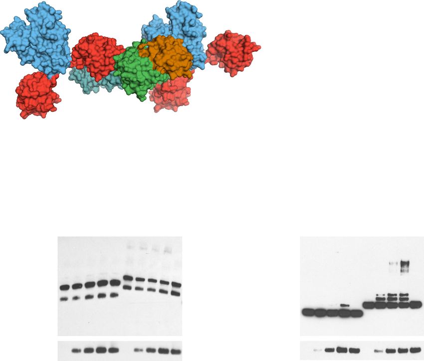
NATURE COMMUNICATIONS | https://doi.org/10.1038/s41467-021-21443-6                                                                           ARTICLE

                                                                                                                                    RING’’
     a                                                                    b                                                                     RING’’’
                                                                                                     Ub        Ube2N
                                                                                                                            Ub’’
                                                                                           RING’
                          RING’       RING
                                               Ub                                       Ub’

                                                                                                                                                     Ub’‘’
     Ub
      be2V2’                                                  Ube2V2

                                                     Ube2N                                    RING                Ube2V2
           Ube2N’ Ub’                                                                                Ub
                                                     Ub
                      Ub’
                                                                          c                                                          RING’’
                                                                                                      donor Ub      acceptor Ub
                                                                                                                                                RING’’’
                                                Ub
            Ube2V2’               Ub’                                                      RING’
                                   RING’’                 Ube2N
                                                                                     Ub’

                                        RING                                               RING           Ube2N
           Ube2N’                                                                                                          Ube2V2            Ub’‘’
                                                     Ube2V2
                            Ub’
                            Ub
                                                                                                             Ub

Fig. 1 Structure of initiation of RING-anchored ubiquitin chain elongation. a Side and top view of the Ub-R:Ube2N~Ub:Ube2V2 structure (Ub-R, Ub in red,
R in blue, Ube2N~Ub, Ube2N in green, Ub in orange, and Ube2V2 in teal). Chains drawn as cartoon represent the asymmetric unit. b The canonical model
of initiation of RING-anchored ubiquitin chain elongation. c Schematic cartoon, representing the canonical model of RING-anchored ubiquitin chain
elongation shown in b. Symmetry mates are denoted by ′ next to the label. Ub ubiquitin, Ub-R ubiquitin-RING.

structure and subsequent conformational selection enables                       between the acceptor ubiquitin and Ube2N (Fig. 2a and
ubiquitin to interact with many different binding partners29.                   Supplementary Fig. 5c), that position the nucleophile K63 much
In our structure, we found the donor ubiquitin β1–β2 loop in its                nearer to the active site (4.8 vs. 7.5 Å). This is achieved because
loop-in configuration, and loop-out to be incompatible with                      Ube2N N123 and D124 contact ubiquitin via the amide of
formation of the closed conformation (Supplementary Fig. 4a,                    ubiquitin K63 and the sidechains of S57 and Q62, respectively
b). Conversely, the acceptor ubiquitin was in a loop-out                        (Fig. 2a). The ~3-fold reduction in kcat (Fig. 2e) for the mutants
configuration (Supplementary Fig. 4c), which appears to be                       Ube2NN123A and Ube2ND124A suggest that the function of these
the default state in ubiquitin28. Donor and acceptor ubiquitin                  residues is to finetune the ubiquitination reaction by aiding
also have distinct B-factor profiles (Supplementary Fig. 4d),                    orientation of the nucleophile. Taken together, the features of
perhaps reflecting some other aspect of their different roles in                 our structure trapped in the process of ubiquitin chain
catalysis. Interestingly, the β1–β2 loop conformation also                      formation provide mechanistic insight into how the RING E3
appears to be critical in ubiquitin-like proteins, such as Nedd8,               promotes catalysis, by simultaneously activating Ube2N for
when activating cullin-RING-ligases (CRL)2.                                     ubiquitin discharge and allowing Ube2V2 to precisely orient the
   RING E3s act by locking the normally very dynamic E2~Ub                      acceptor ubiquitin.
species in a closed conformation, thereby priming it for catalysis
(Fig. 2a)22–24. Comparison with our previously determined
TRIM21 R:Ube2N~Ub structure17 shows scarcely any difference                     The mechanism of RING-anchored ubiquitination. Next, we
between the donor ubiquitin C-termini and the Ube2N active                      sought to understand how RING-anchored ubiquitin chains are
site (Supplementary Fig. 5a). Nonetheless, formation of the                     formed. In our crystal structure, one RING dimer is positioned so
closed Ube2N~Ub conformation alone is not sufficient for                         as to mediate the elongation of another mono-ubiquitinated
catalysis, as this also requires the presence of Ube2V2 (ref. 5),               RING in trans (Figs. 1b, c and 3a). Importantly, this mechanism
which binds and orients the acceptor ubiquitin8–10. We gained                   depends only on binding of the RING-anchored acceptor ubi-
additional insight into how Ube2V2 positions the acceptor                       quitin to Ube2N/Ube2V2, as no contacts with the RING itself
ubiquitin by analyzing a Ube2N~Ub:Ube2V2 complex that we                        could be observed in our crystal structure (Supplementary Fig. 1).
solved at 2.5 Å resolution (Supplementary Fig. 6 and Supple-                    The relative topology of the different RING domains (enzyme
mentary Table 1). By invoking crystal symmetry, this structure                  and substrate) is thus mostly dictated by the catalytic interfaces,
shows the orientation of an acceptor ubiquitin by Ube2V2, so                    resulting in a ~9 nm separation between the enzyme and sub-
that its K63 is pointed toward the active site of Ube2N                         strate RINGs (Fig. 3a). We refer to this arrangement as the cat-
(Supplementary Fig. 6), an orientation comparable with a                        alytic RING topology, in which a RING dimer acts as an enzyme
structure of yeast Ube2N~Ub:Ube2V2 that was solved in a                         and at least one further RING acts as the substrate for ubiquiti-
different crystal lattice9. Without a RING present, the donor                   nation. This topology is not rigid since the linkers between the
ubiquitin is not in the closed conformation and our Ube2N~Ub:                   acceptor ubiquitin and the RING (~3 nm apart), and the RING
Ube2V2 structure thus represents an inactive complex. Align-                    and the next (B-box) domain in the TRIM ligase (~3.5 nm apart)
ment to our Ub-R:Ube2N~Ub:Ube2V2 structure (Supplemen-                          likely provide additional flexibility (Fig. 3a, b and Supplementary
tary Fig. 5b–d) reveals that Ube2N and Ube2V2 are packed                        Fig. 1c). In our structure, it is clear that initiation of TRIM21-
more closely against each other, resulting in additional contacts               anchored chain elongation cannot occur in cis, as the priming

NATURE COMMUNICATIONS | (2021)12:1220 | https://doi.org/10.1038/s41467-021-21443-6 | www.nature.com/naturecommunications                                     3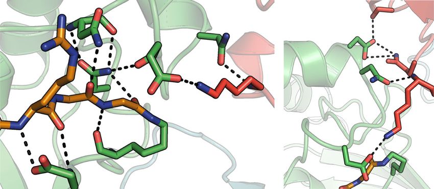
ARTICLE NATURE COMMUNICATIONS | https://doi.org/10.1038/s41467-021-21443-6
a c
9.4
9.2
9.0
pKa
8.8
8.6
N116 S57 8.4
N123 D124
Q62 8.2
N79
d 1000
N123
D119
R74 K63 K63
G75
KM (µM)
G76
D119 100
K87
K87
G76
10
b D119 e 0.01
D119
O O H 0.001
H K63 O O
kcat (s-1)
N
O H H K63
G76 H O N 0.0001
S G76
H
N
H C87 N S
O H
HN O C87 0.00001
T
D A
9N
3A
4A
O HN
W
9
11
12
12
11
D
N
D
O
Ube2N
Fig. 2 Chemical mechanism of ubiquitination. a Magnified regions of the active site of Ube2N~Ub/Ube2V2 (Ube2N in green, donor Ub in orange,
acceptor Ub in red, and Ube2V2 in teal). Stereo-images are shown in Supplementary Fig. 1b. b Chemical scheme for the activation of the acceptor lysine.
c Acid coefficients (pKa), d KM, and e kcat of di-ubiquitin formation by Ube2N/V2 are presented as best fit + standard error. Fits were performed using data
of n = 3 technical replicates, and are shown in Supplementary Fig. 2 and Source data. Ub ubiquitin.
ubiquitin cannot reach the Ube2N/Ube2V2 binding surface TRIM21 constructs lacking the B-box and coiled-coil (Ub-R-PS
(Fig. 3a). Consistent with this, we found that TRIM21 ubiquitin and Ub-R-R-PS). Fc is capable of recruiting two of these
transfer in trans can occur in principle (Supplementary Fig. 7), in constructs, thereby locating their RINGs within ~9 nm (Fig. 3c
line with previous work on TRIM5 (ref. 20). and Supplementary Fig. 8), the distance required for the catalytic
To investigate the spatial requirements of TRIM21 RING RING topology (Fig. 3a, c). Addition of Fc to Ub-R-PS led to
domains for self-anchored ubiquitination experimentally, we weak self-ubiquitination. This low level of activity is likely
established a substrate-dependent ubiquitination assay. TRIM21 because Ub-R-PS can only provide a monomeric RING as the
is recruited by Fc, which is an obligate dimer in solution and can enzyme, while a monomeric RING on the second Ub-R-PS acts as
be bound by two PRYSPRY (PS) domains30 (Supplementary the substrate. TRIM RING dimerization is known to greatly
Fig. 8). To test for the catalytic RING topology, we designed a increase ligase activity21,31–33. We therefore repeated these
series of mono-ubiquitinated TRIM21 constructs that vary the experiments using a Ub-R-R-PS construct. We predicted that
number of RINGs available and their distance to each other when this should allow the catalytic RING topology observed in our
bound to Fc (Fig. 3b and Supplementary Fig. 8). To suppress crystal structure to form upon substrate binding, as the Fc will
background activity, TRIM21 was used at low concentrations bring two RING dimers into close proximity (Fig. 3a, c). Indeed,
(100 or 50 nM) and the reaction was incubated for 5 min only. addition of Fc to Ub-R-R-PS resulted in the efficient formation of
Full-length TRIM proteins form antiparallel homodimers via TRIM21-anchored ubiquitin chains (Fig. 3d). Importantly, while
their coiled-coil domains, resulting in the separation of the two anchored ubiquitination occurred very efficiently, hardly any free
TRIM21 RING domains by ~17 nm even when bound to Fc ubiquitin chains could be observed (Supplementary Fig. 9c). Since
(Supplementary Fig. 8). According to our model, addition of Fc self-ubiquitination only requires E2~Ub to be recruited by the
alone should therefore not induce the catalytic RING topology ligase, this explains its high efficiency relative to free ubiquitin
(Fig. 3c). Indeed, addition of Fc did not stimulate ubiquitination chain formation, as the latter would require recruitment of both
of the full-length Ub-TRIM21 (Ub-RING-Box-coiled-coil- E2~Ub and (poly-) ubiquitin. Indeed, Ub-R-R-PS worked
PRYSPRY or Ub-R-B-CC-PS, Fig. 3d). Even when adding an efficiently in our substrate-induced ubiquitination assay even at
additional RING domain to make the full-length protein a reduced TRIM21 concentrations (Supplementary Fig. 9d). Thus,
constitutive RING dimer (Ub-R-R-B-CC-PS), formation of the inducing formation of the catalytic RING topology by substrate
catalytic RING topology is excluded (Fig. 3c) and no induction of binding enables robust and selective formation of self-anchored
self-ubiquitination is observed upon addition of Fc (Fig. 3d, and ubiquitin chains. Moreover, the catalytic RING topology is only
Supplementary Figs. 8 and 9). As a next step, we designed achieved when the separate requirements of an active enzyme (a
4 NATURE COMMUNICATIONS | (2021)12:1220 | https://doi.org/10.1038/s41467-021-21443-6 | www.nature.com/naturecommunicationsNATURE COMMUNICATIONS | https://doi.org/10.1038/s41467-021-21443-6 ARTICLE
a b
~9 nm RING
RING
RING RING
acceptor Ub Ub Ub R B CC PS
Ub R R B CC PS
Ub R PS
donor Ub Ub R R PS
Ube2V2 Ube2N Ub
Ub
c
Ub-R-B-CC-PS Ub-R-R-B-CC-PS Ub-R-PS Ub-R-R-PS
d
Ube2N/V2: + + + + - + + + + - Ube2N/V2: + + + + - + + + + -
Fc: - + - + Fc: - + - +
kDa: kDa:
250 Ubn-TRIM21
150 250
100 150 Ubn-TRIM21
75 100
Ub-R-R-B-CC-PS 75
50
Ub-R-B-CC-PS
* 50
*
Ub-R-R-PS
37 37 Ub-R-PS
25
25
anti-TRIM21 anti-TRIM21
37 37
Fc Fc
25 anti-Fc 25 anti-Fc
Ub-R-B-CC-PS Ub-R-R-B-CC-PS Ub-R-PS Ub-R-R-PS
Fig. 3 The mechanism of RING-anchored ubiquitination in trans. a Surface representation of the canonical model of the Ub-R:Ube2N~Ub:
Ube2V2 structure (Ub-R, Ub in red, R in blue, Ube2N~Ub, Ube2N in green, Ub in orange, and Ube2V2 in teal). b Domain architecture of TRIM21 constructs
used in biochemical assays. c Cartoon models of substrate (Fc, gray) engagement by TRIM21 constructs (blue). The structural basis for these models is
shown in Supplementary Fig. 8. d Substrate (Fc)-induced self-ubiquitination assay of 100 nM Ub-TRIM21 constructs. Reactions were incubated for 5 min at
37 °C. Further data can be found in Supplementary Fig. 9. * (asterisk) indicates a TRIM21 degradation product that could not be removed during
purification. Western blots are representative of n = 3 independently performed experiments. Uncropped blots are provided in Source data. Ub ubiquitin, R
RING, B Box, CC coiled-coil, PS PRYSPRY, kDa kilo Dalton.
dimeric RING) and a correctly positioned substrate (a third occurred when two Ub-R-R-PS constructs were colocalized by
RING) are fulfilled. their binding to Fc to satisfy the requirements of the catalytic
We next considered how long a TRIM21-anchored ubiquitin RING topology (Fig. 3c, d). To confirm the switch in self-
chain would have to be for cis ubiquitination to become sterically ubiquitination from trans to cis experimentally, we generated
possible. Using our Ub-R:Ube2N~Ub:Ube2V2 structure, we TRIM21 R-R-PS constructs, wherein their N-termini were fused
created models with increasing numbers of K63-linked ubiquitin to up to four linearly connected ubiquitin molecules. Due to their
chains conjugated to the TRIM21 RING domain. These models high structural similarity34, we assumed a linear chain would
suggested that a chain of four ubiquitin molecules would be mimic a K63-linked ubiquitin chain in length and flexibility
necessary and sufficient for self-ubiquitination in cis (Fig. 4a and sufficiently well. Upon testing these new constructs, we observed
Supplementary Fig. 10). Thus, after addition of the priming that only TRIM21 modified with tetra-ubiquitin became
ubiquitin, three ubiquitin molecules must be added in trans, independent of Fc for self-ubiquitination (Fig. 4b). All the other,
before the chain could be further elongated in cis. Consistent with shorter, constructs remained rate-limited by first having to self-
this, we only observed very long TRIM21-anchored ubiquitin ubiquitinate in trans, before switching to cis. This biochemical
chains or species carrying one, two, or three ubiquitin molecules data is in agreement with our structure, showing the initiation of
in our Fc-dependent TRIM21 ubiquitination experiments (Fig. 3d RING-anchored ubiquitination in trans and our model of
and Supplementary Fig. 9c, d). With the addition of a fourth polyubiquitinated RING elongation in cis.
ubiquitin, the reaction appears to progress much more quickly, as Finally, we considered whether the catalytic RING topology is
would be expected for a switch from trans to cis, rapidly an arrangement specific to Ube2N or one that also works with
consuming the tetra-ubiquitin species and converting it into a other E2 enzymes. Thus, we tested whether addition of Fc could
long chain. In the above experiments, self-ubiquitination only induce self-ubiquitination of Ub-TRIM21 in presence of Ube2D1,
NATURE COMMUNICATIONS | (2021)12:1220 | https://doi.org/10.1038/s41467-021-21443-6 | www.nature.com/naturecommunications 5ARTICLE NATURE COMMUNICATIONS | https://doi.org/10.1038/s41467-021-21443-6
a in our structure is thus specific for Ube2N/Ube2V2, explaining
Ub4
why this enzyme16 and not Ube2D1 is required for TRIM21’s
cellular function17. Moreover, this may explain why TRIM21, and
Ube2N
Ube2V2 Ub RING other TRIMs, such as TRIM5, build K63- and not K48-linked
ubiquitin chains when first activated. Their mechanism of
activation, induction of the catalytic RING topology, only results
in formation of self-anchored K63 chains by using Ube2N/
Ube2V2. Collectively, these data identify formation of a catalytic
Ub3 Ub
trans RING topology, as the driving force behind self-
ubiquitination of TRIM21 with Ube2N/Ube2V2.
RING
Catalytic RING topology drives targeted protein degradation.
Ub1
Ub2 Having established the RING topology necessary for self-
anchored ubiquitination in vitro, we next investigated if this
same arrangement is required for TRIM21 activity in cells. We
designed a similar series of TRIM21 constructs for cellular
expression as above, which control for the number of RINGs
available and their distance to each other when bound to Fc
RING Ub4 (Fig. 5a). We expressed these constructs in TRIM21 knockout
RING K63 RPE-1 cells together with GFP-tagged Fc and monitored GFP-Fc
degradation as a readout for TRIM21 activity, in a targeted
protein degradation experiment. Consistent with the inability to
form anchored chains when engaged with Fc in vitro, full-length
TRIM21 did not degrade GFP-Fc in cells (Fig. 5b, c). Degradation
Ub3 could not be rescued by addition of another RING to the N-
terminus, presumably because in this case the RINGs are dimeric
Ube2V2 but still separated by the coiled-coil, with the consequence that no
Ube2N
“substrate” RING is available for ubiquitination. Thus, RING
Ub
dimerization is not sufficient for cellular TRIM21 activity. In the
Ub1 Ub2 R-PS construct, the RINGs are within ~9 nm, and thus within the
range compatible with activity as defined by our structure
b (Fig. 3a). Despite this, no degradation was observed (Fig. 5b, c),
likely because the RINGs can either form a single dimer, or one
n(Ubn-R-R-PS): 1 2 3 4
monomer RING would have to act as the enzyme and the other
Ube2N/V2: - + + - + + - + + - + +
RING as the substrate. This is consistent with the inefficient self-
Fc: - - + - - + - - + - - +
kDa:
ubiquitination of a comparable construct in our biochemical
experiments (Fig. 3d). Only R-R-PS showed efficient GFP-Fc
250
Ubn-TRIM21
degradation (Fig. 5b, c). When this construct engages Fc, two
150 RING dimers can form in close proximity, so that one RING
100 dimer is available to mediate the ubiquitination of the other, thus
75 Ub4-R-R-PS fully satisfying the requirements of the catalytic RING topology.
Ub3-R-R-PS All the constructs were expressed at comparable levels and were
Ub2-R-R-PS
50 active in classical Trim-Away targeted protein degradation assays
Ub1-R-R-PS
37 (Fig. 5d, e and Supplementary Fig. 12), suggesting that the only
anti-TRIM21 difference is the number and relative distance of RING domains
37
when engaged with the GFP-Fc construct. This also agrees with
Fc
25 anti-Fc
our biochemical data, where a similar construct shows strong self-
ubiquitination upon substrate binding (Fig. 3d). Therefore, the
Fig. 4 The mechanism of RING-anchored ubiquitination in cis. a For Fc-induced self-ubiquitination assay in vitro provides a good
ubiquitination in cis, the RING-anchored (blue) ubiquitin chain (red) must prediction for the cellular activity. Our crystal structure of the
be sufficiently long to reach the active site on Ube2N~Ub/Ube2V2 (Ube2N initiation of RING-anchored ubiquitin chain elongation therefore
in green, Ub in orange, and Ube2V2 in teal). The chain can go around two precisely visualizes how this process can work in a physiological
different routes, one shown here and the other in Supplementary Fig. 10. context.
The ubiquitin chain was modeled using the Ub-R:Ube2N~Ub:
Ube2V2 structure and a K63-linked Ub2 structure (2JF5 (ref. 34)) using
PyMol. b Substrate (Fc)-induced self-ubiquitination assay of 100 nM Ubn- Discussion
TRIM21 constructs. Reactions were incubated for 5 min at 37 °C. Western Protein ubiquitination is one of the most abundant posttransla-
blots are representative of n = 3 independently performed experiments. tional modifications, affecting essentially all cellular events. A
Uncropped blots are provided in Source data. Ub ubiquitin, R RING, PS precise understanding of its underlying mechanisms, and how
PRYSPRY, kDa kilo Dalton. they achieve selectivity, is desirable for several reasons. First,
malfunction in the ubiquitin system often manifests in severe
disease35. Second, the use of small molecules (PROTACs, mole-
a highly promiscuous E2 enzyme. However, even after extended cular glues, etc.) has emerged as a promising new approach for
reaction times hardly any TRIM21 modification was detected, targeted degradation of disease-causing proteins in patients36–41.
while in contrast free ubiquitin chains could be observed Despite recent advances in the field, it remains unclear how RING
(Supplementary Fig. 11). The catalytic RING topology we observe E3 ligases achieve specific substrate ubiquitination and how a
6 NATURE COMMUNICATIONS | (2021)12:1220 | https://doi.org/10.1038/s41467-021-21443-6 | www.nature.com/naturecommunicationsNATURE COMMUNICATIONS | https://doi.org/10.1038/s41467-021-21443-6 ARTICLE
a ~17 nm ~17 nm ~9 nm ~9 nm
GFP-Fc
R-B-CC-PS R-R-B-CC-PS R-PS R-R-PS
b c
S
R CC S
-P
S
- -P
-P C
-R -P
R -CC
R B-C
S
-P ns ns ns **
S
h
-
-R
-B
C
kDa: 1.0
Median fuoroscent intensity
m
R
75
(-MG132 / +MG132)
mEGFP-Fc 0.9
50
anti-Fc
100 0.8
75
R-R-B-CC-PS
50 R-B-CC-PS 0.7
37 R-R-PS
R-PS 0.6
25
anti-TRIM21
0.5
250
150
S
S
S
S
S
Vinculin
-P
-P
-P
-P
-P
100
C
C
C
R
-R
anti-Vinculin
C
-C
-C
R
h-
-B
-B
C
R
-R
m
R
S
-P
R CC S
R -B- S
d e
C
-B -P
-R -P
-P C
R CC
S
-P
R S
-
R-B-CC-PS + Vhh-Fc -
-R
-B
TRIM21:
R
R-R-B-CC-PS + Vhh-Fc VhhGFP-FcH433A: + - - - -
VhhGFP-Fc: - + + + +
R-PS + Vhh-Fc
kDa:
R-R-PS+ Vhh-Fc 50 Cav1-GFP
R-B-CC-PS+ Vhh-FcH433A 37
1.2 25
20 Cav1
1.0 anti-Cav1
Normalized GFP
75
R-R-B-CC-PS
fluorescence
0.8
50 R-B-CC-PS
0.6 37 R-R-PS
R-PS
0.4 25
20 anti-TRIM21
50
0.2
VhhGFP-Fc
37
anti-Fc
0.0
0 1 2 3 4 5 150
Vinculin
100
Time (h) anti-Vinculin
ubiquitin chain is formed after the priming ubiquitin has been structure reveals the domain arrangement required for the elon-
transferred. gation reaction, in other words a catalytic RING E3 topology that
Here, we provide a structural framework for understanding enables the extension of a mono-ubiquitinated RING into a K63-
RING E3-anchored ubiquitin chain formation. We were able to linked, RING-anchored ubiquitin chain (Figs. 3 and 4). In this
capture a snapshot of this process in a crystal structure of Ub-R arrangement, two RINGs form a dimer and act as an enzyme on a
with the ubiquitin-charged heterodimeric E2 enzyme Ube2- third RING domain, which acts as the substrate in this reaction.
N~Ub/Ube2V2 (Fig. 1), showing the chemical activation of the We observe that while rigidity is required to position all the
acceptor ubiquitin, exemplified by the deprotonation of the important catalytic residues in the E2 active site optimally (Fig. 2
acceptor lysine by Ube2N D119 (Fig. 2). Most importantly, our and Supplementary Fig. 1), formation of the substrate-anchored
NATURE COMMUNICATIONS | (2021)12:1220 | https://doi.org/10.1038/s41467-021-21443-6 | www.nature.com/naturecommunications 7ARTICLE NATURE COMMUNICATIONS | https://doi.org/10.1038/s41467-021-21443-6
Fig. 5 Catalytic RING topology drives targeted protein degradation. a Schematic cartoons showing the topology of TRIM21 (blue) on GFP-Fc (green and
gray, respectively). b, c GFP-Fc degradation assay. b Western blot of RPE-1 TRIM21-knockout cells transiently expression GFP-Fc and a series of TRIM21
constructs. Western blots are representative of n = 2 independently performed experiments. c Shown is the flow cytometry analysis of green fluorescence
of RPE-1 TRIM21-knockout cells transiently expressing GFP-Fc and a series of TRIM21 constructs. After electroporation, each population of cells was split in
two and either treated with MG132 or DMSO. Data are presented as mean ± standard error of the mean. Each data point in the graph represents one
biologically independently performed experiment (n = 3 (for mCh-CC-PS, R-R-B-CC-PS, and R-PS) or 4 (R-B-C-C-PS and R-R-PS)). A two-tailed unpaired
Student’s T test was performed to assess the significance of fluorescence reduction relative to mCh-CC-PRYSPRY (P values: R-B-CC-PRYSPRY, 0.0797
(ns); R-R-B-CC-PRYSPRY, 0.02366 (ns); R-PRYSPRY, 0.4964 (ns); and R-R-PRYSPRY, 0.0035 (**)). d, e Trim-Away of Caveolin-1-mEGFP (Cav1-GFP) in
NIH 3T3 GFP-Cav-1-knock in cells66. Shown in d is the normalized GFP fluorescence (error bars represent ± SEM of four images) and in e the western blot
after the experiment. Data in d, e are representative of n = 2 independent experiments. Uncropped blots and raw data are provided in Source data. R RING,
B Box, CC coiled-coil, PS PRYSPRY, mCh mCherry, kDa kilo Dalton, ns not significant.
ubiquitin chain likely requires conformational flexibility between signals45. A strategy of mixed, branched chains might be essential
domains that is provided by the unique topology of TRIM pro- for TRIM21 to act as both an immune sensor and effector13.
teins (Fig. 3). Substrate-induced self-ubiquitination of TRIM21 is The catalytic RING topology we describe is consistent with
highly efficient, even at low ligase concentration, in contrast to data showing that TRIM proteins can undergo higher-order
free ubiquitin chain formation (Fig. 3 and Supplementary Fig. 9). assembly. In the case of TRIM5α46, three TRIM5α RINGs are
This implies that physiological ubiquitin signals may not be brought into close proximity when the protein is incubated with
produced as free chains but mainly on substrates, due to the the HIV capsid46–48 (Fig. 6a, b and Supplementary Fig. 13). This
higher reaction efficiency. positioning would fulfill the catalytic RING topology we describe,
Our data establishes that the RING-anchored K63-chain is and would be consistent with the ability of TRIM5α to restrict
first formed in a trans mechanism, where a RING dimer acti- retroviruses49,50 and activate the innate immune response via
vates a Ube2N~Ub molecule, thereby acting as an E3 ligase. self-anchored K63-ubiquitination12,51. The functional require-
An additional mono-ubiquitinated RING acts as a substrate ment for multiple TRIM molecules is also suggested by the fact
for ubiquitination and accepts the donor ubiquitin (Fig. 3). that potent antibody-mediated neutralization of adenovirus
Only after four ubiquitin molecules have been added to the by TRIM21 requires multiple antibodies bound per virus52. In
RING in trans, is the chain sufficiently long for ubiquitin chain addition, TRIM21 was shown to be activated by substrate-
formation in cis (Fig. 4). While ubiquitin chain elongation in induced clustering, resulting in multiple TRIM21:antibody com-
cis occurs at much higher rates, the initial need for a trans plexes on the substrate53. The unique TRIM architecture, in
arrangement may represent an important regulatory mechan- which the RINGs are located at either end of a coiled-coil, and the
ism suppressing TRIM21 activity in absence of a substrate. flexibility provided by the hinge region of the antibody, may be
In the case of TRIM21 or TRIM5α, activation is driven by crucial in enabling TRIM21 molecules bound onto the surface of
substrate binding, which is needed for trans ubiquitination. a virus to engage with each other (Fig. 6c). To fulfill the catalytic
Interestingly, substrate modification with linear ubiquitin RING topology on the virus, two RINGs need to dimerize and a
chains by the RBR (RING-in between-RING) ligase HOIP is third has to be within ~9 nm of the RING dimer, enabling self-
regulated by its partner RBR HOIL, which mono-ubiquitinates anchored ubiquitination and subsequent virus neutralization
all three LUBAC components HOIP, HOIL, and SHARPIN. (Fig. 6d). Since higher-order assembly has been associated with
These ubiquitin primers are then elongated in cis by HOIP, many other K63 ubiquitin chain forming RING E3 ligases, such
thereby outcompeting trans ubiquitination of substrates42. as TRAF6 (ref. 54), RIPLET55, and others, we propose that the
Thus, switching between cis and trans mechanisms of ubiqui- mechanism presented here is thus likely to be found more widely
tination may be a regulatory system exploited by many different within the realm of RING E3 ligases.
types of E3 ligases.
The catalytic RING topology observed in our structure predicts Methods
the requirements for TRIM21-mediated targeted protein degra- Plasmids. Bacterial expression constructs: Ube2V2 and TRIM21 expression con-
dation in cells (Fig. 5). Upon substrate recognition, TRIM21 structs, but full-length were cloned into pOP-TG vectors and full-length TRIM21
forms a K63-linked ubiquitin chain on its N-terminus13. Loss of constructs into HLTV vectors. Ube2N constructs were cloned into pOP-TS, Ube1
into pET21, and ubiquitin into pET17b. Ube2D1 was cloned into pET28a. For
this K63-linked ubiquitin chain prevents virus neutralization, cloning Ub4/3/2-TRIM21 constructs, a linear Ub3 sequence was codon optimized,
immune signaling, and Trim-Away17. Our GFP-Fc degradation ordered as synthetic DNA (Integrated DNA technologies, Coralville, Iowa, USA)
experiment shows that only the TRIM21 construct (R-R-PS) that and inserted into the UbG75/76A-TRIM21 construct. All constructs for mRNA
can form the catalytic RING topology under these conditions production were cloned into pGEMHE vectors56. Constructs were cloned by
enables degradation (Fig. 5). Interestingly, specific orientation of Gibson Assembly and mutations were inserted by mutagenesis PCR. For mCherry-
TRIM21ΔRING-Box, TRIM21382–1428 was amplified by PCR and cut by EcoRI and
the E3 ligase CRLVHL relative to its substrate was also shown to NotI. A 743 bp fragment carrying mCherry was cut by AgeI and EcoRI from V60
be critical for targeted protein degradation43. Nevertheless, how a (pmCherry-C1, Clonetech) and both fragments were ligated into pGEMHE. Pri-
RING-anchored K63-chain leads to degradation of the RING and mers, a complete list of all plasmids used in this study, and the primary structure of
its bound substrate remains mysterious. A mechanism could all purified proteins and mRNA that was expressed in cells are given in Supple-
mentary Data 1.
be envisioned, whereby this chain provides the scaffold for a
branching event that allows the synthesis of degradation com-
petent K48-linked ubiquitin chains. Indeed, TRIM21 can be Expression and purification of recombinant proteins. Ubiquitin-TRIM21,
TRIM21 RING (residues 1–85), Ube2N, and Ube2V2 constructs were expressed in
modified with both K63- and K48-linked ubiquitin chains and Escherichia coli BL21 DE3 cells. Ubiquitin and Ube1 were expressed in E. coli
K48 chain formation is dependent upon, and occurs subsequent Rosetta 2 DE3 cells. All cells were grown in 2×TY media supplemented with 2 mM
to, K63-ubiquitination13. Degradation of the proapoptotic reg- MgSO4, 0.5% glucose and 100 µg mL−1 ampicillin (and 35 µg mL−1 chlor-
ulator TXNIP for instance was shown to be mediated by K48- amphenicol for expression is Rosetta 2 cells). Cells were induced at an OD600 of 0.7.
For TRIM proteins, induction was performed with 0.5 mM IPTG and
chains that were assembled on substrate-bound K63 chains44. 10 µM ZnCl2, for ubiquitin and Ube1 with 0.2 mM IPTG and for E2 enzymes with
K63–K48-chains have also been shown to amplify NF-κB 0.5 mM IPTG. After centrifugation, cells were resuspended in 50 mM Tris pH 8.0,
8 NATURE COMMUNICATIONS | (2021)12:1220 | https://doi.org/10.1038/s41467-021-21443-6 | www.nature.com/naturecommunicationsNATURE COMMUNICATIONS | https://doi.org/10.1038/s41467-021-21443-6 ARTICLE
a ~20 nm
c
B B
CC CC R R
B B
PS
90˚
R 90˚
Adenovirus
B CC B
Antibody ~17 nm
Retrovirus R R
PS TRIM21 CC
TRIM5 B B
PS PS
R
R
~40 nm
~9 nm
~20 nm
R
~100 nm
b d
RR R
R R
R R
R
R R
R
90˚ 90˚
R
Ub Ube2N~Ub Ube2V2
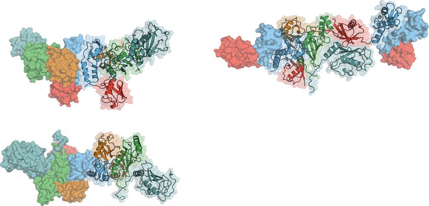
Fig. 6 TRIM protein assembly on viruses. Cartoon models of the assembly of TRIM5 (a, b) and TRIM21 (c, d) on viral capsids. a Shown is the hexagonal
assembly of TRIM5 on HIV-1 capsid as imaged by cryo-electron tomography48. c Assembly of TRIM21:antibody complexes on adenovirus capsid
(adenoviral measurements are based on 6B1T68). b, d Cartoons visualizing how the TRIM protein assembly on the viral capsid enables the formation of the
catalytic RING topology. R RING, B Box, CC coiled-coil, PS PRYSPRY, Ub ubiquitin.
150 mM NaCl, 10 µM ZnCl2, 1 mM DTT, 20% Bugbuster (Novagen), and cOm- Aldrich, St. Louis, USA)), a total concentration of 0.5% percloric adic was added to the
plete protease inhibitors (Roche, Switzerland). Lysis was performed by sonication. stirring lysate at 4 °C. The (milky) lysate was incubated for another 30 min on a stirrer
TRIM proteins and Ube2V2 were expressed with N-terminal GST-tag and purified at 4 °C to complete precipitation. Next, the lysate was centrifuged (50,000 × g) for
via glutathione sepharose resin (GE Healthcare) equilibrated in 50 mM Tris pH 8.0, 30 min at 4 °C. The supernatant was dialyzed overnight (3500 MWCO) against 3 L
150 mM NaCl, and 1 mM DTT. The tag was cleaved on beads overnight at 4 °C. 50 mM sodium acetate (NaOAc) pH 4.5. Afterward, Ub was purified via cation
In case of ubiquitin-TRIM21 constructs, the eluate was supplemented with 10 mM exchange chromatography using a 20 mL SP column (GE Healthcare) and using a NaCl
imidazole and run over 0.25 mL of Ni-NTA beads to remove His-tagged TEV. gradient (0–1000 mM NaCl in 50 mM NaOAc pH 4.5). Finally, size-exclusion
Ube2N and Ube1 were expressed with an N-terminal His-tag and were purified via chromatography was carried out on a HiLoad 26/60 Superdex 75 prep grade column
Ni-NTA resin. Proteins were eluted in 50 mM Tris pH 8.0, 150 mM NaCl, 1 mM (GE Healthcare) in 20 mM Tris pH 7.4.
DTT, and 300 mM imidazole. For Ube2N, TEV-cleavage of the His-tag was per- All proteins were flash frozen in small aliquots (30–100 µL) and stored at −80 °C.
formed overnight by dialyzing the sample against 50 mM Tris pH 8.0, 150 mM
NaCl, 1 mM DTT, and 20 mM imidazole. Afterward, His-tagged TEV protease was
Formation of an isopeptide-linked Ube2N~Ub. Ube2NC87K/K92A charging with
removed by Ni-NTA resin. The cleavage left an N-terminal tripeptide scar (GSH)
WT ubiquitin was performed as normal E1-mediated charging, but in a high pH to
on recombinantly expressed TRIM proteins, an N-terminal G scar on Ube2N and
ensure K87 deprotonation10,17. The isopeptide charging reaction was carried out in
an N-terminal GSQEF scar on Ube2V2. Finally, size-exclusion chromatography
50 mM Tris pH 10.0, 150 mM NaCl, 5 mM MgCl2, 0.5 mM TCEP, 3 mM ATP, 0.8
was carried out on a HiLoad 26/60 Superdex 75 prep grade column (GE Health-
µM Ube1, 100 µM Ube2N, and 130 µM ubiquitin at 37 °C for 4 h. After conjuga-
care) in 20 mM Tris pH 8.0, 150 mM NaCl, and 1 mM DTT.
tion, Ube2NC87K/K92A~Ub was purified by size-exclusion chromatography
Full-length TRIM21 (Ub-R-B-CC-PS or Ub-R-R-B-CC-PS) were expressed as
(Superdex S75 26/60, GE Healthcare) that was equilibrated in 20 mM Tris pH 8.0
His-Lipoyl-fusion proteins in E. coli BL21 DE3 cells. Cells in 2×TY were grown to
and 150 mM NaCl.
an OD600 of 0.8 and induced with 0.5 mM IPTG and 10 µM ZnCl2. Cells were
further incubated at 18 °C, 220 r.p.m. overnight. After centrifugation, cells were
resuspended in 100 mM Tris pH 8.0, 250 mM NaCl, 10 µM ZnCl2, 1 mM DTT, Crystallization. In total, 5 mg mL−1 of human UbG75/76A-TRIM211–85,
20% Bugbuster (Novagen), 20 mM imidazole, and cOmplete protease inhibitors Ube2NC87K/K92A~Ub, and Ube2V2 in 20 mM Tris pH 8.0, 150 mM NaCl, and 1
(Roche, Switzerland). Lysis was performed by sonication. His-affinity purification mM DTT were subjected to sparse matric screening in sitting drops at 17 °C (500
was performed as described above. Immediately afterward, the protein was applied nL protein was mixed with 500 nL reservoir solution). Crystals were obtained in
to an S200 26/60 column (equilibrated in 50 mM Tris pH 8.0, 200 mM NaCl, and Morpheus II screen58 in 0.1 M MOPSO/bis-tris pH 6.5, 12.5% (w/v) PEG 4 K, 20%
1 mM DTT) to remove soluble aggregates. After concentration determination, the (v/v) 1,2,6-hexanetriol, and 0.03 M of each Li, Na, and K.
His-Lipyol tag was cleaved using TEV protease overnight. Since full-length For the Ube2NC87K/K92A~Ub:Ube2V2 structure, 10 mg mL−1 TRIM211–85,
TRIM21 is unstable without tag, the protein was not further purified but used for Ube2NC87K/K92A~Ub, Ube2V2, and Ub in 20 mM Tris pH 8.0, 150 mM NaCl, and
assays. 1 mM DTT were subjected to sparse matrix screening in sitting drops at 17 °C (200
Ubiquitin purification was performed following the protocol established by the nL protein was mixed with 200 nL reservoir solution). Crystals were obtained in the
Pickart lab57. After cell lysis by sonication (lysis buffer: 50 mM Tris pH 7.4, 1 mg mL−1 Morpheus III screen59 in 0.1 M bicine/Trizma base pH 8.5, 12.5% (w/v) PEG 1000,
Lysozyme (by Sigma Aldrich, St. Louis, USA) and 0.1 mg mL−1 DNAse (by Sigma 12.5% (w/v) PEG 3350, 12.5% (v/v) MPD, and 0.2% (w/v) of each anesthetic
NATURE COMMUNICATIONS | (2021)12:1220 | https://doi.org/10.1038/s41467-021-21443-6 | www.nature.com/naturecommunications 9ARTICLE NATURE COMMUNICATIONS | https://doi.org/10.1038/s41467-021-21443-6
alkaloids (lidocaine HCl · H2O, procaine HCl, proparacaine HCl, and tetracaine determine the pKa, the data was fit to Eq. (2):
HCl). Crystals were flash frozen for data collection without the use of additional
cryo-protectant. VHA 10pH þ VA 10pKa
V¼ ð2Þ
10pKa þ 10pH
Crystal data collection, structure solution, and refinement. Data were collected as given in ref. 65, where V is the measured velocity, VA- the velocity for the basic
at the Diamond Light Source beamline i03, equipped with an Eiger2 XE 16 M detecter species, and VHA the velocity for the acidic species.
of a wavelength of 0.9762 Å. For UbG75/76A-TRIM21:1–85Ube2NC87K/K92A~Ub:
Ube2V2, diffraction images were processed using XDS60 to 2.2 Å resolution. The
crystals belong to space group number 5 (C2) with each of the components present as In vitro transcription and RNA purification. For in vitro transcription of mRNA,
a single copy in the asymmetric unit. Analysis of the raw data revealed moderate constructs were cloned into pGEMHE vectors56. Plasmids were linearized, using
anisotropy in the data. The structure was solved by molecular replacement using AscI. Capped (but not polyA-tailed) mRNA was synthesized with T7 polymerase,
PHASER-MR implemented in the Phenix suite61. Search models served TRIM21 using the HiScribe™ T7 ARCA mRNA Kit (New England Biolabs) according to the
RING and Ube2N from 6S53 (ref. 17), ubiquitin from 1UBQ62 and Ube2V2 from 1J74 manufacturer’s instructions.
(ref. 7). Model building and real-space refinement was carried out in coot63, and
refinement was performed using phenix-refine64. The anisotropy in the data could be Cell lines. NIH3T3-Caveolin-1-EGFP66 were cultured in DMEM medium (Gibco;
observed in parts of the map that were less well resolved. While all interfaces show 31966021) supplemented with 10% calf serum and penicillin–streptomycin. RPE-1
clear high-resolution density, particularly parts of Ube2V2 (chain A) that were next to cells (ATCC) were cultured in DMEM/F-12 medium (Gibco; 10565018) supple-
a solvent channel proved challenging to build. The structure is deposited in the mented with 10% calf serum and penicillin–streptomycin.
Protein Data Bank under the accession code 7BBD. All cells were grown at 37 °C in a 5% CO2 humidified atmosphere and regularly
For Ube2NC87K/K92A~Ub:Ube2V2, diffraction images were processed using checked to be mycoplasma free. The sex of NIH3T3 cells is male. The sex of RPE-1
XDS60 to 2.54 Å resolution. The crystals belong to space group number 145 (P32) cells is female. Following electroporation, cells were grown in medium
with each component present three times in the asymmetric unit, related by supplemented with 10% calf serum without antibiotics. For live imaging with the
translational non-crystallographic symmetry. The structure was solved by IncuCyte (Sartorius), cell culture medium was replaced with Fluorobrite (Gibco;
PHASER-MR implemented in the Phenix suite61. Search models used were Ube2N A1896701) supplemented with 10% calf serum and GlutaMAX (Gibco; 35050061).
from 6S53 (ref. 17), ubiquitin from 1UBQ62, and Ube2V2 from 1J74 (ref. 7). Model RPE-1 TRIM21 knockout cells were generated using the Alt-R CRISPR-
building and real-space refinement was carried out in coot63, and refinement was Cas9 system from Integrated DNA technologies (IDT) with a custom-designed
performed using phenix-refine64. The structure is deposited in the Protein Data crRNA sequence (ATGCTCACAGGCTCCACGAA). Guide RNA in the form of
Bank under the accession code 7BBF. crRNA-tracrRNA duplex was assembled with recombinant Cas9 protein (IDT
#1081060) and electroporated into RPE-1 cells together with Alt-R Cas9
Ubiquitin chain formation assay. Ubiquitin chain formation assays were per- Electroporation Enhancer (IDT #1075915). Two days post electroporation, cells
formed in 50 mM Tris pH 7.4, 150 mM NaCl, 2.5 mM MgCl2, and 0.5 mM DTT. were plated one cell per well in 96-well plates and single-cell clones screened by
The reaction components were 2 mM ATP, 0.25 µM Ube1, 80 µM ubiquitin, western blotting for TRIM21 protein. A single clone was chosen that contained no
0.5 µM Ube2N/Ube2V2, or Ube2D1 together with the indicated concentration of detectable TRIM21 protein and confirmed TRIM21 knockout phenotype in a
E3. Samples were taken at the time points indicated and the reaction was stopped Trim-Away assay.
by addition of LDS sample buffer at 4 °C. The samples were boiled at 90 °C for For the proteasome inhibition experiments, MG132 (Sigma; C2211) was used at
2 min and resolved by LDS–PAGE. Ubiquitin chains were detected in the western a final concentration of 25 µM.
blot using an anti-Ub-HRP (Santa Cruz, sc8017-HRP P4D1, 1:5000), TRIM21 by
rabbit anti-TRIM21PRYSPRY D101D ST#9204 (1:1000), and Fc by goat anti-human Transient protein expression from mRNA. To enable precise control of protein
IgG-Fc broad 5211–8004 (1:2000). expression levels, constructs were expressed from in vitro transcribed mRNA.
mRNA was delivered into cells by electroporation using the Neon Transfection
Kinetics of di-ubiquitin formation. Kinetic measurements of di-ubiquitin for- system (Invitrogen). For each electroporation reaction, 8 × 10⁵ RPE-1 TRIM21-
mation were measured for Michaelis–Menten and pKa analysis. The experiment knockout or NIH3T3-Caveolin-1-EGFP cells suspended in 10.5 µl of resuspension
was performed in a pulse-chase format, where the first reaction generated buffer R were mixed with 2 µL of the indicated mRNA in water. After electro-
Ube2N~His-Ub and was chased by Ub1–74. Under these conditions, Ub1–74 only poration, cells were transferred into antibiotic-free DMEM or DMEM/F-12 media
acts as acceptor, as it cannot be charged onto the E1 enzyme. His-tagged ubiquitin supplemented with 10% FBS and left to incubate for 5 h before cells were harvested.
on the other hand serves as donor. Although, theoretically His-Ub could also act as Typically, expression could be detected from 30 min after electroporation and
an acceptor, the high concentrations of Ub1–74 outcompete His-Ub as an acceptor. lasted for ~24 h.
Initially, we determined the linear range of the reaction for all different constructs,
so as to later measure only one point on this trajectory as a representative for the Trim-Away. For each electroporation reaction, 8 × 10⁵ NIH 3T3 Cav1-knock in
initial velocity (v0). For Michaelis–Menten kinetics, we used the following length: cells66 suspended in 10.5 µl of resuspension buffer R were mixed with the indicated
WT, 3 min; D119A, 100 min; D119N, 30 min; N123A, 3 min; D124A. 3 min, and amount of antibody mixture diluted in 2 µL of PBS. mRNAs were added imme-
for pKa measurements the following: WT, 40 s; D119A, 5 min; D119N, 60 s; diately prior to electroporation, to limit the degradation by potential RNAse
N123A, 40 s; D124A, 40 s. activity. Cav1-GFP mRNA encoding Vhh-Fc (WT or PRYSPRY binding-deficient
First, Ube2N charging was performed in 50 mM Tris pH 7.0, 150 mM NaCl, 20 H433A mutant) and TRIM21 were electroporated. The cell mRNA mixtures were
mM MgCl2, 3 mM ATP, 60 µM His-ubiquitin, 1 µM GST-Ube1 (Boston Biochem), taken up into 10 µL Neon electroporation pipette tips (Invitrogen) and electro-
and 40 µM Ube2N. The reaction was incubated at 37 °C for 12 min and stored porated using the following settings: 1400 V, 20 ms, 2 pulses (as described in
afterward at 4 °C until use (within 1 h). refs. 18,67). Electroporated cells were transferred to antibiotic-free Fluorobright
For Michaelis–Menten kinetic analysis, the reaction was conducted in 50 mM media supplemented with 10% FBS and left to incubate for 5 h in an incubator
Tris pH 7.4, 150 mM NaCl with the indicated amount of Ub1–74 (0–400 µM), while before the cells were harvested for immunoblotting. GFP fluorescence measured
for pKa determination in 50 mM Tris and the indicated pH (7.0–10.5), 50 mM using an Incucyte® (essenbioscience) and was normalized to the control (Vhh-
NaCl, and 250 mM Ub1–74. Apart from the buffer, the reaction mix contained 2.5 FcH433A). Protein detection was performed using the following antibodies: Fc: goat
µM Ube2V2. The reaction was initiated by addition of charging mix that was anti-hIgG Fc broad 5211–8004 (1:2000); TRIM21: rabbit anti-TRIM21 D101D
diluted 1 in 20, resulting in 2 µM Ube2N in the reaction. The reaction was stopped (ST#9204) (1:1000), Vinculin: rabbit anti-Vinculin EPR8185 ab 217171 (1:50,000);
by addition of 4× LDS loading buffer. The samples were boiled at 90 °C for 2 min and Caveolin-1: rabbit anti-Cav1 (BD: 610059, 1:1000).
and resolved by LDS–PAGE. Western blot was performed with anti-His antibody
(Clontech, 631212, 1:5000) via the LiCor system, leading to detection of the
mEGFP-Fc degradation assay. For mEGFP-Fc degradation assay, 0.4 µM
following species: His-Ub, His-Ub-Ub1–74, Ube2N~His-Ub, Ube2N~(His-Ub)2 (a
mEGFP-Fc mRNA together with 1.2 µM of the indicated TRIM21 mRNA were
side product of the charging reaction that shows ubiquitination rates similar to
electroporated into 8 × 10⁵ cells, as described above. Electroporated cells were
Ube2N~His-Ub), and E1-His-Ub. The concentration of His-Ub-Ub1–74 was
transferred to antibiotic-free DMEM supplemented with 10% FBS. For western
determined by dividing the value for His-Ub-Ub1–74 by the sum of all bands
analysis only, cells were incubated for 5 h in an incubator before harvest. For flow
detected and multiplying this by the total concentration of His-Ub in the reaction
cytometry analysis, the half of the cells were taken and treated with 25 µM MG132,
(3 µM). Experiments were performed in technical triplicates. Michaelis–Menten
while the other half were treated with DMSO. Then cells were incubated for 5 h in
kinetics data were fit to Eq. (1):
an incubator before being harvested. Cells were fixed before being subjected to flow
cytometry. The same antibodies were used as for Trim-Away (see above).
Et kcat S
V¼ ð1Þ
KM S Flow cytometry. Cells were fixed prior to flow cytometry. For this, cells were
resuspended in FACS fixative (4% formaldehyde, 2 mM EDTA in PBS) and
where V is the measured velocity, Et the total concentration of active sites (2 µM), incubated at room temperature for 30 min. Afterward, cells were centrifuged and
and S the substrate concentration. The curve was fit to determine kcat and KM. To resuspended in FACS buffer (2% FBS, 5 mM EDTA in PSB) and stored at 4 °C,
10 NATURE COMMUNICATIONS | (2021)12:1220 | https://doi.org/10.1038/s41467-021-21443-6 | www.nature.com/naturecommunicationsNATURE COMMUNICATIONS | https://doi.org/10.1038/s41467-021-21443-6 ARTICLE
wrapped in aluminum foil until use. Flow cytometry was performed using an 17. Kiss, L. et al. A tri-ionic anchor mechanism drives Ube2N-specific recruitment
Eclipse (iCyt) A02-0058. Cells were measured using forward and side scattering to and K63-chain ubiquitination in TRIM ligases. Nat. Commun. 10, 4502
assess live cells. In addition, green fluorescence was measured. Live cells were (2019).
selected based on forward and side scattering and only the median GFP fluores- 18. Clift, D. et al. A method for the acute and rapid degradation of endogenous
cence of live cells was used for further analysis (example data shown in Supple- proteins. Cell 171, 1692–1706.e1618 (2017).
mentary Fig. 12d). 19. Sakamoto, K. M. et al. Protacs: chimeric molecules that target proteins to the
Skp1-Cullin-F box complex for ubiquitination and degradation. Proc. Natl
Software. Figures were created using the following software: Graphpad Prism 7.0d, Acad. Sci. USA 98, 8554–8559 (2001).
Pymol 1.8.2.3, Adobe Illustrator v24.2.3, Adobe Photoshop CS6 13.0.6 ×64, Image 20. Fletcher, A. J. et al. Trivalent RING assembly on retroviral capsids activates
Studio Lite 5.2.5, and ImageJ/FIJI 2.0.0-rc-69/1.52p. For flow cytometry, we used TRIM5 ubiquitination and innate immune signaling. Cell Host Microbe 24,
ec800 v1.3.6, which is the operating software for the Eclipse (iCyt) A02-0058. For 761–775 e766 (2018).
cell imaging using the Incucyte, we used the default software Incucyte S3. For 21. Dickson, C. et al. Intracellular antibody signalling is regulated by
crystallography, we used XDS (Version 31. January 2019), Phenix 1.18.2_3874 and phosphorylation of the Fc receptor TRIM21. Elife 7, e32660 (2018).
1.14-3260 (in these versions we used the implemented programs: Phaser, Phe- 22. Plechanovova, A., Jaffray, E. G., Tatham, M. H., Naismith, J. H. & Hay, R. T.
nix_Refine), and Coot 0.8.9 and 0.9. Structure of a RING E3 ligase and ubiquitin-loaded E2 primed for catalysis.
Nature 489, 115–120 (2012).
Reporting summary. Further information on research design is available in the Nature 23. Dou, H., Buetow, L., Sibbet, G. J., Cameron, K. & Huang, D. T. BIRC7-E2
Research Reporting Summary linked to this article.
ubiquitin conjugate structure reveals the mechanism of ubiquitin transfer by a
RING dimer. Nat. Struct. Mol. Biol. 19, 876–883 (2012).
24. Pruneda, J. N. et al. Structure of an E3:E2~Ub complex reveals an allosteric
Data availability mechanism shared among RING/U-box ligases. Mol. Cell 47, 933–942 (2012).
The crystal structures of Ub-R:Ube2N~Ub:Ube2V2 and Ube2N~Ub:Ube2V2 are 25. Yunus, A. A. & Lima, C. D. Lysine activation and functional analysis of E2-
deposited in the Protein Data Bank under the accession codes 7BBD and 7BBF, mediated conjugation in the SUMO pathway. Nat. Struct. Mol. Biol. 13,
respectively. All other relevant data are available from the corresponding authors upon 491–499 (2006).
reasonable request. Source data are provided with this paper. 26. Lide, D. R. CRC Handbook of Chemistry and Physics: a Ready-reference Book
of Chemical and Phyical Data, Vol. 72 (CRC Press, 1991).
27. Llopis, J., McCaffery, J. M., Miyawaki, A., Farquhar, M. G. & Tsien, R. Y.
Received: 30 September 2020; Accepted: 26 January 2021; Measurement of cytosolic, mitochondrial, and Golgi pH in single living cells
with green fluorescent proteins. Proc. Natl Acad. Sci. USA 95, 6803–6808
(1998).
28. Hospenthal, M. K., Freund, S. M. & Komander, D. Assembly, analysis and
architecture of atypical ubiquitin chains. Nat. Struct. Mol. Biol. 20, 555–565
(2013).
References 29. Lange, O. F. et al. Recognition dynamics up to microseconds revealed from
1. Komander, D. & Rape, M. The ubiquitin code. Annu Rev. Biochem. 81, an RDC-derived ubiquitin ensemble in solution. Science 320, 1471–1475
203–229 (2012). (2008).
2. Baek, K. et al. NEDD8 nucleates a multivalent cullin-RING-UBE2D ubiquitin 30. James, L. C., Keeble, A. H., Khan, Z., Rhodes, D. A. & Trowsdale, J. Structural
ligation assembly. Nature 578, 461–466 (2020). basis for PRYSPRY-mediated tripartite motif (TRIM) protein function. Proc.
3. Streich, F. C. Jr & Lima, C. D. Capturing a substrate in an activated RING E3/ Natl Acad. Sci. USA 104, 6200–6205 (2007).
E2-SUMO complex. Nature 536, 304–308 (2016). 31. Koliopoulos, M. G., Esposito, D., Christodoulou, E., Taylor, I. A. & Rittinger,
4. Scott, D. C. et al. Structure of a RING E3 trapped in action reveals ligation K. Functional role of TRIM E3 ligase oligomerization and regulation of
mechanism for the ubiquitin-like protein NEDD8. Cell 157, 1671–1684 catalytic activity. EMBO J. 35, 1204–1218 (2016).
(2014). 32. Sanchez, J. G. et al. Mechanism of TRIM25 catalytic activation in the antiviral
5. Hofmann, R. M. & Pickart, C. M. Noncanonical MMS2-encoded ubiquitin- RIG-I pathway. Cell Rep. 16, 1315–1325 (2016).
conjugating enzyme functions in assembly of novel polyubiquitin chains for 33. Yudina, Z. et al. RING dimerization links higher-order assembly of
DNA repair. Cell 96, 645–653 (1999). TRIM5alpha to synthesis of K63-linked polyubiquitin. Cell Rep. 12, 788–797
6. VanDemark, A. P., Hofmann, R. M., Tsui, C., Pickart, C. M. & Wolberger, C. (2015).
Molecular insights into polyubiquitin chain assembly: crystal structure of the 34. Komander, D. et al. Molecular discrimination of structurally equivalent
Mms2/Ubc13 heterodimer. Cell 105, 711–720 (2001). Lys 63-linked and linear polyubiquitin chains. EMBO Rep. 10, 466–473
7. Moraes, T. F. et al. Crystal structure of the human ubiquitin conjugating (2009).
enzyme complex, hMms2-hUbc13. Nat. Struct. Biol. 8, 669–673 (2001). 35. Popovic, D., Vucic, D. & Dikic, I. Ubiquitination in disease pathogenesis and
8. McKenna, S. et al. An NMR-based model of the ubiquitin-bound human treatment. Nat. Med. 20, 1242–1253 (2014).
ubiquitin conjugation complex Mms2.Ubc13. The structural basis for lysine 63 36. Ito, T. et al. Identification of a primary target of thalidomide teratogenicity.
chain catalysis. J. Biol. Chem. 278, 13151–13158 (2003). Science 327, 1345–1350 (2010).
9. Eddins, M. J., Carlile, C. M., Gomez, K. M., Pickart, C. M. & Wolberger, C. 37. Winter, G. E. et al. DRUG DEVELOPMENT. Phthalimide conjugation as a
Mms2-Ubc13 covalently bound to ubiquitin reveals the structural basis of strategy for in vivo target protein degradation. Science 348, 1376–1381 (2015).
linkage-specific polyubiquitin chain formation. Nat. Struct. Mol. Biol. 13, 38. Bondeson, D. P. et al. Catalytic in vivo protein knockdown by small-molecule
915–920 (2006). PROTACs. Nat. Chem. Biol. 11, 611–617 (2015).
10. Branigan, E., Plechanovova, A., Jaffray, E. G., Naismith, J. H. & Hay, R. T. 39. Zengerle, M., Chan, K. H. & Ciulli, A. Selective small molecule induced
Structural basis for the RING-catalyzed synthesis of K63-linked ubiquitin degradation of the BET bromodomain protein BRD4. ACS Chem. Biol. 10,
chains. Nat. Struct. Mol. Biol. 22, 597–602 (2015). 1770–1777 (2015).
11. Christensen, D. E., Brzovic, P. S. & Klevit, R. E. E2-BRCA1 RING interactions 40. Verma, R., Mohl, D. & Deshaies, R. J. Harnessing the power of proteolysis for
dictate synthesis of mono- or specific polyubiquitin chain linkages. Nat. Struct. targeted protein inactivation. Mol. Cell 77, 446–460 (2020).
Mol. Biol. 14, 941–948 (2007). 41. Schapira, M., Calabrese, M. F., Bullock, A. N. & Crews, C. M. Targeted protein
12. Fletcher, A. J. et al. TRIM5alpha requires Ube2W to anchor Lys63-linked degradation: expanding the toolbox. Nat. Rev. Drug Discov. 18, 949–963
ubiquitin chains and restrict reverse transcription. EMBO J. 34, 2078–2095 (2019).
(2015). 42. Fuseya, Y. et al. The HOIL-1L ligase modulates immune signalling and cell
13. Fletcher, A. J., Mallery, D. L., Watkinson, R. E., Dickson, C. F. & James, L. C. death via monoubiquitination of LUBAC. Nat. Cell Biol. 22, 663–673 (2020).
Sequential ubiquitination and deubiquitination enzymes synchronize the dual 43. Smith, B. E. et al. Differential PROTAC substrate specificity dictated by
sensor and effector functions of TRIM21. Proc. Natl Acad. Sci. USA 112, orientation of recruited E3 ligase. Nat. Commun. 10, 131 (2019).
10014–10019 (2015). 44. Ohtake, F., Tsuchiya, H., Saeki, Y. & Tanaka, K. K63 ubiquitylation triggers
14. Marin, I. Origin and diversification of TRIM ubiquitin ligases. PLoS ONE 7, proteasomal degradation by seeding branched ubiquitin chains. Proc. Natl
e50030 (2012). Acad. Sci. USA 115, E1401–E1408 (2018).
15. Mallery, D. L. et al. Antibodies mediate intracellular immunity through 45. Ohtake, F., Saeki, Y., Ishido, S., Kanno, J. & Tanaka, K. The K48-K63
tripartite motif-containing 21 (TRIM21). Proc. Natl. Acad. Sci. USA 107, branched ubiquitin chain regulates NF-kappaB Signaling. Mol. Cell 64,
19985–19990 (2010). 251–266 (2016).
16. McEwan, W. A. et al. Intracellular antibody-bound pathogens stimulate 46. Ganser-Pornillos, B. K. et al. Hexagonal assembly of a restricting TRIM5alpha
immune signaling via the Fc receptor TRIM21. Nat. Immunol. 14, 327–336 protein. Proc. Natl Acad. Sci. USA 108, 534–539 (2011).
(2013).
NATURE COMMUNICATIONS | (2021)12:1220 | https://doi.org/10.1038/s41467-021-21443-6 | www.nature.com/naturecommunications 11You can also read