Cell biology of diabetic nephropathy: Roles of endothelial cells, tubulointerstitial cells and podocytes
←
→
Page content transcription
If your browser does not render page correctly, please read the page content below
REVIEW ARTICLE

Cell biology of diabetic nephropathy: Roles
of endothelial cells, tubulointerstitial cells
and podocytes
Yoshiro Maezawa1,2, Minoru Takemoto1,2, Koutaro Yokote1,2*
1
 Department of Clinical Cell Biology and Medicine, Chiba University Graduate School of Medicine and 2Division of Diabetes, Metabolism and Endocrinology, Chiba University
Hospital, Chiba, Japan

Keywords                                                                ABSTRACT
Cell type specific, Conditional                                          Diabetic nephropathy is the major cause of end-stage renal failure throughout the world
targeting, Diabetic nephropathy                                         in both developed and developing countries. Diabetes affects all cell types of the kidney,
                                                                        including endothelial cells, tubulointerstitial cells, podocytes and mesangial cells. During
*Correspondence                                                         the past decade, the importance of podocyte injury in the formation and progression of
Koutaro Yokote                                                          diabetic nephropathy has been established and emphasized. However, recent findings
Tel.: +81-43-222-7171                                                   provide additional perspectives on pathogenesis of diabetic nephropathy. Glomerular
Fax: +81-43-226-2095
                                                                        endothelial damage is already present in the normoalbuminuric stage of the disease
E-mail address: kyokote@faculty.
                                                                        when podocyte injury starts. Genetic targeting of mice that cause endothelial injury leads
chiba-u.jp
                                                                        to accelerated diabetic nephropathy. Tubulointerstitial damage, previously considered to
J Diabetes Invest 2015; 6: 3–15                                         be a secondary effect of glomerular protein leakage, was shown to have a primary signifi-
                                                                        cance in the progression of diabetic nephropathy. Emerging evidence suggests that the
doi: 10.1111/jdi.12255                                                  glomerular filtration barrier and tubulointerstitial compartment is a composite, dynamic
                                                                        entity where any injury of one cell type spreads to other cell types, and leads to the
                                                                        dysfunction of the whole apparatus. Accumulation of novel knowledge would provide a
                                                                        better understanding of the pathogenesis of diabetic nephropathy, and might lead to a
                                                                        development of a new therapeutic strategy for the disease.

INTRODUCTION                                                                                                          pathway activation, renin–angiotensin system activation, reac-
Diabetic nephropathy is a potentially fatal diabetic                                                                  tive oxygen species (ROS), activation of the protein kinase C
vascular complication characterized by slowly increasing                                                              pathway, increase of advanced glycation end-product (AGE)
proteinuria and a gradual decrease in renal function.                                                                 and glomerular hyperfiltration2,3. These changes lead to various
Diabetic nephropathy is the leading cause of end-stage                                                                cellular responses, expression of secretory factors and extracellu-
renal failure in Japan and Western countries, affecting                                                               lar matrices that ultimately result in disruption of the glomeru-
alarmingly large numbers of people worldwide1. In Japan,                                                              lar filtration barrier, and histological changes including
diabetic nephropathy accounts for 44% of newly-induced                                                                mesangial expansion, nodular glomerular sclerosis and tubulo-
hemodialysis, and a governmental survey estimated that                                                                interstitial fibrosis (Figure 1)4.
there are approximately 100,000 hemodialysis patients.                                                                   During the past decade, ‘podocentric’ experiments have
The number of patients is increasing rapidly, making                                                                  accumulated a huge amount of knowledge on the roles of
diabetic nephropathy a critical social and economical                                                                 podocytes in diabetic nephropathy. In contrast, recent findings
problem.                                                                                                              have provided an additional perspective that other cell types
   The pathogenesis of diabetic nephropathy has been intensely                                                        are also affected at very early stages of diabetic nephropathy
investigated, and the roles of various mechanisms has been                                                            and contribute to the progression of the disease. To emphasize
established, and include the effect of high glucose, polyol                                                           this concept, the current review will highlight the emerging
                                                                                                                      roles of glomerular endothelial cells and tubulointerstitial cells
                                                                                                                      in the pathogenesis of diabetic nephropathy, and juxtapose
Received 8 May 2014; accepted 15 May 2014                                                                             them to the roles of podocytes.

ª 2014 The Authors. Journal of Diabetes Investigation published by Asian Association of the Study of Diabetes (AASD) and Wiley Publishing Asia Pty Ltd      J Diabetes Invest Vol. 6 No. 1 January 2015   3
This is an open access article under the terms of the Creative Commons Attribution-NonCommercial License, which permits use, distribution and
reproduction in any medium, provided the original work is properly cited and is not used for commercial purposes.
REVIEW ARTICLE
Maezawa et al. http://onlinelibrary.wiley.com/journal/jdi
Non-cell type specific
Polyol pathway Hyperglycemia RAA
AGEs DAG/PKC
ROS Hyperfiltration
Tgfβ Matrix production Inflammation
Podocytes
Endothelial cells
Tubular/interstitial
Vegf Angpt 1 cells
Angpt 2
Glycocalyx Insulin Hypoxia
NO mTOR
signaling
Inflammation
Reduced fenestration Detachment
Reduced charge barrier apoptosis Fibrosis
Figure 1 | Schematic diagram on pathogenesis of diabetic nephropathy. In diabetic conditions, high glucose, activation of the polyol pathway,
glomerular hyperfiltration, activation of the renin–angiotensin–aldosterone system (RAA), increase of advanced glycation end-product (AGE),
increased reactive oxygen species (ROS), activation of diacylglycerol (DAG)/protein kinase C (PKC) pathway and an increase in Tgfb lead to
abnormal cellular responses, such as overproduction of extracellular matrices and inflammation in the kidney. Diabetes also affects the production
of nitric oxide (NO), Angpt2 and glycocalyx in the glomerular endothelial cells, and leads to endothelial injury. In podocytes, reduced vascular
endothelial growth factor (VEGF) A expression increased mechanistic target of rapamycin (mTOR) signaling, and insulin resistance leads to podocyte
dysfunction resulting in podocyte detachment and apoptosis. Furthermore, recent findings show that tubulointerstitial fibrosis also plays significant
roles. These cell types are tightly connected together, and dysfunctions in one compartment can spread to other cell types.
GLOMERULAR ENDOTHELIAL CELLS albumin without detectable change of the ultrastructure9. In
Glomerular endothelial cells are highly fenestrated, with 50- to disease models, chronic infusion of hyaluronidase causes pro-
80-nm pores that go through the cytoplasm5. The luminal sur- teinuria in apolipoprotein E (ApoE) knockout mice10, and Adri-
face of endothelial cells is covered by a thick layer of glycocalyx amycin injection largely decreases the thickness of the
that includes proteoglycans (PGs), such as syndecan, glypican, glycocalyx and proteoglycan expression in the glomeruli11. These
perlecan and versican, as well as their glycosaminoglycan experiments provide strong evidence that the endothelial glyco-
(GAG) side chains, heparan sulfate and chondroitin sulfate6. calyx forms a significant part of the glomerular filtration barrier.
Endothelial GAGs maintain the negative charge of the endothe-
lial glycocalyx, and are believed to be a significant component Role of Endothelial Cells in Diabetic Nephropathy
of the glomerular charge barrier6. Several diabetic animal models show reduced endothelial glyco-
calyx. In non-obese diabetic (NOD) mice, long-term diabetes
Endothelial Cells are a Significant Part of the Glomerular causes a threefold increase in the fractional clearance for nega-
Filtration Barrier tively charged albumin compared with controls. In contrast, the
Because of the huge size of fenestration compared with that of fractional clearance for neutral Ficoll that has a similar size to
circulating proteins, such as albumin, it has been believed that albumin is not increased. This change is accompanied by a
glomerular endothelial cells do not contribute to the filtration decrease in glycocalyx proteins including versican and deco-
of macromolecules. However, emerging studies show its impor- rin12. Therefore, the authors concluded that the charge barrier,
tance in the glomerular filtration barrier. Digestion of GAGs rather than the size barrier, is affected in NOD mice glomeruli.
with heparanase, chondroitinase and hyaluronidase decrease the Disruption of the charge barrier before that of the size barrier
thickness of the glycocalyx layer, and the negative charge den- might show that endothelial damage occurs before podocyte/
sity of the glomerular filtration barrier, resulting in the increase glomerular basement membrane (GBM) damage in NOD mice.
of the fractional clearance of albumin7,8. In addition, elusion of In addition, staining for lectin that binds to hyaluronic acid
non-covalently bound components of the endothelial glycocalyx and heparin sulfates of the endothelial glycocalyx is attenuated
by infusion of hypersonic sodium chloride into the renal artery in streptozotocin (STZ)-induced diabetic rats13 and Zucker fatty
leads to a 12-fold increase in the fractional clearance of rats14.
4 J Diabetes Invest Vol. 6 No. 1 January 2015 ª 2014 The Authors. Journal of Diabetes Investigation published by AASD and Wiley Publishing Asia Pty LtdREVIEW ARTICLE
http://onlinelibrary.wiley.com/journal/jdi Cell biology of diabetic nephropathy
How about human beings? It has long been known that
(a)
endothelial injury, assessed by elevation of plasma von Wille-
brand factor, is seen in both type 1 and type 2 diabetic
patients15,16. An extensive study using state-of-the-art technol-
ogy showed that the volume of endothelial glycocalyx in the
total body and the thickness of the glycocalyx layer in retinal
and sublingual arteries are reduced in type 1 diabetic patients.
In that study, the decrease of endothelial glycocalyx was more
severe in the group with albuminuria17. A similar reduction of
the endothelial glycocalyx was also observed in type 2 diabetic
patients18. In addition, very important studies have shown mor-
phological abnormalities of renal glomerular endothelium in
diabetic patients. Toyoda et al.19 showed that the fraction of
fenestrated endothelium is reduced from 41% in controls to
32% in normo- and microalbuminuric patients, and further to
25% in macroalbuminuric patients with type 1 diabetes.
Recently, Weil et al.20 reported a similar decrease of fenestrated
area in type 2 diabetes patients (Figure 2). Interestingly, podo-
cyte detachment also starts in the normoalbuminuric stage and
correlates to albuminuria20. In that study, the endothelial fenes-
tration fraction correlated with the urinary albumin creatinine
ratio more closely compared with podocyte detachment.
Although podocyte detachment has been considered to be a
critical event in various types of glomerular diseases, including
diabetic nephropathy21, these studies clearly showed that endo- (b)
thelial damage simultaneously occurs when podocytes are dam-
aged. This notion could raise a question against the concept
that podocyte injury is the primary event and endothelial dam-
age occurs as a secondary reaction22,23. These findings rather
support the concept that the glomerular filtration barrier is a
composite multilayered structure, and injury in one layer might
spread to any other layers and affect the whole function of the
glomerular filtration barrier.
Endothelial Dysfunction and Diabetic Nephropathy: Lessons
from Genetically Targeted Mice
Various cytokines and factors are secreted by/have effects on
endothelial cells to maintain the glomerular filtration barrier.
Emerging evidence using genetically targeted mice prove their
importance in diabetic nephropathy.
Nitric Oxide
In diabetic animals and humans, bioavailability of the nitric
oxide (NO) is reduced24. In cultured endothelial cells, high glu-
cose inhibits endothelial NO synthase (eNOS) activity25. How-
ever, in type 2 diabetic patients, eNOS expression is increased
in kidney glomeruli by immunohistochemistry, and the increase
was correlated with more severe vascular complications26. Simi- Figure 2 | Endothelial injury is already present in the
larly, protein expression of eNOS in STZ-induced diabetic rats normoalbuminuric stage. (a) The ultrastructure of glomerular filtration
is increased in afferent arterioles and glomeruli27. In contrast, barrier from a normal subject, note intact endothelial fenestration. (b)
eNOS required cofactors including tetrahydrobiopterin (BH4) Sample from a normoalbuminuric Pima Indian patient with type 2
to produce NO. If eNOS is ‘uncoupled’, peroxynitrite diabetes showing intact podocyte foot processes, but normal
(ONOO–), a putative reactive oxygen species, is produced endothelial fenestrae are absent (arrows; adapted from Weil et al.20
instead of NO. In endothelial cells, high glucose results in a with permission).
ª 2014 The Authors. Journal of Diabetes Investigation published by AASD and Wiley Publishing Asia Pty Ltd J Diabetes Invest Vol. 6 No. 1 January 2015 5REVIEW ARTICLE
Maezawa et al. http://onlinelibrary.wiley.com/journal/jdi
uncoupling of eNOS, reduction of NO production and patients by immunostaining. In contrast, Baelde et al.43 showed
increased superoxide production28. This uncoupling might reduction of Vegfa messenger ribonucleic acid expression in
explain the discrepancy between elevated expression of eNOS human type 2 diabetic glomeruli by Affymetrix microarray43.
and reduced NO production in the diabetic condition. Several additional reports showed that Vegfa expression
The requirement of eNOS in the glomerular endothelium in was decreased in both the glomeruli and tubulointerstitium, and
diabetes was examined using two diabetic animal models com- was correlated with reduced renal microvascular density, tubular
bined with genetic deletion of eNOS. Both db/db and STZ mice epithelial atrophy, mesangial expansion and proteinuria44,45.
bred with eNOS knockout mice showed dramatic albuminuria, These results rather support the notion that VEGF is protective.
increased glomerular basement membrane thickness, mesangial Recently, two genetic mice models shed a new light onto this
expansion, and focal segmental and nodular sclerosis29,30. The controversy. Veron et al.46 produced a mouse model that car-
potential mechanism of the enhanced nephropathy was an ries podocyte-specific inducible overexpression of Vegf164, and
uncoupling of the vascular endothelial growth factor A (VEG- rendered the mice diabetic by STZ injection. The results
FA)–eNOS axis, with enhanced VEGF expression and impaired showed accelerated nephropathy with Kimmelstiel–Wilson like
NO production, which led to excessive endothelial proliferation. nodular glomerulosclerosis and massive proteinuria46. In con-
These phenotypes were at least partially mediated by intraglom- trast, Sivaskandarajah et al.47 showed that inducible deletion of
erular hypertension, because lowering blood pressure rescues VEGFA in adult podocytes results in severe enhancement of
the glomerular lesion in the diabetic eNOS deficient mice31. diabetic nephropathy using a STZ-induced mice model. In that
These results provide robust evidence that endothelial dysfunc- report, the diabetic mutant showed severe glomerulosclerosis,
tion results in enhancement of diabetic nephropathy and enhanced proteinuria and increased apoptosis in the kidneys47.
suggest NO as a potential therapeutic target. These results clearly show that the levels of VEGFA in the
glomeruli require ‘fine tuning’, and either overdose or suppres-
VEGFA sion could result in exacerbation of diabetic nephropathy. This
A large amount of VEGFA is produced by podocytes. The tight and subtle regulation is similar to that of VEGFA in
secreted VEGFA go across the GBM and bind to kinase insert glomerular development.
domain protein receptor (Kdr; also known as VEGF receptor 2)
expressed on the endothelial cells. The VEGFA–Kdr axis is Angiopoietins
essential for the formation and maintenance of the glomerular Another family of angiogenic factors required for maintenance
filtration barrier32,33. Podocyte-specific deletion of VEGFA leads of glomerular endothelial cells is angiopoietin–Tek signaling.
to impaired recruitment of endothelial cells into glomeruli, fail- Angiopoietin 1 (Angpt1) and angiopoietin 2 (Angpt2) are both
ure in formation of glomerular filtration barrier and congenital ligands to Tek tyrosine kinase (Tek/Tie-2)48,49. Angiopoietin 1
nephrotic syndrome33. Mice that carry haploinsufficient VEG- binds to the Tek receptor expressed on endothelial cells, and
FA allele in podocytes show an endothelial swelling and loss of cause its tyrosine phosphorylation, but Angpt2 works as an
fenestration in glomeruli known as endotheliosis – a feature antagonist while not activating any intracellular signaling. How-
seen in thrombotic microangiopathies (TMA)33. Overexpression ever, some data suggest that Angpt2 activates phosphorylation
of VEGFA in podocytes leads to a collapse of the glomerular of Tek in certain conditions50. Angpt1 is believed to stabilize
tuft33. In addition, patients on anti-VEGF therapy sometimes the blood vessel, reduce vascular permeability and support the
develop proteinuria as a result of TMA of the glomeruli34. survival of endothelial cells. Angpt1 conventional knockout
Indeed, deletion of VEGFA alleles from adult mice podocytes mice die at embryonic day 12.549 and Angpt2 knockout is also
results in TMA34. perinatal lethal51.
The role of VEGFA in diabetic nephropathy has been a con- Recent analysis using conditional alleles showed critical roles
troversy. An increase of VEGFA expression was shown in the of Angpt1 in glomeruli. Jeansson et al. showed that deletion of
glomeruli and tubulointerstitium in STZ diabetic rats and in Angpt1 at embryonic day 10.5 resulted in abnormal glomerular
type 2 diabetic mice35–37. As VEGFA is a potent stimulator that development with a single capillary loop without mesangial
destabilizes endothelial cells and induces vascular permeability, migration that is reminiscent to the phenotype of Pdgfb/Pdgfrb
some investigators attempted to block VEGFA signaling to treat mutants52,53. In contrast, loss of the Angpt1 allele at e13.5 does
diabetic nephropathy in mice38,39. In db/db mice, an inhibitor not cause any phenotype, showing that Angpt1 is only required
for tyrosine kinase of Kdr reduced urinary albumin excretion when the vasculature is undergoing dynamic remodeling.
(UAE)40,41. In Zucker diabetic rats, the neutralizing antibody The role of angiopoietins in diabetes has been shown by sev-
for VEGF reduced glomerular hypertrophy, but did not eral reports. In diabetic patients, Angpt2 expression is
improve UAE40. Therefore, these reports appear to support the increased54. On the other hand, diabetic animal models show a
notion that VEGF worsens diabetic nephropathy. decrease of Angpt1 and increase of Angpt2. Furthermore, STZ-
However, reports on VEGFA expression in human diabetic induced diabetic mice with whole-body or glomerular-specific
nephropathy are inconsistent. Hohenstein et al.42 reported Angpt1 deletion develop increased urinary albumin excretion,
enhanced VEGFA expression in glomeruli of type 2 diabetes severe mesangial expansion, glomerular sclerosis and early
6 J Diabetes Invest Vol. 6 No. 1 January 2015 ª 2014 The Authors. Journal of Diabetes Investigation published by AASD and Wiley Publishing Asia Pty LtdREVIEW ARTICLE
http://onlinelibrary.wiley.com/journal/jdi Cell biology of diabetic nephropathy
mortality53. A study using db/db mice showed that treatment diabetic children showed that urinary N-acetyl-b-D-glucosa-
with recombinant adenovirus-expressing cartilage oligomeric minidase (NAG) is increased in diabetes patients compared
matrix protein (COMP)-Ang-1, a potent Angpt-1 variant, with normal controls, and is correlated with urinary albumin
resulted in improvement in diabetic renal damage55. Recently, excretion and glycemic control. This increase was already seen
STZ diabetic mice with podocyte-specific overexpression of in the microalbuminuric stage68,69. Intriguingly, within microal-
Angpt1 also showed a similar protective effect on diabetic buminuric type 1 diabetic patients, a group that shows low lev-
nephropathy56. Taken together, the Angpt1–Tek axis plays an els of urinary NAG and kidney injury molecule 1 (KIM-1)
important protective role in glomerular endothelial cell function tends to show regression of albuminuria after 2 years, suggest-
in the diabetic condition. Drugs that target Angpt1 and its ing that tubular dysfunction is a critical component of the early
downstream molecules might provide potentially useful thera- course of diabetic nephropathy69.
peutic strategies.
Does Tubulointerstitial Change Affect Glomerular Structure
TUBULOINTERSTITIAL CELLS and Function–Tubuloglomerular Feedback?
Although it has been widely accepted that glomerular injury is It has been considered that the glomeruli is the primary site of
the main component of diabetic nephropathy, plenty of evi- injury, and the tubulointerstitial change is a secondary reaction
dence has shown that tubulointerstitial changes are present and to elevated intratubular protein as a result of glomerular leak-
are involved in its progression57. Tubulointerstitium includes age. Recent studies suggested that tubular changes could lead to
the tubular system, interstitial cells and vascular system, and alteration in glomerular structure and function.
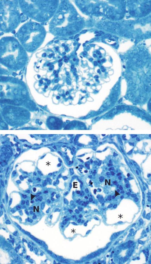
accounts for as much as 90% of kidney volume58. Using genetically modified mice, VEGFA was overexpressed
in renal tubular cells on administration of doxycyline. The
Histological Abnormality of Tubulointerstitium Correlates with mutant mice showed an interstitial fibrosis and tubular cysts
Progression of Diabetic Nephropathy with dense network of peritubular capillaries. The mice did not
Histologically, early diabetic kidneys show tubular hypertrophy, show significant proteinuria; however, they developed glomeru-
thickening of the tubular basement membrane and interstitial lar changes including marked mesangial expansion similar to
inflammation with mononuclear cell infiltration59. When it pro- that seen in diabetic nephropathy. The authors observed down-
gresses, they show tubular atrophy and tubulointerstitial fibrosis. regulation of VEGFA expression in podocytes, and concluded
Interestingly, tubulointerstitial expansion closely correlates with that tubular overproduction of VEGFA resulted in suppression
elevation of serum creatinine in both type 1 and type 2 diabe- of VEGFA in podocytes that led to interference of cross-talk
tes60–62. Taft et al.63 followed 47 patients with diabetes for between podocytes and the endocapillary compartment
4 years, and carried out renal biopsies at the beginning and the (Figure 3)70.
end of the study. The results showed that cortical tubulointersti- Another study also provides evidence for tubuloglomerular
tial fibrosis more closely correlated to the decrease of creatinine feedback. Deletion of sirtuin 1 (Sirt1), a NAD+ regulated
clearance than glomerular changes63. This association seems to deacetylase, specifically in tubules leads to increased albumin-
be weaker in the microalbuminuric stage64, suggesting that tub- uria and effacement of podocyte foot processes. Additionally,
ulointerstitial fibrosis is a good determinant of moderate-to- STZ-induced diabetes aggravated the nephropathy of tubule-
severe renal injury rather than early diabetic nephropathy59. specific Sirt1 knockout mice. Furthermore, overexpression of
In addition, abnormalities in glomerulotubular junctions have Sirt1 in tubules protected the mice from the glomerular ultra-
been reported in diabetic kidneys. A study on renal biopsy structural changes and albuminuria in STZ mice. In human
samples of type 1 diabetes patients showed that 4% of glome- diabetic nephropathy with proteinuria, Sirt1 was downregulated
ruli from microalbuminuric patients show some glomerulotubu- and Claudin1 was upregulated. These results suggest that Sirt1
lar junctional abnormalities, including connections of glomeruli in proximal tubules protects against diabetic nephropathy and
to atrophic tubules. Furthermore, in the macroalbuminuric influence podocyte function71. Taken together, these experi-
stage, 71% of glomeruli showed glomerulotubular junction ments clearly show that tubulointerstitial change can affect glo-
abnormalities, including 8% glomeruli without any tubule con- merular structure and function.
nected (atubular glomeruli)65,66. Type 2 diabetic glomeruli also
show atubular glomeruli and connection to atrophic tubules Epithelial to Mesenchymal Transformation in Diabetic
from the stage of microalbuminuria67. These data suggest that, Nephropathy
even though tubulointerstitial changes are dominantly seen in Epithelial to mesenchymal transformation (EMT) is a cellular
the advanced stage, subtle abnormality is already present in the process through which epithelial cells undergo ‘de-differentia-
microalbuminuric time-point. tion’; lose epithelial characteristics; express mesenchymal mark-
ers, such as alpha-smooth muscle actin, desmin and vimentin;
Increase of Tubular Markers in Urine of Early Diabetic Patients and acquire a matrix-producing fibroblast-like phenotype. A
Data of urinary biomarker also show that tubular damage starts study using genetically tagged proximal tubules showed that up
at an early stage of diabetic nephropathy. Studies on type 1 to 36% of the interstitial fibroblasts were derived from proximal
ª 2014 The Authors. Journal of Diabetes Investigation published by AASD and Wiley Publishing Asia Pty Ltd J Diabetes Invest Vol. 6 No. 1 January 2015 7REVIEW ARTICLE
Maezawa et al. http://onlinelibrary.wiley.com/journal/jdi
(a) overexpression of transforming growth factor beta (Tgfb) in
tubular cells, a multifunctional cytokine that is believed to be
the key mediator of EMT, resulted in total decomposition of
tubular cells and fibrosis, but not mesenchymal transformation
of tubular cells78. Therefore, there is no solid evidence that
tubular cells transform into matrix-producing interstitial fibro-
blasts in diabetic nephropathy. In terms of endothelial to mes-
enchymal transformation, Li et al.79 carried out genetic tag
experiments using Tie2-Cre mice, and showed that infusion of
AGE to mice resulted in fibroblasts derived from endothelial
cells79.
PODOCYTES
Morphology of Podocytes
Podocytes are terminally differentiated cells that wrap the glo-
merular capillary loops from outside, providing physical sup-
port and secretary cytokines. They develop microtubule-based
thick primary foot processes and fine actin-based secondary
(b) foot processes that interdigitate each other. The foot processes
from the neighboring podocytes are connected with each other
by a specialized intercellular junction called a slit diaphragm
(SD). SD consists of proteins including nephrin, podocin, Cd2-
ap and actin-binding protein alpha-actinin 4. The mutations of
these proteins cause focal and segmental glomerulosclerosis in
mice and humans, thus SD is considered to be essential in
macromolecular filtration80.
Diabetic Nephropathy and Podocytes
The early key events in diabetic nephropathy include loss of
podocytes in glomeruli. A landmark study was carried out by
Pagtalunan et al.81, and showed that the number of podocytes
per glomeruli were reduced in renal biopsies from Pima Indian
type 2 diabetes patients with macroproteinuria. The loss of
podocytes was correlated with the flattening of podocyte
foot processes81. Subsequent analysis showed that the decrease
of podocyte number is a good predictor of progression of
Figure 3 | Genetic overexpression of vascular endothelial growth albuminuria82. Similar findings are also reported on the
factor A (VEGFA) in tubules results in glomerular disease. (a) A nephropathy of type 1 diabetic patients19,83.
glomerulus from a control mouse. (b) A glomerulus from a tubular- The mechanism of podocyte loss is considered to be detach-
specific VEGFA overexpression mouse that shows dilated capillaries at ment and apoptosis. Podocyte apoptosis is observed in many
the periphery and nodule-like mesangial expansion, showing that animal diabetic models including Akita mice, db/db84 mice and
tubular dysfunction results in glomerular disease (reproduced from
STZ induced rats85, and could at least partially be mediated by
Hakroush et al.70 with permission).
an increase of Tgfb, Smad786, AGE87, angiotensin II88 and
reactive oxygen species84. In human type 2 diabetic kidneys,
tubules in a unilateral ureter obstruction mouse model72. apoptosis was seen in both tubular and glomerular compart-
A recent analysis reported that 35% of the myofibroblasts were ments, some was obviously in podocytes89. In contrast, under
derived from bone marrow, 10% from endothelial cells and diabetic conditions, some podocytes detach from the GBM
5% from tubular cells through an EMT program in a unilateral and fall into the urine. The detached ‘urinary’ podocytes are
ureter obstruction model73,74. In diabetic nephropathy, EMT- viable and can propagate ex vivo90. Indeed, patients of type 2
like changes of the tubules, such as upregulation of vimentin diabetes show urinary excretion of podocytes, and it worsens
and decrease of E-cadherin, have been observed both in vitro as the disease progresses from normoalbiminuria, microalbu-
and in vivo75,76. In addition, tubular expressions of mesenchy- minuria and to macroalbuminuria91. Interestingly, the urinary
mal markers are also shown in renal biopsy samples from podocyte detachment can be reduced by administration of
human diabetic nephropathy77. In contrast, however, hydroxymethylglutaryl-CoA (HMG-CoA) reductase inhibitors92.
8 J Diabetes Invest Vol. 6 No. 1 January 2015 ª 2014 The Authors. Journal of Diabetes Investigation published by AASD and Wiley Publishing Asia Pty LtdREVIEW ARTICLE
http://onlinelibrary.wiley.com/journal/jdi Cell biology of diabetic nephropathy
Factors Involved in Podocyte Function in Diabetic transplant patients using rapamycin sometimes develop
Nephropathy proteinuria, suggesting the importance of mTor signaling in
Proteins of Slit Diaphragm glomeruli108. Indeed, mice studies showed that the mTor path-
Nephrin is a 180-kDa transmembrane protein that belongs to way is essential for podocyte function. Deletion of mTor or
the immunoglobulin superfamily. In 1998, Karl Triggvasson Rptor specifically from podocytes leads to massive proteinuria
et al.93 group identified the nephrin (NPHS1) gene, and showed and renal failure110,111.
that its mutations caused the congenital nephrotic syndrome of In contrast, mTor activity was increased in renal biopsy
the Finnish type. The nephrin cytoplasmic tail includes three samples of diabetic patients and in diabetic db/db mice111,112.
tyrosine-aspartic acid-x-valine (YDxV) residues. These residues Activation of mTORC1 by deletion of its suppressor, Tsc1, in
are phosphorylated by Src family kinases, recruit SH2/SH3 con- podocytes results in proteinuria, podocyte loss, mesangial
taining Nck adaptor proteins resulting in reorganization of expansion and thickening of glomerular basement mem-
actin cytoskelton. Deletion of Nck1/2 in podocytes leads to brane112. Furthermore, haploinsufficiency of Rptor protected
massive congenital proteinuria94,95. Many reports have shown mice from diabetic nephropathy in STZ mice and db/db mice,
downregulation of nephrin in diabetic nephropathy in rodent showing a decrease in urinary albumin excretion and mesangial
models and human patients96–98. Some reports suggest that expansion111,112. These results show the critical roles of mTor
the decrease of nephrin is partially mediated by the renin– signaling in diabetic nephropathy, and that either overactivation
angiotensin system, AGE and protein kinase C pathway98–101. or elimination of the mTor pathway leads to podocyte dysfunc-
In contrast, there are also several reports showing upregulation tion and glomerular disease. Recent analysis showed that podo-
of nephrin expression in mice diabetic kidneys41,102. The cause cyte-specific ablation of Rictor or Akt2, a potent target of
of this diversity is still unknown, but could be a difference of mTORC2, results in accelerated nephropathy induced by
disease stages. In addition, other slit diaphragm proteins, such heminephrectomy, showing that mTORC2 is also involved in
as podocin and synaptpodin, are also significantly reduced in progression of chronic kidney disease (CKD)113.
human diabetic nephropathy103.
Notch Signaling
Insulin Signaling The Notch pathway regulates cell proliferation, differentiation,
Podocytes express insulin receptor and increase their glucose cell fate specification and organ development, and is conserved
uptake on insulin stimulation104. In addition, knockdown of in all metazoans114. Notch is a transmembrane receptor that
nephrin in podocytes attenuates insulin-dependent glucose binds to cell surface ligands, such as Delta-like or Jagged, on
uptake in podocytes, showing that nephrin is required for insulin neighboring cells. On interaction of these ligands, Notch intra-
function in podocytes105. Recently, a study on a genetic mouse cellular domain (NICD) is cleaved by gamma-secretase, and
model that lacks insulin receptor in podcytes was reported. The then NICD translocates to the nucleus where it forms
mice showed massive proteinuria and histological abnormality transcriptional complexes with deoxyribonucleic acid (DNA)
similar to diabetic nephropathy. In addition, in vitro studies binding proteins including RBPj, CBF1 and Su114.
using an immortalized human podocyte cell line suggest that Although the importance of the Notch pathway has been
insulin sends signals through mitogen-activated protein kinase well documented in renal development, it is mostly silenced in
(MAPK) and phosphoinositide 3-kinase (PI3K), and regulates adult normal kidneys115. However, NICD is increased in podo-
actin cytoskeleton remodeling in podocytes106. These studies cytes of human diabetic nephropathy and focal segmental
show that local insulin resistance affects podocyte function and glomerulosclerosis, and in db/db mice. Conditional overexpres-
leads to progression of diabetic nephropathy, thus demonstrating sion of NICD in podocytes resulted in proteinuria and glomer-
the importance of the therapy that increases insulin sensitivity in ulosclerosis. In addition, deletion of RBPj, an essential
podocytes. transcriptional partner of NICD, or administration of gamma-
secretase inhibitor, protected rats from proteinuric renal models
mTor Signaling including diabetic nephropathy115. The Notch pathway might
The mechanistic target of rapamycin (mTor) is a ubiquitously mediate high glucose-induced apoptosis in podocytes116,117.
expressed serine-threonine kinase. mTor binds to regulatory-
associated protein of mTOR (Rptor), or Rptor independent Wnt/beta-Catenin Signaling
companion of mTor (Rictor), and forms mTORC1 and Wnts are a family of highly-conserved secreted glycoproteins
mTORC2, respectively107. mTORC1 is activated by amino that regulate cell proliferation, cell fate determination, organo-
acids, stress, oxygen and growth factors, and regulates protein genesis and tumorgenesis. Beta-catenin is the central compo-
translation, ribosomal biogenesis, cell growth and autophagy. nent of the canonical Wnt pathway. On binding of Wnts to
mTORC2 responds to growth factors, and regulates cell survival, their receptor, Frizzled, a series of downstream signalings
growth, metabolism and cytoskeletal rearrangement107. Rapamy- including Dishvelled, Axin, adenomatous polyposis coli
cin acutely suppresses mTORC1 function108; however, in certain (APC) and glycogen synthase-3beta (GSK-3beta) are activated,
contexts, it also inhibits mTORC2109. Interestingly, organ and eventually lead to dephosphorylation of beta-catenin.
ª 2014 The Authors. Journal of Diabetes Investigation published by AASD and Wiley Publishing Asia Pty Ltd J Diabetes Invest Vol. 6 No. 1 January 2015 9REVIEW ARTICLE
Maezawa et al. http://onlinelibrary.wiley.com/journal/jdi
Dephosphorylated beta-catenin escapes the ubiquitin-mediated mesenchymal markers, including alpha smooth muscle actin
degradation, then the stabilized beta-catenin translocates to the and vimentin, showing that Tgfb induces de-differentiation in
nucleus where it binds to DNA and regulates transcription of podocytes similar to epithelial mesenchymal transformation128.
various target genes. In addition, it has been shown that Tgfb increases the expres-
Wnt beta-catenin signaling is activated in podocytes of dia- sion of extracellular matrices including fibronectin and type IV
betic nephropathy in humans and STZ-induced diabetic collagen in podocytes, and contributes to the thickening of
mice118,119. Pharmacological activation118, as well as podocyte- GBM129,130.
specific stabilization of beta-catenin, leads to albuminuria119.
Interestingly, deletion of beta-catenin in podocytes and expres- Soluble Flt1
sion of Wnt inhibitor Dickkopf-related protein 1 (Dkk1) result Soluble fms-related tyrosine kinase 1 (sFlt1) is an alternatively
in enhancement of diabetic nephropathy induced by STZ119, spliced soluble form of VEGF receptor 1 (VEGFR-1)/Flt-1, and
suggesting that both hypo- and hyperactivation of the beta-cate- works as a decoy ligand to Kdr. An increase of sFlt1 using ade-
nin pathway promotes proteinuric diseases. In vitro studies novirus results in a marked decrease of endothelial fenestrae in
showed that Wnt/beta-catenin signaling regulates podocyte mice glomeruli131. sFlt blood levels are higher in pre-eclampsia
motility, adhesion, apoptosis and expression of podocyte differ- patients, and injection of sFlt1 to pregnant rats causes hyper-
entiation markers including nephrin119. Interestingly, beta-cate- tension and proteinuria132. A recent report showed a surprising
nin is a key molecule that regulates the cell fate determination additional function of sFlt1 on non-endothelial cells through
when podocytes differentiate from renal vesicles120. Kdr-independent mechanisms. Deletion of Flt1 in podocytes
leads to reorganization of their cytoskeleton, massive protein-
Tgfb uria and renal failure. The allele that lacks the intracellular
Many reports show that Tgfb is a central factor that contributes kinase domain of Flt1 rescues this proteinuric phenotype, sug-
to the progression of glomerulosclerosis. Increased expression gesting that full-length protein is not critically required. The
of Tgfb1 is observed in human diabetic kidneys at early and authors showed that sFlt binds to the lipid microdomain on
late stages of nephropathy, and the expression of Tgfb is corre- the podocyte surface, and promote cellular adhesion and actin
lated with glycemic control121,122. In db/db mice, elevation of reorganization133. Interestingly, mice with overexpression of
Tgfb, its receptor Tgf beta receptor 2 (Tgfbr2) and nuclear sFlt1 in podcytes were protected from STZ-induced changes in
translocation of Smad3, a main signaling transducer of Tgfb glomeruli. Transgenic diabetic mice show a decrease in urinary
signaling, were observed123. Tgfb level was elevated in the albumin excretion, mesangial expansion, podocyte foot pro-
glomeruli of STZ-induced diabetic rats by micropuncture124. In cesses fusion and glomerular basement membrane thicken-
addition, overexpression of Tgfb in hepatocytes results in multi- ing134. These results show an interesting autocrine function of
ple organ phenotype including glomerulonephritis and renal sFlt1 that protects podocytes in addition to its role as a decoy
fibrosis125. Recently, overexpression of Tgfb receptor 1 specifi- on endothelial VEGFA signaling. However, systemic overex-
cally in podocytes resulted in massive glomerulosclerosis126. pression of sFlt1 will not be useful, because intramuscular injec-
Interestingly, this phenotype was mediated by Endothelin1 tion of adeno-associated virus 1 encoding human soluble Flt1
release by podocytes, which leads to mitochondrial oxidative resulted in improvement in podocyte injury, but exacerbated
stress in adjacent endothelial cells in a paracrine manner, thus tubulointerstitial damage, showing a conflicting effect of sFlt1
emphasizing the importance of cross-talk between podocytes in podocytes and interstitial compartment135.
and endothelial cells.
As mentioned earlier, Tgfb is reported to induce podocyte Rho GTPases
apoptosis through the P38-MAPK pathway86. Smad7 is one of Rho guanosine triphosphatases (GTPases) are known as master
the downstream targets of Tgfb signaling, and is also known as regulators of actin cytoskeleton. The classical members of this
a potent inhibitor of the classical Smad signaling pathway, family are Rho, Rac1 and Cdc42. Activation of Cdc42 leads to
forming a negative feedback. Smad7 knockout mice show exac- fillipodia formation, Rac1 promotes lamellipodia formation and
erbated diabetic nephropathy with increased albuminuria, RhoA mainly regulates formation of stress fibers136. Many
enhanced renal fibrosis and increased expressions of inflamma- reports suggested its importance in podocyte biology. Podocyte-
tory factors. In the diabetic Smad7 knockout kidneys, both the specific deletion of Cdc42 leads to a congenital nephrotic syn-
Tgfb–Smad pathway and nuclear factor-kappa B pathway are drome and renal failure with defect in actin polymerization at
activated. Overexpression of Smad7 by gene transfer specifically intracellular domain of nephrin137, whereas the other two Rho
to kidneys attenuated microalbuminuria127. Although Smad7 is guanosine triphosphatases did not show any proteinuria when
reported to induce podocyte apoptosis86, these data suggest that deleted in podocytes. Activation of RhoA in podocytes results
Smad7 plays a protective role in diabetic nephropathy, highly in focal segmental glomerulosclerosis138. It has been shown that
likely through suppression of Tgfb signaling pathways. RhoA is activated in the renal cortex of diabetic db/db mice139.
In the immortalized podocyte cell line, Tgfb decreased Several trials have been carried out to suppress RhoA in dia-
podocyte markers, such as nephrin and Zo-1, and increased betic rodent models. Administration of Fasudil, a Rho-kinase
10 J Diabetes Invest Vol. 6 No. 1 January 2015 ª 2014 The Authors. Journal of Diabetes Investigation published by AASD and Wiley Publishing Asia Pty LtdREVIEW ARTICLE
http://onlinelibrary.wiley.com/journal/jdi Cell biology of diabetic nephropathy
inhibitor, resulted in attenuation of urinary albumin excretion, hyaluronidase infusion induces proteinuria in
ultrastructural chances of glomerular filtration barrier and me- apolipoprotein E-deficient mice. PLoS ONE 2010; 5: e14262.
sangial expansion in db/db mice139. A similar protective effect 11. Jeansson M, Bjorck K, Tenstad O, et al. Adriamycin alters
was also observed in STZ-induced diabetic mice and rats140–142. glomerular endothelium to induce proteinuria. J Am Soc
These effects included suppression of Tgfb1 and connective Nephrol 2009; 20: 114–122.
tissue growth factor, and hypoxia-inducible factor 1 alpha 12. Jeansson M, Granqvist AB, Nystrom JS, et al. Functional
signaling141,142. and molecular alterations of the glomerular barrier in
long-term diabetes in mice. Diabetologia 2006; 49:
CONCLUSION 2200–2209.
Diabetic nephropathy affects many cellular functions, some of 13. Satoh M, Kobayashi S, Kuwabara A, et al. In vivo
them are common across cell types and some are cell-type spe- visualization of glomerular microcirculation and
cific. Recent studies showed distinct roles of each compartment, hyperfiltration in streptozotocin-induced diabetic rats.
and factors that are required for cross-talk between cell types. Microcirculation 2010; 17: 103–112.
An accumulation of a better understanding on the pathogenesis 14. Kuwabara A, Satoh M, Tomita N, et al. Deterioration of
of diabetic nephropathy would provide a novel opportunity for glomerular endothelial surface layer induced by oxidative
the development of a new therapeutic strategy. stress is implicated in altered permeability of
macromolecules in Zucker fatty rats. Diabetologia 2010; 53:
ACKNOWLEDGMENTS 2056–2065.
We deeply thank all the members of the diabetes/metabolism 15. Lamberton RP, Goodman AD, Kassoff A, et al. Von
group at Chiba University Graduate School of Medicine, Clini- Willebrand factor (VIII R:Ag), fibronectin, and insulin-like
cal Cell Biology for fruitful discussion. The authors declare no growth factors I and II in diabetic retinopathy and
conflict of interest. nephropathy. Diabetes 1984; 33: 125–129.
16. Vukovich TC, Schernthaner G, Knobi PN, et al. The effect of
REFERENCES near-normoglycemic control on plasma factor VIII/von
1. Atkins RC, Zimmet P. Diabetic kidney disease: Act now or Willebrand factor and fibrin degradation products in
pay later. Nephrol Dial Transplant 2010; 25: 331–333. insulin-dependent diabetic patients. J Clin Endocrinol Metab
2. Forbes JM, Cooper ME. Mechanisms of diabetic 1989; 69: 84–89.
complications. Physiol Rev 2013; 93: 137–188. 17. Nieuwdorp M, Mooij HL, Kroon J, et al. Endothelial
3. Rask-Madsen C, King GL. Vascular complications of glycocalyx damage coincides with microalbuminuria in
diabetes: Mechanisms of injury and protective factors. Cell type 1 diabetes. Diabetes 2006; 55: 1127–1132.
Metab 2013; 17: 20–33. 18. Broekhuizen LN, Lemkes BA, Mooij HL, et al. Effect of
4. Kolset SO, Reinholt FP, Jenssen T. Diabetic nephropathy sulodexide on endothelial glycocalyx and vascular
and extracellular matrix. J Histochem Cytochem 2012; 60: permeability in patients with type 2 diabetes mellitus.
976–986. Diabetologia 2010; 53: 2646–2655.
5. Maezawa Y, Cina D, Quaggin SE. Chapter 22 Glomerular 19. Toyoda M, Najafian B, Kim Y, et al. Podocyte detachment
Cell Biology. Seldin and Giebisch’s the Kidney: Physiology & and reduced glomerular capillary endothelial fenestration
Pathophysiology. Elsevier Inc., Academic Press, Amsterdam, in human type 1 diabetic nephropathy. Diabetes 2007; 56:
Boston, 2012. 2155–2160.
6. Haraldsson B, Nystrom J, Deen WM. Properties of the 20. Weil EJ, Lemley KV, Mason CC, et al. Podocyte detachment
glomerular barrier and mechanisms of proteinuria. Physiol and reduced glomerular capillary endothelial fenestration
Rev 2008; 88: 451–487. promote kidney disease in type 2 diabetic nephropathy.
7. Jeansson M, Haraldsson B. Morphological and functional Kidney Int 2012; 82: 1010–1017.
evidence for an important role of the endothelial cell 21. Hanamura K, Tojo A, Fujita T. Urinary and glomerular
glycocalyx in the glomerular barrier. Am J Physiol Renal podocytes in patients with chronic kidney diseases. Clin
Physiol 2006; 290: F111–F116. Exp Nephrol 2014; 18: 95–103.
8. Jeansson M, Haraldsson B. Glomerular size and charge 22. Satchell SC. The glomerular endothelium emerges as a
selectivity in the mouse after exposure to key player in diabetic nephropathy. Kidney Int 2012; 82:
glucosaminoglycan-degrading enzymes. J Am Soc Nephrol 949–951.
2003; 14: 1756–1765. 23. Salmon AH, Satchell SC. Endothelial glycocalyx dysfunction
9. Friden V, Oveland E, Tenstad O, et al. The glomerular in disease: Albuminuria and increased microvascular
endothelial cell coat is essential for glomerular filtration. permeability. J Pathol 2012; 226: 562–574.
Kidney Int 2011; 79: 1322–1330. 24. Nakagawa T, Tanabe K, Croker BP, et al. Endothelial
10. Meuwese MC, Broekhuizen LN, Kuikhoven M, et al. dysfunction as a potential contributor in diabetic
Endothelial surface layer degradation by chronic nephropathy. Nat Rev Nephrol 2011; 7: 36–44.
ª 2014 The Authors. Journal of Diabetes Investigation published by AASD and Wiley Publishing Asia Pty Ltd J Diabetes Invest Vol. 6 No. 1 January 2015 11REVIEW ARTICLE
Maezawa et al. http://onlinelibrary.wiley.com/journal/jdi
25. Du XL, Edelstein D, Dimmeler S, et al. Hyperglycemia dysfunction in experimental diabetes. J Am Soc Nephrol
inhibits endothelial nitric oxide synthase activity by 2001; 12: 993–1000.
posttranslational modification at the Akt site. J Clin Invest 39. Flyvbjerg A, Dagnaes-Hansen F, De Vriese AS, et al.
2001; 108: 1341–1348. Amelioration of long-term renal changes in obese type 2
26. Hohenstein B, Hugo CP, Hausknecht B, et al. Analysis of diabetic mice by a neutralizing vascular endothelial growth
NO-synthase expression and clinical risk factors in human factor antibody. Diabetes 2002; 51: 3090–3094.
diabetic nephropathy. Nephrol Dial Transplant 2008; 23: 40. Schrijvers BF, Flyvbjerg A, Tilton RG, et al. A neutralizing
1346–1354. VEGF antibody prevents glomerular hypertrophy in a
27. Sugimoto H, Shikata K, Matsuda M, et al. Increased model of obese type 2 diabetes, the Zucker diabetic fatty
expression of endothelial cell nitric oxide synthase (ecNOS) rat. Nephrol Dial Transplant 2006; 21: 324–329.
in afferent and glomerular endothelial cells is involved in 41. Sung SH, Ziyadeh FN, Wang A, et al. Blockade of vascular
glomerular hyperfiltration of diabetic nephropathy. endothelial growth factor signaling ameliorates diabetic
Diabetologia 1998; 41: 1426–1434. albuminuria in mice. J Am Soc Nephrol 2006; 17: 3093–
28. Brodsky SV, Gao S, Li H, et al. Hyperglycemic switch from 3104.
mitochondrial nitric oxide to superoxide production in 42. Hohenstein B, Hausknecht B, Boehmer K, et al. Local VEGF
endothelial cells. Am J Physiol Heart Circ Physiol 2002; 283: activity but not VEGF expression is tightly regulated during
H2130–H2139. diabetic nephropathy in man. Kidney Int 2006; 69:
29. Zhao HJ, Wang S, Cheng H, et al. Endothelial nitric oxide 1654–1661.
synthase deficiency produces accelerated nephropathy in 43. Baelde HJ, Eikmans M, Doran PP, et al. Gene expression
diabetic mice. J Am Soc Nephrol 2006; 17: 2664–2669. profiling in glomeruli from human kidneys with diabetic
30. Nakagawa T, Sato W, Glushakova O, et al. Diabetic nephropathy. Am J Kidney Dis 2004; 43: 636–650.
endothelial nitric oxide synthase knockout mice develop 44. Bortoloso E, Del Prete D, Dalla Vestra M, et al. Quantitave
advanced diabetic nephropathy. J Am Soc Nephrol 2007; and qualitative changes in vascular endothelial growth
18: 539–550. factor gene expression in glomeruli of patients with type 2
31. Kosugi T, Heinig M, Nakayama T, et al. Lowering blood diabetes. Eur J Endocrinol 2004; 150: 799–807.
pressure blocks mesangiolysis and mesangial nodules, but 45. Lindenmeyer MT, Kretzler M, Boucherot A, et al. Interstitial
not tubulointerstitial injury, in diabetic eNOS knockout vascular rarefaction and reduced VEGF-A expression in
mice. Am J Pathol 2009; 174: 1221–1229. human diabetic nephropathy. J Am Soc Nephrol 2007; 18:
32. Eremina V, Baelde HJ, Quaggin SE. Role of the VEGF–a 1765–1776.
signaling pathway in the glomerulus: Evidence for crosstalk 46. Veron D, Bertuccio CA, Marlier A, et al. Podocyte vascular
between components of the glomerular filtration barrier. endothelial growth factor (Vegf(1)(6)(4)) overexpression
Nephron Physiol 2007; 106: 32–37. causes severe nodular glomerulosclerosis in a mouse
33. Eremina V, Sood M, Haigh J, et al. Glomerular-specific model of type 1 diabetes. Diabetologia 2011; 54:
alterations of VEGF-A expression lead to distinct congenital 1227–1241.
and acquired renal diseases. J Clin Invest 2003; 111: 47. Sivaskandarajah GA, Jeansson M, Maezawa Y, et al. Vegfa
707–716. protects the glomerular microvasculature in diabetes.
34. Sequeira-Lopez ML, Weatherford ET, Borges GR, et al. The Diabetes 2012; 61: 2958–2966.
microRNA-processing enzyme dicer maintains 48. Maisonpierre PC, Suri C, Jones PF, et al. Angiopoietin-2, a
juxtaglomerular cells. J Am Soc Nephrol 2010; 21: 460–467. natural antagonist for Tie2 that disrupts in vivo
35. Cooper ME, Vranes D, Youssef S, et al. Increased renal angiogenesis [see comments]. Science 1997; 277: 55–60.
expression of vascular endothelial growth factor (VEGF) 49. Suri C, Jones PF, Patan S, et al. Requisite role of
and its receptor VEGFR-2 in experimental diabetes. angiopoietin-1, a ligand for the TIE2 receptor, during
Diabetes 1999; 48: 2229–2239. embryonic angiogenesis [see comments]. Cell 1996; 87:
36. Tsuchida K, Makita Z, Yamagishi S, et al. Suppression of 1171–1180.
transforming growth factor beta and vascular endothelial 50. Yuan HT, Khankin EV, Karumanchi SA, et al. Angiopoietin 2
growth factor in diabetic nephropathy in rats by a novel is a partial agonist/antagonist of Tie2 signaling in the
advanced glycation end product inhibitor, OPB-9195. endothelium. Mol Cell Biol 2009; 29: 2011–2022.
Diabetologia 1999; 42: 579–588. 51. Gale NW, Thurston G, Hackett SF, et al. Angiopoietin-2 is
37. Cohen MP, Chen S, Ziyadeh FN, et al. Evidence linking required for postnatal angiogenesis and lymphatic
glycated albumin to altered glomerular nephrin and VEGF patterning, and only the latter role is rescued by
expression, proteinuria, and diabetic nephropathy. Kidney Angiopoietin-1. Dev Cell 2002; 3: 411–423.
Int 2005; 68: 1554–1561. 52. Betsholtz C, Lindblom P, Bjarnegard M, et al. Role of
38. de Vriese AS, Tilton RG, Elger M, et al. Antibodies against platelet-derived growth factor in mesangium development
vascular endothelial growth factor improve early renal and vasculopathies: Lessons from platelet-derived growth
12 J Diabetes Invest Vol. 6 No. 1 January 2015 ª 2014 The Authors. Journal of Diabetes Investigation published by AASD and Wiley Publishing Asia Pty LtdREVIEW ARTICLE
http://onlinelibrary.wiley.com/journal/jdi Cell biology of diabetic nephropathy
factor and platelet-derived growth factor receptor mellitus: Relation to microalbuminuria, retinopathy and
mutations in mice. Curr Opin Nephrol Hypertens 2004; 13: glycaemic control. Diabete Metab 1988; 14: 653–658.
45–52. 69. Gibb DM, Tomlinson PA, Dalton NR, et al. Renal tubular
53. Jeansson M, Gawlik A, Anderson G, et al. Angiopoietin-1 is proteinuria and microalbuminuria in diabetic patients. Arch
essential in mouse vasculature during development and in Dis Child 1989; 64: 129–134.
response to injury. J Clin Invest 2011; 121: 2278–2289. 70. Hakroush S, Moeller MJ, Theilig F, et al. Effects of increased
54. Lim HS, Blann AD, Chong AY, et al. Plasma vascular renal tubular vascular endothelial growth factor (VEGF) on
endothelial growth factor, angiopoietin-1, and fibrosis, cyst formation, and glomerular disease. Am J
angiopoietin-2 in diabetes: Implications for cardiovascular Pathol 2009; 175: 1883–1895.
risk and effects of multifactorial intervention. Diabetes Care 71. Hasegawa K, Wakino S, Simic P, et al. Renal tubular Sirt1
2004; 27: 2918–2924. attenuates diabetic albuminuria by epigenetically
55. Lee S, Kim W, Moon SO, et al. Renoprotective effect of suppressing Claudin-1 overexpression in podocytes. Nat
COMP-angiopoietin-1 in db/db mice with type 2 diabetes. Med 2013; 19: 1496–1504.
Nephrol Dial Transplant 2007; 22: 396–408. 72. Iwano M, Plieth D, Danoff TM, et al. Evidence that
56. Dessapt-Baradez C, Woolf AS, White KE, et al. Targeted fibroblasts derive from epithelium during tissue fibrosis.
glomerular angiopoietin-1 therapy for early diabetic kidney J Clin Invest 2002; 110: 341–350.
disease. J Am Soc Nephrol 2014; 25: 33–42. 73. Zeisberg EM, Potenta SE, Sugimoto H, et al. Fibroblasts in
57. Bonventre JV. Can we target tubular damage to prevent kidney fibrosis emerge via endothelial-to-mesenchymal
renal function decline in diabetes? Semin Nephrol 2012; 32: transition. J Am Soc Nephrol 2008; 19: 2282–2287.
452–462. 74. LeBleu VS, Taduri G, O’Connell J, et al. Origin and function
58. Gilbert RE, Cooper ME. The tubulointerstitium in of myofibroblasts in kidney fibrosis. Nat Med 2013; 19:
progressive diabetic kidney disease: More than an 1047–1053.
aftermath of glomerular injury? Kidney Int 1999; 56: 75. Burns WC, Twigg SM, Forbes JM, et al. Connective tissue
1627–1637. growth factor plays an important role in advanced
59. Najafian B, Alpers CE, Fogo AB. Pathology of human glycation end product-induced tubular epithelial-to-
diabetic nephropathy. Contrib Nephrol 2011; 170: 36–47. mesenchymal transition: Implications for diabetic renal
60. Bader R, Bader H, Grund KE, et al. Structure and function disease. J Am Soc Nephrol 2006; 17: 2484–2494.
of the kidney in diabetic glomerulosclerosis. Correlations 76. Holian J, Qi W, Kelly DJ, et al. Role of Kruppel-like factor 6
between morphological and functional parameters. Pathol in transforming growth factor-beta1-induced epithelial-
Res Pract 1980; 167: 204–216. mesenchymal transition of proximal tubule cells. Am J
61. Lane PH, Steffes MW, Fioretto P, et al. Renal interstitial Physiol Renal Physiol 2008; 295: F1388–F1396.
expansion in insulin-dependent diabetes mellitus. Kidney 77. Rastaldi MP, Ferrario F, Giardino L, et al. Epithelial-
Int 1993; 43: 661–667. mesenchymal transition of tubular epithelial cells in
62. Ueno M, Kawashima S, Nishi S, et al. Tubulointerstitial human renal biopsies. Kidney Int 2002; 62: 137–146.
lesions in non-insulin dependent diabetes mellitus. Kidney 78. Koesters R, Kaissling B, Lehir M, et al. Tubular overexpression
Int Suppl 1997; 63: S191–S194. of transforming growth factor-beta1 induces autophagy
63. Taft JL, Nolan CJ, Yeung SP, et al. Clinical and histological and fibrosis but not mesenchymal transition of renal
correlations of decline in renal function in diabetic patients epithelial cells. Am J Pathol 2010; 177: 632–643.
with proteinuria. Diabetes 1994; 43: 1046–1051. 79. Li J, Qu X, Yao J, et al. Blockade of endothelial-
64. Fioretto P, Steffes MW, Sutherland DE, et al. Sequential mesenchymal transition by a Smad3 inhibitor delays the
renal biopsies in insulin-dependent diabetic patients: early development of streptozotocin-induced diabetic
Structural factors associated with clinical progression. nephropathy. Diabetes 2010; 59: 2612–2624.
Kidney Int 1995; 48: 1929–1935. 80. Greka A, Mundel P. Cell biology and pathology of
65. Najafian B, Crosson JT, Kim Y, et al. Glomerulotubular podocytes. Annu Rev Physiol 2012; 74: 299–323.
junction abnormalities are associated with proteinuria in 81. Pagtalunan ME, Miller PL, Jumping-Eagle S, et al. Podocyte
type 1 diabetes. J Am Soc Nephrol 2006; 17: S53–S60. loss and progressive glomerular injury in type II diabetes.
66. Najafian B, Kim Y, Crosson JT, et al. Atubular glomeruli and J Clin Invest 1997; 99: 342–348.
glomerulotubular junction abnormalities in diabetic 82. Meyer TW, Bennett PH, Nelson RG. Podocyte number
nephropathy. J Am Soc Nephrol 2003; 14: 908–917. predicts long-term urinary albumin excretion in Pima
67. White KE, Marshall SM, Bilous RW. Prevalence of atubular Indians with Type II diabetes and microalbuminuria.
glomeruli in type 2 diabetic patients with nephropathy. Diabetologia 1999; 42: 1341–1344.
Nephrol Dial Transplant 2008; 23: 3539–3545. 83. White KE, Bilous RW, Marshall SM, et al. Podocyte number
68. Watts GF, Vlitos MA, Morris RW, et al. Urinary N-acetyl-beta- in normotensive type 1 diabetic patients with albuminuria.
D-glucosaminidase excretion in insulin-dependent diabetes Diabetes 2002; 51: 3083–3089.
ª 2014 The Authors. Journal of Diabetes Investigation published by AASD and Wiley Publishing Asia Pty Ltd J Diabetes Invest Vol. 6 No. 1 January 2015 13You can also read