CircRNA inhibits DNA damage repair by interacting with host gene
←
→
Page content transcription
If your browser does not render page correctly, please read the page content below
Xu et al. Molecular Cancer    (2020) 19:128
https://doi.org/10.1186/s12943-020-01246-x

 RESEARCH                                                                                                                                          Open Access

CircRNA inhibits DNA damage repair by
interacting with host gene
Xiaolong Xu1†, Jingwei Zhang2†, Yihao Tian1†, Yang Gao1†, Xin Dong1, Wenbo Chen1, Xiaoning Yuan1,
Weinan Yin1, Jinjing Xu2, Ke Chen3*, Chunjiang He1,4* and Lei Wei1,5*

  Abstract
  Background: Deregulated circular RNAs (circRNAs) are associated with the development of cancer and therapy
  resistance. However, functional research of circRNAs mostly focus on potential miRNA or protein binding and more
  potential regulation of circRNA on host gene DNA in cancers are yet to be inspected.
  Method: We performed total RNA sequencing on clinical breast cancer samples and identified the expression
  patterns of circRNAs and corresponding host genes in patient blood, tumor and adjacent normal tissues. qPCR,
  northern blot and in situ hybridization were used to validate the dysregulation of circRNA circSMARCA5. A series of
  procedures including R-loop dot-blotting, DNA-RNA immunoprecipitation and mass spectrum, etc. were conducted
  to explore the regulation of circSMARCA5 on the transcription of exon 15 of SMARCA5. Moreover,
  immunofluorescence and in vivo experiments were executed to investigate the overexpression of circSMARCA5
  with drug sensitivities.
  Results: We found that circRNAs has average higher expression over its host linear genes in peripheral blood.
  Compared to adjacent normal tissues, circSMARCA5 is decreased in breast cancer tissues, contrary to host gene
  SMARCA5. The enforced expression of circSMARCA5 induced drug sensitivity of breast cancer cell lines in vitro and
  in vivo. Furthermore, we demonstrated that circSMARCA5 can bind to its parent gene locus, forming an R-loop,
  which results in transcriptional pausing at exon 15 of SMARCA5. CircSMARCA5 expression resulted in the
  downregulation of SMARCA5 and the production of a truncated nonfunctional protein, and the overexpression of
  circSMARCA5 was sufficient to improve sensitivity to cytotoxic drugs.
  Conclusion: Our results revealed a new regulatory mechanism for circRNA on its host gene and provided evidence
  that circSMARCA5 may serve as a therapeutic target for drug-resistant breast cancer patients.
  Keywords: Breast cancer, circRNA, DNA damage repair, R-loop, Host gene

* Correspondence: kechen@hust.edu.cn; che@whu.edu.cn;
leiweifr@hotmail.com
†
 Xiaolong Xu, Jingwei Zhang, Yihao Tian and Yang Gao contributed equally
to this work.
3
 Department of Urology, Tongji Hospital, Tongji Medical College, Huazhong
University of Science and Technology, Wuhan 430030, China
1
 School of Basic Medical Sciences, Wuhan University, Wuhan 430071, Hubei,
China
Full list of author information is available at the end of the article

                                       © The Author(s). 2020 Open Access This article is licensed under a Creative Commons Attribution 4.0 International License,
                                       which permits use, sharing, adaptation, distribution and reproduction in any medium or format, as long as you give
                                       appropriate credit to the original author(s) and the source, provide a link to the Creative Commons licence, and indicate if
                                       changes were made. The images or other third party material in this article are included in the article's Creative Commons
                                       licence, unless indicated otherwise in a credit line to the material. If material is not included in the article's Creative Commons
                                       licence and your intended use is not permitted by statutory regulation or exceeds the permitted use, you will need to obtain
                                       permission directly from the copyright holder. To view a copy of this licence, visit http://creativecommons.org/licenses/by/4.0/.
                                       The Creative Commons Public Domain Dedication waiver (http://creativecommons.org/publicdomain/zero/1.0/) applies to the
                                       data made available in this article, unless otherwise stated in a credit line to the data.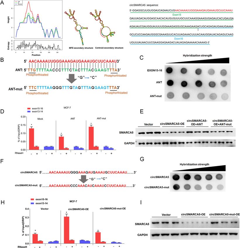
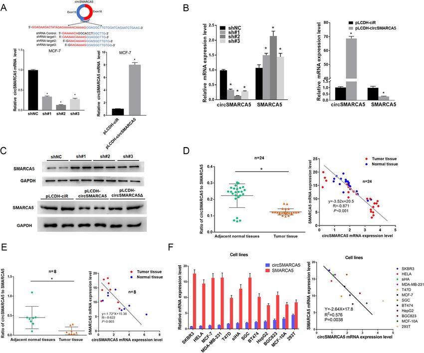
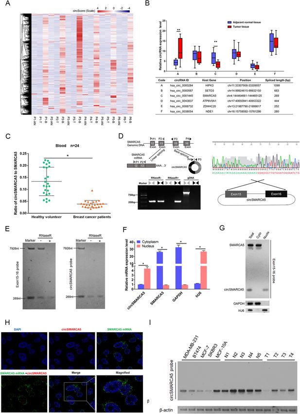
Xu et al. Molecular Cancer   (2020) 19:128                                                                     Page 2 of 19

Introduction                                                   contribute to the therapeutic implications for cytotoxic
Circular RNAs (circRNAs) are novel RNAs that have              drug-resistant breast cancer patients.
been ubiquitously discovered in many species by high-
throughput sequencing in recent years [1, 2]. CircRNAs         Results
are generated by the back-splicing of intronic, exonic or      Identification of expression of circRNAs in breast cancer
intergenic regions. circRNAs are resistant to RNase R,         We performed high throughput sequencing on tumor
and the stability of their structures makes these mole-        (T) and adjacent normal tissue (AN) and peripheral
cules ideal candidates for disease [3]. Extensive studies      blood (B) of six breast cancer patients. Total RNA with
have revealed that dysregulated circRNAs are involved          rRNA-depleted library were constructed and then
in the development of various cancers. In gastric cancer,      circRNAs expressed in those samples were identified.
circRNAs, such as circPVT1, circLARP4, has_circ_               Compared to tumor and adjacent normal tissue, we ob-
0000096, and circ_100269, have been shown to play a            served average higher CIRCscore (expression of circRNA
role in promoting tumor growth, and their expression is        / linear host genes) in blood than both tumor and
correlated with high TNM stage and poor prognosis [4–7].       adjacent normal tissue. In all 8312 circRNAs which were
In colon and hepatic carcinoma, ciRS-7 promoted tumor          expressed across all six patients, we observed average
development and progression by activating the EGFR and         CIRCscores from 0.23 to 1.28 in blood, which is higher
PI3K/Akt pathway [8, 9]. CircRNAs, such as circKIF4A,          than tumor (0.05 to 0.11) and adjacent normal tissue
hsa_circ_0001944, hsa_circ_0001481 and circRNA_0025202,        (0.08 to 0.18) in six patients (Fig. 1a). This result indi-
have been implicated in molecular typing, brain metastasis     cated average higher expression of circRNAs than their
and drug resistance in breast cancer [10–12]. Although great   host genes in peripheral blood, comparing to tissues,
progress has been made, the roles of circRNA and relevant      which might contribute to the exploration of diagnostic
molecular mechanisms remain largely unknown.                   biomarker for breast cancer. We then selected six
  Previous studies have shown that circRNAs exert their        circRNAs with high CIRCscores (average 0.22 to 15.8 in
functions in different ways. As noncoding RNAs, cir-           6 patients) and performed further experimental valid-
cRNAs regulate the expression of other genes by serving        ation in 24 patients. Real-time PCR results established
as sponges for microRNA and RNA-binding proteins               two circRNAs (circHIPK3 and circSMARCA5) were
[13, 14]. In addition, some circRNAs have been shown           significantly differentially expressed between tumor and
to be translated into functional proteins [15, 16]. In         adjacent normal tissue (Fig. 1b and Figure S1A). Espe-
addition, circRNAs have also been shown to directly            cially, circSMARCA5 was lower expressed in tumor
interact with the genomic DNA of the host gene in              samples and less studied in previous work. Furthermore,
plant, which results in altered parent gene expression         the ratio of circ-to-linear (expression of circRNA / linear
[17]. However, the interaction of circRNAs and host            host genes) of circSMARCA5 in blood sample of 24 health
gene DNA were less studied in human cancers.                   volunteers were significantly higher than those of 24
  SMARCA5 is a member of the SWI/SNF complex with              breast cancer patients (P < 0.05) (Fig. 1c and Figure S1B).
ATP-dependent chromatin remodeling activity [18–20].           We next examined the ratio of circ-to-linear of
In the process of DNA damage repair, SMARCA5 is                circSMARCA5 and clinical relevance in patients with
involved in chromatin remodeling in DNA damage                 breast cancer and observed significant difference in the
regions, providing a structural basis for the recruitment      distribution of the patients according to pathologic T (P =
of DNA damage repair factors [21, 22]. In tumors,              0.038) (Table S1). Together, these results indicating the
SMARCA5 is highly expressed in hepatic carcinoma and           potential function and candidate biomarker attributes of
prostate cancer, and its expression level is inversely         circSMARCA5 in breast cancer.
related to tumor radiosensitivity [23, 24].                       To characterize and functionally investigate cir-
  In this study, we established circRNAs have average          cSMARCA5, we firstly detected the expression of cir-
higher expression than their host genes in peripheral          cSMARCA5 in cell lines. circSMARCA5 is derived from
blood, comparing to tissues. Then we identified a              the back-splicing of exon 15 and exon 16 of SMARCA5
circRNA derived from SMARCA5 (circSMARCA5) is                  (Fig. 1d). As expected, endogenous circSMARCA5, but
significantly decreased in breast cancer cell lines and        not pre-mRNA, was resistant to RNase R digestion
breast cancer samples. Different to previous works             (Fig. 1d). In addition, the existence of the 269 nt
revealing circSMARCA5 can also function as a com-              circSMARCA5 was further confirmed by Northern blot
peting endogenous RNAs by binding with miRNA                   assay (Fig. 1e). Furthermore, we found that cir-
molecules [25–28], our mechanism exploration                   cSMARCA5 was mainly present in the nucleus, whereas
displayed circSMARCA5 is involved in regulating DNA            its parent mRNA was present exclusively in the cyto-
repair capacity by binding exon DNA directly. And              plasm, as evidenced by qPCR, Northern blotting and
further functional investigation of this circRNA may           RNA in situ hybridization (Fig. 1f-h and Figure S2).
Xu et al. Molecular Cancer    (2020) 19:128   Page 3 of 19

 Fig. 1 (See legend on next page.)
Xu et al. Molecular Cancer       (2020) 19:128                                                                                                 Page 4 of 19

 (See figure on previous page.)
 Fig. 1 Identification of circRNAs in breast cancer. a Heatmap of CIRCscore (FBPcirc/FBPlinear) in tumor (T), adjacent normal tissue (AN) and blood
 sample (B) from six breast cancer patients. b Expression of six circRNAs with high CIRCscore were validated by RT-qPCR assay in breast tumor and
 adjacent normal tissue. ** represents P < 0.01. CircRNAs IDs are according to circBase through their genomic coordinates. c The ratio of circ-to-linear of
 circSMARCA5 in blood sample of breast cancer patients and health volunteers. Total RNA from blood sample of breast cancer patients and health
 volunteers was extracted and detected by RT-qPCR. The expression level was normalized with β-actin as reference. *: P < 0.05 was considered
 statistically significant. d Schematic illustration showing the genomic region of circSMARCA5 derived from exons 15 and 16 of the SMARCA5 gene.
 Convergent (gray) and divergent (black) primers were designed to amplify the linear or back-splicing products (upper). Total RNA from MCF-7 cells
 with or without RNase R treatment was subjected to RT-PCR (lower) and further validated by Sanger sequencing (Right). e Northern blot using a
 junction-specific probe or an exon 15-16 probe showing the endogenous existence of circSMARCA5 and SMARCA5 mRNA from MCF-7 cells with or
 without RNase-R treatment (R+ or R-). The 7926 bp marker indicates the SMARCA5 full-length transcript transcribed in vitro. The 269 bp marker
 indicates exon 15 and exon 16 of SMARCA5 transcribed in vitro. f The nucleus and cytoplasm mRNA of MCF-7 were extracted, and SMARCA5 and
 circSMARCA5 expression levels were quantitated by RT-PCR. GAPDH and hU6 serve as internal references of the cytoplasm and nucleus, respectively.
 “**” indicates P < 0.01. g The nucleus and cytoplasm mRNA of MCF-7 were extracted, SMARCA5 and circSMARCA5 were examined by Northern
 blotting, and the SMARCA5 exon 15-16 probe was applied in this experiment. h Subcellular localization of circSMARCA5 and SMARCA5 in MCF-7 cells.
 The signals were examined by indirect RNA FISH and confocal microscopy. The nucleus was counterstained with DAPI. The circSMARCA5 probe was
 labled by biotin, while the SMARCA5 probe was labled by DIG. They were stained with red and green fluorescent secondary antibodies, respectively (I)
 The expression of circSMARCA5 detected by northern blot. MDA-MB-231, BT474, MCF-7, SKBR3 are breast cancer cell lines. MCF-10A are normal breast
 cell line. N1,N2,N3,N4,N5 are adjacent normal tissues. T1,T2,T3,T4 are breast cancer tissues. “**” indicates P < 0.01

Next, we examined the expression of circSMARCA5 in                              cirSMARCA5 terminates the transcription of SMARCA5 at
various breast cancer cell lines (MCF7, SKBR3, BT474,                           exon 15
MDA-MB-231) and immortalized but nontransformed                                 We further investigated the mechanism of circSMARCA5
breast epithelial cells (MCF-10A) as well as in adjacent                        in regulating the expression of SMARCA5. Interestingly,
normal tissues and breast cancer tissues. Northern blot                         we found that the overexpression of circSMARCA5 in-
results revealed that the expression levels of cir-                             deed decreased the expression of SMARCA5 exons 15–24
cSMARCA5 in MCF-10A and normal adjacent tissues                                 but had minimal effects on the expression of exons 1–14
are higher than breast cancer cell lines and cancer tis-                        (Fig. 3a). Next, we designed a primer location in exon 13
sues (Fig. 1i). These results indicated that circSMARCA5                        for the amplification of 3′ cDNA ends by rapid amplifica-
is downregulated in breast cancer tissues and cells.                            tion of cDNA ends (RACE) PCR (Fig. 3b, left). As shown
                                                                                in Fig. 3b, SMARCA5 can give rise to multiple isoforms.
                                                                                Importantly, we found a decrease in a band of ~ 5000 bp
circSMARCA5 decreases the expression of SMARCA5 in                              upon circSMARCA5 overexpression, while an ~ 250 bp
cancer cells                                                                    band displayed the opposite phenomenon (Fig. 3b, right).
To clarify the mechanisms of circSMARCA5, we investi-                           Sanger sequencing showed that the ~ 5000 bp band and
gated its effects on the expression of its parent gene                          the ~ 250 bp band are derived from full-length and trun-
SMARCA5. The expression levels of circSMARCA5 and                               cated mRNA (exons 1 to 14), respectively, of the SMAR
SMARCA5 were detected by the primers of junction                                CA5 gene (Fig. 3c). Consistent with the RACE results,
sequence and 22–23 exons sequence, respectively.                                Northern blot assay further demonstrated that ectopic
Knockdown of circSMARCA5 increased both mRNA                                    circSMARCA5 expression decreased SMARCA5 levels
and protein levels of SMARCA5, while conversely,                                and promoted truncated mRNA levels (Fig. 3d). The ob-
circSMARCA5 overexpression decreased SMARCA5                                    servations gathered thus far have led us to hypothesize
levels (Fig. 2a-c and Figure S3). Consistently, the protein                     that circSMARCA5 prevents transcription from exon 15
of SMARCA5 was high expressed in breast tumor                                   of SMARCA5. Indeed, ChIP analysis indicated that the
samples as compared with the corresponding controls                             binding of pol II to exons 1–14 of SMARCA5 was higher
(Figure S4). Moreover, the ratio of circ-to-linear of                           than that to exons 15–24 (Fig. 3e, left), and the ectopic ex-
circSMARCA5 was significantly lower in breast and                               pression of circSMARCA5 decreased the binding of Pol II
renal tumor tissue than the corresponding adjacent                              to exons 15–24 of SMARCA5 (Fig. 3e, right). To further
tissue specimens (Fig. 2d-e and Figure S1C). Besides, a                         address whether circSMARCA5 could terminate the tran-
significant negative correlation was also found between                         scriptional elongation of SMARCA5, we cloned a series of
circSMARCA5 and SMARCA5 expression in various cell                              exons of SMARCA5 in a luciferase plasmid reporter
lines and primary cancer tissues (Fig. 2d-f and Figure                          (Fig. 4a, upper). The transient transfection of these lucifer-
S5), which corroborates our observation that cir-                               ase reporters containing the 15–16 exon sequence re-
cSMARCA5 decreased the expression of SMARCA5 in                                 vealed that luciferase activity was significantly decreased
cancer cells.                                                                   when circSMARCA5 was overexpressed (Fig. 4a, lower).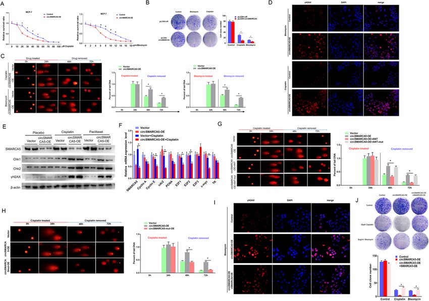
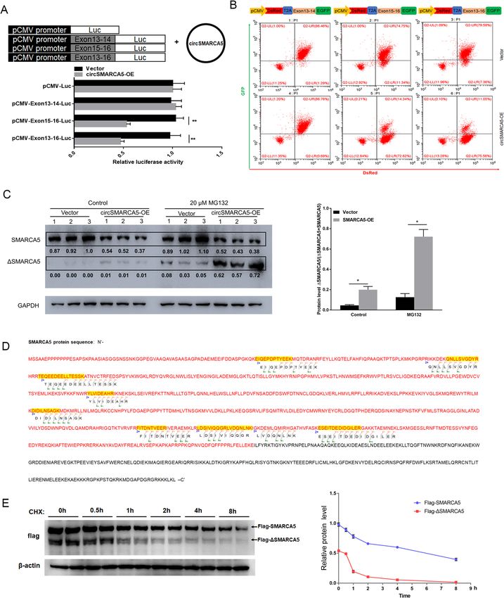
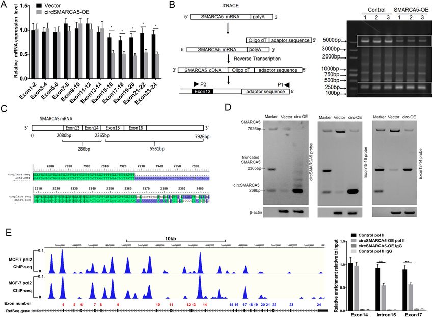
Xu et al. Molecular Cancer    (2020) 19:128                                                                                          Page 5 of 19

 Fig. 2 circSMARCA5 decreases the expression of SMARCA5 in cells. a Generation of circSMARCA5-knockdown and circSMARCA5-overexpressing
 cells. MCF-7 cells were infected with lentiviruses expressing shRNA against circSMARCA5 (sh-circSMARCA5; three different oligonucleotides) or
 circSMARCA5 (pLCDH-circSMARCA5). RT-qPCR was performed to evaluate the expression of circSMARCA5. GAPDH was used as an internal control.
 b RT-qPCR showing the levels of circSMARCA5 and SMARCA5 in MCF-7 cells stably expressing sh-NC, sh-circSMARCA5, pLCDH-ciR (control), or
 pLCDH-circSMARCA5. c Western blot showing the levels of SMARCA5 in MCF-7 cells stably expressing sh-NC, sh-circSMARCA5, pLCDH-ciR
 (control), pLCDH-circSMARCA5, pLCDH-circSMARCA5Δ (without splicing-inducing sequence). GAPDH was used as an internal control. (D-F) The
 ratio of circ-to-linear of circSMARCA5 in tumor tissue were significantly lower than normal tissue in breast cancer samples (d) and RCC samples
 (e) (P < 0.05). A negative correlation between circSMARCA5 and SMARCA5 expression was observed in breast cancer samples (d), RCC samples
 (e), and various cell lines (f)

To further confirm the effect of circSMARCA5 on the                       which was confirmed by mass spectrometry (Fig. 4d).
transcriptional elongation of SMARCA5, we inserted                        Moreover, we found that ΔSMARCA5 is more sus-
exons of SMARCA5 between DsRED and EGFP as in-                            ceptible to proteolysis by the proteasome than SMAR
dicated (Fig. 4b, upper). The EGFP level was signifi-                     CA5 (Fig. 4e). Together, these results show the role
cantly decreased by circSMARCA5 when exons 15–16                          of circSMARCA5 in the termination of transcriptional
were present (Fig. 4b, lower). We further investigated                    elongation at exon 15 of SMARCA5.
the role of circSMARCA5 in the regulation of SMAR
CA5 at the protein level. As expected, circSMARCA5                        circSMARCA5 can form R-loops with its parent gene
overexpression downregulated the protein levels of                        To further dissect the mechanism of SMARCA5 tran-
SMARCA5 and upregulated truncated SMARCA5                                 scriptional termination mediated by circSMARCA5, we
(ΔSMARCA5) protein levels (Fig. 4c and Figure S6),                        investigated whether circSMARCA5 can bind genomic
Xu et al. Molecular Cancer    (2020) 19:128                                                                                         Page 6 of 19

 Fig. 3 cirSMARCA5 terminates the transcription of SMARCA5 at exon 15. a RT-qPCR analysis of the expression of SMARCA5 in MCF-7 cells using a
 series of paired primers. “*” indicates P < 0.05. b Rapid amplification of cDNA ends (RACE) PCR analysis of SMARCAC5 transcripts. The PCR
 products were readily identified by agarose gel electrophoresis. Each set of samples was repeated three times. c Sanger sequencing of two
 transcripts of SMARCAC5 that are regulated by circSMARCA5 overexpression. d Northern blotting using the junction-specific probes for exons 13-
 14 and 15-16 to show the expression levels of the transcripts of SMARCAC5 mRNA from MCF-7 cells stably expressing control vector or pLCDH-
 circSMARCA5 (circ-OE). e CircSMARCA5 prevents transcription from exon 15 of SMARCA5. ChIP-seq analysis showing that the binding of pol II to
 exons 1-14 of SMARCA5 was higher than that to exons 15-24. ChIP-qPCR showed that the ectopic expression of circSMARCA5 decreased the
 binding of Pol II to exons 15-24 of SMARCA5

SMARCA5 DNA to form an R-loop. Dot-blotting with                          A series of fragments from exons 15–16 were hybridized
R-loop-specific S9.6 antibody supported our hypothesis                    with circSMARCA5 for the dot-blotting assay. As shown
that circSMARCA5 can bind exons 15–16 of SMARCA5                          in Fig. 5e, the 67 bp fragment of the 5′ end of exon 15
genomic DNA (Fig. 5a). Additionally, we performed                         plays important role in interacting with circSMARCA5.
DNA-RNA immunoprecipitation (DRIP) qPCR and con-                          Moreover, the secondary structure of circSMARCA5
firmed the interaction between circSMARCA5 and                            was determined by the software MFOLD [29], which
exons 15–16; pretreatment with RNase H ablated this                       revealed the sequence 5′-AACAAAAUUGGGAAAGAU
interaction, confirming that the interaction is R-loop-                   GAAAUGCUUCAAAU-3′ from the 5′ end of exon 15
specific (Fig. 5 b and Figure S7). The interaction of cir-                located in the loop region of circSMARCA5 (Fig. 6a).
cSMARCA5 with the DNA of SMARCA5 was directly                             We thus hypothesized that this sequence might play a
verified by fluorescence in situ hybridization (Fig. 5c).                 key role in mediating the circSMARCA5-DNA inter-
Consistent with previous findings [17], dot-blotting of                   action. To this end, we synthesized the wild-type and
the genome without RNA digest revealed that the bind-                     mutant phosphorylated DNA fragments, ANT and
ing of circRNA to genomic DNA may be widely present                       ANT-mut, respectively, corresponding to this sequence
in cancer cells (Fig. 5d). We next determined the specific                (Fig. 6b). Dot-blotting demonstrated that wild-type
sequence of exons 15–16 required for R-loop formation.                    oligonucleotides (ANT) can bind to circSMARCA5, but
Xu et al. Molecular Cancer    (2020) 19:128   Page 7 of 19

 Fig. 4 (See legend on next page.)
Xu et al. Molecular Cancer     (2020) 19:128                                                                                            Page 8 of 19

 (See figure on previous page.)
 Fig. 4 cirSMARCA5 blocks the transcription of SMARCA5 and promotes the generation of a truncated SMARCA5 protein (ΔSMARCA5). a
 Schematics of luciferase reporter constructs containing the SMARCA5 exon sequence as indicated (upper). The SMARCA5 exon 15-16 sequence
 plays an important negative role in mediating the effect of circSMARCA5 overexpression on luciferase activity (lower). b Schematics of
 fluorescence reporter constructs containing the SMARCA5 exon sequence as indicated (upper). MCF-7 cells were transiently transfected with
 these fluorescence reporters along with or without circSMARCA5 co-overexpression. After transfection for 48 hours, the reporter transcription
 activities were measured by flow cytometry assay. c circSMARCA5 overexpression downregulated the protein levels of SMARCA5 while
 upregulating truncated SMARCA5 (ΔSMARCA5) protein levels. MCF-7 cells stably overexpressing circSMARCA5 or control cells were treated with
 DMSO or MG132. Western blot analysis was performed using an antibody targeting the N-terminus of SMARCA5 to evaluate the expression of
 SMARCA5 and ΔSMARCA5. GAPDH was used as an internal control. d The ΔSMARCA5 protein was identified by mass spectrometry, and detected
 SMARCA5 peptides were showed in the map. The red-labeled portion is the amino acid sequence of the translated defective transcript. e MCF-7
 cells expressing Flag-SMARCA5 and Flag-ΔSMARCA5 were treated with cycloheximide (CHX, 50 μg/ml). The cell lysates were subsequently
 harvested at sequential time points (0, 0.5, 1, 2, 4 or 8 h) after treatment, and then the cell lysates were immunoblotted with anti-Flag or anti-
 Actin antibody

mutant oligonucleotides (ANT-mut) cannot bind to cir-                       affect the function of DNA damage repair capacity.
cSMARCA5 (Fig. 6c). As expected, DRIP-qPCR showed                           CCK8 and clone formation assays revealed that
that ANT inhibited circSMARCA5 binding to the DNA                           circSMARCA5 overexpression increased sensitivity to
at exons 15–16, whereas ANT-mut had no effect on this                       cisplatin or bleomycin in MCF-7 cells (Fig. 7a, b). Next,
interaction (Fig. 6d). Furthermore, the transfection of                     MCF-7 cells were treated with the indicated concentra-
ANT prevented the decrease in SMARCA5 protein                               tion of cisplatin or bleomycin for 24 h and then the
levels in MCF-7 cells stably expressing circSMARCA5,                        DNA damage was evaluated by single cell gel electro-
whereas ANT-mut had no effect on SMARCA5 protein                            phoresis (SCGE) at 48 and 72 h. MCF-7 cells expressing
levels (Fig. 6e). Importantly, the mutation of the key                      circSMARCA5 showed significantly lower repair
sequence in circSMARCA5 impaired the interaction                            capacity than did control cells (Fig. 7c). In parallel,
with its parent gene, which was confirmed by dot-                           DNA damage was examined after 72 h of treatment
blotting and DRIP-qPCR assays (Fig. 6f-h and Figure                         with cisplatin or bleomycin by using an anti-γH2AX
S8). Unlike circSMARCA5, circSMARCA5-mut had little                         antibody. Consistent with the SCGE results, the γH2AX
effect on SMARCA5 protein levels (Fig. 6i). These                           signal in MCF-7 cells expressing circSMARCA5 was
results suggested that circSMARCA5 formed R-loops                           significantly higher than that in MCF-7 cells, as
with its parent gene to inhibit the expression of SMAR                      evidenced by immunostaining (Fig. 7d). Consistently,
CA5 in cancer cells.                                                        cisplatin significantly enhanced the levels of DNA
                                                                            damage response proteins Chk1 and Chk2 in MCF-7
circSMARCA5 inhibits DNA damage repair function                             cells expressing circSMARCA5 (Fig. 7e), whereas
To explore the roles of circSMARCA5 in cancer pro-                          several key cell-cycle genes were reduced specifically
gression, we overexpressed and depleted circSMARCA5                         upon circSMARCA5 overexpression (Fig. 7f). To test
in MCF-7 cells by lentiviral vectors and then examined                      whether circSMARCA5 R-loop formation is necessary
the effect of circSMARCA5 on cell proliferation, migra-                     for its DNA repair function, we transfected ANT or
tion, and apoptosis. However, the results showed that                       ANT-mut into circSMARCA5-expressing cells. The
both overexpressed and depleted circSMARCA5 had no                          SCGE assay and γH2AX measurement showed that
effect on these three activities (Figure S9). Previous                      ANT significantly enhanced the DNA repair capacity,
studies have indicated that SMARCA5 plays an import-                        while ANT-mut had no effect on this activity (Fig. 7g
ant role in regulating the DNA repair process and main-                     and Figure S11). Furthermore, ANT significantly
taining the stability of the genome [22, 30–32].                            decreased the degree of colocalization between cir-
Consistent with previous reports, SMARCA5 overex-                           cSMARCA5 and its cognate DNA locus (Figure S12).
pression improved DNA repair capacity and reduced                           In addition, unlike circSMARCA5, the overexpression
the expression of Chk1 and Chk2 after DNA damage                            of circSMARCA5-mut had little effect on the DNA
repair (Figure S10A). Given that circSMARCA5 can                            repair rate (Fig. 7h and Figure S13A, B). Next, we deter-
promote the production of the truncated ΔSMARCA5                            mined whether SMARCA5 could mediate the effects of
protein, we tested whether the truncated protein is also                    circSMARCA5 in preventing DNA damage repair. As
functional. The overexpression of Flag-ΔSMARCA5                             shown in Fig. 7i, the γH2AX signal was much lower in
had a minimal effect on the expression of Chk1 and                          circSMARCA5-expressing cells complemented with
Chk2 after DNA damage repair (Figure S10B), suggest-                        SMARCA5 than that in cells expressing circSMARCA5
ing that ΔSMARCA5 is a nonfunctional protein                                alone. As expected, ΔSMARCA5 could not rescue the
product. We next assessed whether circSMARCA5 can                           inhibition of DNA damage repair function induced by
Xu et al. Molecular Cancer (2020) 19:128 Page 9 of 19 Fig. 5 (See legend on next page.)
Xu et al. Molecular Cancer (2020) 19:128 Page 10 of 19
(See figure on previous page.)
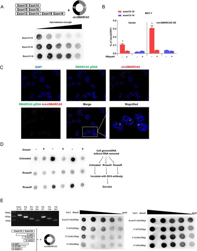
Fig. 5 circSMARCA5 interacts with its site of transcription. a circSMARCA5 interacts with the 15-16 exon sequence of the SMARCA5 locus. A series
of exon DNA fragments were hybridized with circSMARCA in vitro. The DNA-RNA hybridization strength was quantified by dot-blot with R-loop-
specific S9.6 antibody. Hybridization stringency was altered by decreasing ionic strength (80-0.1 mM NaCl). b DRIP-qPCR analysis of the 15-16
exon sequence of SMARCA5 to detect the association of circSMARCA5 in MCF-7 cells. RNase H-treated and/or DRIP-qPCR analysis of the 15-16
exon sequence as a control. c CircSMARCA5 partially localized at its site of transcription. Double FISH of circSMARCA5 (red) and its parent DNA
region (green). The nucleus was stained by DAPI. d Dot-blot of R-loops in MCF-7 cell genomic DNA preparations treated with DNase I, RNase H,
or RNase R. The DNA-RNA hybrids in genome DNA were analyzed by S9.6 antibody. e Mapping of the R-loop formation region of circSMARCA5.
A series of exon 15-16 deletion mutants were hybridized with circSMARCA5 for the dot-blotting assay. The DNA-RNA hybridization intensity was
analyzed by dot-blot with an S9.6 antibody targeting the DNA-RNA hybrid strand. Hybridization stringency was altered by decreasing ionic
strength (80-0.1 mM NaCl)
circSMARCA5 (Figure S13C, D). Moreover, the overex- clinical relevance because chemotherapy with cisplatin
pression of SMARCA5 could significantly rescue the and bleomycin remains the standard of care in breast
growth defects of cells expressing circSMARCA5, as cancer [36–38]. Hence, the restoration of circSMARCA5
demonstrated by a colony formation assay (Fig 7 j). levels provides an approach to overcome treatment
Together, these results demonstrated the roles of resistance in breast cancer patients.
circSMARCA5 in regulating the DNA repair process in SMARCA5, also known as SNF2H, is a member of the
MCF-7 cells. SWI/SNF chromatin-remodeling complex. During DNA
damage repair processes, SMARCA5 is recruited to
DNA damage sites where it induces the ubiquitination
circSMARCA5 overexpression enhances the cisplatin
and phosphorylation of histone H2A, which facilitates
response in breast cancer
chromatin remodeling and DNA damage repair [18, 19].
To further evaluate the therapeutic potential of
In this study, we show that circSMARCA5 expression
circSMARCA5 in breast cancer in vivo, we established
resulted in the downregulation of SMARCA5, and the
circSMARCA5 overexpression clones in MCF-7 cells. As
effect of circSMARCA5 overexpression on DNA repair
shown in Fig. 8a, the overexpression of circSMARCA5
capacity was reversed by concomitant SMARCA5 over-
efficiently enhanced the sensitivity of MCF-7 xenografts
expression, suggesting that the effect of circSMARCA5
to concurrent cisplatin treatment (Fig. 8a, b). The over-
on DNA repair capacity is mediated through SMARCA5.
expression of circSMARCA5 was confirmed by in situ
circRNAs exert functions in various ways, such as form-
hybridization and qPCR analysis (Fig. 8c), along with de-
ing an R-loop with DNA to regulate splicing and tran-
creased SMARCA5 protein levels and increased γH2AX
scriptional pausing [17]. For example, circSEPALLATA3
levels (Fig. 8d). In addition, qPCR analysis demonstrated
regulates the splicing of its parent mRNA through R-
that circSMARCA5 can be detected in the blood,
loop formation [17]. In addition, circRNAs are a novel
suggesting that circSMARCA5 is a secretory molecule.
class of ceRNAs that sponge miRNAs, thus positively
Collectively, these data demonstrate that circSMARCA5
regulating gene expression [13, 14]. Additionally, cir-
could serve as a potential therapeutic target to restore
cRNAs, such as exon-intron circRNAs, regulate gene
sensitivity to cisplatin therapy in breast cancer.
expression through specific RNA-RNA interactions with
U1 snRNA [39]. Furthermore, circRNAs also exert
Discussion functions by binding to proteins and regulating their
Previous studies have indicated that circRNAs have mul- activities [40]. We identified one mechanism by which
tiple functions in cancer development and progression circSMARCA5 regulates the drug sensitivity of breast
[33–35]. In this study, we identified multiple expressed cancer cells to cisplatin and bleomycin through the
circRNAs in breast cancer samples and observed average downregulation of SMARC5. circSMARCA5 is recruited
higher abundance of circRNAs over their host genes in to its parent gene locus, leading to R-loop formation,
peripheral blood than tissues, which might contribute to transcription termination, nonfunctional truncated
the exploration of diagnostic biomarker for breast ΔSMARCA5 protein upregulation, and decreased SMAR
cancer. We then identified that circSMARCA5 is signifi- CA5 expression. This regulatory mechanism has also
cantly decreased in breast cancer tissues using RNA-seq. been verified in cervical cancer (Hela cells) (Figure S14).
More importantly, we define a critical role for cir- However, our evidence demonstrates that circSMARCA5
cSMARCA5 in the regulation of DNA damage repair has no significant effect on the proliferation, migration
capacity and the drug sensitivity of breast cancer cells and apoptosis of breast cancer cells, suggesting that this
in vitro and in vivo through the negative regulation of molecule functions in a cell-type and context-dependent
its parent gene SMARCA5. These findings are of high manner. Notably, we provide evidence thatXu et al. Molecular Cancer (2020) 19:128 Page 11 of 19 Fig. 6 circSMARCA5 can form an R-loop with its parent gene. a Secondary structure prediction for circSMARCA5 using the Mfold program. The sequence KEY shared by the minimum free energy structure and the thermodynamic ensemble structure is marked by red. b The thiophosphorus nucleic acid analog ANT complementary to KEY and its mutant ANT-mut were synthesized in vitro. c Dot-blot verifying the interaction between circSMARCA and ANT or ANT-mut. d DRIP-qPCR analysis on 15-16 exon or 13-14 exon sequences of SMARCA5 to detect the association of circSMARCA5 in MCF-7 cells overexpressing ANT or ANT-mut, RNase H-treated and/or DRIP-qPCR analysis of the 15-16 exon sequence as a control. “*” indicates P < 0.05. e Western blot analysis shows that transfection of ANT into circSMARCA5-overexpressing cells can restore SMARCA5 protein levels but ANT-mut cannot. (f, g) Dot-blot analysis quantifying R-loop strength between the SMARCA5 locus and circSMARCA5 or circSMARCA5-mut (guanine converted to cytosine of the KEY sequence). h DRIP-qPCR in MCF-7 cells transfected with circSMARCA5 or circSMARCA5-mut. RNase H-treated genomic DNA and qPCR of exon13-14 were treated as controls. “*” indicates P < 0.05. i Western blot analysis shows that overexpression of circSMARCA5 to MCF-7 cells can decrease SMARCA5 protein levels but circSMARCA5-mut cannot
Xu et al. Molecular Cancer     (2020) 19:128                                                                                           Page 12 of 19

 Fig. 7 circSMARCA5 decreases DNA repair capacity. a circSMARCA5 increases sensitivity to cisplatin or bleomycin in MCF-7 cells. MCF-7 cells
 stably expressing control vector or pLCDH-circSMARCA5 were treated with cisplatin or bleomycin for 24 h, and CCK8 was used to measure cell
 viability. b Relative colony formation units of MCF-7 cells stably expressing control vector or pLCDH-circSMARCA5 treated with 20 μM cisplatin or
 6 μg/ml bleomycin. After 24 hours, the drugs were replaced by fresh medium. The number of colonies was quantified. c, d, e Single cell gel
 electrophoresis (SCGE) assay indicating that circSMARCA5 overexpression inhibits cell recovery from DNA damage. MCF-7 cells stably expressing
 control vector or pLCDH-circSMARCA5 treated with 20 μM cisplatin or 6 μg/ml bleomycin. After incubation for 24 h, the cells were recovered
 with fresh medium for 24 or 48 hours and then collected for SCGE analysis (c), immunofluorescence assay using an anti-γH2AX antibody (d), and
 western blot assay with the indicated antibodies (e). f RT-qPCR assay showing the relative levels of several key cell cycle genes in MCF-7 cells
 stably expressing control vector or pLCDH-circSMARCA5 treated with DMSO or 20 μM cisplatin for 24 h and replaced with fresh medium for 72 h.
 “**” indicates P < 0.01. g SCGE assay showing that the cotransfection of ANT in circSMARCA5-overexpressing cells can restore the DNA repair
 capacity but the cotransfection of ANT-mut cannot. h SCGE assay showing that the overexpression of circSMARCA5 in MCF-7 cells can decrease
 DNA repair capacity but the overexpression of circSMARCA5-mut cannot. i SMARCA5 abrogates γH2AX levels induced by circSMARCA5. MCF-7
 cells were infected with different combinations of lentivirus as indicated and treated with 6 μg/ml bleomycin. After incubation for 24 h, the cells
 were recovered with fresh medium for 48 hours and then collected for immunofluorescence assay using a γH2A antibody. j The cells as in i were
 treated with 30 μM cisplatin or 8 μg/ml bleomycin. After incubation for 24 h, the cells were recovered with fresh medium for 48 hours and then
 collected for colony formation assays. “*” represents P < 0.05

circSMARCA5 plays a key role in the DNA repair path-                        circRNAs may serve as potential biomarkers for various
way and drug resistance, making it a promising thera-                       diseases [3, 41, 42]. Altogether, the findings in this study
peutic target for breast cancers.                                           confirmed that the overexpression of circSMARCA5 was
   CircSMARC5 has been shown to be downregulated in                         sufficient to improve the chemosensitivity of breast can-
hepatocellular carcinoma tissues, and its downregulation                    cer cells in vitro and in vivo, indicating the important
is associated with aggressive characteristics and unfavor-                  regulation mechanism of this circRNA in breast cancer.
able outcomes [25]. We also confirmed a low abundance                       And our analysis revealed the negative correlation is also
of circSMARCA5 in breast cancer and renal cancer                            existed in other cancers, suggesting a general regulation
tissues compared with matched adjacent normal tissues.                      of circSMARCA5 on host gene in transformed cells. Fur-
   Due to the features of circRNA, including stability,                     thermore, we found that the ratio of circ-to-linear (ex-
tissue specificity and abundance in bodily fluids,                          pression of circRNA / linear host genes) of
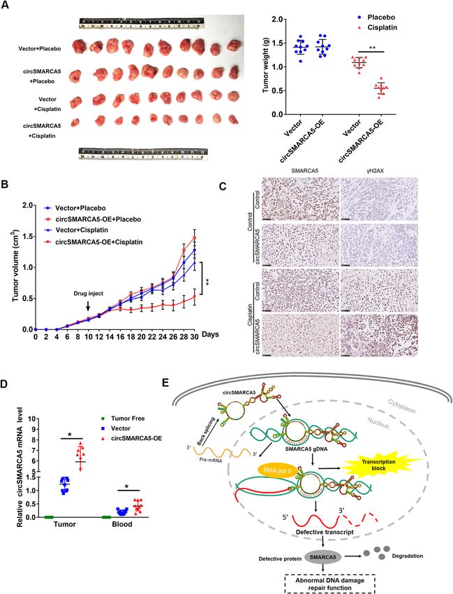
Xu et al. Molecular Cancer    (2020) 19:128                                                                                        Page 13 of 19

 Fig. 8 Figure 8. circSMARCA5 overexpression enhances the cisplatin response in breast cancer. a MCF-7 cells expressing control vector or pLCDH-
 circSMARCA5 were transplanted into mice. After the average tumor volume reached 0.3 cm3, the mice were injected with cisplatin or placebo.
 (Left) Representative images of the isolated tumors from injected mice. (Right) Tumor weight was calculated according to the formula provided
 in the Materials and Methods section. b After injection with cisplatin or placebo, the mean tumor volume was measured by calipers on the
 indicated days. c Representative immunofluorescence staining images (scale bars, 100 μm) and SMARCA5 and γH2AX expression in xenograft
 tumors derived from MCF-7 cells as in A. d RT-qPCR assay showing the relative levels of circSMARCA5 in the tumor tissues and blood of
 xenograft mice. The blood of tumor-free mice was treated as a control. “*” represents P < 0.05. e Proposed model for circSMARCA5
 downregulation of its parent gene expression

circSMARCA5 in the blood sample of breast cancer pa-                      Conclusion
tients were significantly lower than health volunteers                    We observed average higher expression of circRNAs
(Fig. 1c and S1B), which indicating that circSMARCA5                      than their host genes in blood samples comparing to
may serve as an indicator for the evaluation of breast                    breast cancer and adjacent normal tissues. And for the
cancer liquid biopsy. Further clinical research is needed                 first time, we validated the interaction between circRNA
to clarify whether circSMARCA5 is a useful biomarker                      and its host gene DNA in cancer, through a regulation
in breast cancer.                                                         model of circRNA and the transcription of partial exons
Xu et al. Molecular Cancer   (2020) 19:128                                                                 Page 14 of 19

of host gene. Furthermore, we found circRNA could             utilized for detecting the back-splice junction sites of
affect the sensitivity of tumor to chemotherapy drug and      circRNAs. And the fragments per billion mapped bases
our result indicated this circRNA is involved in DNA          (FPB) of circRNA and its linear host gene were extracted
damage repair in human cancer. Our results revealed a         to calculate the CIRCscore (FPBcirc/FPBlinear), which
new regulatory mechanism for circRNA on its host gene         could indicate the expression ratio of circRNA to their
and provided evidence that circSMARCA5 may serve as           host genes. Genome assembly GRCh37 and GENCODE
a therapeutic target for drug-resistant breast cancer         (version 19) gene annotation were used. Total 8312
patients.                                                     circRNAs were detected with expression across all six
                                                              patients.
Materials and methods
Cell culture and cell transfection                            Northern blot analysis
All cell lines, including 293 T, MCF-10A, MCF-7,              Northern blot (NB) analysis was performed according to
SKBR3, BT474, T47D and MDA-MB-231, were purchased             the manufacturer’s instructions (DIG Northern Starter
from the China Center for Type Culture Collection             Kit, Roche). Digoxigenin (DIG)-labeled riboprobes were
(CCTCC, Chinese Academy of Sciences, Shanghai, China).        transcribed by DIG RNA labeling Mix (Roche, USA) and
The cells were cultured according to the protocols from       the HiScribe T7 In Vitro Transcription Kit (NEB, USA),
the American Type Culture Collection (ATCC; https://          and then purified by phenol-chloroform extraction and
www.atcc.org/). Plasmid and phosphorylated deoxyribo-         ethanol precipitation. Ten micrograms of total RNA or
nucleic acid single strand transfection were carried out by   1 mg of in vitro synthesized linear or circRNA was elec-
Zlipo2000 (Zomanbio, Beijing, China) or liposomal trans-      trophoresed on a denaturing urea polyacrylamide gel,
fection reagent (Yeasen, Shanghai, China).                    transferred to a Hybond-N+ membrane (GE, USA) by a
                                                              positive capillary transfer system and UV-crosslinked as
Sample collection                                             a standard protocol. The membrane was then hybridized
Breast cancer tissues, adjacent tissues and blood samples     with specific DIG-labeled riboRNA probes. NB probes
were collected from breast cancer patients treated at         are listed in Table S2.
Zhongnan Hospital of Wuhan University (Wuhan,
China) after written informed consent was obtained.           DNA and RNA fluorescence in situ hybridization (FISH)
One milliliter of EDTA blood from different breast            The cells were fixed with 4% paraformaldehyde. After
cancer patients was collected before surgery. The blood       permeabilization with proteinase K pretreatment, the
samples were stored in Trizol at − 80 °C until use. In the    DNA was denatured with 2X SSC solution containing
RNA-seq study, 6 women (mean age: 52 years, range:            70% formamide at 72 °C and then incubated for 12 h at
45–60 years) diagnosed with luminal-B subtype breast          37 °C in hybrid solution [50% formamide, 2X SSC, 0.25
cancer were included. The amplification and overexpres-       mg/mL Escherichia coli transfer RNA, 0.1 mg/mL sal-
sion of HER2 were found in 3 patients, while the other        mon sperm DNA (Sigma, USA), 2.5 mg/mL BSA (Sigma,
patients were negative in HER2 status. In addition,           USA), and DIG- or biotin-labeled 100 pM probes tran-
cancer and adjacent normal tissues and blood samples          scribed by T7 In Vitro Transcription Kit (NEB, USA)].
from 24 breast cancer patients with clinical pathologic       After washing with hybrid solution supplemented with
information, and blood samples from 24 health volun-          RNase A to remove excessive probes, the cells were sub-
teers were collected for the qPCR validation.                 jected to indirect immunofluorescence using FITC/
                                                              AF594 coupled with anti-DIG antibody or FITC/AF594
Whole transcriptome sequencing                                coupled with streptavidin. The nuclei were stained with
Total RNA was extracted from the tumor, adjacent              DAPI. The probes are listed in Table S2.
normal tissue and blood samples of six breast cancer
patients, followed by rRNA depletion from total RNA.          Plasmid constructs
Then, the RNA was reverse transcribed to cDNA and             The sequence for exons 15–16 of SMARCA5 was
constructed into a strand-specific library. Illumina HiSeq    PCR amplified using primers F (5′-CGGAATTCTG
XTen was performed for sequencing. All raw data can           AAATATGCTATCTTACAG GGAGGCTTGTGGAT
be accessed in the NCBI GEO database (https://www.            CAGAAT.
ncbi.nlm.nih.gov/geo/query/acc.cgi?acc=GSE133998, ac-           CTG-3′) and R (5′-CGGGATCCTCAAGAAAAA
cession code: wxmjuumulvsbbuf).                               ATATATTCACCTTTTGTTTTTC.
                                                                TCTATAGTCTCTCC − 3′), digested by EcoRI and
Identification of circRNA expression                          BamHI, and ligated into pLCDH-ciR (Geneseed, China)
To identify the expression of circRNAs from RNA-Seq,          and pLCDH-ciR-GFPmut (GFP mutated by site-directed
one extensively used algorithm Circexplorer3 [43] were        mutagenesis) to create two circSMARCA5-overexpressing
Xu et al. Molecular Cancer (2020) 19:128 Page 15 of 19
plasmids. The pCMV-DsRed-T2A-MSC-EGFP reporter 24:23:1), with 20 μg/ml proteinase K (Sigma, USA) and
vector was constructed by cloning the coding region of 20 μg/ml RNase A (Sigma, USA) applied to degrade pro-
DsRed and T2A into psi-EGFP-N1(Addgene, USA). Exons teins and RNA in the genome, respectively. The DNA
13–14, 15–16 and 13–16 of SMARCA5 were PCR ampli- was digested with 6 U EcoRI, EcoRV, XbaI, BamHI, and
fied and cloned into the pCMV-DsRed-T2A-MSC-EGFP SspI (NEB, USA) at 37 °C overnight or treated with 0.4
reporter by in-Fusion cloning. To overexpress SMARCA5 U/μl RNase H (Diamond, China) as a negative control
and ΔSMARCA5 protein, the SMARCA5 complete and for R-loop validation. The purified DNA fragment at
defective transcript sequences were cloned into pFLAG- 10 μg was incubated with 10 μg S9.6 antibody (Kerafast,
CMV2 (Addgene, USA). Gene-specific shRNA target se- USA) at 4 °C for 2 h in 0.2 ml IP buffer (20 mM HEPES-
quences were synthesized and cloned into the pLKO.1 KOH (pH 7.5), 150 mM NaCl, 10 mM MgCl2, 0.5% Tri-
plasmid. The primers for making these constructs are pro- ton X-100), with mouse IgG (CST, USA) as a control.
vided in Table S3. The mixture was incubated with 20 μg protein A/G
agarose beads (GE, USA) at 4 °C overnight. The beads
RNA secondary structure prediction were washed three times with IP buffer and treated with
The circSMARCA5 complete sequence was input into 20 ng proteinase K at 45 °C for 30 min. The DNA was
the RNAfold web server (http://rna.tbi.univie.ac.at/cgi- purified and applied by PCR or qPCR. The primers are
bin/RNAWebSuite/RNAfold.cgi). The minimum free en- listed in Table S3.
ergy structure and thermodynamic ensemble structure
were predicted by this website. Mass spectrum analysis
To detect ΔSMARCA5, a defective protein of SMAR
The 3′ rapid amplification of cDNA ends (3′ RACE) CA5, SDS-PAGE of the target protein molecule weight
The total RNA was extracted from cells, and the in- region was used to analyze the protein mass spectrum.
tegrity was verified. Oligo dT with a tag sequence In brief, MCF-7 cells overexpressing circSMARCA5
was used for reverse transcription. The 3′ end se- were treated with 30 μM MG132 for 24 h. The cells were
quences of mRNA were PCR amplified using primers harvested, and the total protein was extracted for SDS-
located in SMARCA5 exon 2 and the tag sequence. PAGE. When the 90 kd band and 110 kD protein
Fragments with different expression levels were identified marker band were sufficiently separated, the electro-
by one-generation sequencing. All primers are listed in phoresis was stopped, and the SDS-PAGE gel was ex-
Table S3. cised between 70 and 90 kD for analysis by mass
spectrometry. Mass spectrometry data were analyzed by
R-loop dot-blotting Mascot 2.3 and aligned with UniProt human_20190102_
Dot-blotting was performed using S9.6 monoclonal anti- 177661.fasta protein sequence data.
body according to previously published protocols [17, 44].
DNase I, RNase H and RNase R (Epicentre Technologies)
Real-time quantitative PCR (qPCR)
treatments were performed essentially as described using a
For RT-qPCR, total RNA was extracted from breast can-
standard hybridization buffer (10 mM Tris-HCl, pH 7.5, 50
cer tissues and adjacent tissues, rRNA was depleted, and
mM NaCl, 0.1 mM EDTA). RNA for exons 13–14, 15–16
RNase R was used to digest the linear RNA. qPCR was
and 15–16 mutation templates of SMARCA5 were tran-
carried out as described [46]. In brief, total RNA was ex-
scribed with a T7 RNA Polymerase kit (NEB, USA) and
tracted by TRIzol reagent (Invitrogen, Carlsbad, CA),
circularized by splint ligation with T4 RNA ligase with cir-
and cDNA was synthesized by random primers or oligo-
cRNAs purified from linear RNAs by RNase R digestion.
dT (Yeasen ,China). qPCR was performed on a CFX 96
DNA templates, including SMARCA5 exons 13–14, 15–16,
real-time PCR system using SYBR Green Real-time PCR
13–16 and their mutation templates, were amplified by
Master Mix (Bio-Rad, USA). The expression levels of
PCR. The strength of the R-loops was assessed by increas-
target genes were normalized by 2-ΔΔCt with GAPDH
ing the hybridization stringency through a stepwise
and β-actin as references. Each experiment was repeated
decrease in ionic strength from 80 to 10 mM NaCl. The
three times. The primers used in qPCR are listed in
dot-blot experiments were repeated three times, with
Table S3.
quantification performed with Image Lab software ver 5.2.1
(mean, background normalized values presented).
Single cell gel electrophoresis
Drip A comet assay was carried out as described [47]. In brief,
DRIP was carried out as described [45] with some modi- 1 × 10⁵ cells were mixed with 1 ml agarose (0.8% agarose
fications. In brief, 40 μg of genomic DNA was extracted in PBS). The suspension of agarose cells was dripped
from cells by PCI (phenol:chloroform:isoamyl alcohol = onto the slide to form a uniform film. The slide wasXu et al. Molecular Cancer (2020) 19:128 Page 16 of 19
incubated at 4 °C for 10 min, and then placed in a slide hematoxylin. A Panoramic MIDI automatic digital slide
box, followed by the addition of e precooled cracking scanner (3DHISTECH Ltd., Budapest, Hungary) was
liquid (NaCl 14.6 g, 100 mm EDTA 500 μl, 0.4 M Tris used for image processing and quantification. The ex-
250 μl, Triton X 100 ml, DMSO 10 ml, adjust solution pression levels of the target proteins (Smarca5, γH2AX)
pH to 10 with NaOH). Avoiding light and using a con- in each tissue sample were examined based on the inten-
stant 20 V voltage, electrophoresis was performed for 30 sity of immunohistochemical staining.
min. The slide was removed after electrophoresis and
soaked 2 times in 0.4 m Tris solution (formula 9.69 g Flow cytometry
Tris with constant volume to 200 ml, and the solution The cell cycle was analyzed by flow cytometry. The
pH was adjusted to 7.5 ml) for 5 min each. Then, the Ex13–14, Ex15–16, and Ex13–16 sequences were
slides were removed and stained in 2 μg/ml EB or acrid- inserted into a series of sequences of PCMV-DsRed-
ine orange solution for 5 min, washed twice with pure T2A-MSC-EGFP from the MSC site in the form of
water for 10 min, and observed under a fluorescence recombinant vectors. After 48 h of transfection in the
microscope. The cell trailing rate and mean tail length experimental group, the three recombinant vectors were
were analyzed and calculated by CASP software. stably transfected into a cell line expressing cir-
cSMARCA5 and a control cell line. The cells were
harvested with trypsin, fully resuspended in a single cell
Xenografts in nude mice
suspension, and the negative control tubes and specimen
Female BALB/cnu/nu nude mice, 28–30 days old and
tubes were sequenced according to the order of the sam-
weighing 16–18 g, were maintained under sterile condi-
ples, and the fluorescence was detected by flow cytome-
tions and fed sterile feed and water. The nude mice were
try. The two detection channels used were FITC-A and
divided into 4 groups, with 6 mice in each group. Each
PE- A, respectively. For cell cycle, the cells were stained
mouse was injected subcutaneously with prepared cells
with PI Cell cycle kit (Yeasen, China) and detected by
(1 × 10⁷) at a single site. The tumor volume was measured
PE channel, which has the same excitation wavelength
with calipers and calculated using the formula VT = 1/
with PI. For the BrdU staining, 107 cells were labeled
2(L╳ W ╳ W) (L: the maximum of tumor; W: the mini-
with 10 μM BrdU for 2 h at 37 °C. Cells were fixed and
mum length of tumor). When the tumor volume was 0.1
intracellularly stain with Anti-BrdU-APC antibody. The
cm3, two groups were injected with cisplatin (10 mg/kg/
cells were counterstained with PI, and the stained cells
day), and the other two groups were given 1X PBS as a
were analyzed by flow cytometry. A total of 10,000 cells
placebo. When the tumor of the control group grew to
were detected, and the number of cells in each fluores-
200 μl, all mice were sacrificed, and the xenograft was
cent region was analyzed. For cell transcription analysis,
removed, weighed and measured to determine tumor
cells transfected with pDsRed-C1 or pEGFP-C1
volume. All operations are carried out following the
(Addgene, USA) were used as channel gates to analyze
Guidelines for Animal Experimentation of Wuhan Univer-
the proportion of cells in each region.
sity. Our protocol was approved by the Ethics Committee
for Animal Experimentation and was performed on the
Chromatin Immunoprecipitation (ChIP)
basis of the Guidelines for Animal Experimentation of
ChIP was carried out as described [48]. In brief, the cells
Wuhan University and the National Institute of Health
were crosslinked with formaldehyde at a final concentra-
Guide for the Care and Use of Laboratory Animals.
tion of 1% for 10 min, and then glycine was added to a
final concentration of 125 mM and incubated with shak-
Immunohistochemistry ing for 5 min. The cells were resuspended in cell lysis
Tissue sections (5 μm) were deparaffinized in a gradient buffer (5 mM PIPES (pH 8.0), 85 mM KCl, 0.5% Nonidet
dilution of xylene and then hydrated in a gradient P-40) and incubated on ice for 10 min to allow the re-
dilution of absolute ethanol. Subsequently, endogenous lease of nuclei. The nuclear pellet was resuspended in
peroxidase activity was blocked by the freshly prepared MNase digestion buffer (0.32 M sucrose, 50 mM Tris-Cl
solution of 3% H2O2 for 10 min at room temperature. (pH 7.6), 4 mM MgCl2, 1 mM CaCl2, 0.1 mM PMSF) for
After antigen retrieval was performed in 0.02 M PBS 1 h on ice. EDTA was added to 50 mM to stop the diges-
buffer (pH 7.2–7.6), 5% BSA blocking was conducted at tion. The supernatant was then sonicated. After measur-
37 °C for 30 min. After washing with PBS, the primary ing the DNA concentration, fragmented chromatin
antibodies (Including anti-Smarca5, diluted 1:200, R; (300–700 bp) was diluted with dilution buffer (10 mM
anti-γH2AX, diluted 1:100, R) were incubated at 4 °C Tris-Cl (pH 8.0), 0.5 mM EGTA, 1% Triton X-100, 140
overnight. The secondary antibody (diluted 1:1000, R) mM NaCl) supplemented with 1 mM PMSF. The cleared
was incubated for 2 h at room temperature. Finally, the supernatant was incubated with 2 μg of rabbit anti-Rpb1
slides were stained with DAB and counterstained with antibody (RNA polymerase II, CST, Danvers, MA, USA)Xu et al. Molecular Cancer (2020) 19:128 Page 17 of 19
or 2 μg of anti-rabbit IgG (CST, Danvers, MA, USA) on and then permeabilized with 0.5% Triton X-100 in PBS for
a rocker overnight. After adding 40 μL protein A/G 10 min at room temperature. After treating the cells with
Dyna-beads (Life Technologies, Carlsbad, CA), the reac- 5% BSA for 30 min at room temperature, the slides were in-
tions were incubated for 2 h at 4 °C. The beads were cubated with antibody (anti-SMARCA5 or anti-γH2AX)
washed with cell lysis buffer, low-salt wash buffer (200 overnight at 4 °C. After washing with 1X PBST (0.1% Tween
mM NaCl, 50 mM Tris-HCl (pH 8.0), 5 mM MgCl2, 1% 20 in PBS) 3 times, the cells were subjected to indirect im-
Triton X-100), high-salt wash buffer (50 mM HEPES (pH munofluorescence using a fluorescence-labeled antibody
7.9), 500 mM NaCl, 1 mM EDTA, 0.1% SDS, 1% Triton X- (rabbit 594/488, Proteintech, USA). The nuclei were stained
100, 0.1% deoxycholate), LiCl buffer (250 mM LiCl, 100 with DAPI. The images were taken using a fluorescence
mM Tris-HCl (pH 8.0), 5 mM EDTA, 0.5% Na- microscope (Olympus, Japan).
deoxycholate, 0.5% Triton X-100) and TE buffer (100 mM
Tris-Cl (pH 7.5). The bound DNA was eluted and reverse- Nuclear and cytoplasmic separation
crosslinked using elution buffer (50 mM Tris-Cl (pH 8.0), The cells were collected and resuspended in cell lysate
10 mM EDTA, 1% SDS, 20 ng proteinase K) at 65 °C over- buffer (1% NP40, 5 nM EDTA, and 0.5% sodium deoxy-
night. The DNA was purified and analyzed by real-time cholate) for 5 min, followed by centrifugation at 4000
qPCR assay. The specific primers (Table S3) were applied. rpm for 1 min at 4 °C. The cytoplasmic proteins were
present in the supernatant. The pellet was washed with
Cycloheximide chase assay cell lysate buffer for 10 min at 4 °C and then centrifuged
The cycloheximide assay was carried out as described [49]. for 5 min at 4 °C to collect the nuclei.
MCF-7 cells were cultured in 6-well plates at a density of
1 × 10⁵ cells/well and then transfected with plasmid Statistical analyses
pFLAG-CMV-SMARCA5 or FLAG-CMV-ΔSMARCA5 All statistical analysis was done with the software Graph-
during the logarithmic phase. Forty-eight hours after trans- pad Prism (GraphPad Software, La Jolla, CA) and R
fection, cycloheximide was added to the medium at a final packages (www.r-project.org).
concentration of 50 μg/ml. MCF-7 cells were harvested
after treatment with cycloheximide for 0.5, 1, 2, 4, or 8 h. A
western blot assay was applied to detect the expression Supplementary information
Supplementary information accompanies this paper at https://doi.org/10.
level of SMRCA5 or ΔSMARCA5. A mAb against flag (AB 1186/s12943-020-01246-x.
clonal, Cambridge, MA, USA, dilution 1:1000) was used to
detect SMARCA5 expression, and the rAb SMARCA5-N- Additional file 1: Figure S1. The expression level of SMARCA5 and
terminal region (ABclonal, USA, dilution 1:1000) was used circSMARCA5 in blood and tisssues sample of breast cancer patients
health volunteers and tisssues sample of renal cancer. Figure S2. RNA
to detect ΔSMARCA5 expression. FISH showing circSMARCA5 was mainly expressed in the nucleus. Figure
S3. circSMARCA5 decreases the expression of SMARCA5 in MCF-7 cells.
Phosphorthioate modification of DNA Figure S4. The protein level of SMARCA5 in breast cancer and adjacent
normal tissues. Figure S5. The expression correlation of circSMARCA5
To detect the target sequence of circSMARCA5 binding and SMARCA5in different tumors. Figure S6. circSMARCA5 overexpres-
to exon 15–16 DNA, phosphorylated DNA sequences sion downregulated the protein levels of SMARCA5 while upregulating
were applied. In brief, the DNA sequence 5′-ATTTGA the truncated SMARCA5 (ΔSMARCA5) protein levels. Figure S7. Frag-
mented genomic DNA for DRIP-qPCR experiments. Figure S8. The sec-
AGCATTTCATCTTTCCCAATTTTGTT-3′ was syn- ondary structure of circSMARCA5 and circSMARCA5-mut. Figure S9.
thesized with phosphorthioate modification of three circSMARCA5 has no significant effect on the proliferation and migration
bases at the 5′ and 3′ ends and named ANT. The muta- ability of breast cancer cells. Figure S10. The truncated protein
ΔSMARCA5 is a nonfunctional protein product. Figure S11. Immuno-
tion of ANT was named ANT-mut, with cytosine con- fluorescence assay using a γH2AX antibody showing that the cotransfec-
verted to guanine. The ANT phosphorthioate-modified tion of ANT in circSMARCA5-overexpressing cells can abrogate γH2AX
DNA was applied to a dot-blot assay at a concentration levels inducedby circSMARCA5 but the cotransfection of ANT-mut cannot.
Figure S12. ANT significantly decreased the degree of colocalization be-
of 10 μg/ml to examine the binding ability to cir- tween circSMARCA5 and its cognate DNA locus. Figure S13. (A) MCF-7
cSMARCA5. Then, 10 pmol of ANT phosphorthioate- cells expressing control vector, circSMARCA5 or circSMARCA5-mut were
modified DNA was transfected into MCF-7 cells in each treated with cisplatin or bleomycin in concentration gradient for 24 h,
and CCK8 was used to measure cell viability. Figure S14. circSMARCA5
well of a six-well plate for 48 h, and the SMARCA5 pro- downregulate SMARCA5 and suppress DNA damage repair in Hela cell.
tein expression level was detected by western blotting. Additional file 2: Table S1. The ratio of circ-to-linear of circSMARCA5
(expression of circRNA / linear host genes) and clinical pathologic charac-
Immunofluorescence teristics of breast cancer.
The cells were inoculated on the slide and cultured in a 24- Additional file 3: Table S2. Probes for circRNA detection.
well plate. After DNA damage drugs (cisplatin or bleomycin) Additional file 4: Table S3. Probe for transcribed RNA and expression
plasmid.
were applied for 24 h, the cells were washed with PBS. Sub-
sequently, the cells were fixed with 4% paraformaldehydeYou can also read