Developmental Analysis of a Medicago truncatula smooth leaf margin1 Mutant Reveals Context-Dependent Effects on Compound Leaf Development W OA ...
←
→
Page content transcription
If your browser does not render page correctly, please read the page content below
The Plant Cell, Vol. 23: 2106–2124, June 2011, www.plantcell.org ã 2011 American Society of Plant Biologists. All rights reserved.

Developmental Analysis of a Medicago truncatula smooth leaf
margin1 Mutant Reveals Context-Dependent Effects on
Compound Leaf Development                                                W OA

Chuanen Zhou,a Lu Han,a,1 Chunyan Hou,a Alessandra Metelli,a Liying Qi,a Million Tadege,b Kirankumar S. Mysore,c
and Zeng-Yu Wanga,2
a Forage  Improvement Division, Samuel Roberts Noble Foundation, Ardmore, Oklahoma 73401
b Department  of Plant and Soil Sciences, Oklahoma State University, Stillwater, Oklahoma 74078
c Plant Biology Division, Samuel Roberts Noble Foundation, Ardmore, Oklahoma 73401

Compound leaf development requires highly regulated cell proliferation, differentiation, and expansion patterns. We
identified loss-of-function alleles at the SMOOTH LEAF MARGIN1 (SLM1) locus in Medicago truncatula, a model legume
species with trifoliate adult leaves. SLM1 encodes an auxin efflux carrier protein and is the ortholog of Arabidopsis thaliana
PIN-FORMED1 (PIN1). Auxin distribution is impaired in the slm1 mutant, resulting in pleiotropic phenotypes in different
organs. The most striking change in slm1 is the increase in the number of terminal leaflets and a simultaneous reduction in
the number of lateral leaflets, accompanied by reduced expression of SINGLE LEAFLET1 (SGL1), an ortholog of LEAFY.
Characterization of the mutant indicates that distinct developmental domains exist in the formation of terminal and lateral
leaflets. In contrast with the pinnate compound leaves in the wild type, the slm1 sgl1 double mutant shows nonpeltately
palmate leaves, suggesting that the terminal leaflet primordium in M. truncatula has a unique developmental mechanism.
Further investigations on the development of leaf serrations reveal different ontogenies between distal serration and
marginal serration formation as well as between serration and leaflet formation. These data suggest that regulation of the
elaboration of compound leaves and serrations is context dependent and tightly correlated with the auxin/SLM1 module in
M. truncatula.

INTRODUCTION                                                                    shape are determined by cell expansion. The distal portion
                                                                                normally displays secondary morphogenesis earlier than the
Leaves are the main photosynthetic organs of flowering plants                   proximal portion in a developing leaf. As a result, different
and show considerable diversity in shape and size. Diverse leaf                 developmental stages can be observed at the same time in a
forms can be categorized into two major types: simple leaves                    leaf (Hagemann and Gleissberg, 1996; Ori et al., 2007). In simple
and compound leaves. Simple leaves often have a single unit of
                                                                                leaf species, such as Arabidopsis thaliana, the blade expands
undivided blade. Compound leaves consist of multiple discon-
                                                                                from a region at the edge of the leaf primordium termed the
tinuous blade subunits, termed leaflets, that are attached to a
                                                                                marginal blastozone, which maintains morphogenetic activity
rachis and display different forms such as pinnate and palmate
                                                                                (Hagemann and Gleissberg, 1996). In compound-leafed species,
compound leaves (Kim et al., 2003a). Simple and compound leaf
                                                                                such as tomato (Solanum lycopersicum), leaflet primordia that
morphology can be further characterized based on leaf margins,
                                                                                are marked by rapid cell division can also initiate from the
such as entire, serrated, or lobed (Goliber et al., 1999).
                                                                                marginal blastozone, resulting in discrete leaflets (Hagemann
   Leaves are derived from a pluripotent cell population named
                                                                                and Gleissberg, 1996; Koenig et al., 2009).
the shoot apical meristem (SAM). The leaf founder cells at the
                                                                                   The plant hormone auxin is known to regulate the initiation of
flanks of SAM are specified and grow into leaf primordia. Leaf
                                                                                organs from the SAM, the formation of leaf serrations, and the
development proceeds through primary morphogenesis, during
                                                                                patterning of leaf veins (Benková et al., 2003; Reinhardt et al.,
which leaflets and serrations are produced by cell division, and
                                                                                2003; DeMason and Chawla, 2004; Hay et al., 2006; Scarpella
secondary morphogenesis, during which final leaf size and
                                                                                et al., 2006; Barkoulas et al., 2008; DeMason and Polowick,
                                                                                2009; Koenig et al., 2009; Bilsborough et al., 2011). Auxin
1 Current address: School of Medical and Life Science, University of            distribution follows a polar gradient with the actions of influx
Jinan, Jinan 250022, China.
2 Address correspondence to zywang@noble.org.                                   and efflux transporters. Previous work on PIN-FORMED1 (PIN1)
The author responsible for distribution of materials integral to the            has shown that PIN1 actively directs auxin efflux in Arabidopsis
findings presented in this article in accordance with the policy described      (Benková et al., 2003; Reinhardt et al., 2003). An auxin maximum
in the Instructions for Authors (www.plantcell.org) is: Zeng-Yu Wang            can be generated in the L1 surface layer of meristem via PIN1
(zywang@noble.org).
W
  Online version contains Web-only data.
                                                                                localization toward the auxin convergence point at the center of
OA
   Open Access articles can be viewed online without a subscription.            the incipient primordium (Benková et al., 2003; Reinhardt et al.,
www.plantcell.org/cgi/doi/10.1105/tpc.111.085464                                2003; Heisler et al., 2005; Hay et al., 2006). Therefore, an auxin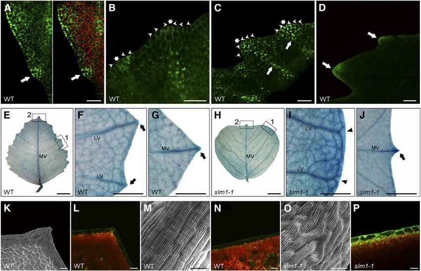
Context-Specific Effects Revealed by slm1        2107

maximum is the earliest marker of a new lateral organ primordium            compound-leafed plants, KNOX1 expression is reestablished
(Heisler et al., 2005; Barkoulas et al., 2008; Bayer et al., 2009;          later in developing primordia (Hareven et al., 1996; Bharathan
DeMason and Polowick, 2009; Koenig et al., 2009). Loss of                   et al., 2002; Kim et al., 2003b; Uchida et al., 2007; Shani et al.,
function of PIN1 leads to defects in initiation and separation of           2009). For example, in tomato, KNOX1 is expressed in develop-
lateral organs, such as fused cotyledons and leaves, pin-like               ing leaf primordia rather than only in the SAM (Hareven et al.,
inflorescences, and abnormal branches (Vernoux et al., 2000;                1996). In C. hirsuta, an Arabidopsis relative with dissected leaves,
Reinhardt et al., 2003). In addition, the auxin/PIN1 module that            transgenic lines with reduced expression of KNOX1 have fewer
triggers initiation of the leaf primordium at the flanks of the SAM is      leaflets, and ectopic KNOX1 expression leads to increased
probably redeployed in leaves to regulate leaf shape (Scarpella             leaflet number, suggesting that KNOX1 proteins are required
et al., 2010; Bilsborough et al., 2011). The PIN1 convergence               for leaflet formation in this species (Hay and Tsiantis, 2006).
points in the epidermis are associated with auxin activity maxima           Furthermore, leaflet formation in C. hirsuta involves auxin activity
at the tips forming serrations, and the sites of lateral vein               maxima accompanied by downregulation of KNOX1 gene ex-
formation are defined by internalizing auxin through the center             pression, implying a manner similar to the leaf initiation process
of the serrations (Hay et al., 2006; Scarpella et al., 2006;                at the SAM (Barkoulas et al., 2008).
Kawamura et al., 2010). As a result, the pin1 mutant has a                     Genetic regulation of compound leaf development is complex
smooth leaf margin (Hay et al., 2006). A recent study shows that            in various compound-leafed species. The FLORICAULA (FLO)/
two feedback loops are involved in Arabidopsis leaf margin                  LEAFY (LFY) putative orthologs, such as UNIFOLIATA/SINGLE
development. The first one relates to the transport of auxin                LEAFLET1 (SGL1) in some leguminous plants belonging to the
regulated by its own distribution via PIN1. In the second loop,             inverted repeat lacking clade (IRLC), including pea, alfalfa
CUP-SHAPED COTYLEDON2 (CUC2) promotes the generation                        (Medicago sativa), and Medicago truncatula, may function in
of auxin activity maxima while auxin represses CUC2 expression              place of KNOX1 to regulate compound leaf development (Hofer
(Bilsborough et al., 2011). In addition, CUC3 also plays a role in          et al., 1997; Wojciechowski et al., 2004; Champagne et al., 2007;
sculpting leaf margin serrations (Hasson et al., 2011).                     Wang et al., 2008). The leaves of the pea uni mutant have one
   In compound-leafed species, such as tomato, hairy bittercress            to three leaflets, which are simpler than wild-type leaves.
(Cardamine hirsuta), and pea (Pisum sativum), the initiations of            Neither rachises nor tendrils are formed (Hofer et al., 1997). In
leaflet primordia are correlated with local peaks of auxin response.        M. truncatula, leaves of the sgl1 mutant turn into a simple form
Perturbation of auxin transport by 1-N-naphthylphthalamic acid              (Wang et al., 2008). In these IRLC species, the expression of
(NPA) or inactivation of PIN1 orthologs inhibited the formation of          KNOX1 genes is not associated with compound leaves, although
leaflets in tomato and C. hirsuta (Barkoulas et al., 2008; Koenig           overexpression of KNOX1 in alfalfa results in an increase in leaflet
et al., 2009). In addition, differential auxin distribution is capable of   number (Hofer et al., 2001; Champagne et al., 2007). On the other
delineating the initiation of lobes and patterning blade outgrowth          hand, downregulation of the expression of the FLO/LFY gene in
in tomato (Koenig et al., 2009). Adult leaves of pea possess both           the non-IRLC legumes, such as soybean (Glycine max) and Lotus
leaflets and tendrils and the tendril is probably an abaxialized            japonicus, leads to moderate simplifications of compound leaves
leaflet (Hofer et al., 2009). Auxin is tightly associated with the          (Dong et al., 2005; Champagne et al., 2007). These data suggest
initiation of pinna primordia during compound leaf development in           that the FLO/LFY putative orthologs in IRLC species play an
pea (DeMason and Polowick, 2009). In NPA-treated plants, ter-               important role in compound leaf development.
minal tendrils were converted to leaflets in some cases, and the               Independent studies in different species shed light on the
number of lateral pinna pairs was reduced (DeMason and Chawla,              elaboration mechanisms of branches, leaves, leaflets, lobes,
2004; DeMason and Hirsch, 2006). Furthermore, the development               and serrations, in which the auxin/PIN1 module is extensively
of axillary meristem and the outgrowth of axillary buds require             involved. However, thus far, all mutants that have been found to
auxin synthesis and transport (Reinhardt et al., 2003; Ongaro and           affect lateral leaflet development do not affect the initiation of the
Leyser, 2008; Balla et al., 2011). The recently described model for         terminal leaflet (Efroni et al., 2010). Furthermore, although it has
pea bud outgrowth indicates that auxin is involved in the deter-            been reported that PIN1 is involved in the regulation of LFY via
mination of plant architecture (Balla et al., 2011). These results          local accumulation of auxin in Arabidopsis (Vernoux et al., 2000), it
demonstrate that auxin distribution and auxin response are central          is not clear how the putative orthologs of PIN1 and LFY interact
to the regulation of plant growth.                                          in compound-leafed species. M. truncatula is a model legume
   Much effort has been devoted to the identification of regulators         species whose adult leaves are trifoliates with serrations on the
for compound leaf development. Several mechanisms have                      leaf margin. The mechanism of leaf development in this species is
been shown to be involved in the developmental window to                    largely unknown, and only a few genes have been identified (Wang
elaborate leaf formation (Braybrook and Kuhlemeier, 2010; Efroni            et al., 2008; Chen et al., 2010). In this study, a mutant with a
et al., 2010). As the first homeodomain factors identified in               smooth leaf margin, smooth leaf margin1 (slm1), was isolated from
plants, Class I KNOTTED1-like homeobox (KNOX1) genes are                    a Tnt1 retrotransposon-tagged mutant population of M. trunca-
essential for the regulation of indeterminacy of SAM, but their             tula. SLM1 was identified by thermal asymmetric interlaced-PCR
expression is excluded from incipient leaf primordia in both                and association analysis. Molecular analysis shows that SLM1
simple-leafed and compound-leafed plants (Hake et al., 2004).               encodes an auxin efflux carrier protein Mt PIN10 in M. truncatula
The repression of KNOX1 genes persists during leaf formation in             (Schnabel and Frugoli, 2004) and is the ortholog of Arabidopsis
simple-leafed plants, such as Arabidopsis (Byrne et al., 2000; Ori          PIN1. SLM1 loss of function causes diffuse auxin distribution,
et al., 2000; Hay and Tsiantis, 2006; Uchida et al., 2007). In some         ultimately resulting in pleiotropic phenotypes in different regions,
2108      The Plant Cell

such as leaves, leaf margins, and flowers. The most striking                 leaflet number was reduced (Figures 1G, 1I, to 1K; see Supple-
feature of the slm1 mutant is the formation of multiple terminal             mental Figure 1M online). In the slm1-1 mutant, 42% of adult
leaflets and a simultaneous reduction in the number of lateral               leaves (n = 100) did not produce any lateral leaflets, and 45% of
leaflets accompanied with reduced SGL1 expression. In the slm1               adult leaves (n = 100) developed more than one terminal leaflet.
sgl1 double mutant, only the formation of lateral leaflets was               Additionally, the terminal leaflet length of the mutant was reduced
affected, suggesting that distinct developmental domains exist in            (Table 1). Fused leaflets were observed in rare cases (3%, n = 100)
the initiation of lateral and terminal leaflets. In addition, different      in slm1-1, resulting in a malformed leaflet (Figure 1J, arrowhead).
ontogenies were observed between distal serration and marginal               Fusion between petioles, which still show distinct domains of
serration formation, as well as leaflet formation. We present a              adaxial and abaxial sides, was frequently observed in slm1-1 and
possible model for the regulation of elaboration of compound                 was confirmed by anatomical analysis (Figures 1L and 1M).
leaves in M. truncatula, which is context dependent and tightly                 To better characterize compound leaf defects in slm1-1, scan-
correlated with the auxin/SLM1 module.                                       ning electron microscopy analysis of leaf development was
                                                                             performed. In M. truncatula, lateral leaflet primordia and the
                                                                             terminal leaflet primordium do not develop at the same stage
RESULTS                                                                      (Wang et al., 2008). A common leaf primordium (CM) that has the
                                                                             potential to differentiate into leaflet primordium developed first
SLM1 Is Required for Lateral Organ Development at the                        (stages 1 and 2) (Wang et al., 2008). Then, a pair of lateral leaflet
Vegetative Stage                                                             primordia (LL) emerged at the proximal end of the common leaf
                                                                             primordium at stage 3 (Figure 1N). Last, the common leaf primor-
To identify mutants with defects in compound leaf development,               dium differentiated into a single terminal leaflet primordium (TL) at
;10,000 independent lines of Tnt1 retrotransposon-tagged M.                  stage 4 (Wang et al., 2008). The ontogeny of compound leaf
truncatula populations were screened. Three mutant lines with                development probably implies that the developmental identities
obvious changes in leaf margin were identified. In contrast with             between lateral leaflet primordia and the terminal leaflet primor-
the wild type, which exhibits serrations on the leaf margin (Figure          dium are different in M. truncatula. At stage 5, the wild type
1A), these mutants showed an obvious smooth leaf margin                      showed a single terminal leaflet primordium, two lateral leaflet
phenotype (Figure 1B). The mutants were named slm1 (slm1-1,                  primordia, and two stipule primordia (ST) (Figure 1O). In slm1-1, at
slm1-2, and slm1-3).                                                         least two terminal leaflet primordia were initiated from a common
   Alterations in SLM1 activity not only affected leaf margin but            leaf primordium during the development of some adult leaves
also dramatically affected the formation of lateral organs, such             (Figure 1P). In some cases, three terminal leaflet primordia were
as cotyledons, leaves, flowers, and branches, and showed                     developed and no lateral leaflet primordium was formed at stage 5
increased indeterminacy throughout plant growth. slm1-1 seed-                (Figure 1Q), resulting in a compound leaf with three terminal
lings showed abnormal cotyledons, in which 24% displayed                     leaflets and without lateral leaflets (Figure 1R). Accompanying
fused cotyledons and 11% displayed triple cotyledons (Figures                these changes was an increase of the petiole and rachis length of
1C to 1E, n = 50), suggesting that SLM1 affects the initiation of            mature leaves in 8-week-old slm1-1 plants, suggesting that the
cotyledons or the partitioning of the embryo apical domain. In               proximal-distal axis of slm1-1 was also altered (Table 1).
addition, the elaboration of veins was abnormal in cotyledons of                Shoot branching of slm1-1 was also altered. In the wild type,
slm1-1, indicating that SLM1 is required for vascular patterning in          one node bears one trifoliate and one higher order branch
cotyledons (see Supplemental Figures 1A to 1C online).                       (Figures 1S and 1W). By contrast, the development of higher-
   In the wild type, the juvenile leaf (first true leaf) has a simple leaf   order branches and leaves on some nodes of slm1-1 was
morphology, and all other adult leaves are in trifoliate form                abolished, suggesting defects in the initiation of axillary meri-
(Figures 1C and 1F; see Supplemental Figure 1D online). In adult             stems and/or in the outgrowth of axillary buds (Figure 1T).
leaves of the wild type, a single terminal leaflet develops on the           Moreover, multiple leaves and branches frequently developed
distal end of the petiole/rachis and a pair of lateral leaflets              at the distal portion of stem in slm1-1 (Figures 1U to 1W). These
develops on the sides of the petiole (Figure 1H). The epicotyl               observations indicate that SLM1 is required for the determinacy
length of slm1-1 was increased compared with that of the wild                of shoot branching in M. truncatula. As a result, the architecture
type (see Supplemental Figures 1G to 1K online). In the slm1-1               of the slm1-1 plant was affected (Figure 1W) and displayed a
mutant, the juvenile leaf did not develop in most cases (Figures             semidwarf phenotype at the reproductive stage (see Supple-
1D and 1E; see Supplemental Figure 1L online). However, adult                mental Figure 1F online). In addition, all three alleles of SLM1
leaves in slm1-1 could be produced continuously (see Supple-                 exhibited the same defects in leaves with variation of leaflet
mental Figure 1E online). In addition, the first adult leaf developed        number and showed a semidwarf phenotype at the vegetative
at almost the same time in the wild type and slm1-1. Therefore,              stage (Table 1; see Supplemental Figure 1N online).
the initiation of the juvenile leaf in slm1-1 was abolished instead
of being skipped. This observation suggests that the formation               SLM1 Is Required for Flower Development
of the juvenile leaf is more sensitive than that of adult leaves in
slm1-1 and implies that loss of function of SLM1 has a greater               Wild-type M. truncatula enters the reproductive stage after ;60
impact on the juvenile stage than adult stage.                               d of growth. At the reproductive stage, a node in the wild type
   A striking change in slm1-1 is the development of multiple                bears one to three open flowers besides shoot branches and
terminal leaflets at the distal portion of rachis, while the lateral         leaves (Figure 2A). The wild-type flowers are comprised of a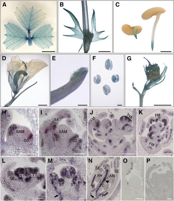
Context-Specific Effects Revealed by slm1            2109

Figure 1. The slm1-1 Mutant of M. truncatula Shows Developmental Defects at the Vegetative Stage.
(A) and (B) Leaf margin of the wild type (WT) (A) and slm1-1 (B).
(C) to (E) Four-day-old seedlings of the wild type (C) and slm1-1 ([D] and [E]). The arrow points to the first true leaf in the wild type (C). Note that the
development of the first true leaf was abolished in slm1-1 ([D] and [E]). The arrowhead points to a cotyledon fusion (D).
(F) and (G) Five-week-old plants of the wild type (F) and slm1-1 (G). Arrowheads point to three adult leaves of slm1-1. Two of the adult leaves have
double terminal leaflets developed at the distal end of petiole, and one has three terminal leaflets (G). No lateral leaflets developed in all three marked
adult leaves (G). Rac, rachis; Pet, petiole; TL, terminal leaflet; LL, lateral leaflet.
(H) to (K) Adult leaves of the wild type (H) and slm1-1 ([I] to [K]). Note that three terminal leaflets developed on the petiole and no lateral leaflets
were produced (J). Petiole fusion could be observed in (K). Two terminal leaflets developed on the distal end of each petiole, respectively (K).
Arrows indicate asymmetric lateral leaflets on the petiole (I). Arrowhead indicates two fused terminal leaflets (J). TL, terminal leaflet; LL, lateral
leaflet.
(L) and (M) Transverse sections of petioles in the wild type (L) and slm1-1 (M); the sectioning regions are shown in (H) and (K) by white lines,
respectively. AD, adaxial side; AB, abaxial side.
(N) to (R) Scanning electron micrographs of leaf primordia in the wild type at stage 3 (N) and stage 5 (O) and in slm1-1 at stage 3 (P) and stage 5 (Q), and
2110        The Plant Cell

Table 1. Phenotypic Characterization of Wild-Type and slm1 Plants of M. truncatula

Genotype           Terminal Leaflet Length (cm)            Terminal Leaflet Width (cm)           Petiole Length (cm)           Rachis Length (cm)

Wild type          1.80   6   0.20                         2.13   6   0.12                       6.53   6   0.58               0.95   6   0.13
slm1-1             1.47   6   0.17*                        2.24   6   0.15                       7.41   6   0.43*              1.64   6   0.15*
slm1-2             1.40   6   0.18*                        2.10   6   0.19                       7.44   6   0.55*              1.61   6   0.17*
slm1-3             1.51   6   0.26*                        2.17   6   0.16                       7.38   6   0.73*              1.42   6   0.37*
Leaflet length was measured from tip to base of the terminal leaflet. Leaflet width was measured from margin to margin of the terminal leaflet. Petiole
and rachis length were measured on the first fully expanded trifoliate of 8-week-old plants. Numbers are presented as mean 6 SD. The number of
observations in each mean is 35. Asterisks indicate that the differences between the wild type and slm1 are statistically significant at P < 0.05.

central carpel enclosed by a stamina tube, vexillum, sepal, and                  slm1-2 or slm1-3 could not be associated with the mutant
fused alae and keel (Figures 2B to 2F). By contrast, the lesion in               phenotype. Further analysis with the slm1-1 mutant identified
SLM1 resulted in abnormal flowers with mild, moderate, and                       19 flanking sequences, and one was confirmed to be associated
severe phenotypes in slm1-1 (Figures 2G to 2K), but flowering                    with the mutation. BLAST analysis using this flanking sequence
time of the mutant was not affected. Moreover, malformed floral                  was performed against the M. truncatula genome from the
organs and fused floral organs were frequently observed (Figures                 National Center for Biotechnology Information. A full-length
2L to 2P), suggesting that SLM1 regulates the initiation and                     genomic sequence of 2476 nucleotides was obtained. The full-
separation of floral organs. In addition, similar to shoot branching             length coding sequence (CDS) of SLM1 was cloned by RT-PCR
at the vegetative stage, flower arrangement was severely af-                     and found to contain 1776 nucleotides. Alignment between the
fected in slm1-1, indicating that branching is also abnormal at the              cDNA and genomic sequences of SLM1 revealed that SLM1
reproductive stage in slm1-1 (Figure 2Q). To further investigate                 consists of six exons and five introns (Figure 3A).
the defects in floral organs in slm1-1, the developmental pro-                      PCR amplification of the SLM1 genomic sequence from the
cesses of flowers between the wild type and slm1-1 were com-                     three mutant lines and the wild type revealed that only slm1-1
pared by scanning electron microscopy. At stage 6 (Benlloch                      carried a single 5.3-kb Tnt1 retrotransposon insertion, resulting in
et al., 2003), floral organ primordia were completely differenti-                the interrupted expression of SLM1 (Figures 3B and 3D). Amplifi-
ated and carpel suture became visible in the wild type (Figure                   cation of the full-length CDS of SLM1 from slm1-2 and slm1-3 and
2R). As for mutants, slm1-1 exhibited defects in the separation of               sequence comparison revealed that both slm1-2 and slm1-3
floral primordia, which could be seen as early as at stage 4                     contained lesions in this gene, which were not due to the Tnt1
(Figure 2U). At stage 6, fused floral primordia were more obvious                insertion. slm1-2 carried a single-base-pair deletion mutation in the
(Figure 2V), resulting in fused floral organs (Figures 2W to 2Y),                first exon, which caused a shift in the reading frame and resulted in
which are distinct from those of the wild type (Figure 2S). At the               premature termination of the encoded protein (Figure 3A). More-
late stage of flower development, anthers of slm1-1 dehisced                     over, this change introduced an additional AseI restriction site. This
normally, similar to those of the wild type (Figures 2T and 2Y), and             allowed us to amplify the sequence spanning the mutation site and
their pollen was viable, as revealed by pollen staining (Figure 2Z).             generate a cleaved polymorphic sequence marker, which yielded
The three alleles of SLM1 showed the same defective phenotype                    products of 1169 and 518 bp after AseI digestion of the PCR
of flowers. However, variation in fertility among the three slm1                 product in slm1-2 but not in the wild type (Figure 3C). The other
alleles was observed. slm1-1 was infertile, while slm1-2 and                     mutant, slm1-3, has a single-base-pair substitution (G to A) in the
slm1-3 could occasionally produce seedpods and seeds with                        intron splicing site, resulting in altered mRNA splicing (Figure 3A).
normal germination ability (see Supplemental Figure 2 online).                   To verify this, the CDS of SLM1 in slm1-3 was amplified. Longer
                                                                                 mRNA molecules were indeed amplified in slm1-3, confirming the
Molecular Cloning of SLM1                                                        altered splicing of SLM1 transcripts in this allele (Figure 3D).

To identify the gene responsible for the developmental defects,                  SLM1 Complements the Mutant Phenotype of slm1
thermal asymmetric interlaced-PCR was performed to recover
the flanking sequences from the three mutant lines. Surprisingly,                As Tnt1 retrotransposon-tagged lines of M. truncatula generally
except for slm1-1, the flanking sequences recovered from either                  contain 20 to 50 insertions (Tadege et al., 2008), two backcrosses

Figure 1. (continued).

the developing leaf in slm1-1 at stage 9 (R). Arrowheads point out that at least two terminal leaflet primordia initiated from a common leaf primordium
(P). CM, common leaf primordium; TL, terminal leaflet primordium; LL, lateral leaflet primordium; ST, stipule primordium; Pet, petiole.
(S) to (V) Development of branches in the wild type (S) and slm1-1 ([T] to [V]). Arrow points to the node that bears one trifoliate and a higher-order branch
in the wild type (S). Arrowheads point to nodes without branches and leaves in (T) and to the distal portion of stem with radial multiple leaves and
branches in (U). Scanning electron micrograph shows the SAM with radial lateral organs in slm1-1 (V). ST, stipule.
(W) A schematic illustration of branch arrangement in the wild type (left) and slm1-1 (right) at the vegetative stage.
Bars = 5 mm in (A) to (K) and (S) to (U), 200 mm in (L) and (M), and 50 mm in (N) to (R) and (V).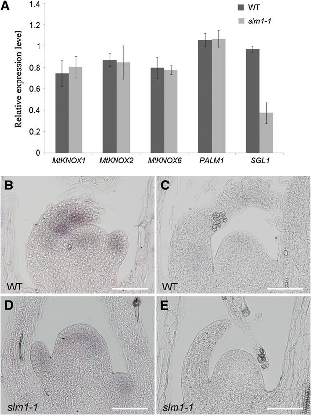
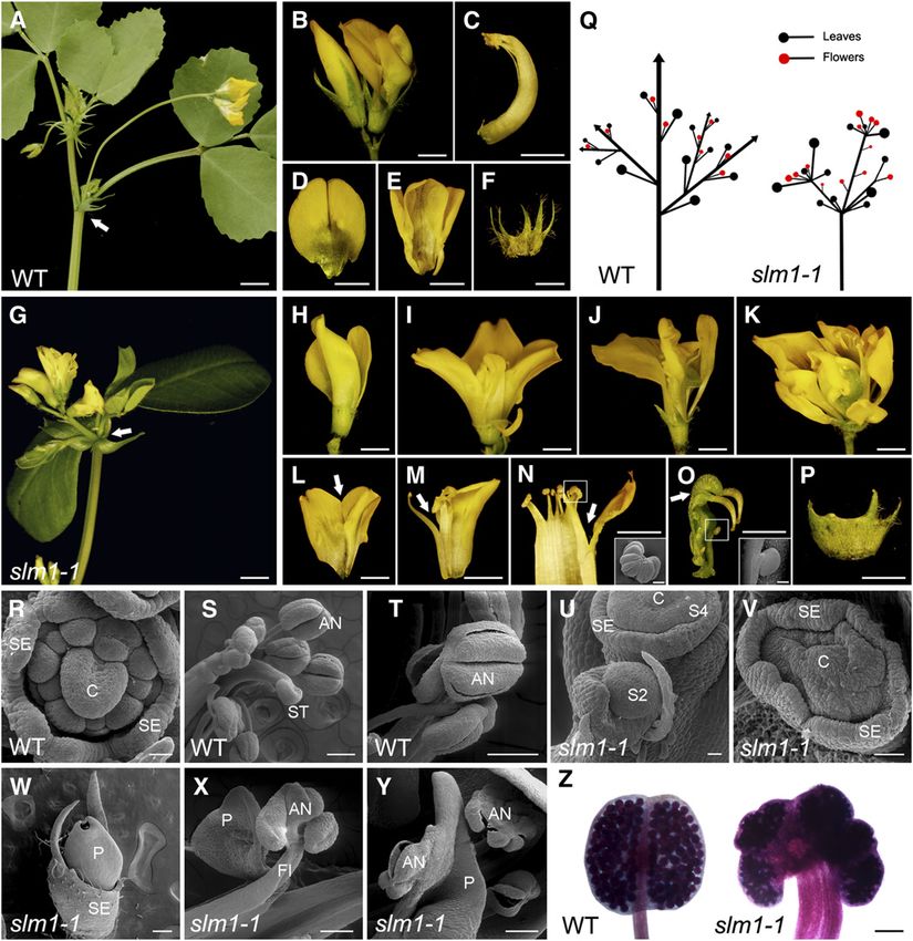
Context-Specific Effects Revealed by slm1             2111

Figure 2. The slm1-1 Mutant of M. truncatula Shows Developmental Defects at the Reproductive Stage.

(A) Flower development in the wild type. Arrow indicates a node that bears two open flowers and one fully expanded trifoliate.
(B) Flower phenotype in the wild type. The flowers of the wild type show bilateral symmetry along the dorsal-ventral axis.
(C) to (F) Dissected floral organs of the wild type. The side view of the central carpel (C), top view of vexillum (D), alae and keel (E), and sepal (F).
(G) Flower development in slm1-1. Arrow indicates that flowers and leaves develop radially at the distal portion of stem.
(H) to (K) Flower phenotype in slm1-1 with mild (H), moderate ([I] and [J]), and severe (K) alterations.
(L) to (P) Dissected floral organs of slm1-1. Fusions between floral organs were frequently observed; for example, the fusion between vexillums (L),
between stamen and petal (M), between anthers (N), and between pistils (O). The sepal is also abnormal (P). The insets in (N) and (O) show fused
anthers and exposed ovules by scanning electron microscopy, respectively. Arrows in (L) to (O) indicate the fusion of floral organs.
(Q) A schematic illustration of branch arrangement of the wild type (left) and slm1-1 (right) at the reproductive stage.
(R) to (T) Scanning electron microscopy analysis of floral organs in the wild type. Representative images show floral primordia at stage 6 (R), anthers
and stigma (S), and dehiscing anthers (T) in a mature flower.
(U) to (Y) Scanning electron microscopy analysis of floral organs in slm1-1. Representative images show floral primordia at stage 2 ([U], S2), stage 4
([U], S4), and stage 6 (V). At the late stage of floral development, fully fused petals (W), fused filament and petal (X), and dehiscing anther (Y) were
observed.
(Z) Pollen staining in the wild type (left) and slm1-1 (right).
C, carpel; SE, sepal; P, petal; ST, stigma; AN, anther; FI, filament; S2, stage 2; S4, stage 4; WT, wild type. Bars = 5 mm in (A) and (G), 2 mm in (B) to (F)
and (H) to (P), 200 mm in (S), (T), (W) to (Y), and the insets in (N) and (O), 100 mm in (Z), and 50 mm in (R), (U), and (V).
2112      The Plant Cell

Figure 3. Molecular Characterization of SLM1 in M. truncatula.

(A) Schematic diagram of the gene structure of SLM1. The positions of the ATG start and TGA stop codons are shown. Vertical arrows mark the
nucleotide changes in various slm1 alleles. Numbers indicate nucleotide positions of the site of mutations. Boxes represent exons and lines represent
introns. A single base, T (thymine), was deleted in slm1-2.
(B) PCR amplification of SLM1 from the wild type (WT) and slm1-1. A single insertion of the tobacco Tnt1 retrotransposon (;5.3 kb) was detected in
slm1-1.
(C) Transcripts of SLM1 from the wild type and slm1-2 were amplified by RT-PCR and digested by AseI, resulting in length polymorphism because of a
single-base-pair deletion mutation in slm1-2. Three technical replicates were performed.
(D) RT-PCR analysis of SLM1 transcripts in the wild type and slm1 alleles. Altered splicing of transcript in slm1-3 is shown. Actin was used as a loading
control. Three technical replicates were performed.

were performed to obtain a segregation population of slm1-1 to                 5 online). Based on these results, SLM1 is identified as a putative
confirm that the mutant phenotype was caused by Tnt1 insertion in              ortholog of Arabidopsis PIN1.
a single gene. The mutants and wild-type-like plants showed a                     To assess the function of SLM1 as an auxin transporter, the
segregation ratio of 1:3, suggesting that the mutant phenotype was             Arabidopsis pin1 mutant was transformed with a construct con-
associated with a single recessive locus (see Supplemental Figure              taining the SLM1 CDS driven by the Arabidopsis PIN1 promoter.
3A online). To further confirm that the mutant phenotype was                   This construct was capable of fully rescuing the defects of the pin1
caused by the mutation of this gene, a construct carrying a 5.2-kb             mutant, suggesting that SLM1 is a functional auxin efflux trans-
genomic fragment containing the promoter region and SLM1 open                  porter (see Supplemental Figure 6 online). To examine the ability of
reading frame was transformed into slm1-1 plants. Phenotypic                   Arabidopsis PIN1 to suppress the loss-of-function phenotype
observation confirmed that complementary SLM1 expression fully                 seen in slm1-1, the Arabidopsis ProPIN1:PIN1:GFP (green fluo-
rescued leaf and floral defects in slm1-1 at different developmental           rescent protein) construct was introduced into the slm1-1 mutant.
stages (see Supplemental Figure 3B online).                                    It has been shown that this construct was sufficient to rescue the
                                                                               C. hirsuta pin1 mutant (Barkoulas et al., 2008). The defects of
                                                                               slm1-1 were fully suppressed in eight transgenic plants and
SLM1 Is an Ortholog of Arabidopsis PIN1                                        partially suppressed in seven other transgenic plants, suggesting
BLASTX analysis using the SLM1 CDS revealed several hits                       functional conservation between SLM1 and PIN1.
belonging to the auxin efflux carrier protein family. Most known
members of this family are PIN components of auxin efflux                      Expression Pattern of SLM1
facilitators in plants. These carriers are auxin specific and local-
ized to the basal ends of auxin transport-competent cells                      The expression pattern of SLM1 in different tissues and organs
(Kramer, 2004; Blakeslee et al., 2005). Phylogenetic analyses                  was analyzed using the M. truncatula Gene Expression Atlas
with 18 members of the PIN family from Arabidopsis (At), C.                    (http://mtgea.noble.org/v2). The relative expression level of
hirsuta (Ch), P. sativum (Ps), Triticum aestivum (Ta), Oryza sativa            SLM1 was obtained using the probe set Mtr.47942.1.S1, which
(Os), and Brassica juncea (Bj) revealed that SLM1 was evolu-                   represented SLM1 in the microarray chip. The data revealed that
tionarily closer to the PIN1 family and showed 65% identity with               SLM1 was expressed in almost all tissues. The expression of
At PIN1, suggesting a possible effect of SLM1 on polar auxin                   SLM1 was relatively high in the vegetative bud, root tip, and
transport (see Supplemental Figure 4 and Supplemental Data                     developing nodule (see Supplemental Figure 7 online). To de-
Set 1 online). Sequence alignment was also performed among                     terminate the expression pattern comprehensively, an SLM1
At PIN1, Ch PIN1, Ps PIN1, Ta PIN1, Os PIN1, Bj PIN1, and SLM1.                promoter-b-glucuronidase (GUS) construct was introduced into
SLM1 shared high sequence similarity with PIN1 proteins at the                 wild-type M. truncatula, and GUS activity was examined in
conserved C- and N-terminal domains (see Supplemental Figure                   transgenic plants. The transgenic plants showed GUS expression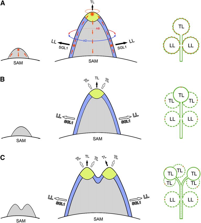
Context-Specific Effects Revealed by slm1 2113
in leaf veins, the basal region of leaflets (Figure 4A), the stem, primordia develop at stage 3 (Benlloch et al., 2003). High SLM1
stipule (Figure 4B), and root tip of germinating seeds (Figure 4C). expression was detected in the developing common primordia
GUS expression was also detected in the basal region of the at stage 3 (Figure 4L) and in the developing petals, stigma,
flower (Figure 4D), stigma (Figure 4E), anther (Figure 4F), and and stamens at stage 5 (Figure 4M). At stage 7, SLM1 mainly
young seedpod (Figure 4G). accumulated in petals and inside the carpel, where ovules
The spatial and temporal localizations of SLM1 at the vegeta- were under development (Figure 4N). In addition, SLM1 mRNA
tive and reproductive stages were examined further by in situ was detected in the vascular bundles (Figures 4M and 4N,
hybridization analysis in the wild type. SLM1 mRNA was de- arrows).
tected in the cells that give rise to leaf primordia at the SAM and
in the developing leaf primordia (Figures 4H and 4I). Strong SLM1
Local Auxin Activity Maxima Facilitate the Initiation of Leaf
expression was observed in floral meristems and restricted to the
and Floral Primordia
site that would give rise to floral organ primordia (Figures 4J and
4K). In M. truncatula, sepal primordia initiate first in floral mer- To investigate whether the initiation of leaf and floral primordia in M.
istems. Then, common primordia that produce petal and stamen truncatula is related to auxin transportation and accumulation, PIN1
Figure 4. Expression Pattern of SLM1 in M. truncatula.
(A) to (G) Promoter-GUS fusion studies of SLM1 expression in transgenic M. truncatula. SLM1 promoter driven GUS is expressed in the adult leaf (A),
stem and stipule (B), root tip of germinating seeds (C), flower (D), stigma (E), anther (F), and 5-d-old seedpod (G).
(H) to (P) In situ hybridization analysis of SLM1 mRNA in vegetative and reproductive apices of the wild type.
(H) and (I) Longitudinal sections of the SAM at stage 1 (S1; [H]) and stage 4 (S4; [I]).
(J) and (K) Longitudinal section (J) and transverse section (K) of floral apices at stage 2.
(L) to (N) Longitudinal sections of the floral apical meristem at stage 3 (L), stage 5 (M), and stage 7 (N).
(O) and (P) The sense probe was hybridized and used as control. Arrows indicate vascular bundles. Arrowhead indicates the inside of the carpel.
FM, floral meristem; SE, sepal; P, petal; C, carpel; ST, stamen; AN, anther. Bars = 5 mm in (A), 2 mm in (B) to (D) and (G), 200 mm in (E) and (F), and 50
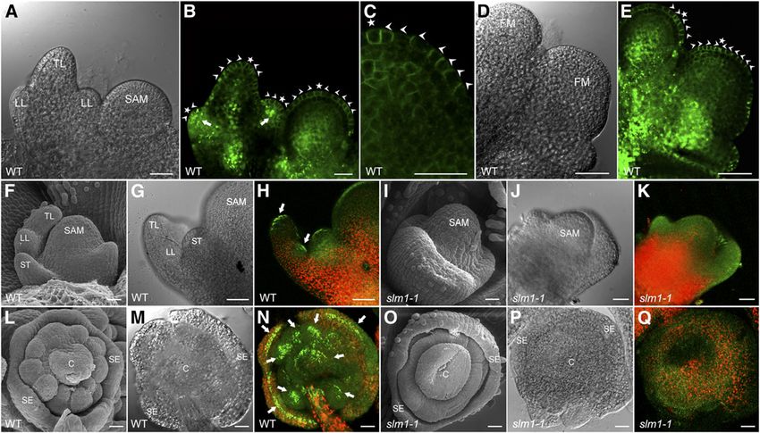
mm in (H) to (P).2114 The Plant Cell localization was examined in M. truncatula plants transformed SLM1 Regulates Leaf Margin and Leaf Marginal with the Arabidopsis ProPIN1:PIN1:GFP reporter (Benková et al., Cell Development 2003). The results showed that PIN1 is apically localized at the epidermal cells of the leaf and floral meristem and mark the site One prominent phenotype of slm1-1 was the conversion of the of incipient primordia formation at the meristem flank toward serrated leaf margin to smooth leaf margin. To understand how the primordia tips (Figures 5A to 5E). In addition, GFP expression was leaf serrations are formed in M. truncatula, the DR5rev:GFP auxin also upregulated in initiating lateral leaflet primordia (Figure 5B, response reporter and the ProPIN1:PIN1:GFP reporter were trans- arrows). These observations suggest that the local auxin activity formed into wild-type plants. DR5rev:GFP expression was detected maxima generated by PIN1/SLM1 probably facilitate the formation in the tips of initiating serrations (Figure 6A). To examine whether of both leaves and floral primordia. To verify this hypothesis, the auxin accumulation is generated by PIN1/SLM1-directed auxin DR5rev:GFP auxin response reporter was transformed into the wild efflux, localization of the ProPIN1:PIN1:GFP reporter was exam- type and slm1-1, respectively. A gradient of DR5 activity, with a ined. Polar expression of ProPIN1:PIN1:GFP was observed in the maximum at the tips of leaves and floral organ primordia, was epidermal cells, predicting that the flow of auxin converged to the detected by GFP signal in the wild type, indicating that auxin site of serration initiation (Figure 6B). As the leaf serrations were maxima are required for the proper development of primordia expanding, ProPIN1:PIN1:GFP expression displayed evidence (Figures 5F to 5H and 5L to 5N). However, auxin distribution was that the direction of auxin flux was toward the tips of serrations disturbed in slm1-1, which was defective in the positioning and (Figure 6C) where auxin accumulated (Figure 6D). These observa- separation of lateral organ primordia (Figures 5I to 5K and 5O to tions demonstrate that auxin transportation and activity gradients 5Q). These results demonstrate that the defects of slm1-1 are are important for the formation of leaf marginal serrations. caused by disorders of auxin transportation and distribution, To further elucidate the role of auxin in leaf margin morpho- which are tightly correlated with SLM1. genesis, the DR5:GUS auxin response reporter (Ulmasov et al., Figure 5. PIN1/SLM1-Dependent Auxin Gradients in Leaf and Floral Organ Formation in M. truncatula. (A) Leaf primordia of the wild type (WT) at stage 4. (B) and (C) Distribution of the ProPIN1:PIN1:GFP marker (green signal) in leaf primordia (B) and a close view of the localization of ProPIN1:PIN1:GFP marker in the SAM (C). Arrowheads mark the direction of PIN1 polarization. Asterisks indicate the auxin convergence points that mark the site of incipient primordium initiation. (D) Floral primordia of the wild type at stage 2. FM, floral meristem. (E) Distribution of the ProPIN1:PIN1:GFP marker in floral primordia. Arrowheads point to the direction of PIN1 polarization. Asterisks indicate the auxin convergence points. (F) to (K) Leaf primordia of the wild type ([F] to [H]) and slm1-1 ([I] to [K]). Leaf primordia harboring the auxin response marker DR5 (DR5rev:GFP) were observed by scanning electron microscopy ([F] and [I]), light-field microscopy ([G] and [J]), and confocal microscopy ([H] and [K]). Arrows point to auxin accumulation at the tip of lateral and terminal leaflet primordia. (L) to (Q) Floral primordia of wild-type ([L] to [N]) and slm1-1 ([O] to [Q]). Arrows point to auxin accumulation at the tip of floral organ primordia. TL, terminal leaflet primordium; LL, lateral leaflet primordium; ST, stipule; C, carpel; SE, sepal; WT, wild type. Bars = 25 mm.
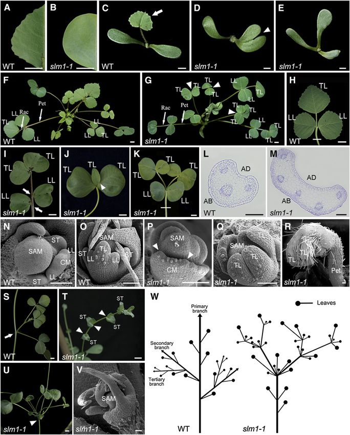
Context-Specific Effects Revealed by slm1             2115

1997) was introduced into wild-type and slm1-1 plants to reflect                 observations indicate that SLM1 regulates the elaboration of leaf
relative auxin levels. Terminal leaflets were used to compare the                shape and the pattern of leaf venation by directing auxin distri-
configuration of the leaf margin and veins of both the wild type                 bution. Moreover, a smooth leaf margin was also observed in the
and slm1-1, since the terminal leaflet and lateral leaflet showed                NPA-treated plants, confirming that SLM1 is involved in polar
similar DR5:GUS expression patterns. In the wild type, auxin                     auxin transport, which is correlated with the formation of the leaf
accumulated in the midvein and at the tips of serrations (Figure                 margin (see Figure 8K). In addition, we noticed that the distal
6E). Furthermore, local auxin maxima at the tips of serrations                   serration associated with the midvein was intact in slm1-1,
were tightly associated with the positioning of lateral veins.                   similar to as in the wild type, implying that different develop-
Among the lateral veins, the auxin level gradually decreased from                mental mechanisms exist between distal serration and marginal
the tips of serrations to the midvein (Figures 6E and 6F). Auxin                 serrations (Figures 6G and 6J). On the other hand, the surface of
accumulation was also observed in the midvein of slm1-1.                         marginal cells changed in the fully expanded leaves of slm1-1.
However, auxin distribution in lateral veins was diffuse. In con-                The ridge-like structure was distorted due to the loss of function
trast with the correlation between lateral veins and serrations in               of SLM1, compared with that of the wild type (Figures 6K, 6M,
the wild type, higher-order and free-ending veins developed at                   and 6O). Furthermore, auxin accumulation was observed within
the distal end of lateral veins in slm1-1 (Figures 6H and 6I). These             marginal cells of both the wild type and slm1-1 by assaying

Figure 6. Involvement of SLM1 in Leaf Margin Development in M. truncatula.

(A) DR5rev:GFP expression maximum at the site of serration initiation of the leaf margin (green signal, left) and an overlay image with chlorophyll
autofluorescence (red signal, right) in the wild type (WT). Arrows point to the site of serration initiation.
(B) and (C) PIN1:PIN1-GFP expression during the development of leaf serrations. The localization of ProPIN1:PIN1-GFP reporter is polar at the site of
serration initiation (B) and developing serrations (C). Asterisks indicate auxin flow converging at the tip of a serration. Arrowheads indicate the
orientation of auxin flow predicated by PIN1/SLM1. Arrows point to the location of lateral vein formation.
(D) DR5rev:GFP expression in developing leaf serrations. Arrows indicate auxin accumulation at the tip of serrations.
(E) to (G) DR5:GUS expression in the fully expanded terminal leaflet of the wild type (E). Close views of marginal serration (empty box 1) and distal
serration (empty box 2) are shown in (F) and (G), respectively. Arrows mark auxin accumulation at the tip of serrations. MV, midvein; LV, lateral vein.
(H) to (J) DR5:GUS expression in a fully expanded terminal leaflet of slm1-1 (H). Close views of leaf margin (empty box 1) and distal serration (empty box
2) are shown in (I) and (J), respectively. Arrowheads point to lateral veins, which do not terminate at the margins. Arrow indicates auxin accumulation at
the tip of the distal serration. MV, midvein; LV, lateral vein.
(K) to (P) Observation of marginal cells in the wild type ([K] to [N]) and slm1-1 ([O] and [P]). Scanning electron microscopy analysis of the surface of
marginal cells at the tip (K) and the side (M) of serrations in the wild type and the surface of marginal cells in slm1-1 (O). DR5rev:GFP expression is shown
in the marginal cells at the same location in the wild type ([L] and [N]) and slm1-1 (P).
Bar = 25 mm in (A) to (C), 5 mm in (E) and (H), 1 mm in (F), (G), (I), and (J), 150 mm in (D), 50 mm in (K), (L), (N), and (P), and 20 mm in (M) and (O).
2116 The Plant Cell
DR5rev:GFP expression (Figures 6L, 6N, and 6P). GFP expression
level was higher in slm1-1, suggesting that more auxin accumu-
lated in the marginal cells of these plants. These observations
imply that the development of marginal cells also requires proper
auxin activity gradients.
SGL1 Is Partially Involved in Lateral Leaflet Defects in slm1
Defects in compound leaf development in slm1-1 suggest that
SLM1 is required for the correct formation of both the lateral
leaflet and terminal leaflet. Previous studies indicate that multiple
genes are involved in leaf development (Champagne et al., 2007;
Wang et al., 2008; Chen et al., 2010). Quantitative RT-PCR (qRT-
PCR) analysis was performed to determine the expression of
M. truncatula genes that have been proposed to regulate this
process. These genes included M. truncatula homologs of the
Class I KNOX1 homeobox gene family, Mt KNOX1 (SHOOT
MERISTEMLESS-like), Mt KNOX6 (SHOOT MERISTEMLESS-
like), Mt KNOX2 (KNAT1/BREVIPEDICELLUS-like) (Di Giacomo
et al., 2008), M. truncatula PALMATE-LIKE PENTAFOLIATA1
(PALM1) (Chen et al., 2010), and SGL1 (Wang et al., 2008). The
expression of SGL1 was suppressed in slm1-1, whereas the
expression of other genes remained essentially unchanged
(Figure 7A). The spatial localization of SGL1 in slm1-1 during
leaf development was further examined by in situ hybridization
analysis. mRNA expression of SGL1 was detected in the SAM
and young leaf primordia in the wild type (Figure 7B). In slm1-1,
the reduction in SGL1 expression supported the qRT-PCR
results, illustrating that endogenous SGL1 expression was down-
regulated (Figure 7D). As a negative control, a sense probe did not
give any hybridization signal (Figures 7C and 7E). Since SGL1 Figure 7. Expression Analysis of Genes Related to Compound Leaf
regulates lateral leaflet development, these observations Development in M. truncatula.
indicate that downregulated expression of SGL1 probably con-
(A) Transcript levels of the M. truncatula KNOX1, PALM1, and SGL1
tributes to the reduced lateral leaflet number in slm1-1.
genes in the wild type (WT) and slm1-1. Transcript levels were measured
by qRT-PCR using leaf meristems from 6-week-old plants. Means 6 SE
The Development of the Terminal Leaflet Is Regulated by are shown (n = 3).
SLM1 Independently of SGL1 (B) to (E) In situ hybridization and expression patterns of SGL1 in leaf
primordia of the wild type (B) and slm1-1 (D). SGL1 sense probes were
The role of SLM1 in promoting leaflet development was further used as a negative control in the wild type (C) and slm1-1 (E). Bars = 50 mm.
examined by generating double mutants with the sgl1 mutant
(Figures 8A to 8J; see Supplemental Figure 8 online). In slm1-1,
the number of lateral leaflets decreased, but the number of the same medium developed two (Figure 8K, bottom panel, a) or
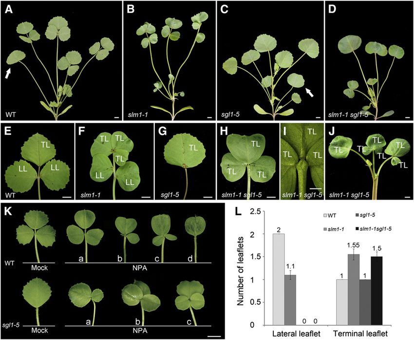
terminal leaflets increased (Figures 8B, 8F, and 8L). In sgl1-5, all three (Figure 8K, bottom panel, b and c) terminal leaflets. Taken
adult leaves were simple and only terminal leaflets were pre- together, these observations reveal that the local gradients of
served (Figures 8C and 8G). The slm1-1 sgl1-5 double mutant did auxin activity, generated by SLM1, are differentially required for
not produce lateral leaflets but developed multiple terminal the development of lateral and terminal leaflets in M. truncatula.
leaflets whose number was similar to that of slm1-1 (Figure 8L). They also demonstrate that the development of terminal leaflets
To examine whether local auxin activity gradients are required for is independent of SGL1 activity.
the development of leaflets, the gradients were perturbed by
growing wild-type and sgl1-5 plants on medium containing 50
mM NPA. The results showed that the phenotype of wild-type DISCUSSION
plants treated with NPA mimicked the slm1-1 phenotype. The
leaf margin of all adult leaves became smooth and various leaf- SLM1 Is the M. truncatula Putative Ortholog of
let numbers were noticed (Figure 8K, top panel, a to d). The Arabidopsis PIN1
following leaflet variations were observed: one lateral leaflet
degenerated (Figure 8K, top panel, b); three terminal leaflets slm1, identified as a recessive mutant by segregation analysis, is
developed (Figure 8K, top panel, c), and a simple leaf formed defective in leaf and floral development. In this study, three
(Figure 8K, top panel, d). In addition, the sgl1-5 mutants grown on independent SLM1 alleles were found. They showed the sameContext-Specific Effects Revealed by slm1 2117 Figure 8. SLM1 and SGL1 Regulate Compound Leaf Development in M. truncatula. (A) to (D) Four-week-old plants of the wild type (WT) (A), slm1-1 (B), sgl1-5 (C), and slm1-1 sgl1-5 (D). Arrows indicate the juvenile leaf in (A) and (C). Note that the juvenile leaf did not develop in (B) and (D). (E) to (J) Adult leaves of the wild type (E), slm1-1 (F), sgl1-5 (G), and slm1-1 sgl1-5 ([H] and [I]). Close view of the basal region of terminal leaflets of slm1-1 sgl1-5 (I). Note that three terminal leaflets were developed on the distal end of petiole (I). Radial multiple leaves developed at the distal portion of the stem in slm1-1 sgl1-5 (J). TL, terminal leaflet; LL, lateral leaflet. (K) Adult leaf phenotype of wild-type (top) and sgl1-5 (bottom) plants grown on MS medium supplemented with 50 mM NPA. Control plants were grown on MS medium supplemented with the same concentration of DMSO. The letters a to d indicate variations of compound leaf forms in the wild type and sgl1-5 under NPA treatment. (L) Number of lateral leaflets and terminal leaflets in the wild type and the mutants. Fifty-day-old plants were used for calculating the leaflet numbers of adult leaves. Means 6 SE are shown (n = 100). Bars = 5 mm in (A) to (H), (J), and (K), and 2 mm in (I). defects in the development of compound leaves and flowers 2000). Third, Arabidopsis PIN1 is an auxin efflux carrier required for except for fertility. slm1-1 is infertile, and the expression of SLM1 polar auxin transport. Auxin distribution at the meristem of pin1 or in this allele is completely interrupted by a Tnt1 insertion, indi- NPA-grown plants is diffuse (Benková et al., 2003), which is similar cating that slm1-1 is a null allele. slm1-2 and slm1-3 are point to the auxin distribution pattern in slm1. Fourth, the Arabidopsis mutations and retain low fertility. The maintenance of partial ProPIN1:PIN1:GFP construct has been used for PIN1 localization fertility in slm1-2 and slm1-3 is probably because the SLM1 in different species to investigate the initiation of leaf/leaflet pri- proteins in the mutants contain partially conserved N-terminal mordia and the development of the leaf margin (Benková et al., domains (see Supplemental Figure 5 online). 2003; Barkoulas et al., 2008; Koenig et al., 2009). The Arabidopsis SLM1 is identified as the M. truncatula putative ortholog of ProPIN1:PIN1:GFP construct is capable of fully rescuing the slm1 Arabidopsis PIN1 by the following lines of evidence. First, some of mutant, suggesting conserved function between PIN1 and SLM1. the defects in slm1 were similar to the classical Arabidopsis pin1 Cross-species complementation of PIN1 was also found between phenotypes, such as triple cotyledons, fused lateral organs, ab- Arabidopsis and C. hirsuta (Barkoulas et al., 2008), indicating that normal branching, and smooth leaf margin (Gälweiler et al., 1998; the promoter of PIN1 can be trans-activated in both M. truncatula Vernoux et al., 2000; Reinhardt et al., 2003; Hay et al., 2006). and C. hirsuta. Fifth, the SLM1 CDS driven by the Arabidopsis PIN1 Second, the expression patterns of SLM1 revealed by the SLM1 promoter could complement the pin1 mutant phenotype, suggest- promoter-GUS reporter and in situ hybridization are similar to those ing that SLM1 is a functional auxin efflux transporter and can of PIN1 in Arabidopsis (Palme and Gälweiler, 1999; Vernoux et al., restore polar auxin transport in a heterologous system.
2118      The Plant Cell

Developmental Domains in the Elaboration of Lateral and                    SGL1-dependent manner (Figure 9A). In slm1, the founder cells
Terminal Leaflets in M. truncatula                                         cannot be identified in the marginal blastozone without auxin
                                                                           activity maxima, resulting in reduced lateral leaflet number
The development of compound leaves has been documented in                  (Figures 9B and 9C). On the other hand, a terminal zone is
several species such as tomato, C. hirsuta, pea, and M. trunca-            located at the apex of a common primordium and is probably
tula (Hareven et al., 1996; Hofer et al., 1997; DeMason and                more likely to resemble the SAM with a radial prepattern than to
Chawla, 2004; Hay and Tsiantis, 2006; Champagne et al., 2007;              have dorsiventral polarity. In the wild type, the terminal zone is
Barkoulas et al., 2008; Blein et al., 2008; Wang et al., 2008;             competent for the formation of multiple terminal leaflet primordia
DeMason and Polowick, 2009; Koenig et al., 2009; Shani et al.,             but is prevented from doing so by drainage of auxin into the tip of
2009; Chen et al., 2010). Several key genes were uncovered by              the terminal zone, which results in a single terminal leaflet
analyses of various mutants with defects in compound leaf                  primordium (Figure 9A). By contrast, multiple terminal leaf pri-
formation and development. However, to date, all the mutants               mordia can initiate from the terminal zone in slm1 due to its
identified to be defective in the initiation of lateral leaflets do not    diffuse auxin distribution, resembling leaf primordia initiated
affect the formation of the terminal leaflet (Efroni et al., 2010). In     from SAM and resulting in multiple terminal leaflets (Figures 9B
our experiments, the mutation in SLM1 reveals a novel pheno-               and 9C).
type of increased terminal leaflet number, suggesting that a
unique mechanism is involved in compound leaf development in
M. truncatula.                                                             Different Molecular Mechanisms in the Marginal Blastozone
   In compound-leafed species, lateral leaflets are considered             and Terminal Zone in IRLC Legumes
to be formed from a region at the primordium margin named                  Class I KNOX1 genes are expressed in the SAM and involved in
the marginal blastozone, which has meristematic potential                  acquiring and maintaining SAM activity (Hay and Tsiantis,
(Hagemann and Gleissberg, 1996; Dengler and Tsukaya, 2001).                2009). The auxin and AS1 pathway repress the expression of
In addition, the initiation of lateral leaflet primordia is associ-        the KNOX gene BREVIPEDICELLUS to promote leaf fate (Hay
ated with local peaks of auxin response (Barkoulas et al., 2008;           et al., 2006). KNOX1 proteins are involved in compound leaf
DeMason and Polowick, 2009; Koenig et al., 2009). In pea, auxin            patterning in a number of species (Bharathan et al., 2002; Hay
peaks are also tightly associated with the initiation of pinna             and Tsiantis, 2006; Barkoulas et al., 2008; Shani et al., 2009) but
primordia that will differentiate into leaflets or tendrils (DeMason       excluded from leaflet formation in IRLC legumes (Champagne
and Polowick, 2009). In accordance with these findings, we                 et al., 2007). The expression level of M. truncatula homologs of
found that local auxin activity gradients generated by SLM1                KNOX1 genes remained essentially unchanged in slm1, indi-
facilitate the initiation of lateral leaflets in M. truncatula. In our     cating that these genes are not involved in the defects in leaf
experiments, auxin activity maxima were also observed at the               development.
apex of the common leaf primordium (Figure 5H). Here, we name                 A reduction in LFY expression in the inflorescence apices of
the apex of the common leaf primordium the terminal zone, in               the pin1 mutant was reported in Arabidopsis previously, indicat-
reference to the concept of the marginal blastozone. In slm1, the          ing that PIN1 probably regulates LFY expression indirectly via
number of terminal leaflets increased, while the number of lateral         local accumulation of auxin (Vernoux et al., 2000). SGL1, the
leaflets decreased. This observation suggests that the develop-            putative ortholog of LFY in M. truncatula, is required for the
mental characteristics of lateral leaflet primordia and the terminal       initiation of lateral leaflet primordia (Wang et al., 2008). It has
leaflet primordium are probably different, implying that distinct          been proposed that FLO/LFY may function in place of KNOX1
developmental domains exist in the elaboration of lateral and              genes in the regulation of compound leaf development in IRLC
terminal leaflets in M. truncatula. This hypothesis is also sup-           legumes (Champagne et al., 2007). Thus, the reduced SGL1
ported by the ontogenic analysis that the lateral leaflet and              expression in slm1 implies that SGL1 is likely partially responsi-
terminal leaflet have their own ontogenies with distinct develop-          ble for the defects in lateral leaflet development. In addition, our
mental status in M. truncatula.                                            data demonstrate that the downregulated expression of SGL1 is
   Previous studies showed that the leaf common primordium                 not caused by PALM1, which is a repressor of SGL1 (Chen et al.,
developed from an existing radial prepattern of SAM accompa-               2010), since the expression level of PALM1 did not change in
nied by the establishment of dorsiventral polarity (Hagemann               slm1. These findings suggest that the expression of SGL1 is
and Gleissberg, 1996). Then, lateral leaflet primordia initiate from       sensitive to local auxin activity gradients generated by SLM1 in
a common leaf primordium that has an existing dorsiventral                 compound leaf development and also imply that the change in
prepattern (Efroni et al., 2010). By clonal analysis and examina-          SGL1 expression is probably a secondary effect.
tion of auxin maxima, a recent study showed that only one to four             As mentioned above, the marginal blastozone and terminal
founder cells of the marginal cell files are involved in lateral leaflet   zone are associated with the formation of lateral leaflets and
initiation in C. hirsuta (Barkoulas et al., 2008; Efroni et al., 2010).    terminal leaflets, respectively. In the slm1 sgl1 double mutant, the
Based on our data and previous research, we propose a model to             formation of lateral leaflets was fully repressed, but the multiple
explain possible differences between the marginal blastozone               terminal leaflets were unaffected. This phenotype was also
and terminal zone in M. truncatula. The marginal blastozone has            confirmed by the ectopic formation of terminal leaflets in the
existing dorsiventral polarity, although it appears to function in a       sgl1 mutant, where auxin transport was perturbed by treatment
manner that is mechanistically similar to SAM. Local auxin                 with auxin transport inhibitors. The expression of SGL1 can be
maxima mark the founder cells to initiate lateral leaflets in an           detected in the common leaf primordia at the early stage (this
You can also read