Dysfunctional polycomb transcriptional repression contributes to Lamin A/C dependent muscular dystrophy - JCI
←
→
Page content transcription
If your browser does not render page correctly, please read the page content below
Dysfunctional polycomb transcriptional repression contributes
to Lamin A/C dependent muscular dystrophy
Andrea Bianchi, … , Claudia Bearzi, Chiara Lanzuolo
J Clin Invest. 2020. https://doi.org/10.1172/JCI128161.

 Research   In-Press Preview Muscle biology     Stem cells

Graphical abstract

Find the latest version:
https://jci.me/128161/pdf
Dysfunctional Polycomb transcriptional repression contributes to Lamin A/C

dependent muscular dystrophy

Andrea Bianchi1,   2*
                        , Chiara Mozzetta2*§, Gloria Pegoli3, Federica Lucini1,3, Sara

Valsoni1,3, #, Valentina Rosti4, Cristiano Petrini5, Alice Cortesi1, Francesco Gregoretti6,

Laura Antonelli6, Gennaro Oliva6, Marco De Bardi3, Roberto Rizzi1, 4, Beatrice

Bodega1, Diego Pasini7, Francesco Ferrari5, 8, Claudia Bearzi1,            9
                                                                               and Chiara

Lanzuolo3, 4

1.      Istituto Nazionale di Genetica Molecolare “Romeo ed Enrica Invernizzi”,

Milan, Italy

2.      CNR Institute of Cellular Biology and Neurobiology, Rome, Italy

3.      IRCCS Santa Lucia Foundation, Rome, Italy

4.      CNR Institute of Biomedical Technologies, Milan, Italy

5.      IFOM, the FIRC Institute of Molecular Oncology, Milan, Italy

6.      CNR Institute for High Performance Computing and Networking, Naples, Italy

7.      European Institute of Oncology, Milan, Italy

8.      CNR Institute of Molecular Genetics "Luigi Luca Cavalli-Sforza", Pavia, Italy

9.      CNR Istituto di Biochimica e Biologia Cellulare, Rome, Italy

§.      Present address: CNR Institute of Molecular Biology and Pathology (IBPM),

        Rome, Italy

#.      Present address: San Raffaele Telethon Institute for Gene Therapy (SR-

        Tiget), IRCCS San Raffaele Scientific Institute, Milan, Italy

*       Equal contribution

Conflict of interest statement

The authors have declared that no conflict of interest exists.

                                                                                         1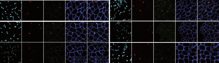
Corresponding author: Lanzuolo Chiara. Via Fratelli Cervi, 93, 20090 Segrate (MI),

Italy; Phone: +39x 02 00660358; e-mail: chiara.lanzuolo@cnr.it

Keywords

Muscle Stem Cells (MuSCs), Lamin A, Polycomb, cell fate, differentiation, premature

senescence, muscular dystrophy.

Running head

PcG dysfunctions in lamin dystrophy

                                                                                 2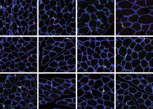
Abstract

Lamin A is a component of the inner nuclear membrane that, together with epigenetic

factors, organizes the genome in higher order structures required for transcriptional

control. Mutations in the Lamin A/C gene cause several diseases, belonging to the

class of laminopathies, including muscular dystrophies. Nevertheless, molecular

mechanisms involved in the pathogenesis of Lamin A-dependent dystrophies are still

largely unknown. Polycomb group of proteins (PcG) are epigenetic repressors and

Lamin A interactors, primarily involved in the maintenance of cell identity. Using a

murine model of Emery-Dreifuss Muscular Dystrophy (EDMD), we showed here that

Lamin A loss deregulated PcG positioning in muscle satellite stem cells leading to

de-repression of non-muscle specific genes and p16INK4a, a senescence driver

encoded in the Cdkn2a locus.      This aberrant transcriptional programme caused

impairment in self-renewal, loss of cell identity and premature exhaustion of

quiescent satellite cell pool. Genetic ablation of Cdkn2a locus restored muscle stem

cell properties in Lamin A/C null dystrophic mice. Our findings established a direct

link between Lamin A and PcG epigenetic silencing and indicated that Lamin A-

dependent muscular dystrophy can be ascribed to intrinsic epigenetic dysfunctions of

muscle stem cells.

                                                                                   3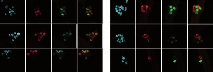
Introduction

The nuclear lamina (NL) is located in the inner part of the nuclear membrane and is

made up of a complex network of type V filament proteins, the lamins (1, 2). In

vertebrates lamin proteins are divided into A and B types, based on sequence

homologies. A growing body of evidence suggests that lamins are directly involved in

the functional control of the genome, by organizing its three dimensional positioning

in the nuclear space, through the association with transcriptionally repressed large

genomic regions, called Lamina-associated domains (LADs) (3). The crucial function

of lamins is attested by an entire class of genetic diseases, called laminopathies,

where specific components of the NL are altered (4). In particular, the study of Lamin

A/C is gaining an increasing interest for three reasons: i) Lamin A/C plays an

undisputed role in several cellular processes from mechanotransduction to cell

differentiation; ii) Lamin A/C has a peculiar intranuclear distribution being present in

the nucleoplasm as well as in the nuclear periphery (5); iii) Lamin A/C interacts with

several epigenetic factors, exerting a functional control on transcriptional regulation

(3, 6). One of the most studied Lamin A/C dependent cellular process is myogenesis

because mutations in the LMNA gene lead to muscular dystrophies, as in the case of

Emery Dreifuss Muscular Dystrophy (EDMD) (7). However, epigenetic mechanisms

involved in lamin-dependent dystrophy are still largely unknown. PcG proteins are

epigenetic repressors originally discovered for their central roles in development and

cell differentiation (8) and recently described as functional partners of Lamin A/C (9-

14). In the last years several evidence demonstrated that PcG proteins are involved

in the regulation of adult stem cells (15, 16), safeguarding cell identity and preventing

cell fate transition. In multipotent stem cells, PcG proteins ensure the correct balance

between self-renewal and lineage-specific differentiation, promptly responding to the

environmental changes. At the molecular level this is achieved through PcG binding

at bivalent domains, genomic regions containing active and repressive epigenetic

signatures and a poised RNA polymerase II (17). This epigenetic condition allows a

                                                                                       4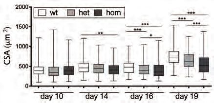
rapid transition from one transcriptional state to another, ensuring the correct
expression of unique and specific cell lineage genes. Defects in these fine-tuned
mechanisms lead to lack of cell identity (18) or pathological reprogramming (19).
Given their key role in regulating stem cells fate decisions and tissue homeostasis, it
is conceivable that PcG dysfunctions contribute to lamin-dependent, tissue-specific
human diseases. Here, we examined how the absence of Lamin A/C impacts muscle
stem (satellite) cells (MuSCs) in vivo, and the role of PcG proteins in lamin muscular
dystrophy. We found that MuSCs lacking Lamin A/C redistribute PcG-dependent
histone marks leading to transcriptional upregulation of crucial PcG-target genes,
such as non-muscle related genes. This leads to lack of MuSC cell identity and
cellular senescence, determining a premature exhaustion of the muscular stem cell
niche. Genetic ablation of the PcG-regulated Cdkn2a locus in lamin dystrophic mice
restores MuSCs defects.
Results
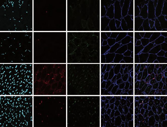
Lamin A is required to preserve quiescent muscle stem cells (MuSC) pool.
We analysed the muscle stem cell niche composition in the severe dystrophic Lmna
Δ8-11 -/- mice (homozygous, hom), together with their unaffected littermates, wild
type (wt, Lmna Δ8-11 +/+) or heterozygous (het, Lmna Δ8-11 +/-), during dystrophy
progression at 10, 14, 16 and 19 days after birth. In early stages of post-natal growth
(d10 and d14) no differences were found in the relative amounts of quiescent (QSCs;
PAX7+/MYOD-) and activated (ASCs; PAX7+/MYOD+) MuSCs (Figure 1A, 1B and
S1A) among distinct genotypes. Conversely, starting from d16, an unbalance of
MuSCs becomes evident in Lmna Δ8-11 -/- muscles, with a decreased proportion of
QSCs compared to ASCs, mirroring a decline in myofibers cross sectional area
(CSA) (Figure 1C). Of note, the overall amount of PAX7+ MuSCs was not
significantly altered across the different genotypes (Figure S1B) and Ki67 staining at
d19 confirmed that in Lmna Δ8-11 -/- muscles a lower amount of QSCs
5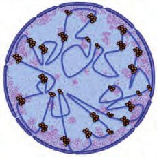
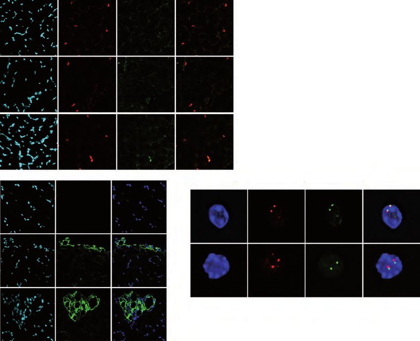
(PAX7+/Ki67-) is present (Figure S1C). These findings suggested that Lmna Δ8-11 -

/- MuSCs may be deficient in self-renewal capacity. To test this hypothesis, we

isolated single myofibers at d19 and cultured them for 96h, monitoring their ability to

give    rise   to   self-renewing   PAX7+/MYOD-,          activated   PAX7+/MYOD+         and

differentiating PAX7-/MYOD+ cells (Figure 1D and 1E). In fibers isolated from Lmna

Δ8-11 -/- muscles, we observed a decrease in the number of self-renewing

PAX7+/MYOD- cells compared to wt, paralleled by a diminished number of

differentiating cells (PAX7-/MYOD+) and an increased number of activated satellite

cells. Immunostaining with the myogenic marker MYOG, involved in later stages of

differentiation, highlighted a lower number of MYOG+/PAX7- cells in Lmna Δ8-11 -/-

(Figure 1F and 1G) accompanied by proliferation defects ex vivo (Figure S1D).

These findings suggest a defect in muscle differentiation, as described in (20), and a

previously     unreported     self-renewal   impairment.      Interestingly,   the   healthy

heterozygous Lmna Δ8-11 +/- mice, although not developing muscular dystrophy

(21),   presented    an     intermediate   self-renewal    phenotype    between      wt   and

homozygous Lmna Δ8-11 -/- (Figure 1D and 1E), suggesting that proper Lamin A

levels are important for MuSCs homeostasis to preserve their self-renewal capacity.

To further address this, we performed repeated muscle injuries on adult

heterozygous Lmna Δ8-11 +/- mice, which show less Lamin A at mRNA and protein

level (Figure S2A and S2B). Analysis of MuSC populations revealed a lower amount

of QSCs in Lmna Δ8-11 +/- muscles upon repeated injuries (Figure 2A and 2B;

Injured) and a decline in Pax7+ cells (Figure 2C; Injured), suggesting that Lamin A

affects MuSCs self-renewal in a dose dependent manner.

Lmna Δ8-11 -/- dystrophic MuSCs display chromatin redistribution of PcG

dependent signature

Our recent results showed a Lamin A/C-PcG crosstalk along in vitro myogenesis

(10). We thus wondered if the altered MuSCs balance observed in Lmna Δ8-11 -/-

                                                                                           6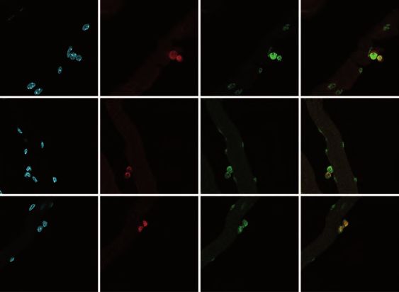
muscles might be ascribed to aberrant PcG functions. We first performed

immunostaining of Ezh2, the catalytic subunit of Polycomb Repressive Complex 2

(PRC2) (Figure S3A) in d19 MuSCs. We fixed MuSCs before FACS-Sorting to

preserve the nuclear architecture of Lamin A-deficient cells (see methods). We found

a general intranuclear diffusion of Ezh2 in Lmna Δ8-11 -/- MuSCs, ascertained by

measuring PcG bodies parameters (22) (Figure S3A, S3B and S3C). We also

measured Ezh2 expression both in MuSCs and whole muscles (Figure S3D) and we

analysed Ezh2 protein levels in whole muscles (Figure S3E and S3F). We found no

major differences between Lmna Δ8-11 +/+ and -/- mice. To further analyse the

Ezh2 intranuclear distribution in QSCs, we performed triple immunostaining on

muscle cryosections (Figure S3G and S3H). Ezh2 levels, assessed measuring

fluorescence intensity, were similar in Lmna Δ8-11 +/+ and -/- MuSCs both in

PAX7+/Ki67- and PAX7+/Ki67+ cells. Since Ezh2 is hardly detectable in adult

quiescent satellite cells (23-25), this result suggests that during post-natal growth

developmental signals might instead contribute to maintain Ezh2 expression in non-

proliferating MuSCs.

On the other hand, evaluation of the number of PcG bodies on the same sections

highlighted a decrease in the number of Ezh2 bodies in the mutant (Figure S3I and

S3J), leading us to conclude that the absence of Lamin A/C does not affect Ezh2

protein levels but influence its nuclear distribution. To gain further insights into

possible PcG-dependent transcriptional defects, we performed RNA-sequencing on

freshly isolated MuSCs at d19, finding 1424 upregulated genes and 1842

downregulated genes in the Lmna Δ8-11 -/- MuSCs as compared to the wt (Figure

S4A). Interestingly, performing a Gene Set Enrichment Analysis (GSEA) based on

differential expression generated after conditional ablation of Ezh2 in MuSCs (24)

and Lmna Δ8-11 -/- up-regulated genes, we found a significant association between

the two datasets suggesting that Lamin A absence impairs Ezh2 function (Figure

                                                                                   7
S4B). We also followed the deposition of the Ezh2 dependent H3K27me3 histone

mark in Lmna Δ8-11 mice by quantitative spike-in ChIP-seq (26) (see additional

methods, Figure S4C and S4D). Integrative analysis of RNA-seq and ChIP-seq

revealed that up-regulated genes in Lmna Δ 8-11 -/- condition are significantly

enriched for H3K27me3 targets (identified in wt condition) (Figure 3A). Indeed,

analysis of H3K27me3 distribution around the Transcription Start Sites (TSS) and

along the body of genes indicated a decrease of this repressive mark in Lmna Δ8-11

-/- MuSCs compared to wt (Figure 3B and 3C), which was not accompanied by a

statistical decrease in H3K27me3 global levels in MuSCs (Figure S5A and S5B) and

in whole muscle (Figure S5C and S5D). In contrast, a deep analysis of H3K27me3

ChIP-seq reads coverage in the intergenic genomic regions between the known

H3K27me3 enrichment peaks interestingly showed a higher average coverage in the

Lmna Δ8-11 -/- MuSCs compared to wt counterparts (Figure 3D). These results are

compatible with a diffusion of PcG proteins along the chromatin fibers rather than a

complete PcG displacement. To identify the PcG targets mostly affected by Lamin A

deficiency, genes were grouped according to their transcription level in wt MuSCs.

We thus defined 4 equally sized groups of genes based on expression level quartiles

(Figure 3E). For each expression category, we reanalysed the H3K27me3

distribution along the body of genes and the TSS and the percentage of upregulated

genes in the Lmna Δ8-11 -/- MuSCs (Figure 3E, 3F and S5E). In quartile I we found

only a small percentage (0.65%) of upregulated genes in Lmna Δ8-11 -/-, suggesting

that the H3K27me3 decrease/redistribution is not sufficient to activate transcription in

highly repressed genes (Figure 3F, S5E and S6A). In contrast, quartiles II, III and IV

are more affected by the diminished H3K27me3 levels in Lmna Δ8-11 -/- (Figure 3F,

S5E and S6B), showing a percentage of upregulated genes between 5 and 9%.

Specifically, we noticed that in wt MuSCs, H3K27me3 ChIP-seq signal enrichment

around the TSS and within the body of genes is progressively lower in quartiles of

                                                                                      8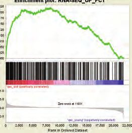
higher expression, as expected (Figure 3F, III and IV quartiles and S5E). However,

for Lmna Δ8-11 -/- mice the decrease of H3K27me3 signal inside the gene body is

relatively less marked than in wt mice, in fact the average enrichment is slightly

higher. We quantified and confirmed this observation by considering the ratio of

H3K27me3 ChIP-seq enrichment signal at the TSS and 2.5Kb downstream of the

TSS, for each gene, in wt and Lmna Δ8-11 -/- mice (Figure S7A), showing that this

ratio is significantly different for higher expression quartiles (Figure S7B).

Lamin A-dependent PcG redistribution determines de-repression of non-

muscle related bivalent genes

The altered PcG binding observed in Lmna Δ8-11 -/- MuSCs, prompted us to

examine more in detail the bivalent genes, a subgroup of PcG targets whose

expression is more susceptible to variations of PcG occupancy (27). Bivalent genes

are characterized by the concurrent presence of both H3K27me3 and H3K4me3

marks around TSS and have an intermediate gene expression state (28). We first

performed H3K27me3 and H3K4me3 ChIP-seq in wt MuSCs (Figure S8A and S8B)

and we defined bivalent genes using the parameters described in (17) for the

H3K4me3 window at TSS (Figure S8C). Then, we tested the association between

bivalent and up-regulated genes in the Lmna Δ8-11 -/- by means of the Fisher exact

test. We observed a significant over-representation of bivalent genes among up-

regulated ones in the Lmna Δ8-11 -/- MuSCs (Figure 4A). To gain more insights into

the biological relevance of deregulated genes in the mutant mice, we performed

semantic similarity analysis of all GO terms associated with upregulated genes

(Figure 4B) together with GSEA (Figure S8D and S8E). These analyses showed a

negative correlation with muscle specification (Figure S8D) together with an

acquisition of markers related to lipid metabolic processes (Figure 4B and S8E).

Notably, Fisher exact test analysis highlighted a significant overlap between genes

                                                                                 9
with bivalent promoter and the Lmna Δ8-11 -/- MuSCs up-regulated genes involved
in adipogenesis (Figure 4C), suggesting that Lamin A is involved in preserving
MuSCs identity by ensuring the correct PcG-mediated transcriptional repression of
non-muscle genes.
PPARγ is aberrantly expressed in Lmna Δ8-11 -/- dystrophic MuSCs
Given this strong association between bivalent reactivation and adipogenesis
markers (Figure 4C), we analysed different lipid-related GO categories, finding
among the top GO terms, PPARG (Peroxisome proliferator-activated receptor
gamma) (Supplementary Table S1). This master transcription factor for adipose cell
differentiation (29, 30) was found statistically upregulated in Lmna Δ8-11 -/- MuSCs
(FDR< 0.05). Moreover, PPARG gene is a Polycomb target and has a bivalent
signature in wt MuSCs (Supplementary Table S1 and Figure S9A). These
observations prompted us to analyse PPARG transcriptional deregulation. We
stained muscles with PAX7 and PPARγ to directly test if MuSCs displayed aberrant
expression of PPARγ in the absence of Lamin A. Strikingly, we found about 10% of
Lmna Δ8-11 -/- MuSCs that simultaneously express both muscular and adipogenesis
markers being PAX7+/ PPARγ+ (Figure 5A and 5B). Accordingly, the genomic region
of the PPARG gene showed a decrease of H3K27me3 enrichment around the TSS in
the Lmna Δ8-11 -/- MuSCs, accompanied by a transcriptional up-regulation (Figure
S9B). To evaluate if the aberrant expression of adipogenic genes in MuSCs of
mutant mice culminate with fatty infiltration we performed an immunofluorescence for
Perilipin 1, a protein present on the surface of lipid droplets (31) (Figure 5C and 5D),
on cryosections of muscles derived from 19 days-old Lmna Δ8-11 mice. We found
large areas of adipose accumulation between myofibers of LaminA/C +/- and -/-
muscles, which were instead undetectable in the wt mice. Considering the key role of
PcG proteins in mediating the formation of chromatin loop structures (32, 33), we
10reasoned that the loss of H3K27me3 and transcriptional up-regulation of PPARG
locus could be related to the alteration of chromatin 3D structure. The genome 3D
architecture is organized in structurally separated Topologically Associated Domains
(TADs), chromosomal structures that favour intra-domain looping interactions (34).
TADs can be identified by genome-wide chromosome conformation capture (Hi-C)
and are largely conserved across different cell types. We verified that the PPARG
locus is included in a TAD encompassing a region extending also upstream of the
PPARG locus itself, using high-resolution Hi-C data on mouse embryonic stem cells
(35) and the 3D Genome Browser (36) (Figure S10). Then, we performed 3D
multicolor DNA FISH analysis on pre-fixed MuSCs using one BAC probe overlapping
the PPARG promoter and a second probe annealing at the other TAD border. We
observed an overlap of the signals from the two regions in the wt Lmna Δ8-11 +/+
MuSCs that indicates the presence of a DNA looping in cis (Figure 5E and 5F). By
contrast, in Lmna Δ8-11 -/- MuSCs the distance between the signals was higher,
definitely suggesting the lack of DNA/DNA interaction. Indeed, from the analysis of
H3K27me3 ChIPseq tracks we noticed in the Lmna Δ8-11 -/- MuSCs a reduction of
H3K27me3 peaks upstream the PPARG locus (Figure S10). FISH analysis also
highlighted that in wt the entire genomic region is close to the nuclear periphery
(Figure 5E and 5G) while in Lmna Δ8-11 -/- is re-located in the nuclear interior,
suggesting that Lamin A absence interferes with chromatin anchoring to the nuclear
lamina and PcG dependent DNA conformation.
Lmna Δ8-11 -/- MuSCs undergo premature senescence
Taken together, these results clearly point towards a role of Lamin A in mediating
PcG-transcriptional repression in MuSCs to safeguard their identity and regenerative
capacity. This lack of cell identity and the impairment of self-renewal displayed by
Lmna Δ8-11 -/- MuSCs are all features reminiscent of the phenotype described for
Ezh2-null MuSCs (24). Moreover, the impairment in self-renewal and the progressive
11decline of MuSCs pool are also typical traits of aged MuSCs (37) in which both
Lamin A/C and PcG proteins play a key role (38, 39). A major cellular mechanism
that ensures self-renewal and hence the maintenance of the MuSC pool is the
asymmetric division (40). At the molecular level, in aged mice, the accumulation of
activated form of P38 (phospho-p38, Ph-P38) and its symmetric distribution in MuSC
doublets heavily compromise the self-renewal capacity leading to MuSCs functional
decline (41, 42). To test whether premature exhaustion of quiescent Lmna Δ8-11 -/-
MuSCs cells could be ascribed to a defective asymmetric division, we stained
myofibers-associated MuSCs for Ph-p38 after 48h of culture, a timing at which, 19
days-post-natal myofibers formed MuSCs-derived doublets (Figure 6A). In contrast to
wt, Lmna Δ8-11 -/- MuSC doublets showed a preferential symmetric distribution of
ph-p38, quantified by relative fluorescence intensity (Figure 6A and 6B) often
accompanied by a planar orientation with respect to myofibers (see additional
methods) (Figure 6C). This highlights problems in asymmetric division, which should
be instead characterized by apico-basal orientation (43). In line with this result, in
Lmna Δ8-11 -/- muscle sections we found higher amount of Ph-P38+ (Figure S11A
and S11B) MuSCs and sign of genomic instability, as measured by increased γH2AX
DNA repair signal foci (Figure S11C and S11D), not accompanied by apoptosis or
necrosis as evidenced by Annexin staining (Figure S12A). To test if defective
asymmetric division is associated to premature senescence we then analysed
RNAseq to determine if Lmna Δ8-11 -/- MuSCs share the same transcriptional
signature of MuSCs isolated from aged mice. We performed a GSEA using two
different RNA datasets from MuSCs of 24 months old mice (25, 44) and Lmna Δ8-11
-/- up-regulated genes. In line with our hypothesis, we found that 19 days-old Lmna
Δ8-11 -/- MuSCs present an upregulated transcriptome similar to 20-24-months-old
MuSCs (Figure 6D and S12B), but different from geriatric 28-32-months-old MuSCs
(Figure S12C). At the molecular level, the senescence program is supported by
upregulation of some PcG-regulated Cyclin-dependent Kinase Inhibitors (CDKIs) (45)
12as p21, involved in cellular senescence and in cell cycle arrest. p21 maintains the
viability of DNA damage‐induced senescent cells (46) and aberrant expression of
p21 has been observed in EDMD derived human myoblasts (47). ChIP-seq and
RNA-seq analysis of Cdkn1a/p21 locus showed a displacement of Ezh2 from the
promoter accompanied by an upregulation of p21 transcript in Lmna Δ8-11 -/-
MuSCs (Figure 6E).
Genetic ablation of Cdkn2a locus partially rescues self-renewal defects in
Lmna Δ8-11 -/- dystrophic mice.
To further corroborate our findings, we also analysed the Cdkn2a locus, a PcG target
primarily involved in muscular senescence (44) (Figure S13A). Two transcripts,
p16INK4a and p19ARF originate from Cdkn2a locus (48). Interestingly, it has been
recently reported that p16INK4a expression is a second event, subsequent to p21
upregulation, in the cellular senescence progression (49). In line with these
observations, p16INK4a expression is specifically induced in geriatric 28-32-months-old
MuSCs (not in 24-months-old MuSCs) (44). Moreover, depletion of p16INK4a is
sufficient to reduce senescence-associated gene expression in geriatric MuSCs.
RNA-seq and qRT-PCR on 19-days old Lmna Δ8-11 -/- MuSCs did not reveal any
transcription of p16INK4a in wt or Lmna Δ8-11 -/- (Figure 7A). However, qRT-PCR
analysis performed in older mice (26 days from birth), revealed higher levels of
p16INK4a transcript in Lmna Δ8-11 -/- MuSCs and whole muscles compared to the wt
counterpart (Figure 7A), suggesting a transition during dystrophy progression toward
geriatric condition. We thus decided to test whether the genetic ablation of Cdkn2a
locus could reverse Lmna Δ8-11 -/- MuSCs premature aging, by crossing Lmna Δ8-
11 +/- with Cdkn2a -/- mice (50). Analysis performed in Lmna Δ8-11 +/+ background
showed no differences in the percentage of QSCs and ASCs, nor on CSA,
suggesting that Cdkn2a is dispensable for post-natal muscle development (Figure
7B, 7C and 7D; LMNA+/+). On the other hand, Cdkn2a Lmna Δ8-11 -/- mice partially
13rescued the quiescent MuSC pool and CSA defects observed in the absence of
Lamin A (Figure 7B, 7C and 7D; LMNA-/-; S13B and S13C) emphasizing that Lamin
A-dependent muscular dystrophy might be due to progressive MuSCs functional
decline caused by acquisition of premature aging features.
Discussion
Lamin A-dependent muscular dystrophy pathogenesis has been classically ascribed
to nuclear fragility that renders myonuclei more prone to mechanical stress and
damage imposed by myofibre contraction (4). However, the evidence that Lamin A/C
is expressed also by MuSCs has led to suggest that satellite cell dysfunction might
contribute to EDMD progression (51), yet experimental evidence of this hypothesis
was still lacking.
Cell fate choice during muscle differentiation is governed by epigenetic factors
controlling the sequential restriction of transcriptional programs (52). Any dysfunction
in this finely tuned epigenetic regulation could lead to impaired or aberrant cell fate
determination (53). Here, we show that Lamin A/C is indeed crucial to preserve
MuSCs identity and regenerative capacity. We demonstrate that cell-autonomous
Lamin A-dependent Polycomb dysfunction leads to MuSCs functional decline, which
culminate with impaired regenerative capacity and dystrophic phenotype (Figure 1).
Traditionally, the role of Lamin A/C in muscle differentiation has been considered to
cause defects in muscle differentiation (54, 55). However, in other conditions, MuSCs
from Lamin A/C null mice showed a normal ability to differentiate and to form
myotubes (20, 56). By moving the viewpoint from differentiation to cell identity we
now propose that in the absence of Lamin A/C, a portion of MuSCs derails from their
fate affecting the quiescent MuSCs pool. At the molecular level, we described a
mechanism of Lamin A-dependent deregulation of PcG targets showing the
spreading of repressive marks along the chromatin fiber (Figure 3), with lack of the
expected higher order structures and consequent de-repression of bivalent genes
14(Figure 4, 5 and 8). Recently it has been proposed that PcG domains can have
distinct size and boundaries characteristics (57): upon differentiation, loci directly
involved in fate specification lose PcG-mediated looping interactions, allowing new
active promoter/enhancer interactions. In parallel, other PcG domains, such as the
Hox clusters, do not change their 3D architecture. Our findings further corroborate
this hypothesis showing that the stability of PcG-interacting domains correlates with
PcG occupancy and depends on Lamin A (Figure 5 and 8). PcG dysfunctions drive
Lmna Δ8-11 -/- MuSCs toward two fates not mutually exclusive (Figure 8): lack of cell
identity, highlighted by the presence of MuSCs co-expressing muscle and adipogenic
markers (Figure 4 and 5), and premature senescence, as shown by defects in
asymmetric division and accumulation of Ph-P38 and γH2AX (Figure 6 and S11).
These epigenetic alterations determine a progressive decline in MuSCs self-renewal
that accompanies the muscular dystrophy progression (Figure 1), ultimately leading
to a geriatric condition characterized by the expression of p16INK4a from Cdkn2a locus
(Figure 7) (58). Genetic ablation of Cdkn2a locus can recover some muscular
dystrophy defects of the Lmna Δ8-11 -/- mouse (Figure 7) thus supporting the
hypothesis that dystrophic and aging muscles share a dysfunction of epigenetic
mechanisms controlling cell cycle and fate decision of MuSCs. Our findings
corroborate recent evidence on PcG dysfunction in human disease (19), showing
that PcG alterations contribute to pathology progression and severity in EDMD. This
will further stimulate future studies on the role of PcG proteins in the dynamics of
stem cell niche, when embedded in a pathological environment.
15Methods
Animals
Heterozygous B6.129S1(Cg)-Lmnatm1Stw/BkknJ mice (Lmna Δ8-11 +/-) (21) and
Cdkn2a +/- mice (50) were used.
Satellite cells extraction, apoptosis evaluation and multiple injuries
Hind-limb muscle were isolated from sacrificed mice and digested 120 minutes in 2,4
U/ml of Dispase II (Roche, 04942078001), 2 ug/ml of Collagenase A (Roche,
1013586001), 0,2 mM CaCl2 (Sigma, C5670), 4 mM MgCl2 (Sigma, M8266), 10
ng/ml DNase I (Roche, 1014159001) in PBS1X (Euroclone, ECB4004L) at 37°C in a
water bath. The sample were resuspended in HBSS (Gibco, 14025-050)
supplemented with 0,1% BSA (Sigma, A7030). Cell suspension was serially filtered
with 70 (Falcon, 352350) and 40 µm (Falcon, 352340), stained with antibodies
indicated in supplementary table S2: PB-CD45 1:50, PB-CD31 1:50, PB-Ter119
1:50, FITC-Sca1 1:50, APC-α7integrin 1:200 and sorted with BD FACS ARIA III for:
PB-CD45-/ PB-CD31-/ PB-Ter119-/ FITC-Sca1-/ APC-α7integrin+.
For multiple injury experiment, 20 µl of Cardiotoxin (CTX) 10 µM (Latoxan, L8102)
were injected in TA muscle each week for 3 weeks. TA muscle was harvested one
week after the last CTX injection.
For apoptosis assay we stained MuSCs with Annexin. 15-20K of satellite cells were
washed in 1 mL cold PBS 1X up, centrifuged 7 minutes at 400g and incubated 20
minutes at room temperature in the dark with 100 µL Annexin V FITC buffer (FITC
Annexin V Apoptosis Detection Kit I-ref. 556547). Then, samples were washed with
500 µL of cold PBS 1X, centrifuged 7 minutes at 400g and incubated 15 minutes at
room temperature in the dark with 300 µL of cold PBS 1X containing 5 µL of
Propidium iodide. Samples were analyzed at BD FACS-CANTO (voltage FSC=357,
SSC=462, medium flow to acquire, 300-400 events/second).
Immunofluorescence
On muscle sections: Tibialis Anterior (TA) muscles were embedded in Killik (Bio-
Optica, 05-9801), immediately frozen in pre-cooled isopentane (Sigma, 277258) and
sectioned (Leica CM1850 cryostat) at 8 µm thin. Sections were fixed 20 minutes in
paraformaldehyde (PFA) 4% (Sigma, P6148) and washed 3x5 minutes in PBS1X
(Euroclone, ECB4004L). To permeabilize tissues pre-cooled methanol (Sigma,
322415) at -20°C was added for 6 minutes. Antigen retrieval was performed 2x5
minutes in hot citric acid 80°C (Sigma, C0759) pH6.0 and washed 2x5 minutes in
16PBS1X. Sections were blocked 1 h in BSA 4% (Sigma, A7030) followed by
incubation 45 minutes with FAB mouse fragment 1:100 (Jackson Immuno Research,
115-007-003)/ PBS1X. Primary antibodies were diluted 1:100 in blocking solution,
except for pparγ diluted 1:75, and incubated O/N at 4°C. The day after, sections were
washed 3x10 minutes in PBS1X/0,1% BSA and incubated with secondary antibodies
in blocking solution 1:200 1h at RT in the dark. Then, sections were washed 3x10
minutes in 0,1% BSA/PBS1X and incubated 2h at RT with Laminin and Pax7 (1:20).
After washing 3x10 minutes in PBS1X/0,1% BSA sections were incubated 45
minutes with Cy5 (1:300) and Biotin (1:500) for Pax7 signal amplification. After
washing 3x10 minutes in PBS1X, sections were incubated 45 minutes with
secondary antibodies Cy3-streptavidin (1:1250). The sections were finally washed
3x10 minutes in PBS1X, stained 5 minutes with dapi (Sigma, D9542), briefly washed
twice in PBS1X and mounted on slide with a drop of Prolong Antifade (Life P36930).
On single myofibers: Tibialis Anterior (TA), Soleus (S), Gastrocnemius (G) and
Exstensor Digitorium Longus (EDL) were isolated from mice and digested 45-50
minutes in 0,2% collagenase type I (Sigma, C0130)/DMEM (Gibco, 10569010) at
37°C. 2 rounds of myofibers washes were performed in pre-coated dishes with 20%
FBS (Gibco, 10500064)/DMEM. Myofibers were let grown in DMEM supplemented
with 20% FBS, 1% Chicken Embryo extract (Seralab, CE650-DL) and 1%
Penicillin/Streptomycin (Euroclone, ECB 3001) for 48h or 96h changing the medium
only after 72h. Myofibers were collected in 2ml tubes pre-coated with 10%
FBS/PBS1X and fixed 15 minutes with 4%PFA followed by 3 washes in PBS1X.
Permeabilization was performed 5 minutes with 0,5% Triton X-100 (Sigma,
93443)/PBS1X followed by 2 washes in PBS1X. Myofibers were incubated 1h in
blocking solution (10% of FBS/PBS1X). Primary antibodies were incubated in
blocking solution ON at 4°C. The day after myofibers were washed in 0,25%
tween/PBS1X twice and incubated 60 minutes with secondary antibodies in blocking
solution. Fibers were washed in 0,1% tween/PBS1X, incubate 5 min with dapi, briefly
washed twice in PBS1X and mounted on slide with a drop of Prolong Antifade
(Thermofisher, P36970).
On satellite cells: In order to preserve the integrity of chromatin architecture,
Muscular stem (Satellite) Cells (MuSCs) suspension was fixed in 1% PFA for 9
minutes and quenched with 125 mM Glycine (Sigma, 8898) before FACS staining
and sorting. SC cells were placed on pre- Poly-L-lisined coverslips (Sigma, P8920) at
density of 100.000/mL for 30 minutes at RT. Coverslips were fixed with PFA at
4%/PBS for 10 minute at RT. Then, cells were washed in 2X5 minutes in 0,05%
Triton X-100/PBS 1X, permeabilized with 0,5% Triton X-100/PBS1X for 10 minutes
17and rinsed in PBS 1X. The slides were let in 20% Glycerol (Sigma, G5516)/PBS 1X
at least 60 minutes followed by 4 round of freeze and thaw: freezed on dry ice (30
seconds) and thawed in 20% Glycerol/PBS 1X at RT. Slides were washed 2X5
minutes in 0,05% Triton X-100 in PBS 1X, 1x5 minutes 0,5% Triton X-100/PBS1X,
incubated in HCl 0,1M (Sigma, H1758) for 15 minutes and rinsed in PBS 1X.
Aspecific signals were blocked with 1% BSA/PBS1X for 30 minutes at room
temperature. Reaction with primary antibodies Ezh2 diluted 1:100 in blocking solution
was performed 12–16 h at 4°C; Lamin A/C diluted 1:200 in blocking solution was
performed the day after at room temperature for 2 hour. Secondary antibodies were
diluted 1:200 in blocking solution for 1 h at room temperature. Washes were done in
PBS1X. DNA was counterstained with dapi, and glasses were mounted in Prolong
Antifade. Primary antibodies are listed in supplementary table S2. Secondary
antibodies were from Jackson Immuno Research: Alexa 488 (711-545-152); Cy5
1:300 (111-115-144); Cy3-streptavidin (016-160-084); Alexa 594 (711-545-150).
Three-dimensional multicolor DNA FISH
To produce probes for 3D multicolor DNA FISH we used the following BAC DNA
clones (BACPAC Resources Program, CHORI): CH29-101F16 (for Pparγ) and
CH29-555O5 (for the upstream region of Pparγ). 1-3 µg of BAC DNA were labelled
with dig-dUTP (Roche, 11093088910) (for the upstream region of Pparγ) or cy3-
dUTP (Thermo Fisher Scientific, C11401) (for Pparγ) through nick translation in 50 µl
of Labelling mix buffer (0.02 mM C-G-A dNTPs: Euroclone, EMR273025,
EMR274025, EMR272025 respectively, 0.01 mM dTTP Euroclone EMR275025, 0.01
mM labelled dUTP, 50 mM Tris-HCl pH 7.8, 5 mM MgCl2, 10 mM b-
mercaptoethanol, 10 ng/µl Bovine serum albumin (BSA), 0.05-0.1 U/µl DNA
Polymerase I (Thermo Fisher Scientific, 18010-017), 0.004-0.001 U/µl Amplification
Grade DNase I (Sigma, D5307) for 30 min-2 h at 16 °C, to obtain an average probe
size of 50 bp. Probes were collected by ethanol precipitation, resuspended in 10 mM
Tris-HCl pH 7.5 and then quantified using a Nanodrop 1000 Spectrophotometer
(Thermo Fisher Scientific). For a single experiment 100-300 ng of each probe was
precipitated with 3.5 µg of Mouse Cot-1 DNA (Thermo Fisher Scientific, 18440-016)
and 20 µg of Deoxyribonucleic acid, single stranded from salmon testes (Sigma,
D7656), and then resuspended in 6 µl of Hybridization solution (50% formamide pH
7.0 (FA)/2X SSC/10% Dextran sulfate). Pre-fixed Satellite cells Cells were plated
directly on poly-L-Lisyned coverslips and fixed with 4% Paraformaldehyde (PFA) in
1X PBS and TWEEN 20 0.1% (PBS-T) for 10 min at room temperature. During the
18last minute few drops of 0.5% Triton X-100 in 1X PBS (PBS) were added and then
cells were washed with 0.01% Triton X-100 in PBS three times for 3 min at room
temperature. Cells were first permeabilized with 0.5% Triton X-100 in PBS for 10 min
at room temperature. In order to remove RNA, samples were treated with RNase
Cocktail Enzyme Mix (Thermo Fisher Scientific, AM2288) for 1 h at 37 °C. Cells were
subjected to other steps of permeabilization with 20% Glycerol in PBS overnight at
room temperature, followed by four cycles of freeze and thaw interleaved by soak
with 20% Glycerol in PBS. Permeabilized cells were washed with PBS three times for
10 min at room temperature. Cells were then incubated in 0.1 M HCl for 5 min at
room temperature, followed by a rinse with 2X SSC and then incubated in 50% FA in
2X SSC for at least 30 min at room temperature. Slides were equilibrated in 2X SSC
for 2 min, washed in PBS for 3 min and then treated with 0.0025-0.0075% pepsin in
0.01-0.03 N HCl for 2-4 min at room temperature to eliminate cytoskeleton. Pepsin
was inactivated with 50 mM MgCl2 in PBS twice for 5 min. Nuclei were post-fixed
with 1% PFA in PBS for 1 min, washed with PBS for 5 min and with 2X SSC twice,
and then back to 50% FA in 2X SSC for at least 30 min at room temperature.
Hybridization solution was loaded on a clean microscopic slide, coverslip with nuclei
was turned upside down on the drop of hybridization mixture and sealed with rubber
cement. Samples were denatured for 4 min at 75 °C and leaved to hybridize in a
metallic box floating in a 37 °C water bath overnight. Samples were washed with 2X
SSC three times for 5 min at 37 °C and with 0.1X SSC three times for 5 min at 60 °C,
followed a rinse with 0.2% TWEEN 20 in 4X SSC. Aspecific binding sites were
blocked with Blocking solution (4% BSA in 4X SSC, 0.2% TWEEN 20) for 20 min at
37 °C. Samples were then incubated in the appropriate concentration of Streptavidin,
DyLight 488 Labeled Anti-Digoxigenin/Digoxin (Vector Laboratories, DI-7488) (1:100)
diluted in Blocking solution for 35 min in a dark and wet chamber at 37 °C. Samples
were washed with 0.2% TWEEN 20 in 4X SSC three times for 3 min at 37 °C,
equilibrated in PBS and post-fixed with 2% formaldehyde in PBS for 10 min at room
temperature. Finally, the 3D-fixed nuclei were washed with PBS three times for 5 min
at room temperature, counterstained with 1 ng/µl DAPI in PBS for 10 min at room
temperature and washed with PBS two times for 5 min at room temperature.
Coverslips were mounted. An Eclipse Ti-E (Nikon Instruments) microscope was used
to scan the nuclei, with an axial distance of between 0.2-0.25 micron consecutive
sections.
Histone extraction and Western blot
19Total proteins were prepared starting from quadriceps muscle. Muscles were
homogenized on ice with tissue ruptor (Qiagen, 902756) in 1ml of extraction buffer
(50 mM Tris HCl, pH 7.5, 150mM NaCl, 1 mM EDTA pH 8.0, 0,1% SDS, 1% Np40
(Sigma, 74385), 0,5% Sodium deoxycholate (Sigma, D6750) 1× protease Inhibitors
(Roche, 04693132001) 2 mM PMSF (Sigma, 93482), 1 mM NaF (Sigma, s7920); 1
mM Na3VO4). After 30 minutes in ice 3 pulses of 10 seconds sonication at 30%
amplitude were performed with Branson digital sonifier 250 to allow dissociation of
protein from chromatin and solubilization. Extracts were analyzed by SDS-PAGE
using a 8–10% gel (29:1 acrylamide solution Sigma, 01708). For histone extractions
approximately 2.000.000 of freshly isolated MuSCs was collected and resuspended
in 15uL of 0.2 N HCl with 1× protease Inhibitors (Roche, 04693132001) 2 mM PMSF
(Sigma, 93482), 1 mM NaF (Sigma, s7920); 1 mM Na3VO4) and incubated overnight
at 4°C with constant rocking. The supernatant was run on 4-12% bis-tris Acrylamide
gel (Thermo Fisher Scientific, NW04125).
Primary antibodies are listed in supplementary table S2.
RNA-seq
Total RNA from freshly isolated satellite cells from 3 mice for each genotype was
extracted in TRI-Reagent (Sigma, T9424) following the relative guidelines. Libraries
were prepared using the Illumina TruSeq Stranded Total RNA with Ribo-Zero GOLD
kit and were quality controlled with an Agilent Bioanalyzer at the Sequencing Facility
of the Institute of Applied Genomics (IGA, Udine, Italy). 125 bp reads were produced
using an Illumina HiSeq2500 machine in paired-end mode to reach a sequencing
depth of about 30 million reads for each sample. RTPCR were performed on total
RNA extracted from single mouse. Primers sequences: gapdh:
gtatgtcgtggagtctactgg, tcgtggttcacacccatcac; p16: gtgtgcatgacgtgcggg,
cagttcgaatctgcaccgtag; p19: gctctggctttcgtgaacatg, tcgaatctgcaccgtagttgag; p21:
acggaggcagaccagcct, acacagagtgagggctaagg.
ChIP-seq
For ChIP analysis, satellite cells from pools of 6-9 mice were used and prefixed as
previously described (Immunofluorescence section). Isolated satellite cells were
resuspended in SDS buffer (100 mM NaCl, 50mM TrisHCl pH8.1, 5 mM EDTA, 0.2%
NaN3, 0.5% SDS) and stored at -80°C (100 µL/106 cells). 2-2.5*106 of fixed
cells/experiment were thawed on ice, resuspended in fresh SDS buffer and
incubated at 4°C in mild agitation for 3 hours, passing them through a 0.50x16mm
syringe needle every hour. Solution was then adjusted to IP Buffer composition (100
20mM Tris pH 8.6, 0.3% SDS, 1.7% Triton X-100, 5 mM EDTA) and cells were
sonicated with Branson Digital Sonifier to 200 bp fragments. For spike-in
experiments, a 5% of sheared drosophila chromatin was added to each sample. The
2% of the total volume from each sample was taken as input chromatin. The
remaining fragmented chromatin was incubated with 1 mM PMSF (Sigma, 93482)
and 4 µg of the antibody of interest on a rotating wheel at 4°C overnight. Primary
antibodies used were: H3K27me3 (Millipore 07-449) and H3K4me3 (Millipore 07-
473). The next day, protein G beads (Life Technology, 1004D) were added (80 µL)
and samples were incubated for additional 2 hours on the rotating wheel at 4°C. The
beads were washed with Low Salt solution (150 mM NaCl, 20 mM TrisHCl pH 8.0, 2
mM EDTA, 0.1% SDS, 1% Triton X-100), High Salt solution (500 mM NaCl, 20 mM
TrisHCl pH 8.0, 2 mM EDTA, 0.1% SDS, 1% Triton X-100), Low Salt solution and
then TE NaCl (50 mM NaCl, 10 mM TrisHCl pH8.0, 1 mM EDTA). Crosslinking was
reversed by incubating the beads at 65°C overnight in Elution buffer (50 mM TrisHCl
pH 8.0, 10 mM EDTA, 1% SDS). Input chromatin was also decrosslinked in Elution
buffer overnight at 65°C. The next day, all samples were diluted with one volume of
TE 10:1, treated with 0.2 ug/mL RNase A (Sigma R6513) for 2 hours at 37°C and
then with 0.2ug/mL Proteinase K (Sigma P2308) for 2 hours at 55°C. DNA was
isolated through standard phenol/chloroform extraction, followed by precipitation and
resuspension in 31 µL of 10 mM Tris-HCl pH 8.0. Before library construction, ChIP
were validated using the primers: hoxd9 ggataatcgcctaggtgtgactt,
catctcttcttgcctctctggg and pax7prom gcgaccccctgaggaaaa,
cgaaaagaagtctccaacgagtatt. Libraries for IP and reference input DNA were created
for each sample using the automation instrument Biomek FX (Beckman Coulter),
then qualitatively and quantitatively checked using Agilent High Sensitivity DNA Kit
(Agilent Technologies, 5067-4627) on a Bioanalyzer 2100 (Agilent Technologies).
Libraries with distinct adapter indexes were multiplexed and, after cluster generation
on FlowCell, were sequenced for 50 bases in the single read mode on a HiSeq 2000
sequencer at the IEO Genomic Unit in Milan.
Real-time PCR analysis
Total RNA was extracted from satellite cells using Maxwell RSC miRNA tissue kit
(Promega, AS1460), while total RNA from muscle tissues was extracted using
TriReagent (Sigma-Aldrich, T9424). 1ug of RNA from each sample was subjected
to cDNA synthesis using QuantiTect reverse transcription kit (Qiagen, 205313)
and amplified in the presence of 8 ul of SYBR select master mix (Thermo fisher,
4472908). Expression was calculated by normalizing on GAPDH and relative to
21the average of the wild type controls samples. Primer sequences used for transcriptional analyses, mouse: mRTP21 5’-acggaggcagaccagcct-3’, 5’- acacagagtgagggctaagg-3’; mrtezh2 5’-caaatacatgtgcagctttctg-3’, 5’- atgcctatcctgtggtcacc-3’; mrtpparg 5’-ttgctgaacgtgaagcccatcgag-3’, 5’- gtccttgtagatctcctggagcag-3’; mrtp16 5’-gtgtgcatgacgtgcggg-3’, 5’- cagttcgaatctgcaccgtag-3’; mrtp19 5’-gctctggctttcgtgaacatg-3’, 5’- tcgaatctgcaccgtagttgag-3’; mrtgapdh 5’-gtatgtcgtggagtctactgg-3’, 5’- tcgtggttcacacccatcac-3’. Bioinformatics and image analysis See additional methods. Data availability The datasets generated during the current study are available in the GEO repository with accession number GSE123725. Statistics All the data are represented using Graph Pad prism 6. The sample size (n) is described for each experiment in the relative figure legend. Multiple comparison between three o more groups were made using one way Anova or two way Anova with significance as P value
FISH experiments. L.A., F.G. and G.O. quantified immunofluorescence and FISH

images. M.D.B. performed FACS. F.F. and C.P. analysed ChIPseq data. C.L.

supervised the project and analysed data. C.L., A.B. and C.M. wrote the

manuscript. All authors edited and approved the manuscript.

Acknowledgements

We thank Maria Vivo, Gisèle Bonne, Federica Marasca, Pierluigi Manti, Davide

Gabellini, Lorenzo Puri, Daniela Palacios, Giovanna Lattanzi, the Italian network of

Laminopathies and members of the laboratory for stimulating discussions and

constructive criticisms. We are grateful to Chiara Cordiglieri and INGM Imaging

Facility for assistance during images acquisition and to Mariacristina Crosti,

Monica Moro and INGM FACS Facility for assistance in cell sorting. This work was

supported by grants from the Italian Minister of Health n. GR-2013-02355413 to

C.L., My First AIRC Grant (MFAG) n. 18535 to C.L., AFM-Telethon n. 21030 to

C.L. and F.F. and Cariplo 2017-0649 to C.L. and F.F.

                                                                                 23
References
1. Shimi T, Kittisopikul M, Tran J, Goldman AE, Adam SA, Zheng Y, Jaqaman
K, and Goldman RD. Structural organization of nuclear lamins A, C, B1,
and B2 revealed by superresolution microscopy. Mol Biol Cell.
2015;26(22):4075-86.
2. Turgay Y, Eibauer M, Goldman AE, Shimi T, Khayat M, Ben-Harush K,
Dubrovsky-Gaupp A, Sapra KT, Goldman RD, and Medalia O. The
molecular architecture of lamins in somatic cells. Nature.
2017;543(7644):261-4.
3. van Steensel B, and Belmont AS. Lamina-Associated Domains: Links with
Chromosome Architecture, Heterochromatin, and Gene Repression. Cell.
2017;169(5):780-91.
4. Zaremba-Czogalla M, Dubinska-Magiera M, and Rzepecki R.
Laminopathies: the molecular background of the disease and the
prospects for its treatment. Cell Mol Biol Lett. 2011;16(1):114-48.
5. Kolb T, Maass K, Hergt M, Aebi U, and Herrmann H. Lamin A and lamin C
form homodimers and coexist in higher complex forms both in the
nucleoplasmic fraction and in the lamina of cultured human cells. Nucleus.
2011;2(5):425-33.
6. Serebryannyy L, and Misteli T. Protein sequestration at the nuclear
periphery as a potential regulatory mechanism in premature aging. J Cell
Biol. 2018;217(1):21-37.
7. Dubinska-Magiera M, Zaremba-Czogalla M, and Rzepecki R. Muscle
development, regeneration and laminopathies: how lamins or lamina-
associated proteins can contribute to muscle development, regeneration
and disease. Cell Mol Life Sci. 2013;70(15):2713-41.
8. Schuettengruber B, Bourbon HM, Di Croce L, and Cavalli G. Genome
Regulation by Polycomb and Trithorax: 70 Years and Counting. Cell.
2017;171(1):34-57.
9. Briand N, and Collas P. Laminopathy-causing lamin A mutations
reconfigure lamina-associated domains and local spatial chromatin
conformation. Nucleus. 2018;9(1):216-26.
10. Cesarini E, Mozzetta C, Marullo F, Gregoretti F, Gargiulo A, Columbaro M,
Cortesi A, Antonelli L, Di Pelino S, Squarzoni S, et al. Lamin A/C sustains
PcG protein architecture, maintaining transcriptional repression at target
genes. J Cell Biol. 2015;211(3):533-51.
11. Marullo F, Cesarini E, Antonelli L, Gregoretti F, Oliva G, and Lanzuolo C.
Nucleoplasmic Lamin A/C and Polycomb group of proteins: an
evolutionarily conserved interplay. Nucleus. 2016:0.
12. Oldenburg A, Briand N, Sorensen AL, Cahyani I, Shah A, Moskaug JO, and
Collas P. A lipodystrophy-causing lamin A mutant alters conformation and
epigenetic regulation of the anti-adipogenic MIR335 locus. J Cell Biol.
2017;216(9):2731-43.
13. Salvarani N, Crasto S, Miragoli M, Bertero A, Paulis M, Kunderfranco P,
Serio S, Forni A, Lucarelli C, Dal Ferro M, et al. The K219T-Lamin mutation
induces conduction defects through epigenetic inhibition of SCN5A in
human cardiac laminopathy. Nat Commun. 2019;10(1):2267.
2414. Zheng X, Hu J, Yue S, Kristiani L, Kim M, Sauria M, Taylor J, Kim Y, and
Zheng Y. Lamins Organize the Global Three-Dimensional Genome from
the Nuclear Periphery. Mol Cell. 2018;71(5):802-15 e7.
15. Chiacchiera F, Rossi A, Jammula S, Piunti A, Scelfo A, Ordonez-Moran P,
Huelsken J, Koseki H, and Pasini D. Polycomb Complex PRC1 Preserves
Intestinal Stem Cell Identity by Sustaining Wnt/beta-Catenin
Transcriptional Activity. Cell Stem Cell. 2016;18(1):91-103.
16. Lapthanasupkul P, Feng J, Mantesso A, Takada-Horisawa Y, Vidal M,
Koseki H, Wang L, An Z, Miletich I, and Sharpe PT. Ring1a/b polycomb
proteins regulate the mesenchymal stem cell niche in continuously
growing incisors. Dev Biol. 2012;367(2):140-53.
17. Bernstein BE, Mikkelsen TS, Xie X, Kamal M, Huebert DJ, Cuff J, Fry B,
Meissner A, Wernig M, Plath K, et al. A bivalent chromatin structure marks
key developmental genes in embryonic stem cells. Cell. 2006;125(2):315-
26.
18. Morata G, and Herrera SC. Cell reprogramming during regeneration in
Drosophila: transgression of compartment boundaries. Curr Opin Genet
Dev. 2016;40(11-6.
19. Lu TT, Heyne S, Dror E, Casas E, Leonhardt L, Boenke T, Yang CH, Sagar,
Arrigoni L, Dalgaard K, et al. The Polycomb-Dependent Epigenome
Controls beta Cell Dysfunction, Dedifferentiation, and Diabetes. Cell
Metab. 2018;27(6):1294-308 e7.
20. Cohen TV, Gnocchi VF, Cohen JE, Phadke A, Liu H, Ellis JA, Foisner R,
Stewart CL, Zammit PS, and Partridge TA. Defective skeletal muscle
growth in lamin A/C-deficient mice is rescued by loss of Lap2alpha. Hum
Mol Genet. 2013;22(14):2852-69.
21. Sullivan T, Escalante-Alcalde D, Bhatt H, Anver M, Bhat N, Nagashima K,
Stewart CL, and Burke B. Loss of A-type lamin expression compromises
nuclear envelope integrity leading to muscular dystrophy. J Cell Biol.
1999;147(5):913-20.
22. Gregoretti F, Cesarini E, Lanzuolo C, Oliva G, and Antonelli L. An
Automatic Segmentation Method Combining an Active Contour Model and
a Classification Technique for Detecting Polycomb-group Proteinsin High-
Throughput Microscopy Images. Methods Mol Biol. 2016;1480(181-97.
23. Boonsanay V, Zhang T, Georgieva A, Kostin S, Qi H, Yuan X, Zhou Y, and
Braun T. Regulation of Skeletal Muscle Stem Cell Quiescence by Suv4-
20h1-Dependent Facultative Heterochromatin Formation. Cell Stem Cell.
2016;18(2):229-42.
24. Juan AH, Derfoul A, Feng X, Ryall JG, Dell'Orso S, Pasut A, Zare H, Simone
JM, Rudnicki MA, and Sartorelli V. Polycomb EZH2 controls self-renewal
and safeguards the transcriptional identity of skeletal muscle stem cells.
Genes Dev. 2011;25(8):789-94.
25. Liu L, Cheung TH, Charville GW, Hurgo BM, Leavitt T, Shih J, Brunet A, and
Rando TA. Chromatin modifications as determinants of muscle stem cell
quiescence and chronological aging. Cell Rep. 2013;4(1):189-204.
26. Orlando DA, Chen MW, Brown VE, Solanki S, Choi YJ, Olson ER, Fritz CC,
Bradner JE, and Guenther MG. Quantitative ChIP-Seq normalization
reveals global modulation of the epigenome. Cell Rep. 2014;9(3):1163-70.
2527. Minoux M, Holwerda S, Vitobello A, Kitazawa T, Kohler H, Stadler MB, and
Rijli FM. Gene bivalency at Polycomb domains regulates cranial neural
crest positional identity. Science. 2017;355(6332).
28. Bernstein E, Duncan EM, Masui O, Gil J, Heard E, and Allis CD. Mouse
polycomb proteins bind differentially to methylated histone H3 and RNA
and are enriched in facultative heterochromatin. Mol Cell Biol.
2006;26(7):2560-9.
29. Barak Y, Nelson MC, Ong ES, Jones YZ, Ruiz-Lozano P, Chien KR, Koder A,
and Evans RM. PPAR gamma is required for placental, cardiac, and
adipose tissue development. Mol Cell. 1999;4(4):585-95.
30. Rosen ED, Sarraf P, Troy AE, Bradwin G, Moore K, Milstone DS,
Spiegelman BM, and Mortensen RM. PPAR gamma is required for the
differentiation of adipose tissue in vivo and in vitro. Mol Cell.
1999;4(4):611-7.
31. Tong J, Li W, Vidal C, Yeo LS, Fatkin D, and Duque G. Lamin A/C deficiency
is associated with fat infiltration of muscle and bone. Mech Ageing Dev.
2011;132(11-12):552-9.
32. Bantignies F, Roure V, Comet I, Leblanc B, Schuettengruber B, Bonnet J,
Tixier V, Mas A, and Cavalli G. Polycomb-Dependent Regulatory Contacts
between Distant Hox Loci in Drosophila. Cell. 2011;144(2):214-26.
33. Lanzuolo C, Roure V, Dekker J, Bantignies F, and Orlando V. Polycomb
response elements mediate the formation of chromosome higher-order
structures in the bithorax complex. Nat Cell Biol. 2007;9(10):1167-74.
34. Dixon JR, Selvaraj S, Yue F, Kim A, Li Y, Shen Y, Hu M, Liu JS, and Ren B.
Topological domains in mammalian genomes identified by analysis of
chromatin interactions. Nature. 2012;485(7398):376-80.
35. Bonev B, Mendelson Cohen N, Szabo Q, Fritsch L, Papadopoulos GL,
Lubling Y, Xu X, Lv X, Hugnot JP, Tanay A, et al. Multiscale 3D Genome
Rewiring during Mouse Neural Development. Cell. 2017;171(3):557-72
e24.
36. Wang Y, Song F, Zhang B, Zhang L, Xu J, Kuang D, Li D, Choudhary MNK, Li
Y, Hu M, et al. The 3D Genome Browser: a web-based browser for
visualizing 3D genome organization and long-range chromatin
interactions. Genome Biol. 2018;19(1):151.
37. Chakkalakal JV, Jones KM, Basson MA, and Brack AS. The aged niche
disrupts muscle stem cell quiescence. Nature. 2012;490(7420):355-60.
38. Gonzalo S, Kreienkamp R, and Askjaer P. Hutchinson-Gilford Progeria
Syndrome: A premature aging disease caused by LMNA gene mutations.
Ageing Res Rev. 2017;33(18-29.
39. Jacobs JJ, Kieboom K, Marino S, DePinho RA, and van Lohuizen M. The
oncogene and Polycomb-group gene bmi-1 regulates cell proliferation
and senescence through the ink4a locus. Nature. 1999;397(6715):164-8.
40. Chang NC, Chevalier FP, and Rudnicki MA. Satellite Cells in Muscular
Dystrophy - Lost in Polarity. Trends Mol Med. 2016;22(6):479-96.
41. Bernet JD, Doles JD, Hall JK, Kelly Tanaka K, Carter TA, and Olwin BB. p38
MAPK signaling underlies a cell-autonomous loss of stem cell self-renewal
in skeletal muscle of aged mice. Nat Med. 2014;20(3):265-71.
42. Cosgrove BD, Gilbert PM, Porpiglia E, Mourkioti F, Lee SP, Corbel SY,
Llewellyn ME, Delp SL, and Blau HM. Rejuvenation of the muscle stem cell
26population restores strength to injured aged muscles. Nat Med.
2014;20(3):255-64.
43. Kuang S, Kuroda K, Le Grand F, and Rudnicki MA. Asymmetric self-
renewal and commitment of satellite stem cells in muscle. Cell.
2007;129(5):999-1010.
44. Sousa-Victor P, Gutarra S, Garcia-Prat L, Rodriguez-Ubreva J, Ortet L, Ruiz-
Bonilla V, Jardi M, Ballestar E, Gonzalez S, Serrano AL, et al. Geriatric
muscle stem cells switch reversible quiescence into senescence. Nature.
2014;506(7488):316-21.
45. Beguelin W, Rivas MA, Calvo Fernandez MT, Teater M, Purwada A,
Redmond D, Shen H, Challman MF, Elemento O, Singh A, et al. EZH2
enables germinal centre formation through epigenetic silencing of
CDKN1A and an Rb-E2F1 feedback loop. Nat Commun. 2017;8(1):877.
46. Yosef R, Pilpel N, Papismadov N, Gal H, Ovadya Y, Vadai E, Miller S, Porat
Z, Ben-Dor S, and Krizhanovsky V. p21 maintains senescent cell viability
under persistent DNA damage response by restraining JNK and caspase
signaling. EMBO J. 2017;36(15):2280-95.
47. Kandert S, Wehnert M, Muller CR, Buendia B, and Dabauvalle MC.
Impaired nuclear functions lead to increased senescence and inefficient
differentiation in human myoblasts with a dominant p.R545C mutation in
the LMNA gene. Eur J Cell Biol. 2009;88(10):593-608.
48. Quelle DE, Ashmun RA, Hannon GJ, Rehberger PA, Trono D, Richter KH,
Walker C, Beach D, Sherr CJ, and Serrano M. Cloning and characterization
of murine p16INK4a and p15INK4b genes. Oncogene. 1995;11(4):635-45.
49. Ito T, Teo YV, Evans SA, Neretti N, and Sedivy JM. Regulation of Cellular
Senescence by Polycomb Chromatin Modifiers through Distinct DNA
Damage- and Histone Methylation-Dependent Pathways. Cell Rep.
2018;22(13):3480-92.
50. Serrano M, Lee H, Chin L, Cordon-Cardo C, Beach D, and DePinho RA. Role
of the INK4a locus in tumor suppression and cell mortality. Cell.
1996;85(1):27-37.
51. Gnocchi VF, Ellis JA, and Zammit PS. Does satellite cell dysfunction
contribute to disease progression in Emery-Dreifuss muscular dystrophy?
Biochem Soc Trans. 2008;36(Pt 6):1344-9.
52. Robinson DCL, and Dilworth FJ. Epigenetic Regulation of Adult
Myogenesis. Curr Top Dev Biol. 2018;126(235-84.
53. Tosic M, Allen A, Willmann D, Lepper C, Kim J, Duteil D, and Schule R. Lsd1
regulates skeletal muscle regeneration and directs the fate of satellite
cells. Nat Commun. 2018;9(1):366.
54. Frock RL, Kudlow BA, Evans AM, Jameson SA, Hauschka SD, and Kennedy
BK. Lamin A/C and emerin are critical for skeletal muscle satellite cell
differentiation. Genes Dev. 2006;20(4):486-500.
55. Solovei I, Wang AS, Thanisch K, Schmidt CS, Krebs S, Zwerger M, Cohen
TV, Devys D, Foisner R, Peichl L, et al. LBR and lamin A/C sequentially
tether peripheral heterochromatin and inversely regulate differentiation.
Cell. 2013;152(3):584-98.
56. Melcon G, Kozlov S, Cutler DA, Sullivan T, Hernandez L, Zhao P, Mitchell S,
Nader G, Bakay M, Rottman JN, et al. Loss of emerin at the nuclear
27You can also read