A new role for SR1 from Bacillussubtilis: regulation of sporulation by inhibition of kinAtranslation
←
→
Page content transcription
If your browser does not render page correctly, please read the page content below
Published online 3 September 2021                                             Nucleic Acids Research, 2021, Vol. 49, No. 18 10589–10603
                                                                                                       https://doi.org/10.1093/nar/gkab747

A new role for SR1 from Bacillus subtilis: regulation of
sporulation by inhibition of kinA translation
                                                                             *
Inam Ul Haq, Sabine Brantl and Peter Müller

Matthias-Schleiden-Institut für Genetik, Bioinformatik und Molekulare Botanik, AG Bakteriengenetik,
Friedrich-Schiller-Universität Jena, Philosophenweg 12, Jena D-07743, Germany

Received March 26, 2021; Revised July 27, 2021; Editorial Decision August 14, 2021; Accepted August 20, 2021

                                                                                                                                                              Downloaded from https://academic.oup.com/nar/article/49/18/10589/6363770 by guest on 22 November 2021
ABSTRACT                                                                         trans-encoded sRNAs are only partially complementary
                                                                                 to their––often multiple––target RNAs yielding only par-
SR1 is a dual-function sRNA from Bacillus subtilis. It                           tial duplexes (rev. in 1). The majority of sRNAs have been
inhibits translation initiation of ahrC mRNA encoding                            found and intensively characterized in Escherichia coli and
the transcription activator of the arginine catabolic                            Salmonella enterica (rev. in 4) whereas only a few well-
operons. Base-pairing is promoted by the RNA chap-                               studied examples are known from Gram-positive bacteria,
erone CsrA, which induces a slight structural change                             among them Bacillus subtilis (rev. in 5–8). Trans-encoded
in the ahrC mRNA to facilitate SR1 binding. Addi-                                sRNAs can employ a variety of regulatory mechanisms, af-
tionally, SR1 encodes the small protein SR1P that in-                            fecting either target RNA translation or stability. They can
teracts with glyceraldehyde-3P dehydrogenase A to                                inhibit translation initiation by direct binding to the target
promote binding to RNase J1 and enhancing J1 activ-                              RBS (ribosome binding site), blocking of a ribosome stand-
ity. Here, we describe a new target of SR1, kinA mRNA                            by site or inducing structural changes around the RBS, ac-
                                                                                 tivate translation, inhibit or promote target RNA degrada-
encoding the major histidine kinase of the sporula-
                                                                                 tion or induce premature transcription termination (rev. in
tion phosphorelay. SR1 and kinA mRNA share 7 com-                                1,5,8). In some instances, translation inhibition and recruit-
plementary regions. Base-pairing between SR1 and                                 ment of an RNase for target RNA degradation are com-
kinA mRNA decreases kinA translation without af-                                 bined. Other mechanisms are target mRNA trapping by
fecting kinA mRNA stability and represses transcrip-                             sponge RNAs (rev. in 1) or interference between sRNA-
tion of the KinA/Spo0A downstream targets spoIIE,                                induced translation inhibition and Rho-dependent tran-
spoIIGA and cotA. The initial interaction between                                scription termination (rev. in 9).
SR1 and kinA mRNA occurs 10 nt downstream of                                        Since trans-encoded sRNAs comprise very short (6–
the kinA start codon and is decisive for inhibition.                             10 nt) regions complementary to their target mRNAs, they
The sr1 encoded peptide SR1P is dispensable for                                  often require RNA chaperones like Hfq or ProQ to facil-
kinA regulation. Deletion of sr1 accelerates sporu-                              itate target RNA binding by either increasing the bind-
                                                                                 ing rate or stabilizing the sRNA/target RNA complex (rev.
lation resulting in low quality spores with reduced
                                                                                 in 1).
stress resistance and altered coat protein composi-                                 Hfq can, in addition, stabilize sRNAs, help releasing
tion which can be compensated by sr1 overexpres-                                 an inhibitory structure that sequesters the target RBS, re-
sion. Neither CsrA nor Hfq influence sporulation or                              cruit RNases to target mRNAs and promote or block Rho-
spore properties.                                                                dependent transcription termination (rev. in 10). The re-
                                                                                 cently discovered ProQ is only encoded in the genomes of
                                                                                 Gram-negative bacteria and seems to play a similar role as
INTRODUCTION                                                                     Hfq but at a different set of targets because it can––in con-
Small regulatory RNAs (sRNAs) are the main posttran-                             trast to Hfq––also bind structured RNAs (11). In contrast
scriptional regulators in all three kingdoms of life and either                  to Gram-negative bacteria, Hfq does not seem to play a gen-
act by base-pairing with their target RNAs and or by pro-                        eral role in sRNA-mediated regulation in Gram-positives
tein binding (rev. in 1). Over the past 20 years, a variety of                   (rev. in 8) but alternative RNA chaperones might fulfill the
approaches have been employed to discover chromosome-                            role of Hfq or ProQ. One of them might be CsrA (see be-
encoded sRNAs in a multitude of Gram-negative and                                low).
Gram-positive species (rev. in 2,3). Whereas cis-encoded                            The majority of sRNAs are untranslated, but a few of
base-pairing sRNAs are completely complementary to their                         them comprise small ORFs and act, in addition to base-
target RNAs and can form complete duplexes with them,                            pairing to their complementary target mRNAs, as protein-

* To   whom correspondence should be addressed. Tel: +49 3641 949575; Email: p.mueller@uni-jena.de


C The Author(s) 2021. Published by Oxford University Press on behalf of Nucleic Acids Research.
This is an Open Access article distributed under the terms of the Creative Commons Attribution License (http://creativecommons.org/licenses/by/4.0/), which
permits unrestricted reuse, distribution, and reproduction in any medium, provided the original work is properly cited.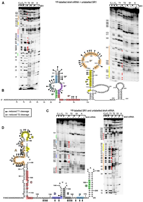
10590 Nucleic Acids Research, 2021, Vol. 49, No. 18
coding mRNAs in the same or a different pathway (rev.
in 12).
The 205 nt long SR1 from Bacillus subtilis is such a dual-
function sRNA (12). It is transcribed from a A -dependent
promoter under gluconeogenic conditions, whereas it is re-
pressed under glycolytic conditions mainly by CcpN and to
a minor extent by CcpA (13–16). Via 7 complementary re-
gions, SR1 base-pairs with ahrC mRNA encoding the tran-
scriptional activator of the arginine catabolic operons ro-
cABC and rocDEF to inhibit its translation (17). Inhibition
occurs by inducing structural changes around the ahrC RBS
(18). In addition, SR1 codes for the small peptide SR1P (39
aa) that interacts with glyceraldehyde-3P-dehydrogenase
Downloaded from https://academic.oup.com/nar/article/49/18/10589/6363770 by guest on 22 November 2021
GapA (19). This interaction increases binding of GapA to
RNase J1 and significantly enhances RNase J1 activity (20–
22). Both functions of SR1––the base-pairing and the pep-
tide encoding––are remarkably conserved over one billion
years of evolution (23). Whereas the RNA chaperone Hfq
is required for ahrC translation (18), the RNA chaperone Figure 1. Role of KinA in sporulation. Central role of KinA in the sporu-
CsrA was recently discovered to slightly restructure ahrC lation cascade. KinA is the major histidine kinase in the sporulation phos-
mRNA to facilitate SR1 binding (24). phorelay whose transcription depends on stationary phase H . In addi-
Bacillus subtilis forms endospores to survive nutrient tion, minor membrane-bound kinases KinB-E (stacked symbols behind
KinA) transfer signals to Spo0A. KinA responds to an as yet unknown
starvation. Over the years, the complex regulatory network signal by autophosphorylating and subsequently transferring phosphate
that governs sporulation and involves more than 500 of to Spo0F, which phosphorylates Spo0B, which passes the phosphate on to
the 4200 B. subtilis genes (25) has been elucidated step by Spo0A, the master regulator of differentiation. At high levels, Spo0A∼P
step (rev. in 26). Five histidine kinases, KinA, KinB, KinC, promotes sporulation by activating transcription of downstream genes,
KinD and KinE, perceive and transmit environmental sig- among them spoIIE, spoIIGA and, further downstream via SigF, SigE,
SigG and SigK, to cotA. Several phosphatases (light orange) like RapA, B,
nals that finally result in phosphorylation of Spo0A, the E or H or Spo0E antagonize Spo0A phosphorylation at various steps. The
central transcriptional regulator of the sporulation genes Rap phosphatases are under control of the small phosphatase regulatory
(rev. in 27). KinA is the major histidine kinase in the phos- proteins (Phr) PhrA, C, E and H. By a direct protein-protein interaction
phorelay that regulates sporulation (28). Its threshold level with KinA, the small protein Sda blocks phosphate transfer to Spo0F, and
is itself degraded by the ClpX protease. By contrast, KipI inhibits KinA au-
governs the entry of Bacillus subtilis into sporulation (29). tophosphorylation and is inhibited by KipA. In addition, transition state
Upon starvation and stress, KinA autophosphorylates regulator AbrB represses transcription of the phosphatase spo0E gene and
and transfers its phosphate via Spo0F and Spo0B to Spo0A is itself repressed by Spo0A∼P.
(rev. in 30, see Figure 1). KinA has three PAS domains that
measure the redox status of the cell. Its transcription is un-
der control of the stationary phase H . So far, only two di- Figure 1). Furthermore, SR1 slows down the sporulation
rect regulators of KinA are known, the 46 aa protein Sda process and impacts spore size and hydrophobicity, stress
(suppressor of DnaA) and KipI (synonym PxpB) (Figure resistance and coat protein composition. In contrast to its
1). Sda couples replication and sporulation and blocks the role in the SR1/ahrC system, neither Hfq nor CsrA affect
phosphate transfer to Spo0F during replication as its bind- sporulation. The physiological importance of sporulation
ing site on KinA overlaps with that of Spo0F (31). The control by SR1 is discussed.
Sda–KinA interaction surface was mapped (32). By con-
trast, KipI inhibits KinA autophosphorylation by affecting MATERIALS AND METHODS
the ATP/ADP reactions but does not impact the phospho-
transferase function of the KinA catalytical domain (33). Enzymes and chemicals
Two KipI monomers bind via their C-terminal domains at Chemicals used were of the highest purity available. Q5
a conserved proline in the KinA dimerization and histidine DNA polymerase, T7 RNA polymerase, CIP and polynu-
phophotransfer (DHp) domain (34). The KipI inhibitory cleotide kinase were purchased from NEB, Firepol Taq
activity is counteracted by KipA (33). polymerase from Solis Biodyne, RNase T1 from Sigma
Here, we report on a new regulator of KinA, the dual- Aldrich, RNase T2 from MoBiTec Göttingen, S1 nuclease
function sRNA SR1. We demonstrate that SR1 base-pairs from Thermo Scientific and RNase A and RNasin from
with kinA mRNA upstream of the RBS and within the 5 Promega.
part of the coding sequence resulting in translation inhi-
bition in vivo without altering the kinA mRNA stability.
Strains, media and growth conditions
The sr1-encoded peptide SR1P is not involved in kinA reg-
ulation. Deletion or overexpression of sr1 affect B. subtilis Escherichia coli strain TG1 and B. subtilis strains DB104
sporulation and transcription of at least three downstream (see Supplementary Table S2) were used. Complex TY
KinA targets, the directly Spo0A-regulated genes spoIIE medium (35) and CSE minimal medium (17) served as cul-
and spoIIGA as well as the K -dependent cotA gene which is tivation media. For E. coli 100 g/ml ampicillin, 100 g/ml
located further down in the Spo0A regulatory cascade (see spectinomycin and 200 g/ml erythromycin were used and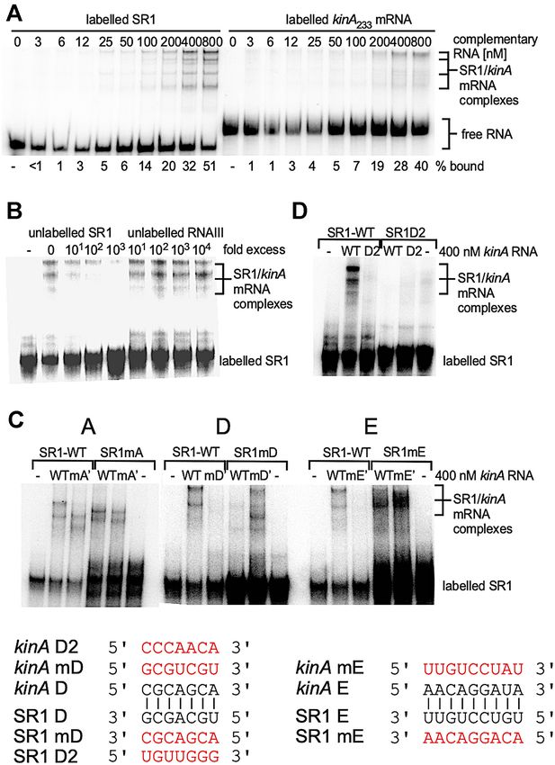
Nucleic Acids Research, 2021, Vol. 49, No. 18 10591
for B. subtilis 100 g/ml spectinomycin, 5 g/ml chloram- TY plates to determine total CFU. The rest was mixed with
phenicol, 5 g/ml erythromycin and 12 g/ml kanamycin. 300 l washed mineral oil and vortexed for 20 s. After 10
min of spontaneous phase separation, 100 l of the aque-
Sporulation assay, purification of endospores and spore coat ous phase were plated on TY plates to determine the per-
proteins and phase-contrast microscopy centage of hydrophilic spores. Plates were incubated at 37◦ C
overnight and CFU counted. Hydrophobicity was calcu-
For sporulation assays, TY cultures inoculated from a fresh
lated as the difference between total spores and hydrophilic
agar plate were grown for 6 h at 37◦ C, diluted in CSE
spores divided by the number of total spores.
medium with 0.05% glucose to OD600 = 0.2 and further
incubated for 24 h in the shaker bath at 37◦ C. Dilutions
were plated on TY plates to count total CFU (colony form- Quantification of AP (alkaline phosphatase) and DPA (dipi-
ing units). In parallel, cultures were incubated at 80◦ C for colinic acid)
20 min and dilutions plated on TY plates for spore count-
Strains were grown for 8 h in 500 ml TY with 0.1% glu-
Downloaded from https://academic.oup.com/nar/article/49/18/10589/6363770 by guest on 22 November 2021
ing. For calculations of the sporulation rate, the number of
cose at 37◦ C, centrifuged, resuspended in the same vol-
spores per total CFUs was considered.
ume of CSE medium without glucose and further culti-
Strains were cultivated for 48 h in CSE with 0.1% glucose
vated in a shaker bath. 1 ml samples were taken at different
(for nutrient starvation) or in TY (for nutrient rich condi-
time-points, shock-frozen and stored at -20◦ C. AP activity
tions) by shaking at 37◦ C. Endospores were purified by re-
was measured as described (37): Frozen samples were cen-
peated centrifugation, washing with ice cold bidist (bidis-
trifuged, washed with and resuspended in 900 l AP buffer
tilled water) and storage at 4◦ C with repeated washing steps
(0.5 M Tris–HCl pH 10.0; 5 mM MgCl2 ; 1 mM ZnCl2 ).
every 2–3 days for 4 weeks. One additional washing step was
100 l of solution 1 (0.5% BCIP and 0.75% NBT in 25%
included before further use.
DMF and 75% bidist) were added and the reaction stopped
Spore coat proteins were prepared as described (36) with
by adding 250 l AP stop solution (1 M KH2 PO4 , 0.5 M
following modifications: Spores were treated for 30 min
EDTA pH 8.0). Samples were centrifuged, pellets washed
in lysozyme/TES buffer at 37◦ C followed by two washing
three times with and resuspended in 500 l bidist. Then,
steps in fresh TES buffer and 2 h incubation at 70◦ C in
500 l DMF were added and cells were lysed for 1 h at 37◦ C.
100 mM sodium borate, 100 mM NaCl, 0.5% SDS and
The optical density (OD630 ) of cleared lysates was measured
50 mM DTT; pH 10.0. After centrifugation, the super-
against bidist/DMF. DPA quantification was performed as
natant was denatured with Laemmli buffer and separated
described (38) with slight modifications: 50 ml samples were
on a 12% Tris–glycine SDS-PAA (polyacrylamide gel).
centrifuged, washed twice with and resuspended in 1 ml
For microscopy, purified spores were applied to an
bidist and stored at –20◦ C until further use. After 15 min
agarose pad (1% agarose in water) and phase con-
treatment with 100 l lysozyme (10 mg/ml) at 37◦ C, sam-
trast images obtained with a Nikon eclipse Ti2 micro-
ples were autoclaved for 30 min at 121◦ C, subsequently acid-
scope equipped with a Nikon Plan-Apochromat 100×/1.45
ified with acetic acid (0.5 M final concentration) and incu-
oil immersion Ph3 objective and a Hamamatsu ORCA-
bated at 37◦ C for 1 h. 800 l of the cleared lysate were mixed
Flash4.0 LT + Digital CMOS camera. For each sample, five
with 200 l 1% (NH4 )2 Fe(SO4 )2 and 1% ascorbic acid in
pictures were taken from different spots. Spore length mea-
bidist, and the optical density measured immediately at 440
surements were performed with ImageJ and manually for
nm.
each spore, after the identity of the pictures was blinded.
For each picture, the length and breadth of only 20 spatially
singular spores were measured and spores of all rotary ori- Primer extension
entations were included. Halos around the spores were not
Primer extension was carried out as described (13) using
included. Identities of the pictures were only revealed after
total RNA from B. subtilis strain DB104 and 5’-labelled
evaluation of the measurements.
primers SB346 and SB3692 (all primers are listed in Supple-
mentary Table S1). For the sequencing reaction, pUCSR1
Analysis of spore resistances and hydrophobicity
(13) served as template.
Purified spores were treated for 10 min in bidist at 70◦ C and
subsequently subjected to different stress factors. To inves-
In vitro transcription, preparation of total RNA and Northern
tigate heat resistance, spores were diluted in TY medium,
blotting
incubated at 86◦ C, time samples taken and plated on TY
plates, which were incubated overnight at 37◦ C for CFU In vitro transcription was performed as described (18). Two-
counting. Survival rates were depicted as relative CFUs step PCRs were employed to introduce mutations by ex-
compared with a pre-treatment sample. For ethanol resis- changing nucleotides of the complementary inner primers
tance, ethanol was added to a final concentration of 70% to (Supplementary Table S1). Preparation of total RNA and
spores kept in bidist followed by incubation at 65◦ C. Time Northern blotting including the determination of RNA
samples were taken and diluted in fresh TY, plated, incu- half-lives were carried out as described (39).
bated and evaluated as above. To analyse spore hydropho-
bicity, white mineral oil was washed five times with equal
cDNA synthesis and qRT-PCR
amounts of bidist by vortexing and subsequent centrifuga-
tion to remove water-soluble components. Spores were di- RNA prepared from 500 l stationary phase TY cultures
luted in bidist to 400 l, of which 100 l were plated on was treated with 5 l DNase I (10 U) in a total volume of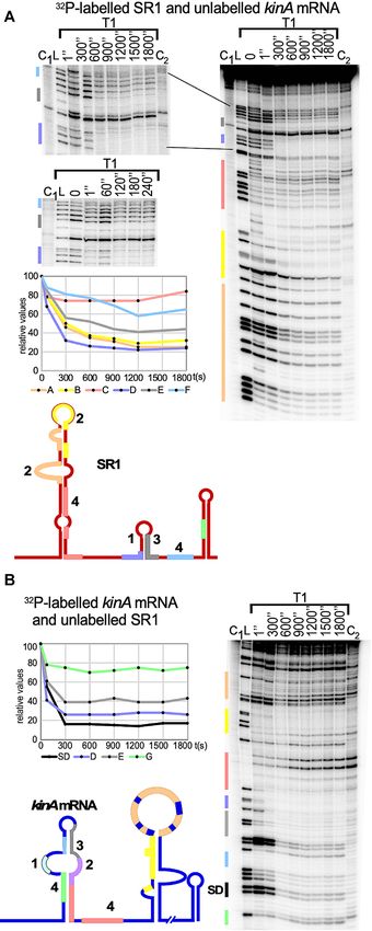
10592 Nucleic Acids Research, 2021, Vol. 49, No. 18
50 l for 2 h at 37◦ C. After phenol and chloroform extrac- was generated on chromosomal DNA of strain DB104
tion followed by ethanol precipitation, cDNA was synthe- with primer pair SB3501/SB3465, cleaved with BamHI
sized with 50 U SuperScript IV reverse transcriptase (In- and EcoRI and inserted into pGAB1 (24) yielding pGAB-
vitrogen) for 120 min at 42◦ C, followed by 10 min diges- kinA. The same approach was used to construct the pGAB-
tion with 1 l RNase A (25 U) at 37◦ C. After one phenol kinAmD , and primer pair SB3651/SB3652 was used to gen-
and two chloroform extractions followed by ethanol precip- erate the mutation. Primer SB3501 adds the weak con-
itation, the pellet was dissolved in 100 l bidist. Quantita- stitutive promoter pI (42) to the 5 end of the kinA se-
tive real-time PCR was performed with the Maxima SYBR quence composed of the native 5 UTR and the first 100
Green/ROX qPCR Master Mix (ThermoFisher) and the codons. pGAB-kinA and pGAB-kinAmD were linearized
Mx3005P system (Stratagene) in 96 well blocks. Each well with ScaI and integrated into the amyE locus of the DB104
contained 9 l bidist, 1.5 l of primer mix (5 pmol/ml each (kinA::kanR ) strain. The -galactosidase activities were de-
in bidist) and 2 l undiluted template cDNA. The reac- termined at 55◦ C as above after 6 h growth at 37◦ C in
tion was started after addition of 12.5 l qPCR MasterMix a shaker bath. Plasmid pGKSR1mD S was constructed by
Downloaded from https://academic.oup.com/nar/article/49/18/10589/6363770 by guest on 22 November 2021
2× in a darkened room. Forty cycles with denaturation for cloning a BamHI/EcoRI fragment generated by a two-step-
30 s at 95◦ C, 30 s annealing at 45◦ C and 30 s extension at PCR with primer pairs SB350/SB3641 and SB3642/SB317
72◦ C were used. Only for 5S rRNA detection, the template into the pGK15 BamHI/EcoRI vector. A list of all plasmids
cDNA was diluted 1000-fold. Immediately after qRT-PCR, used is provided in Supplementary Table S3. DB104 con-
a melting point analysis with MxPro software (Stratagene) taining a start-to stop codon mutation in the sr1 gene was
was carried out to test for product specificity. For final vali- constructed by LFH (long-flanking homology)-PCR (39).
dation, the Ct method was employed defining the num- A 1 kb front cassette with an sr1 start-to stop mutation was
ber of cycles at which the fluorescence exceeds a certain generated by PCR with outer primer pair SB3771/SB3772
threshold as Ct value. For evaluation, the MxProSoftware and internal primer pair SB3769/SB3770 on chromosomal
from Stratagene was used. Each experiment was performed DNA. A 1 kb back cassette was generated with primer pair
with three independent clones per genotype and with a sec- SB3773/SB3774. The chloramphenicol resistance gene was
ond technical replication of each sample. amplified on plasmid PINT8C as template with primer pair
SB2938/SB2939. The final 3 kb fragment generated by com-
Analysis of RNA–RNA complex formation and secondary bining and amplifying the three cassettes with primer pair
structure probing SB3772/SB3774 was directly used for DB104 transforma-
tion. All mutations were confirmed by sequencing.
Both SR1 and kinA233 mRNA were synthesized in vitro
by T7 RNA polymerase from PCR templates. RNA-
RNA complex formation assays were performed as pre- RESULTS
viously (24). RNA secondary structure probing was con- CopraRNA predicts complementary base-pairing between
ducted as described (40) with slight modifications: SR1 SR1 and kinA mRNA
or kinA mRNA (20 000–30 000 cpm) were dissolved in 5
l 1× TMN buffer containing 0.4 g tRNA, the diluted So far, only one primary target of the sRNA SR1 is known,
RNases T1, T2, A or S1 nuclease were added and cleav- ahrC mRNA (17,18). Since the majority of base-pairing
age conducted for 5 min at 37◦ C. For S1 cleavage, 2 mM sRNAs from intergenic regions of bacterial chromosomes
ZnCl2 were added. Reaction mixes were separated along- studied to date have several targets (1), CopraRNA (43) was
side a T1 ladder on denaturing 8% PAA gels. For mapping employed to search for additional targets of SR1. Rank 1
of the SR1-kinA mRNA complex, an excess of unlabelled target was kinA encoding the major kinase of the sporula-
RNA was used and complex formation allowed for 15 or 30 tion phosphorelay that phosphorylates Spo0F (rev. in 26).
min in TMN buffer before addition of RNases. IntaRNA (44) predicted 7 more or less continuous regions
in kinA complementary with SR1 which we designated A’ to
Construction of strains and plasmids and determination of - G’ in kinA and A to G in SR1, among them two stretches, G’
galactosidase activities and F’, flanking the RBS, respectively (Figure 2) and one,
E’, directly downstream of the GUG start codon. The other
To construct transcriptional lacZ fusions for Spo0A down- regions were found between nt 70 and nt 154, i.e. within the
stream targets, PCR fragments were generated on chromo- first 32 codons of the kinA ORF.
somal DNA with primer pairs SB3545/SB3546 (spoIIGA),
SB3543/SB3544 (spoIIE) and SB3547/SB3548 (cotA) each
SR1 and kinA mRNA interact in vitro
comprising –115 to + 10 relative to the transcription start
site and including all known Spo0A binding sites, di- To investigate whether SR1 is able to base-pair with kinA
gested with BamHI and EcoRI and inserted into pMG16 mRNA, we used labelled wild-type SR1 and an excess of
(41) yielding pMGPGA(pspoIIGA ), pMGPE(pspoIIE ) and unlabelled kinA mRNA (comprising the 5 233 nt) and vice
pMGPC(pcotA ), respectively. The -galactosidase activity versa in EMSAs. Up to four SR1-kinA RNA complexes
was measured at 28◦ C as described (24), but transformants most likely representing different conformations of 1:1 com-
were inoculated from fresh agar plates into TY, grown for plexes, could be observed (Figure 3A). 51% of labelled SR1
2 h in the shaker bath at 37◦ C, subsequently diluted in were found in the complex with kinA mRNA at a concentra-
prewarmed TY to OD560 = 0.2 and further cultivated for tion of 800 nM (Figure 3A). A time course experiment with
24 h. Samples were taken after 4, 8, 16 and 24 h. To con- labelled RNA and 400 nM of the unlabelled binding part-
struct a translational kinA-lacZ fusion, a PCR fragment ner demonstrated an increase of complex formation from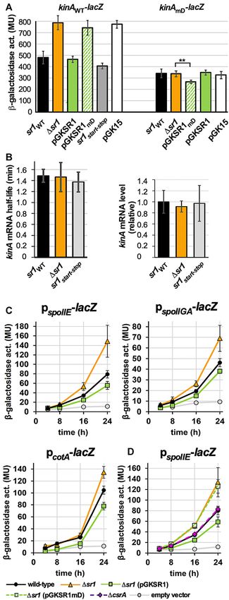
Nucleic Acids Research, 2021, Vol. 49, No. 18 10593

Figure 2. Complementarity between SR1 and kinA mRNA. IntaRNA (44) was used to search for complementary regions between SR1 and kinA mRNA.
The seven mostly uninterrupted complementary regions are highlighted in colour and designated A to G in SR1 and A’ to G’ in kinA mRNA. The RBS is
boxed and the GUG start codon is underlined.

                                                                             unlabelled RNAIII was not even able to do so at a 104 -
                                                                             fold excess. To narrow down complementary regions deci-
                                                                             sive for the SR1-kinA RNA interaction, each of the 7 com-

                                                                                                                                                    Downloaded from https://academic.oup.com/nar/article/49/18/10589/6363770 by guest on 22 November 2021
                                                                             plementary regions A to G were mutated individually in ei-
                                                                             ther kinA mRNA or SR1 (Figure 3C and Supplementary
                                                                             Figure S1B). In each mutant, all nt of the corresponding
                                                                             region (see coloured regions in Figure 2) were replaced by
                                                                             the complementary nt. Interestingly, only mutated regions
                                                                             D and E of kinA prevented the interaction with SR1 while
                                                                             mutations in A, B, C, F and G did not impair binding (Fig-
                                                                             ure 3C and Supplementary Figure S1B). Region D located
                                                                             10 nt downstream of the kinA start codon was decisive for
                                                                             SR1 binding, which was confirmed by compensatory muta-
                                                                             tions (Figure 3C). Even the alteration of 2 nt within region
                                                                             D prevented SR1 binding to kinA mRNA and vice versa,
                                                                             but in contrast to an exchange of 7 nt they could not com-
                                                                             pletely compensate the binding deficiency of the individu-
                                                                             ally mutated region (Figure 3D).
                                                                                In summary, SR1 binds specifically to kinA mRNA in
                                                                             vitro, and complementary region D located 10 nt down-
                                                                             stream of the kinA start codon is decisive for the in vitro
                                                                             SR1-kinA RNA interaction.

                                                                             SR1 represses kinA mRNA translation in vivo by a base-
                                                                             pairing interaction but does not affect kinA mRNA stability
                                                                             To confirm the in vitro interaction between SR1 and kinA
                                                                             mRNA in vivo, a translational kinA-lacZ reporter gene fu-
                                                                             sion under control of the weak constitutive heterologous
                                                                             promoter pI (42) was constructed and integrated into the
                                                                             amyE locus of the B. subtilis chromosome of the kinA and
Figure 3. SR1 and kinA mRNA interact in vitro. EMSAs with 0.15 fmol          the isogenic sr1 strain as well as the sr1 overexpression
32− P [ ␣-UTP]-labelled RNA and increasing concentrations of unlabelled
complementary RNA. Labelled and unlabelled RNA were mixed and in-            strain sr1 (pGKSR1S). As shown in Figure 4A left, dele-
cubated for 30 min at 37◦ C in TMN buffer, followed by separation on         tion of sr1 resulted in a 1.8-fold increase in kinA translation
6% native PAA gels. Autoradiograms of the gels are shown. (A) EMSAs          whereas sr1 overexpression was able to compensate this ef-
with wild-type kinA mRNA and SR1 species. (B) Competition EMSA with          fect. This suggests that SR1 acts on kinA posttranscription-
unlabelled SR1 or heterologous RNAIII. Above, the fold excess of the
competitor RNA is indicated. (C) EMSAs with SR1 and kinA233 mRNA
                                                                             ally. To investigate if the sr1 encoded small protein SR1P
species either wild-type or mutated in region A or A’, D or D’ or E or E’.   is involved in the regulation of kinA, an isogenic B. subtilis
In the mutated RNA (labelled with m) all nt of the corresponding region      strain with a start-to stop codon mutation in the chromoso-
were exchanged by the respective complementary nt as shown below for         mal sr1p ORF was constructed. This mutation had no im-
kinAmD and kinAmE. (D) as (C), but only 2 nt in region D or D’ were          pact on the kinA-lacZ translation indicating that only the
exchanged (see below kinAD2, SR1D2)
                                                                             sRNA SR1 but not SR1P was required for the observed ef-
                                                                             fect (Figure 4A). To analyse a possible influence of SR1 on
                                                                             the kinA mRNA stability, we employed qRT-PCR to deter-
1 min towards 30 min (Supplementary Figure S1). To con-                      mine the half-life of kinA mRNA in the presence and ab-
firm that binding is specific to SR1, a competition experi-                  sence of SR1 as well as in the absence of the sr1-encoded
ment was performed by adding an excess of unlabelled SR1                     peptide SR1P. As shown in Figure 4B left, neither SR1 nor
or unlabelled heterologous RNAIII (42) to labelled SR1                       SR1P affected the half-life of kinA mRNA. In addition,
and an excess of unlabelled kinA mRNA (Figure 3B). While                     the qRT-PCR indicated that the amounts of kinA mRNA
a 100-fold excess of unlabelled SR1 could successfully dis-                  were almost the same in the wild-type, sr1 knockout and
place labelled SR1 from the complex with kinA mRNA,                          sr1start-to stop strain (Figure 4B, right) ruling out an effect of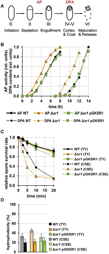
10594 Nucleic Acids Research, 2021, Vol. 49, No. 18
SR1 on the kinA promoter. Within the 5 233 nt of kinA
mRNA we did neither find a potential for alternative fold-
ing nor a Rho-dependent or Rho-independent transcrip-
tion terminator. Furthermore, a previous B. subtilis tran-
scriptome analysis performed in the absence and presence
of Rho (45) did not reveal any premature termination signal
in the presence of Rho in the corresponding region. There-
fore, we can exclude SR1-mediated transcription termina-
tion as alternative mechanism of SR1 action. Consequently,
SR1 represses translation of kinA mRNA without affecting
its stability.
To substantiate the importance of complementary region
D for the SR1-kinA mRNA interaction in vivo, we con-
Downloaded from https://academic.oup.com/nar/article/49/18/10589/6363770 by guest on 22 November 2021
structed pGKSR1mD S carrying the same 7 nt exchange in
region D which was shown in the EMSAs (Figure 3C) to be
important for kinA mRNA binding. This mutation does not
affect the previously demonstrated ability of SR1P to inter-
act with GapA for modulating the B. subtilis degradosome-
like network (20,21). The -galactosidase activities in the
sr1 strain with and without pGKSR1mD S were compara-
ble (Figure 4A), indicating that SR1mD cannot downregu-
late translation of wild-type kinA mRNA. Northern blots
confirmed identical expression levels of SR1, SR1start-to stop
and SR1mD (Supplementary Figure S2).
To confirm a base-pairing interaction between SR1 and
kinA mRNA in vivo, compensatory nt exchanges were in-
troduced in SR1 and kinA in the translational kinA-lacZ
fusion and -galactosidase activities measured (Figure 4A
right). The -galactosidase activities of the kinAmD -lacZ fu-
sion were almost the same in the presence and absence of
wild-type SR1 demonstrating that wild-type SR1 cannot in-
teract with kinAmD mRNA. By contrast, SR1mD expressed
from pGKSR1mD was able to interact with the kinAmD
mRNA indicated by a slightly, but significantly, lower -
galactosidase activity of the kinAmD -lacZ fusion.
From these data, we conclude that the base-pairing in-
Figure 4. SR1 inhibits kinA translation without impacting RNA stabil- teraction between SR1 and kinA mRNA is decisive for the
ity and affects transcription from three Spo0A∼P-dependent promoters. repression of kinA translation in vivo.
(A) Left: A translational kinA-lacZ fusion under the weak constitutive
heterologous promoter pI were integrated into the amyE locus of B. sub-
tilis DB104 (kinA) and the isogenic sr1, sr1start-stop , sr1 (pGKSR1S), Analysis of three promoters regulated by the KinA-
sr1 (pGKSR1mD S) and sr1 (pGK15) strains. Right: A translational downstream target Spo0A corroborates the in vivo role of
kinAmD -lacZ fusion under control of pI was integrated into the DB104
(kinA) strain and the isogenic sr1, sr1 (pGKSR1mD) and sr1 SR1 in sporulation
(pGKSR15) strains. KinAmD and SR1mD are complementary. The - To investigate the impact of SR1 on the transcription of
galactosidase activities were measured from six to eight individual clones
(kinAWT -lacZ) or at least 10 individual clones (kinAmD -lacZ) for each KinA downstream genes, a set of three genes was chosen
strain after 6 h growth in TY medium until OD600 = 4.5. The indicated val- whose transcription is strongly induced by Spo0A∼P dur-
ues are the results of three biological replicates. Error bars represent stan- ing sporulation (45,46). While spoIIE and spoIIGA are F -
dard deviations. pGK15, empty vector control; asterisks label significancy dependent early sporulation genes encoding SpoIIE and
in Student’s t-test (** P < 0,005). (B) Left: The half-life of kinA mRNA was
determined by qRT-PCR as described in Materials and Methods in wild-
SpoIIGA, respectively, cotA is a K -dependent late sporu-
type DB104 and the isogenic (sr1) and sr1start-stop strains. The average lation gene. SpoIIE is a serine phosphatase that dephos-
values obtained from three biological replicates with standard deviations phorylates the anti-anti-sigma factor SpoAA and controls
are shown. Right: The relative amounts of kinA mRNA were determined F activity whereas SpoIIGA is a protease involved in the
by qRT-PCR in the wild-type strain DB104 and the isogenic (sr1) and maturation of E . CotA is an outer spore coat protein (47).
sr1start-stop strains after growth in complex TY medium until OD600 = 4.5.
(C) Transcriptional pspoIIE -lacZ, pspoIIGA -lacZ and pcotA -lacZ fusions were Transcriptional fusions of the pspoIIE , pspoIIGA and pcotA pro-
integrated into the amyE locus of B. subtilis strain DB104, DB104(sr1) moters with the promotorless lacZ gene were constructed
and DB104(sr1) (pGKSR1) and -galactosidase activities measured af- and integrated into the chromosomal amyE locus of B. sub-
ter growth in TY medium after 4, 8, 16 and 24 h. The indicated values tilis strains DB104 and isogenic sr1 and sr1(pGKSR1)
are the results of three biological replicates with seven transformants each.
Error bars represent standard deviations. EV: empty vector control (insert-
strains. Seven independent transformants were grown over
free pMG16 vector integrated in amyE locus). (D) Comparison of the ef- 24 h in complex TY medium, time samples taken and
fect of pGKSR1 and pGKSR1mD as well as CsrA on the transcription from -galactosidase activities determined (Figure 4D). In all
pspoIIE . three cases, deletion of sr1 increased the promoter activity,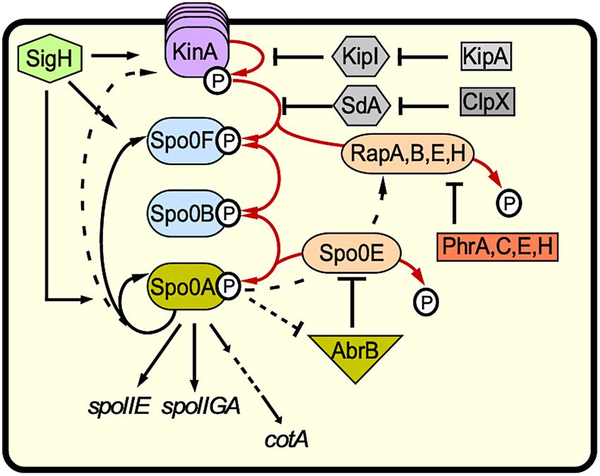
Nucleic Acids Research, 2021, Vol. 49, No. 18 10595

whereas sr1 overexpression under its native promoter from            To investigate protection of kinA mRNA by SR1 and
plasmid pGKSR1 (15 copies/cell) decreased the promoter            the possible induction of structural alterations, 5 labelled
activity. The most pronounced effects were observed for the       kinA233 mRNA was incubated with an excess of unlabelled
spoIIE promoter. After 24 h growth (conditions of sporu-          SR1, the complex was allowed to form at 37◦ C and, subse-
lation) an almost 2-fold increase in pspoIIE activity was ob-     quently, partially digested with RNases T1, T2, A and nu-
served in the absence of SR1, whereas the effects for pspoIIGA    clease S1 (Figure 5A, B). In parallel, 5 end-labelled SR1
and pcotA were only 1.5- and 1.3-fold, respectively. The re-      was incubated with an excess of unlabelled kinA233 mRNA
duction of promoter activity upon sr1 overexpression was          and treated likewise (Figure 5C, D).
generally much smaller, but over the time––after 8, 16 and           Addition of unlabelled SR1 to labelled kinA mRNA re-
24 h––the corresponding values were always lower than             sulted in a clear protection of regions A’, B’, C’, D’ and
those of the wild-type strain. Furthermore, the pspoIIE ac-       E’ (Figure 5A), whereas regions G’ and F’ were already
tivity was compared between the sr1 strain without and           double-stranded in unbound kinA mRNA and could not
with pGKSR1mD S and found to be nearly identical (Figure          be evaluated. Most important, however, was the complete

                                                                                                                                  Downloaded from https://academic.oup.com/nar/article/49/18/10589/6363770 by guest on 22 November 2021
4C) supporting the importance of region D for the SR1-            protection of nt 44–52 covering the kinA RBS which itself is
mediated inhibition of kinA.                                      not complementary to any region in SR1. Apparently, SR1
   From these data, we conclude that the regulation of the        binding to the adjacent complementary regions G’ and F’,
Spo0A downstream genes affected by the major kinase               E’ and D’ impedes the access of the 30S subunit to its bind-
KinA is due to the base-pairing between SR1 and kinA              ing site. This is in good agreement with the observed inhi-
mRNA.                                                             bition of kinA translation by SR1 (Figure 4A). These data
                                                                  were complemented by addition of unlabelled kinA mRNA
                                                                  to labelled SR1, which revealed protection of regions A, B,
Secondary structure probing of kinA mRNA and the SR1-
                                                                  D, E and F (Figure 5C). Regions C and G could not be
kinA mRNA complex
                                                                  assessed, as they remained sequestered by intramolecular
To investigate in more detail how SR1 can inhibit kinA            base-pairing.
translation without directly binding to and protecting the           In summary, secondary structure probing of the SR1-
kinA RBS, we first determined the secondary structure             kinA mRNA complex demonstrated that SR1 binding
of the 233 nt kinA mRNA species that was also used in             makes the kinA RBS inaccessible, which is consistent with
the EMSAs. To this end, we performed limited digestions           SR1 inhibiting translation of a kinA-lacZ fusion.
with structure-specific ribonucleases by treating in vitro syn-
thesized, 5 -end labelled and gel-purified kinA233 mRNA          Binding pathway
with RNase T1 (cleaves 3 of unpaired G residues), RNase
                                                                  The results of the EMSAs (Figure 3) and the translational
T2 (unpaired nucleotides with a slight preference for A
                                                                  lacZ-fusions (Figure 4A) revealed that region D/D’ is deci-
residues), RNase A (unpaired C and U residues) and nu-
                                                                  sive for the SR1-kinA mRNA interaction. To analyse the se-
clease S1 (single-stranded nucleotides). Supplementary Fig-
                                                                  quence of interactions between SR1 and kinA mRNA, two
ure S3A shows the analysis and Supplementary Figure S3B
                                                                  time-course experiments were performed, one with labelled
the schematic presentation of two slightly different kinA
                                                                  SR1 and unlabelled kinA mRNA (Figure 6A), the other vice
mRNA structures derived from the cleavage data, one 5 bp
                                                                  versa (Figure 6B). In both cases, the interaction between D
stem below regions G’ to D’ the other with an 8 bp stem
                                                                  and D’ was the initial one. It occurred already after a few
interrupted by a bulged out U. Most probably, a mix be-
                                                                  seconds as seen in the high-resolution time course in Fig-
tween both structures is present in vitro. However, in both
                                                                  ure 6A. In kinA mRNA, the next protected region was the
structures, the double- or single-strandedness of all comple-
                                                                  RBS, although it is not contacted directly by SR1. As more
mentary regions is identical.
                                                                  G-residues sensitive to RNase T1 cleavage are present in the
   Structure probing revealed that the kinA RBS (nt 46–
                                                                  complementary regions A to G in SR1 compared to A’ to G’
49) is located in a single-stranded region (nt 44 to 52) and
                                                                  in kinA mRNA, T1-cleavages revealed that both the bulge
should, therefore, be accessible to the ribosomal 30S sub-
                                                                  in A and the loop of B bind more or less simultaneously af-
unit. Interestingly, the complementary region G’ upstream
                                                                  ter D, followed by the E and then the F region (Figure 6A).
of the RBS as well as regions F’ and E’ immediately down-
                                                                  In the EMSAs (Figure 3), E was the only other complemen-
stream of the RBS are located in completely (G’, F’) or al-
                                                                  tary region whose mutation in SR1 prevented kinA mRNA
most completely (E’) double-stranded regions. Region D’
                                                                  binding whereas this was not the case in regions A, B, C, F
that proved to be decisive in the EMSA for interaction
                                                                  or G. In kinA, region E bound immediately after D (Figure
with SR1 (Figure 3C,D) displayed 4 single-stranded and
                                                                  6B) and, since B and F are completely double-stranded, no
3 double-stranded nt whose complete (7nt) exchange lead
                                                                  conclusion could be drawn about them.
DB104 (sr1, pGKSR1mD ) behave like the isogenic sr1
                                                                     In conclusion, the SR1–kinA mRNA interaction com-
strain (Figure 4D). Region A’ was fully and C’ partially
                                                                  mences between complementary regions D and D’ located
single-stranded whereas region B’ was completely paired.
                                                                  10 nt downstream of the kinA start codon and leads to an
   The same approach was used for secondary structure
                                                                  immediate protection of the kinA RBS.
probing of 5 -end labelled SR1, and the result confirmed our
previously published SR1 structures (18,24): SR1 regions
                                                                  SR1 affects B. subtilis sporulation
A, B, D, E and F were mostly or fully (region F) single-
stranded and regions C and G were completely base-paired          The translational repression of kinA by SR1 should impact
(Figure 5D).                                                      B. subtilis sporulation. Therefore, we compared sporulation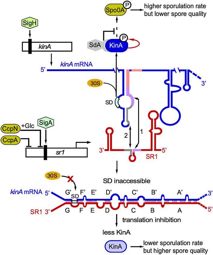
10596 Nucleic Acids Research, 2021, Vol. 49, No. 18

                                                                                                                                                            Downloaded from https://academic.oup.com/nar/article/49/18/10589/6363770 by guest on 22 November 2021

Figure 5. Secondary structure of the kinA mRNA/SR1 complex. (A and C) Secondary structure probing of the kinA mRNA/SR1 complex. 15 nmol of
purified, 5 labelled kinA mRNA or 5 labelled SR1 were incubated with a 166-fold or 133-fold, respectively, excess of the complementary unlabelled RNA,
the complex allowed to form for 30 min at 37◦ C and subjected to limited cleavage of T1 (0.1 U), T2 (0.1 U), S1 (2.2 U) and A (0.4 ng). The digested RNAs
were separated on 8% denaturing gels. Autoradiograms are shown. L, T1 digestion under denaturing conditions. Nucleotide positions are included. Altered
T1 and T2 cleavages are indicated by the symbols shown in the box in (B) and (D). (B and D) Structures of kinA mRNA and SR1, respectively, in which
nucleotides protected by binding of the complementary RNA are indicated.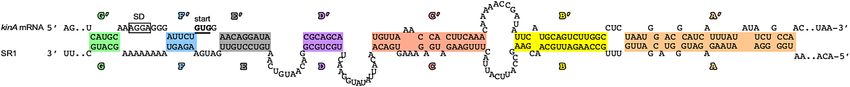
Nucleic Acids Research, 2021, Vol. 49, No. 18 10597

                                                                               of wild-type strain DB104, DB104(sr1) and the sr1 over-
                                                                               expression strain DB104(pGKSR1). Cells were grown for
                                                                               6 h in TY medium, inoculated to OD600 = 0.2 into mini-
                                                                               mal CSE medium with glucose and further cultivated in a
                                                                               shaker bath for 24 h. Afterwards, the ratio of spores and
                                                                               living cells was determined (Figure 7A). As expected, dele-
                                                                               tion of SR1 increased sporulation about 1.8-fold, whereas
                                                                               sr1 overexpression from its native promoter reduced sporu-
                                                                               lation about 1.5-fold. Since CsrA binds SR1 (24), we inves-
                                                                               tigated a potential influence of CsrA on sporulation. How-
                                                                               ever, deletion of csrA had no significant effect on sporula-
                                                                               tion. Likewise, the deletion of hfq - Hfq also binds to SR1
                                                                               (18) - did not affect sporulation (Figure 7A).

                                                                                                                                               Downloaded from https://academic.oup.com/nar/article/49/18/10589/6363770 by guest on 22 November 2021
                                                                               The amount of SR1 affects properties, stress resistance and
                                                                               protein composition of spores
                                                                               First, we performed an experiment to analyse if the amount
                                                                               of SR1 affects spore germination. To this end, purified
                                                                               spores from wild-type DB104 and the isogenic sr1 knock-
                                                                               out and pGKSR1 overexpression strains were treated for 10
                                                                               min at 70◦ C, suspended in TY and cultivated at 37◦ C in a
                                                                               shaker bath. Aliquots were plated on TY agar plates at par-
                                                                               ticular time points, incubated at 37◦ C overnight and CFUs
                                                                               were counted. All three strains showed a lag phase of about
                                                                               190 min indicating that none of them displayed a germina-
                                                                               tion latency (Supplementary Figure S4). Moreover, no dif-
                                                                               ferences in spore outgrowth ratio or kinetics between these
                                                                               strains were observed after germination induction with TY
                                                                               medium in time-lapse microscopy (not shown).
                                                                                  To investigate a possible effect of SR1 on spore size,
                                                                               phase-contrast microscopy was applied and the length of
                                                                               500 spores measured for the wild-type, sr1 knockout and the
                                                                               sr1 overexpression strains. Spores from the sr1 strain were
                                                                               on average ≈ 5% shorter and those from the overexpression
                                                                               strain slightly longer than those from the wild-type strain
                                                                               (Figure 7B).
                                                                                  In addition, we compared purified spores for their out-
                                                                               growth ability after stress treatment. Spores were treated
                                                                               for 10 min at 70◦ C, suspended in TY medium, incubated
                                                                               at 86◦ C and time samples plated on TY agar plates. After
                                                                               overnight incubation at 37◦ C, CFUs were counted. Figure
                                                                               7C reveals that spores from the sr1 overexpression strain
                                                                               showed a much higher heat resistance than those from the
                                                                               wild-type strain DB104 over the entire time, whereas the
                                                                               spores of the sr1 knockout strain were less heat-resistant
                                                                               and formed less colonies, which was detectable already after
                                                                               2.5 min of heat treatment. To evaluate another stress factor,
Figure 6. Time-course of SR1–kinA RNA interaction. The same approach
                                                                               spores were treated with 70% ethanol at 65◦ C and germi-
was used as in Figure 5 for secondary structure probing with either la-        nation investigated as above. A similar effect was observed
belled SR1 or labelled kinA mRNA. However, an excess of the comple-            as for exclusive heat treatment (Figure 7C). Neither 65◦ C
mentary unlabelled RNA was added, incubation started at 37◦ C and sam-         nor 70% ethanol alone had an appreciable effect on spore
ples taken at the indicated incubation times, instantly added to tubes con-    survival, even after a 60 min treatment (not shown). Inter-
taining RNase T1 and subjected to a 5 min digestion at 37◦ C. (A) Time
course with labelled SR1. The second smaller gel shows samples taken at        estingly, in both cases the survival rate of the sr1 strain
shorter time intervals and was used to confirm that region D is bound first.   caught up with the wild-type strain after ≈20 min. This
(B) Time course with labelled kinA mRNA. In both cases, graphs based on        could be an evidence that just a subpopulation of the sr1
the calculation of the corresponding gels show that region D is bound first,   spores had a reduced resistance. A similar result was ob-
whereas regions G and C, that are completely double-stranded in SR1 (G
and C) and partially double-stranded in kinA mRNA (G’), do not play
                                                                               served after treatment with UV light (254 nm). Again, the
a role. In kinA mRNA, immediately after binding of D’, the RBS is pro-         sr1 strain was less resistant compared to wild-type and
tected. Numbers added to the regions in the schematic structures of SR1        overexpression strains but neither the sr1 strain caught
and kinA mRNA indicate the sequence of interactions.                           up with the wild-type strain with increasing UV stress nor
10598 Nucleic Acids Research, 2021, Vol. 49, No. 18
Downloaded from https://academic.oup.com/nar/article/49/18/10589/6363770 by guest on 22 November 2021
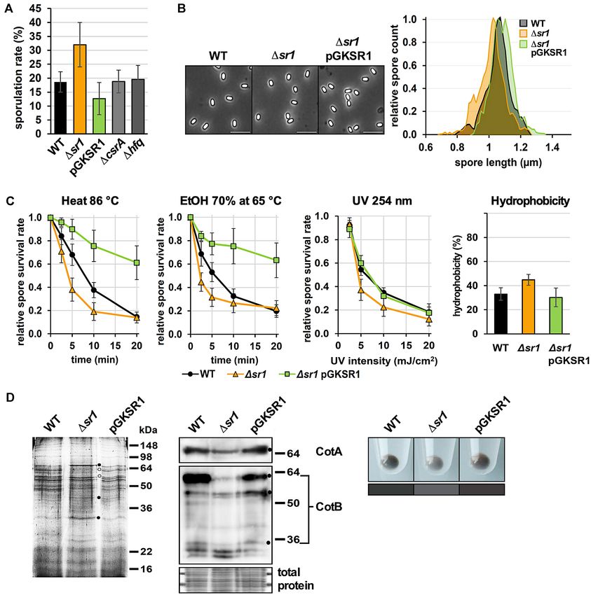
Figure 7. SR1 impacts B. subtilis sporulation, size and stress resistance of spores and abundance of spore coat proteins. (A) The sporulation assay was per-
formed as described in Materials and Methods with strains DB104, DB104(sr1::cat), DB104(pGKSR1), DB104(csrA::cat) and DB104(hfq::cat). In
all cases, the indicated values are the results of three biological replicates. Error bars represent standard deviations. (B) Left: Phase-contrast micrographs of
purified wild-type, Δsr1 and Δsr1 pGKSR1 spores. Right: Spore length distribution of wild-type (black), Δsr1 (orange) and Δsr1 pGKSR1 (green) spores.
The lengths of 500 spores for each phenotype were measured from phase-contrast micrographs as shown left. Depicted are the smoothened histograms of
the length distributions. (C) Resistance of spores obtained from DB104, DB104(sr1) and DB104(sr1, pGKSR1) against heat, ethanol, UV light and
their hydrophobicity were determined in three independent experiments. Standard deviations are indicated. (D) Composition of spore coat proteins from
wild-type, Δsr1 and Δsr1 pGKSR1 strains. Left, Coomassie-blue stained protein gel. Dots depict differences in the abundance of five individual proteins.
Centre, Western blot with antibodies against CotA and CotB. CotB-46 is the main species, and CotB-66 likely a polyphosphorylated form of the protein
(67). Below, the same amounts of total proteins applied for Western blotting were stained with Coomassie-blue as loading control. Right, spore colours.
Below, the mean colours of comparable areas of the picture are represented for better comparison.Nucleic Acids Research, 2021, Vol. 49, No. 18 10599
was the sr1 overexpression strain able to further increase its
UV resistance above wild-type level. Furthermore, the hy-
drophobicity of sr1 spores was about 30% higher than that
of the other two strains (Figure 7C, right).
To analyse a possible influence of SR1 on the protein
composition of the spore coat, spore coat proteins from
the three isogenic strains were prepared and analysed in a
12% SDS-PAA gel (Figure 7D). The abundance of at least
five proteins differed between the three strains (labelled with
dots). As shown by Western blotting, the amount of CotA
and CotB was lower in spores formed in the absence of SR1
(Figure 7D) which might explain their lower stress resis-
tance. In addition, we noticed a less brownish colour of sr1
Downloaded from https://academic.oup.com/nar/article/49/18/10589/6363770 by guest on 22 November 2021
spores.
Taken together we conclude that SR1 has a physiolog-
ical function in spore formation, stress resistance and pro-
tein composition of the spore coat by regulating kinA trans-
lation. Furthermore, we hypothesize that under starvation
conditions SR1 might slow down spore formation to enable
the formation of high-quality spores.
SR1 decelerates spore formation
To investigate our hypothesis, we studied the impact of SR1
on the speed of spore formation. To this end, we chose two
markers, AP (alkaline phosphatase) which is active between
septation (stage II) and engulfment (stage III), and DPA
(dipicolinic acid) detectable only after cortex and coat for-
mation in stages IV and V of the spore formation process
(Figure 8A). We grew the isogenic wild-type, sr1 and sr1
overexpression strains and determined the sporulation spe-
cific AP activity and the presence of DPA during sporula-
tion. As shown in Figure 8B, the AP activity could be mea-
sured ≈1 h earlier in the sr1 knockout compared to the wild-
type and sr1 overexpression strains indicating that engulf-
ment starts earlier in the absence of SR1. Likewise, DPA
could be detected in the sr1 strain ≈ 2h earlier than in the
wild-type and the sr1 overexpression strain. This confirms
our hypothesis that the sr1 knockout strain sporulates ear-
lier and progresses faster through spore formation than the
wild-type strain. From these data, we infer that SR1 slows
down the entire process of spore formation by about 2-fold
repressing kinA translation and, consequently, reducing the
amount of the major histidine kinase KinA.
SR1 is only required under nutrient starvation in minimal
medium for high quality spores
As SR1 is upregulated in the absence of glucose (13,15),
we compared the properties of spores generated in mini- Figure 8. SR1 slows down sporulation and is only required under starva-
mal CSE with those generated in complex TY medium. In tion conditions for production of high-quality spores. (A) Schematic rep-
TY, SR1 is expressed in high amounts after 5 h (13), but resentation of the sporulation process. Sporulation-specific alkaline phos-
the secondary carbon sources of the complex medium pre- phatase (AP) activity emerges between stages II and III. DPA is synthe-
vent the culture from starving. As presented in Figure 8C, sized during stage V. (B) Investigation of the spore formation progress in
wild-type, sr1 knockout and sr1 overexpression strains. AP activity and
spores generated in TY medium were more heat resistant in DPA content were measured over time after resuspension in CSE medium
comparison to those generated in CSE, and heat resistance and are depicted as relative signal strength. The time difference between
did not differ between wild-type, sr1 knockout and sr1 over- onset of AP activity and DPA detectability is representative of the sporu-
expression strains. By contrast, spores generated in CSE lation progress. (C) Heat resistance assay of spores formed in TY medium.
Purified spores of wild-type, sr1 knockout and sr1 overexpression strains
medium were less heat resistant and displayed differences cultured in TY were treated with 86◦ C and spore survival was measured
between the three strains: Whereas spores from the sr1 over- over time. For comparison, the heat resistance of spores from minimal CSE
expression strain generated in CSE were only slightly less re- medium (as in Figure 5C) is depicted in dotted lines. (D) Comparison of
sistant than all spores generated in TY medium, spores from spore hydrophobicity between TY and CSE medium.10600 Nucleic Acids Research, 2021, Vol. 49, No. 18
the wild-type strain showed a 2- to 3-fold lower heat resis- DISCUSSION
tance and those from the sr1 knockout strain were even less
In 2010, it was shown that IPTG-induced KinA synthesis
heat resistant than those from the wild-type strain, at least
beyond a certain threshold can lead to entry into an irre-
after 5–15 min heat treatment. In addition, spores produced
versible sporulation process independent of nutrient avail-
in TY were also less hydrophobic indicating a more efficient
ability (29). This indicates that the amount of KinA has to
crust formation and general condition, and did not reveal
be tightly regulated to prevent sporulation under nutrient-
differences between the three strains (Figure 8D). Together
rich and nonstress conditions. Here, we report on a new
this illustrates that SR1 is only necessary under starvation
regulator of the kinA gene, the trans-encoded sRNA SR1
conditions to enable the formation of high-quality spores.
that shares 7 complementary regions with kinA mRNA des-
ignated A/A’ to G/G’. SR1 inhibits translation of kinA
A sporulation-dependent promoter upstream of psr1 barely mRNA by a base-pairing interaction (Figure 4A). The ef-
contributes to the amounts of SR1 fect of the sr1 deletion could be compensated by overexpres-
Downloaded from https://academic.oup.com/nar/article/49/18/10589/6363770 by guest on 22 November 2021
Previously, we mapped the sr1 transcription start site (TSS) sion of wild-type sr1 but not sr1mD carrying a 7 nt exchange
at a A -dependent promoter (13). Nicolas et al. reported an in region D required for the initial interaction with kinA
additional sporulation-dependent RNA originating ≈130 mRNA. At least in enterobacteria, most translationally in-
nt upstream of this TSS (45, see Supplementary Figure S5). hibited mRNAs are rapidly degraded, but this is an indirect
Primer extension with cultures grown in TY for 6 h (station- effect because they are not protected by ribosomes against
ary phase) or 24 h (sporulation) revealed the TSS of A - endoribonucleases (rev. in 1). By contrast, SR1 does not af-
dependent psr1 after 30 min exposure (Supplementary Fig- fect the stability of kinA mRNA (Figure 4B) excluding both
ure S5A) whereas the additional promoter was detectable degradation as consequence of lower ribosome coverage of
only after 72 h exposure (Supplementary Figure S5B). To kinA mRNA and the recruitment of a 5 -3 exoribonuclease
determine the promoter strength, three transcriptional lacZ like RNase J1 as mechanism of SR1 action. This is con-
fusions were constructed. After 48 h growth at 37◦ C bright sistent with the dispensability of the SR1-encoded peptide
blue colonies were visible for the A -dependent psr1 -lacZ fu- SR1P (Figure 4A, B)––previously shown to impact RNase
sion on TY agar with X-Gal, and dark-blue colonies on 3- J1 activity via GapA binding (20)––for kinA regulation. In-
fold diluted TY agar (Supplementary Figure S5D), whereas terestingly, SR1 only marginally influences the half-life of
colonies with the lacZ fusion of the additional promoter ahrC mRNA either (17,24) suggesting that it might mainly
were white on TY agar and slightly blue on threefold di- employ translational control as mechanism of action. By
luted TY agar, which is in agreement with an ≈144-fold contrast, B. subtilis RoxS inhibits translation and promotes
weaker TSS signal. Due to the extremely low promoter ac- mRNA decay of ppnKB mRNA and sucCD mRNA (48)
tivity, no -galactosidase activity could be measured. Con- whereas it activates translation of yflS mRNA and, inde-
sequently, the very weak promoter upstream of psr1 scarcely pendently, prevents its degradation by RNase J1 (49). The
contributes to the amount of SR1 under sporulation condi- latter is rather unusual as only a few sRNAs have been
tions. shown to directly affect target mRNA degradation, among
them E. coli RyhB (50) and Salmonella enterica MicC (51).
Since the 5 region of kinA mRNA that binds SR1 has
Conservation of the SR1–kinA mRNA interaction among the
neither the potential for alternative folding nor contains a
Bacillales
Rho-dependent or -independent transcription terminator,
In 2012, we discovered 39 SR1/SR1P homologues within we rule out SR1-mediated transcription termination as al-
the Bacillales and further analysed 9 of them (23). A new ternative regulatory mechanism.
BLAST analysis detected SR1 homologues in 139 species, Although CsrA binds both kinA mRNA and SR1 in the
again only in Bacillus and Geobacillus species. In only nine nanomolar range (Supplementary Figure S7B; 24), it nei-
Bacillus species, we found kinA homologues which have ther affected the SR1-kinA mRNA interaction (Supplemen-
substantial sequence similarity to B. subtilis kinA. The other tary Figure S7C) nor induced structural changes into kinA
kinA genes encode a protein of similar size and function, but mRNA in vitro (Supplementary Figure S7D). CsrA had
perhaps different evolutionary origin of at least the 5 part also no impact on sporulation (Figure 7A) or the pspoIIE -
and, therefore, are not complementary to SR1. To analyse lacZ transcriptional fusion (Figure 4D). This is in contrast
if the SR1-kinA mRNA interaction is conserved in the nine to its role in the SR1-ahrC mRNA system, where it intro-
species, we aligned their seven complementary SR1-kinA re- duces slight structural changes into ahrC mRNA around
gions (Supplementary Figure S6). Region D required for the the region required for the initial contact with SR1 (24).
initial interaction between both molecules was identical in Likewise, Hfq did not impact sporulation although it also
the nine SR1 species, and in five of nine kinA homologues, binds to SR1 (18). However, it cannot be excluded that a
only one nt exchange was found in D’. Regions E’, F’ and so far unidentified RNA chaperone promotes the SR1-kinA
the RBS were completely conserved in the nine kinA homo- mRNA interaction in vivo.
logues, and SR1 region F carried only in two species one nt The complementary region G’ is located upstream of the
exchange. By contrast, region G/G’ which was bound last kinA RBS, and regions F’ to A’ immediately downstream of
in our time course experiment (Figure 6) differed in four and within the first 32 codons of kinA (Figure 2). In agree-
species significantly. From these data we conclude that the ment with SR1 inhibiting translation, kinA mRNA bind-
SR1-kinA interaction is highly conserved in these 9 Bacillus ing initiates at the D/D’ region 10 nt downstream of the
species. start codon followed by a rapid protection of the kinA RBSYou can also read