Structural insights into human acid-sensing ion channel 1a inhibition by snake toxin mambalgin1 - eLife
←
→
Page content transcription
If your browser does not render page correctly, please read the page content below
RESEARCH ARTICLE

                                     Structural insights into human acid-
                                     sensing ion channel 1a inhibition by snake
                                     toxin mambalgin1
                                     Demeng Sun1,2†, Sanling Liu1†, Siyu Li1†, Mengge Zhang1†, Fan Yang1, Ming Wen1,
                                     Pan Shi1, Tao Wang3, Man Pan2, Shenghai Chang4, Xing Zhang4,
                                     Longhua Zhang1*, Changlin Tian1,3*, Lei Liu2*
                                     1
                                      Hefei National Laboratory of Physical Sciences at Microscale, Anhui Laboratory of
                                     Advanced Photonic Science and Technology and School of Life Sciences, University
                                     of Science and Technology of China, Hefei, China; 2Tsinghua-Peking Joint Center
                                     for Life Sciences, Ministry of Education Key Laboratory of Bioorganic Phosphorus
                                     Chemistry and Chemical Biology, Department of Chemistry, Tsinghua University,
                                     Beijing, China; 3High Magnetic Field Laboratory, Chinese Academy of Sciences,
                                     Hefei, China; 4School of Medicine, Zhejiang University, Hangzhou, China

                                     Abstract Acid-sensing ion channels (ASICs) are proton-gated cation channels that are involved in
                                     diverse neuronal processes including pain sensing. The peptide toxin Mambalgin1 (Mamba1) from
                                     black mamba snake venom can reversibly inhibit the conductance of ASICs, causing an analgesic
                                     effect. However, the detailed mechanism by which Mamba1 inhibits ASIC1s, especially how
                                     Mamba1 binding to the extracellular domain affects the conformational changes of the
                                     transmembrane domain of ASICs remains elusive. Here, we present single-particle cryo-EM
*For correspondence:                 structures of human ASIC1a (hASIC1a) and the hASIC1a-Mamba1 complex at resolutions of 3.56
zlhustc@ustc.edu.cn (LZ);            and 3.90 Å, respectively. The structures revealed the inhibited conformation of hASIC1a upon
cltian@ustc.edu.cn (CT);             Mamba1 binding. The combination of the structural and physiological data indicates that Mamba1
lliu@mail.tsinghua.edu.cn (LL)       preferentially binds hASIC1a in a closed state and reduces the proton sensitivity of the channel,
†
 These authors contributed           representing a closed-state trapping mechanism.
equally to this work

Competing interests: The
authors declare that no
competing interests exist.
                                     Introduction
                                     Acid-sensing ion channels (ASICs) are a group of voltage-independent proton-gated cation channels
Funding: See page 17                 belonging to the degenerin/epithelial sodium channel (DEG/ENaC) superfamily (Kellenberger and
Received: 20 March 2020              Schild, 2002; Krishtal and Pidoplichko, 1981; Waldmann et al., 1997). These channels are involved
Accepted: 10 September 2020          in diverse physiological processes, including learning and memory (Kreple et al., 2014;
Published: 11 September 2020         Wemmie et al., 2002; Yu et al., 2018), neurodegeneration after ischemic stroke (Gao et al., 2005;
                                     Lee and Chen, 2018; Qiang et al., 2018; Xiong et al., 2004), and pain sensation (Callejo et al.,
Reviewing editor: Leon D Islas,
Universidad Nacional Autónoma
                                     2015; Deval et al., 2010; Deval et al., 2011; Wemmie et al., 2013). Therefore, ASICs have
de México, Mexico                   emerged as potential therapeutic targets in the management of psychiatric disorders, neurodegen-
                                     erative diseases and pain (Baron and Lingueglia, 2015; Rash, 2017; Wemmie et al., 2006;
   Copyright Sun et al. This
                                     Wemmie et al., 2013).
article is distributed under the
                                         Peptide toxins from venom are the most potent and subtype-selective ASIC modulators and thus
terms of the Creative Commons
Attribution License, which           have been very powerful tools for studying the gating and modulation mechanisms of ASICs
permits unrestricted use and         (Baron et al., 2013; Kalia et al., 2015). In the past decade, structures of chicken ASIC1 (cASIC1) in
redistribution provided that the     different states have been reported. These include structures of the apo-form cASIC1 in the inactive
original author and source are       (Jasti et al., 2007), desensitized (Gonzales et al., 2009) and resting (Yoder and Gouaux, 2020;
credited.                            Yoder et al., 2018) states and of venom-bound states: the MitTx-bound open state

Sun, Liu, Li, et al. eLife 2020;9:e57096. DOI: https://doi.org/10.7554/eLife.57096                                                 1 of 21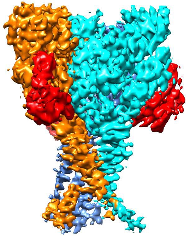
Research article                                                                         Structural Biology and Molecular Biophysics

                                     (Baconguis et al., 2014), and the PcTx1-bound ion-selective, nonselective (Baconguis and Gouaux,
                                     2012), and inactive states (Dawson et al., 2012). Structural studies of cASIC1-toxin complexes, com-
                                     bined with the structure of cASIC1 alone, have revealed a comprehensive molecular mechanism for
                                     proton-dependent gating in ASICs. In the resting state, the position of the thumb domain lies farther
                                     away from the threefold molecular axis, thereby expanding the ‘acidic pocket’ (Yoder et al., 2018).
                                     In the open and desensitized states, the ‘closure’ of the thumb domain into the acidic pocket
                                     expands the lower palm domain (Baconguis et al., 2014; Gonzales et al., 2009), leading to an iris-
                                     like opening of the channel gate.
                                         Mambalgin1 (Mamba1), a 57-residue three-finger toxin isolated from the venom of the black
                                     mamba snake (Dendroaspis polylepis polylepis), has been proven to be a potent, rapid and revers-
                                     ible inhibitor of ASIC1a- or ASIC1b-containing channels in both central and peripheral neurons
                                     (Diochot et al., 2012). Mamba1 has an analgesic effect that is as strong as that of morphine but
                                     does not involve opioid receptors, highlighting its potential utility for the management of pain. A
                                     detailed investigation of the binding of Mamba1 to human ASICs and its resulting modulatory effect
                                     could provide insights into the mechanism of interaction between toxins and ASICs and could offer
                                     crucial clues for the development of new drugs targeting ASICs.
                                         It should be pointed out that, to date, all ASIC structures solved and gating mechanism reported
                                     have been of chicken ASIC1, which can be pharmacologically quite different when compared to
                                     human ASIC1a despite ~89% sequence identity. Functional studies have shown that human ASIC1a
                                     (hASIC1a) and cASIC1 exhibit different responses to the channel activity modulation toxins Mamba1
                                     (Sun et al., 2018). In our own experiments, synthetic Mamba1 was observed to inhibit the channel
                                     currents of both recombinant hASIC1a and cASIC1 in CHO cells (Figure 1a). The inhibitory effects of
                                     Mamba1 on both full-length hASIC1a and cASIC1 are concentration dependent, with IC50 values of
                                     197.3 ± 37.4 and 123.6 ± 28.5 nM, respectively (Figure 1b). Interestingly, in the presence of 500 nM
                                     Mamba1, hASIC1a and cASIC1 showed decreases of 60.4 ± 12.9% and 19.6 ± 6.1% respectively, in
                                     the measured sodium currents (Figure 1a). At saturation (10 mM Mamba1), hASIC1a and cASIC1
                                     showed decreases in the measured sodium currents of 78.9 ± 6.2% and 31.9 ± 4.7% (Figure 1b).
                                     These data indicated that Mamba1 acts as an inhibitor targeting both hASIC1a and cASIC1, with
                                     comparable affinities but different efficacies. These observations suggested that functional and phar-
                                     macological differences exist between chicken ASIC1 and human ASIC1a, thus leading to the ques-
                                     tion of whether the structures of cASIC1 can fully recapitulate the functional states of hASIC1a.
                                     Therefore, structural studies on human ASICs are necessary to define functional states and provide
                                     comprehensive insights into the gating and toxin peptide modulation mechanisms of human ASICs.
                                         In this study, we resolved the cryo-EM structures of hASIC1a in the apo-form and in complex with
                                     the Mamba1 toxin at 3.56 Å and 3.90 Å resolution representing the first structure of human
                                     ASICs, respectively. The structure of apo-hASIC1a was shown to be highly similar to that of cASIC1
                                     in the resting state. Comparison of the structures of hASIC1a in the apo-form and the Mamba1-
                                     bound state revealed minor structural deviations. Electrophysiological studies revealed that Mamba1
                                     prefers to bind hASIC1a in a closed state. Direct interactions between residues in Mamba1 and acid-
                                     sensing residues in the ‘acidic pocket’ of hASIC1a were observed to reduce the apparent proton
                                     sensitivity of the hASIC1a channel, leading to channel inhibition. These data indicate that the mecha-
                                     nism by which Mamba1 inhibits hASIC1a channel is closed-state trapping.

                                     Results
                                     Functional characterization and structure determinations
                                     To facilitate the expression and purification of hASIC1a, a series of truncations of full-length hASIC1a
                                     was performed. Finally, truncated hASIC1a with the 60 carboxyl terminal residues removed (named
                                     hASIC1aDC) was determined to be functional by whole-cell patch-clamp electrophysiology. The hASI-
                                     C1aDC channel exhibits electrophysiological properties very similar to those of the full-length channel
                                     (Figure 1c–d). Mamba1 inhibits hASIC1aDC channels with an IC50 of 106.6 ± 23.6 nM, which is com-
                                     parable to the reported IC50 of full-length hASIC1a in CHO cells (148.6 ± 33.2 nM) (Figure 1b). To
                                     better understand the structure and toxin modulation of human ASICs, we set out to purify hASI-
                                     C1aDC, construct the hASIC1aDC-Mamba1 complex in vitro and subject the protein and protein com-
                                     plex to single-particle cryo-EM studies.

Sun, Liu, Li, et al. eLife 2020;9:e57096. DOI: https://doi.org/10.7554/eLife.57096                                                      2 of 21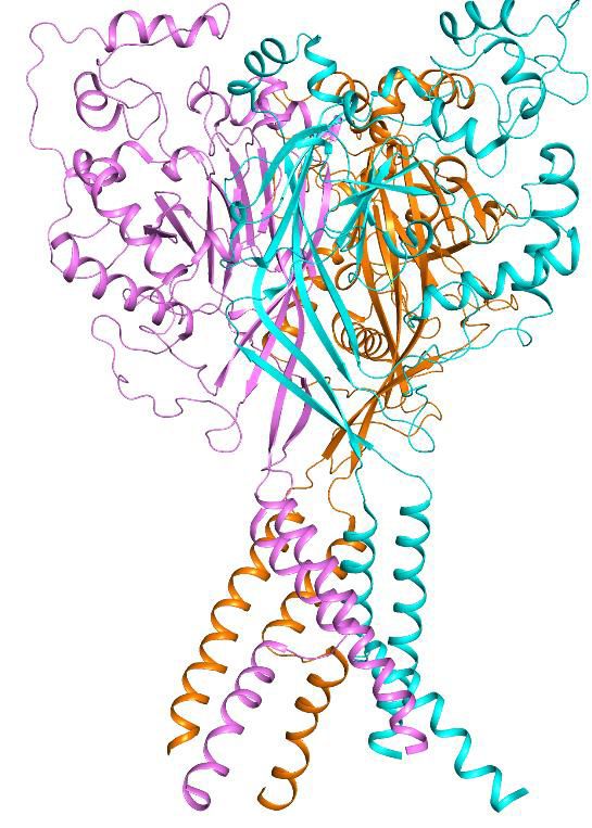
Research article                                                                                     Structural Biology and Molecular Biophysics

    a                                 Mamba1                                Mamba1                                            Mamba1
                      8.0 6.0                                 8.0 6.0                                    8.0 6.0

                                                2 nA

                                                                                                                                   2 nA
                                                       10 s
                                                                                                                                          10 s

                                 hASIC1a WT                       cASIC1 WT                                                  hASIC1aΔC

    b                                                         c   8.0 6.0       8.0 6.0                 d 100
               100
                                                                                                                        80
                      80

                                                                                                         I / Imax (%)
    IMamba1 / I (%)

                                                                                                                        60
                      60

                      40                                                                                                40

                               hASIC1a
                                                                                          2 nA
                      20       cASIC1                                                                                   20    hASIC1a
                               hASIC1a   ΔC
                                                                                                 10 s                         hASIC1a ΔC
                      0                                                                                                 0
                       100        101     102      103        104 hASIC1a WT      hASIC1aΔC                             8.0      7.5        7.0        6.5   6.0   5.5
                                                                                                                                                  pH
                                 Mamba1 concentration (nM)

Figure 1. Functional analysis of hASIC1a and hASIC1aDC. (a) Typical current traces representing the inhibition of recombinant hASIC1a (left), cASIC1
(middle) and hASIC1aDC (right) by Mamba1 toxin in CHO cells. (b) Concentration-response curve showing the inhibition of hASIC1a, hASIC1aDC and
cASIC1 expressed in CHO cells by Mamba1. Imamba1 and I represent the currents elicited by the pH 6.0 solution in the presence and absence of
Mamba1 toxin respectively. (c) Representative whole-cell patch-clamp recordings from wild-type hASIC1a and hASIC1aDC activated by pH 6.0 solution.
(d) pH-dependent activation and inactivation curves of hASIC1a (solid lines) and hASIC1aDC (dash lines). Data were collected from CHO cells
transfected with hASIC1a or hASIC1aDC DNA. Data are presented as the mean ± SD.
The online version of this article includes the following source data and figure supplement(s) for figure 1:
Source data 1. Source data for Figure 1b and d.
Figure supplement 1. Purification of hASIC1aDC for cryo-EM study.

                                                    The hASIC1aDC protein was overexpressed and purified from sf9 insect cells. Mamba1 was
                                                obtained through total chemical synthesis (Fang et al., 2011; Fang et al., 2012; Pan et al., 2014;
                                                Schroeder et al., 2014). The details of protein purification, complex construction, cryo-sample prep-
                                                aration, image acquisition, data processing, model building, and structure refinement can be found
                                                in the Materials and methods section. Briefly, recombinant hASIC1aDC protein was purified from sf9
                                                cells in the presence of 0.05% (w/v) n-dodecyl-b-D-maltoside (DDM) and subjected to cryo-EM stud-
                                                ies (Figure 1—figure supplement 1). Micrographs were collected on a Titan Krios electron micro-
                                                scope equipped with a Gatan K2 Summit detector. A 3D EM map of apo-form hASIC1aDC was
                                                reconstructed to an overall resolution of 3.56 Å (Figure 2—figure supplements 1–2). Following a
                                                similar protocol, the EM map of hASIC1aDC in complex with Mamba1 was obtained at 3.90 Å (Fig-
                                                ure 3—figure supplements 1–2).

                                                Cryo-EM structure of apo-hASIC1aDC
                                                The trimeric hASIC1aDC shows a canonical chalice-like architecture (Figure 2a–b). Each subunit of
                                                hASIC1aDC harbors a cysteine-rich extracellular domain (ECD). The ECD resembles a hand-like archi-
                                                tecture with the palm, knuckle, finger and thumb domains clenching a ‘ball’ of b strands (Figure 2—

Sun, Liu, Li, et al. eLife 2020;9:e57096. DOI: https://doi.org/10.7554/eLife.57096                                                                                  3 of 21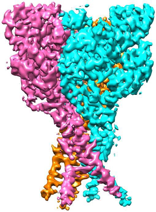
Research article Structural Biology and Molecular Biophysics
a b c d TM2b
TM1
TM2a 90e
TM1
TM2a
G444
S446
A445 S446
G444 A445
‘GAS’ belt
TM2b
Finger
e f g Knuckle
E238 D237
G430
β-ball
D434 D347
Distance along pore (Å)
The Thumb D351
gate The Palm
G437
gate
α4 α5
Acidic
G440 pocket
S446
G444
T449
A445
E452
TMD 90e
Pore radius (Å)
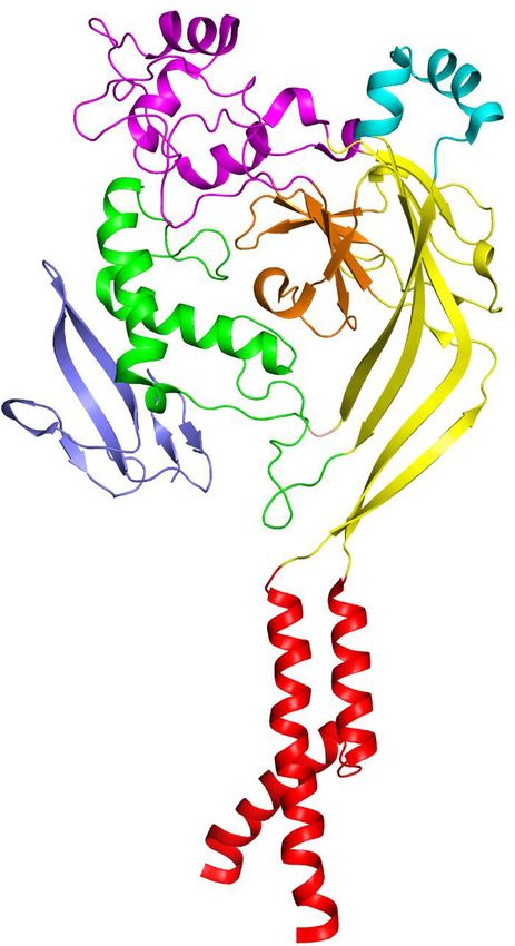
Figure 2. Cryo-EM structure of apo-hASIC1aDC. (a) Cryo-EM density map of apo-hASIC1aDC. The three hASIC1aDC subunits are colored orange, cyan
and pink. (b) Overall structure of trimeric hASIC1aDC, with different colors representing each subunit. (c) Ribbon representation of the hASIC1aDC TMD.
Two subunits are colored red and grey, respectively. The Ca atoms of the ‘GAS belt’ (G444-A445-S446) are shown as spheres. (d) View of the TMD from
the intracellular side. Residues in the GAS belt are shown in stick representations. Distances between the Gly444 carbonyl oxygen atoms are indicated.
(e) Close-up view of the pore domain. Map of solvent-accessible pathway is shown (red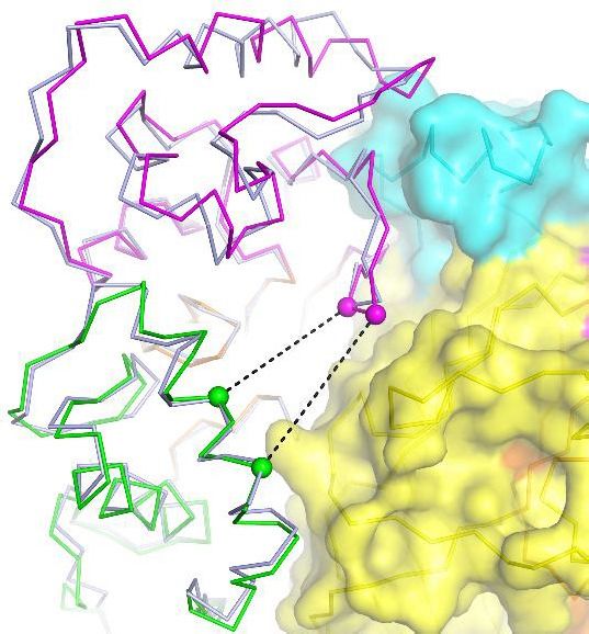
Research article                                                                                                        Structural Biology and Molecular Biophysics

                                                      segments TM2a and TM2b. The TM2b helical element interacts with the cytoplasmic portion of TM1
                                                      of the adjacent subunit (Figure 2c), resulting in a swap of the TM2 helices. Overall, the three copies
                                                      of TM1, TM2a and TM2b define a cavernous, threefold symmetric pore of the hASIC1a channel, in
                                                      which TM2 resides on the periphery of the pore (Figure 2d). Electrostatic mapping of the solvent-
                                                      accessible surface reveals that the ion channel pore of hASIC1aDC harbors a modest negative poten-
                                                      tial conferred by the presence of Asp434 and Gln438, by the carbonyl oxygen atoms of Gly437,
                                                      Gly440, and Gly444 (Figure 2e). The pore profile of hASIC1aDC also shows a closed gate along the
                                                      threefold axis as a result of primary constrictions at Asp434 and Gly437 (Figure 2e–f).

                                                      The resting-state conformation of hASIC1aDC at pH 8.0
                                                      The apo-hASIC1aDC structure at pH 8.0 is observed to have high similarity with the resting-state
                                                      structure of cASIC1 at pH 8.0 (Yoder et al., 2018). The two structures can be well superimposed
                                                      with a root-mean-square deviation (RMSD) of 0.88 Å over 1124 aligned Ca atoms (Figure 2g). Espe-
                                                      cially, the structure of the acidic pockets of hASIC1aDC and cASIC1 is almost identical, giving an
                                                      RMSD of 0.41 Å. In the acidic pocket, the distances between the Ca atoms of Asp347-Glu238 and
                                                      Asp351-Asp237 in hASIC1aDC are measured to be 13.9 and 15.6 Å, respectively, which are

   a                                                                                                              b                                        c
                                                                                                                                                                               D347
                                                                                                                                                                                      D351

                                                          90e                                                                                                    R28
                                                                                                                                  α4

                                                                                                                      Finger-II                                         F27               α4
                                                                                                                                       α5
                                                                                                                                                                                         F352
                                                                                                                                                                              L32
                                                                                                                                                                 Finger-II

                                                                                                                  Finger-III            Finger-I           d
                                                                                                                                                                                     α5
   e                                                            f                                                                                                        α4
                                        ***         ***                                                                 ***
                100
                                              ***         ***                    100            ***                                                                            H6
                                                                                                        **      ***
                      80                                                              80                                                                         D300
                                                                    IMamba1 / I (%)

                                                                                                                                                                                          Y360
    IMamba1 / I (%)

                      60          ***                                                 60                                                                                            Q5
                      40                                                                                                                                            K8
                                                                                      40
                                                                                                                                                                                     Finger-I
                      20                                                              20

                      0                                                               0
                             WT   Q5A H6A K8A F27A R28A                                    WT   D347A D351G F352L Y360A
                                         Mamba1                                                       hASIC1a

Figure 3. Structural basis of Mamba1 binding to hASIC1aDC. (a) Cryo-EM density map of the hASIC1aDC-Mamba1 complex. The three hASIC1aDC
subunits are colored orange, cyan and slate. Mamba1 is colored red. (b) Overall structure of the hASIC1aDC-Mamba1 complex. A single subunit of
hASIC1aDC is shown in cartoon representation, with each domain in a different color. Mamba1 is shown as a slate-colored ribbon. (c, d) Close-up views
of the interactions of the Finger-I (c) and Finger-II (d) regions of Mamba1 with the thumb domain of hASIC1aDC. (e, f) Bar graph representing the
inhibition of wild-type hASIC1a currents by Mamba1 mutants (e) and hASIC1a mutants by wild-type Mamba1 (f). Imamba1 and I represent the currents
elicited by the pH 6.0 solution in the presence and absence of Mamba1 toxin, respectively. Data are presented as the means ± SD. Comparison wild-
type Mamba1 (e) or hASIC1a (f) unless specified, ***p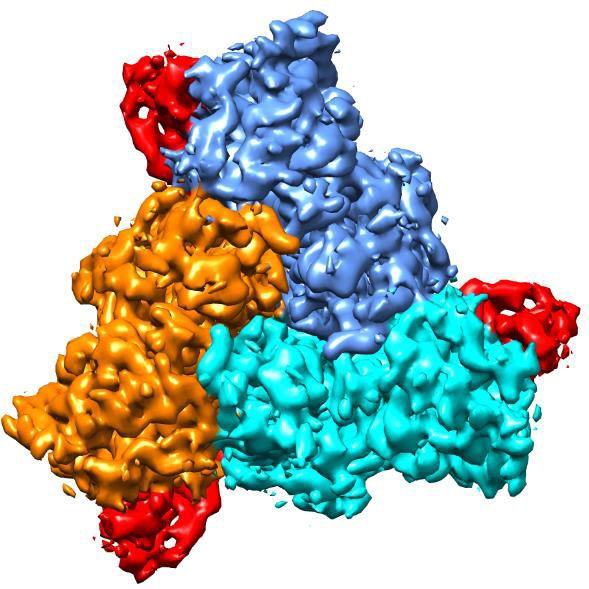
Research article                                                                       Structural Biology and Molecular Biophysics

                                     comparable to the distances measured in the cASIC1 channel (13.0 Å for Asp346-Glu239 and 15.0 Å
                                     for Asp350-Asp238) (Figure 2g). Moreover, the structures of the TMDs of hASIC1aDC and cASIC1
                                     are superimposed well, with an RMSD value of 0.34 Å. The pore profiles of hASIC1aDC and cASIC1
                                     are almost the same (Figure 2f). Our structure shows that hASIC1aDC has all the hallmarks of resting-
                                     state cASIC1, including the expanded acidic pocket, the extended GAS belt and a closed gate.
                                     Combining this information with the electrophysiology data, we conclude that the structure of hASI-
                                     C1aDC reported here represents the resting state of hASIC1a.

                                     Interaction between Mamba1 and hASIC1a
                                     The overall architecture of the hASIC1aDC-Mamba1 complex shows a triskelion-like shape viewed
                                     down the threefold symmetry axis, with one Mamba1 molecule radiating from each hASIC1aDC sub-
                                     unit (Figure 3a). Each of the three Mamba1 molecules binds to the ECD of a subunit of hASIC1aDC,
                                     interacting with the outside of the ECD of hASIC1aDC (Figure 3b). Different with the PcTx1 and
                                     MitTx toxins, Mambal1 was observed to interact with the thumb domain of hASIC1a channel.
                                     Although the interaction between PcTx1 and cASIC1 involves the thumb, palm and b-ball domains
                                     (Figure 3—figure supplement 3a). The interaction between MitTx and cASIC1 involves the palm
                                     domain besides the thumb domain (Figure 3—figure supplement 3b).
                                         Previously, we determined the cryo-EM structure of the cASIC1-Mamba1 complex, which
                                     revealed the binding locations of the toxin to the ECD of cASIC1 (Sun et al., 2018). However, the
                                     relatively low resolution (5.7 Å) could not show a reliable binding interface analysis between
                                     Mamba1 and cASIC1. Herein, the cryo-EM structure of the hASIC1aDC-Mamba1 complex at 3.9 Å
                                     resolution illuminates a more detailed binding interface between Mamba1 and hASIC1aDC. The bind-
                                     ing of Mamba1 to hASIC1aDC induces a conformational change in Finger-II of Mamba1. The tip
                                     region of Finger-II flips to the thumb domain of hASIC1aDC to facilitate the interaction between the
                                     toxin and the channel (Figure 3—figure supplement 2c). In the tip region of Finger-II, Arg28 is ori-
                                     ented toward the a5 helix of the thumb domain, thus becoming closer to Asp347 and Asp351. The
                                     measured distances between the Ca atoms of Arg28-Asp347 and Arg28-Asp351 are 10.5 and 10.2
                                     Å, respectively. Although the side chains of Arg28, Asp347 and Asp351 cannot be clearly assigned,
                                     it seems there could be salt bridges between Arg28 and Asp347 or Asp351 (Figure 3c). Phe352, a
                                     residue that is conserved in ASIC1 orthologues, is nestled within a hydrophobic cluster composed of
                                     Met25, Phe27, Leu32 and Leu33 in Finger-II of Mamba1, mediating a hydrophobic interaction
                                     between Mamba1 and hASIC1aDC (Figure 3c). Moreover, Mamba1 could form multiple polar con-
                                     tacts with hASIC1aDC through its Finger-I region. Mamba1-Gln5 and His6 could form hydrogen
                                     bonds with the side chain of Tyr360 of hASIC1aDC. Mamba1-Lys8 could form salt bridges with
                                     Asp300 (Figure 3d). Notably, Finger-III of Mamba1 is located farther from the thumb domain of
                                     hASIC1aDC, pointing in the opposite direction from the threefold axis of the channel core. Finger-III
                                     thus has no contact with hASIC1aDC (Figure 3b). The upper scaffold region of Mamba1 likewise
                                     does not contact hASIC1aDC.
                                         To verify the toxin-channel interactions, individual mutations were introduced into Mamba1 and
                                     the possible counterpart interaction regions on hASIC1a. The inhibition of hASIC1a activity by
                                     Mamba1 is greatly reduced when residue Mamba1-Gln5, His6, Lys8, Phe27 or Arg28 is mutated to
                                     Ala (Figure 3e). Mamba1 is significantly less effective in inhibiting the hASIC1a mutants Asp347Ala,
                                     Asp351Gly, Phe352Leu and Tyr360Ala (Figure 3f and Figure 3—figure supplement 4). These data
                                     are consistent with previous electrophysiological studies on chicken or rat ASIC1 channels
                                     (Mourier et al., 2016; Salinas et al., 2014; Sun et al., 2018).
                                         Furthermore, the interaction of hASIC1aDC and Mamba1 was validated using 19F-labeled Mamba1
                                     and 19F-NMR spectroscopy in the solution state at ambient temperature. 19F labeling was intro-
                                     duced into Mamba1 by replacing the residue sites Phe18, Phe27, Leu30 or Leu32 in Mamba1 with
                                     19
                                        F-labeled L-4-trifluoromethyl-phenylalanine (19F-tfmF) through chemical synthesis. One-dimen-
                                     sional 19F-NMR spectra indicate a significant change in chemical shift for residues Mamba1-Phe27
                                     (within Finger-II) upon hASIC1aDC protein titration, and slight changes for Leu30 and Leu32, whereas
                                     no change is observed for residue Phe18 (as a negative control) (Figure 3—figure supplement 5).
                                     These data represent that Phe27 in Mamba1 has a significant conformational change when binds to
                                     hASIC1a channel, while the conformation of Leu30, Leu32 and Phe18 are not altered. These data
                                     indicate that Phe27 plays a key role in Mamba1 binding to hASIC1a channel.

Sun, Liu, Li, et al. eLife 2020;9:e57096. DOI: https://doi.org/10.7554/eLife.57096                                                    6 of 21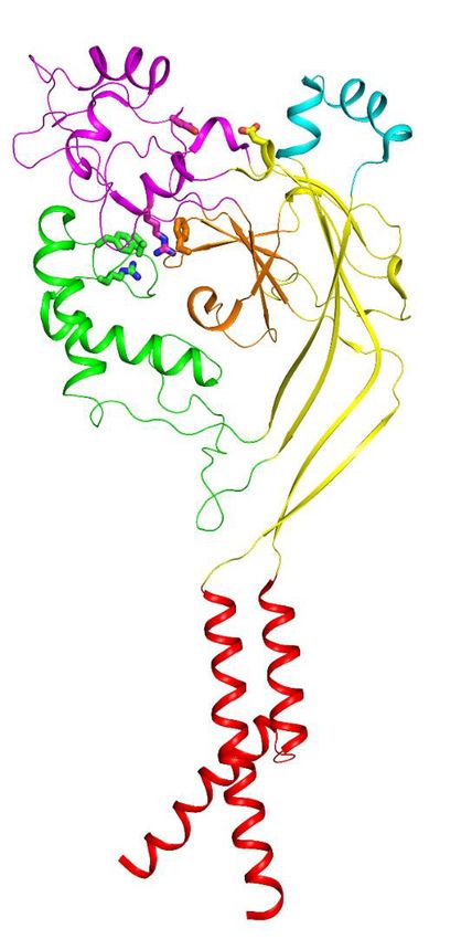
Research article                                                                       Structural Biology and Molecular Biophysics

                                       Collectively, both mutation-based patch-clamp electrophysiology analysis and 19F-NMR measure-
                                     ments support the interaction between Mamba1 and hASIC1a mediated by the Finger-I/II regions of
                                     Mamba1 and the thumb domain of hASIC1a.

                                     The inhibited conformation of hASIC1a upon Mamba1 binding
                                     The structure of apo-hASIC1aDC and hASIC1aDC-Mamba1 made it possible to illustrate the detailed
                                     conformation changes in both the ECD and TMD of hASIC1aDC upon Mamba1 binding. Structure
                                     alignment of the hASIC1aDC-Mamba1 complex and apo-hASIC1aDC gives an RMSD of 0.44 Å2, indi-
                                     cating the globally high similarity of the two structures (Figure 4a and Figure 4—figure supplement
                                     1a–b). However, minor structural differences between the two structures can still be observed.
                                         The snake toxin peptide Mamba1 contacts Asp347, Asp351, and Phe352 located in the a5 helix
                                     of the thumb domain, causing these residues to flip outward from the acidic pocket. The a5 helix
                                     thus deviates by ~5˚ around the central axis, whereas the a4 helix and the finger domain adopt the
                                     same conformation as in the apo-hASIC1aDC structure (Figure 4b). In the hASIC1aDC-Mamba1 com-
                                     plex structure, the distances between the Ca atoms of Asp347-Glu238 and Asp351-Asp237 (13.3
                                     and 12.0 Å, respectively) (Figure 4b) are slightly closer than those in the apo-hASIC1aDC structure
                                     (13.9 and 15.6 Å, respectively).
                                         In the TMD, TM1 undergoes a lateral pivot of ~6˚ around its carboxyl terminus upon Mamba1
                                     binding. Meanwhile, TM2a have a shift of approximately 2.5 Å away from the channel pore, and
                                     TM2b shifts approximately 4 Å at its amino terminus and 8 Å at its carboxyl terminus away from the
                                     pore (Figure 4c). Comparison of the ion channel pore architectures of apo-hASIC1aDC and the hASI-
                                     C1aDC-Mamba1 complex shows that the extracellular vestibule undergoes a slight expansion upon
                                     Mamba1 binding (Figure 4d and Figure 4—figure supplement 1c). However, the overall pore pro-
                                     file of the hASIC1aDC-Mamba1 complex is similar to that of apo-hASIC1aDC. The transmembrane
                                     pore of the hASIC1aDC-Mamba1 complex has a diameter less than 2.0 Å in the gate, indicating a
                                     closed channel. (Figure 4e). The slight shifts of the thumb domain and transmembrane helices of
                                     hASIC1a in complex with Mamba1 cause it to adopt a less compact conformation than that of apo-
                                     form hASIC1a in the resting state, but the expanded conformation of the acid pocket and the closed
                                     pore are not altered.

                                     Mamba1 reduces the proton sensitivity of hASIC1a
                                     Interestingly, we found that the inhibition of hASIC1a by Mamba1 depended on the pH of the toxin
                                     perfusion. When 500 nM Mamba1 was applied at conditioning pH 8.0, before the pH dropped to
                                     6.0 (Figure 5a), the hASIC1a current showed decreases of 50.2 ± 8.8% at the peak (Figure 5b). In
                                     contrast, the coapplication of 500 nM Mamba1 as the pH dropped to 6.0 from pH 8.0 did not pro-
                                     duce as much suppression as preapplication did (peak current showed decreases of 10.6 ± 7.2%)
                                     (Figure 5a and b). We also measured the current amplitudes at the end of such applications. As
                                     shown in Figure 5b, the currents showed decreases of 49.9 ± 19.4% when Mamba1 application was
                                     administered in the absence of the stimulating pH 6.0 application, while showed decreases of 9.3 ±
                                     8.6% in the presence of the stimulating pH 6.0 application. These data suggest that Mamba1 bind-
                                     ing favors a resting closed state of hASIC1a at neutral pH rather than an open or desensitized state
                                     at acidic pH.
                                         Moreover, similar to a previous report (Diochot et al., 2012), in the absence of Mamba1,
                                     hASIC1a showed half-maximal activation at a pH (pH50) of 6.46 ± 0.02. In contrast, in the presence of
                                     Mamba1 (500 nM), the pH50 of activation was 5.89 ± 0.07 (Figure 5c–d). Mamba1 drastically shifts
                                     the activation curve of hASIC1a by 0.57 pH units to a more acidic pH, demonstrating that Mamba1
                                     decreases the apparent H+ affinity of hASIC1a, thus inhibiting channel activation at pH 6.0. These
                                     observations indicate that Mamba1 acts as an inhibitor targeting ASIC by modifying the proton sen-
                                     sitivity of the channel, consistent with previous conclusions (Diochot et al., 2016; Salinas et al.,
                                     2014).
                                         In fact, multiple acidic residues located around the acidic pocket have been found to contribute
                                     to modulation of the proton sensitivity of ASICs, including Asp237, Glu238, Asp347 and Asp351
                                     (Jasti et al., 2007). It has been reported previously that the neutralization of Asp346 and Asp350 in
                                     cASIC1 (corresponding to Asp347 and Asp351 in hASIC1a) has profound effects on either pH50 or
                                     the apparent Hill coefficient, or both (Jasti et al., 2007). The Asp346Asn mutation shifts the pH

Sun, Liu, Li, et al. eLife 2020;9:e57096. DOI: https://doi.org/10.7554/eLife.57096                                                    7 of 21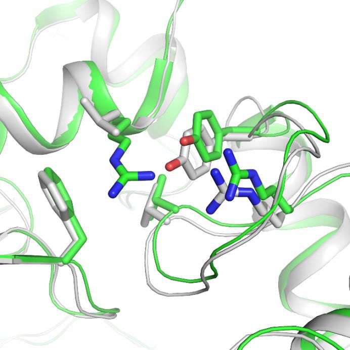
Research article                                                                                                     Structural Biology and Molecular Biophysics

    a                                    b                                                          c
                                                  α5                          E238
                                                                             D237                                                                  TM2a
                                                                 D347

                                                                                                                                TM1                     S446
                                                                    D351
                                                                                                                                           G444        A445
                                                  α4

                                                                                                                                              TM2b

    d                                                                                             e

                                                                                                      Distance along pore (Å)
                                             Mamba1
                              The gate                                  The gate                                                                The gate

                                                                                                                                            Pore radius (Å)
                 hASIC1aDC                              hASIC1aDC -Mamba1

Figure 4. The structure of hASIC1aDC inhibited by Mamba1. (a) Single-subunit superposition of apo form and Mamba1-bound hASIC1aDC shows global
conformational changes. The domains of apo-hASIC1aDC are shown in different colors, and Mamba1-bound hASIC1aDC is colored grey. (b, c)
Conformational changes in the acidic pocket and TMD of hASIC1aDC upon Mamba1 binding. (b) View of the acidic pocket from superposed apo- and
Mamba1-bound hASIC1aDC. hASIC1aDC is shown in cartoon representation and colored as in (a). Ca atoms of Glu238 and Asp347, Asp239 and Asp351
are shown as spheres. (c) View of the TMD from superposed apo- and Mamba1-bound hASIC1aDC. The TMDs of hASIC1aDC and the hASIC1aDC-
Mamba1 complex are shown as ribbons in red and grey, respectively. (d) Pore profiles of hASIC1aDC (left) and the hASIC1aDC-Mamba1 complex (right)
calculated with HOLE software (red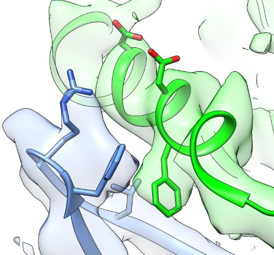
Research article Structural Biology and Molecular Biophysics
a Mamba1 (500 nM) b + Mamba1
at pH 6.0
pH=6.0 + Mamba1
100 at pH 8.0
pH=8.0 100
80 80
IMamba1 / I (%)
IMamba1 / I (%)
60 60
1 nA
40 40
10 s
20 20
0 0
Peak Current Current after 30 pH 6.0 application
c d
100 pH50 Hill coefficient
With With
Without Without
80 Mamba1 Mamba1
Mamba1 Mamba1
(500 nM) (500 nM)
I / Imax (%)
60 hASIC1a hASIC1a-
hASIC1a + Mamba1 6.46f0.02 5.89f0.07 4.23f0.70 2.56f0.28
WT
hASIC1a D347A
40 hASIC1a D347A + Mamba1 hASIC1a-
5.88f0.03 5.72f0.01 2.94f0.37 4.53f0.53
hASIC1a D351G D347A
hASIC1a D351G + Mamba1 hASIC1a-
20 hASIC1a F352L D351G
6.04f0.05 5.79f0.02 2.14f0.32 3.72f0.73
hASIC1a F352L + Mamba1
hASIC1a-
0 6.13f0.01 5.97f0.04 2.69f0.80 2.32f0.43
F352L
7.0 6.5 6.0 5.5 5.0 4.5 4.0
pH
e
D237
E238
D347
R191 R28
F352
D351 F27
+ Mamba1
Expanded
acidic pocket
Expanded
Mamba1 acidic pocket
Resting State Inhibited State
Figure 5. The ‘closed-state trapping’ model for Mamba1 inhibition of hASIC1a activity. (a) Typical traces of hASIC1a currents recorded in CHO cells
with administrations of Mamba1 (500 nM). The Mamba1 toxin applications were administered either in the absence (upper panel) or presence (middle
and lower panels) of the stimulating pH 6.0 application. (b) Bar graph representing the inhibition of the peak currents of hASIC1a statistics with
administrations of Mamba1 at pH 6.0 and pH 8.0 (left panel). The inhibition of current amplitudes at the end of such applications are also measured
and statistically compared (right panel). (c) pH-dependent activation curves of wild-type hASIC1a (WT) and hASIC1aDmutants obtained in the absence
or presence of Mamba1. (d) The measured pH50 and Hill coefficient values of wild-type hASIC1a (hASIC1a-WT) and hASIC1a mutants in the conditions
with or without Mamba1. Data are presented as the mean ± SEM. (e) The closed-state trapping inhibition mechanism of hASIC1a by Mamba1. Mamba1
binding leads to deformation of the proton-sensitive site, then stabilizes the expanded conformation of the acidic pocket and traps the channel in a
Figure 5 continued on next page
Sun, Liu, Li, et al. eLife 2020;9:e57096. DOI: https://doi.org/10.7554/eLife.57096 9 of 21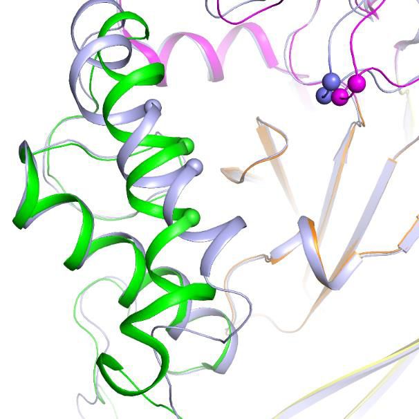
Research article                                                                                   Structural Biology and Molecular Biophysics

Figure 5 continued
closed state. The structure of a single subunit of hASIC1aDC is shown as a cartoon in gray, representing the resting state of the channel (left). The
structure of hASIC1aDC-Mamba1 is shown as a cartoon, with each domain colored differently. Mamba1 is colored blue (right).
The online version of this article includes the following source data for figure 5:
Source data 1. Source date for Figure 5b, c and d.

                                      hASIC1a by 0.59 and 0.42 pH units, respectively, with pH50 values of 5.88 ± 0.03 and 6.04 ± 0.05.
                                      The mutations reduce the Hill coefficient from approximately 4 to 2. Meanwhile, Mamba1 is able to
                                      shift the activation curves of the hASIC1a-Asp347Ala and hASIC1a-Asp351Gly mutants further, with
                                      pH50 values of 5.72 ± 0.01 and 5.79 ± 0.02, respectively (Figure 5c–d). We suggest that probable
                                      interactions between Asp347 and/or Asp351 of hASIC1a and Mamba1 shield the protonation of the
                                      two residues, thus reducing the proton sensitivity of hASIC1a. On the other hand, the interaction
                                      between Asp351 and Mamba1 could hinder the interaction between Asp351 and Arg190 (located at
                                      the b-ball), which is critical for the collapse of the acidic pocket in active or desensitized channels.
                                      Therefore, we suspect that Mamba1 inhibits the hASIC1a channel by hindering proton-induced tran-
                                      sitions from the resting closed states to the active and/or desensitized states in a model of closed-
                                      state trapping (Figure 5e).
                                          Notably, Asp347 and Asp351 are not the only proton-sensing residues in hASIC1a (Vullo et al.,
                                      2017). Glu79, Glu219, Glu409 and Glu418, which are located in the palm domain, which have no
                                      direct contact with Mamba1, were also found to participate either in proton binding or in subse-
                                      quent conformational changes (Jasti et al., 2007; Krauson et al., 2013; Ramaswamy et al., 2013).
                                      This observation may explain why Mamba1 can inhibit only approximately 80% of the hASIC1a cur-
                                      rent and loses its inhibitory effect on hASIC1a at a lower pH, such as pH 4.0.

                                      The intradomain interaction affects the Mamba1 inhibition of ASIC
                                      As shown in Figure 1, although Mamba1 has similar affinity to hASIC1a and cASIC1
                                      (IC50 = 197.3 ± 37.4 and 123.6 ± 28.5 nM, respectively), the toxin shows a stronger inhibitory effect
                                      on hASIC1a than on cASIC1 channels. In fact, the amino acid sequences of hASIC1a and cASIC1 are
                                      highly similar (89% identity), and the sequences of the thumb domain of the two orthologues are
                                      identical (Figure 6—figure supplement 1). This similarity may explain the similar affinity of Mamba1
                                      in targeting hASIC1a and cASIC1. Comparison of the apo-form structure of hASIC1a with the rest-
                                      ing-state structure of the cASIC1 channel, together with the structure of the hASIC1a-Mamba1 com-
                                      plex, showed comparable expansion of the acid pocket between chicken and human ASIC1a
                                      channels (Figures 2g and 4b). The key residues (such as Asp346 and Phe351) on the a5 helix in the
                                      thumb domain of chicken ASIC1 channels that participate in Mamba1 binding are also critical in
                                      human ASIC1a channels. These observations suggest that the different inhibitory efficacies of
                                      Mamba1 on cASIC1 and hASIC1a channels could not be due to the toxin-channel interaction.
                                          Through sequence alignment analysis and structure comparison, several key residues that do not
                                      contribute to Mamba1 binding were found to contribute to the different inhibitory effects of
                                      Mamba1 on hASIC1a and cASIC1. These residues include Gln102, Arg155 (both located in the finger
                                      domain of hASIC1a) and Asp167 (located in the palm domain), which are mapped to Arg103,
                                      Leu156 and Glu168 of cASIC1, respectively (Figure 6a and b). In cASIC1, Leu156 is observed to
                                      interact with the hydrophobic pocket composed of Phe189, Val327 and Tyr334 in the thumb domain
                                      (Figure 6c, Figure 6—figure supplement 2). The hydrophobic contact could participate in the inter-
                                      action between the finger and thumb domains. Meanwhile, Arg103 and Glu168 could form electro-
                                      static interaction pairs with each other (the distance between the Ca atom of the two residues was
                                      measured to be 13.3 Å), mediating the contact between the finger and palm domains (Figure 6d,
                                      Figure 6—figure supplement 2). It was reported that the movement of the finger domain plays a
                                      critical role in the activation of ASICs (Bonifacio et al., 2014; Gwiazda et al., 2015; Krauson and
                                      Carattino, 2016; Ramaswamy et al., 2013; Vullo et al., 2017). The replacement of Leu156 by
                                      Arg155 and of Arg103-Glu168 by Gln102-Asp167 in hASIC1a result in disruption of intradomain
                                      interactions, thus leading to enhanced inhibition of the channel by Mamba1.
                                          To elucidate the contributions of these residues to the inhibition of ASIC1 activity by Mamba1,
                                      mutations were introduced at these sites in hASIC1a channels, and whole-cell patch-clamp

Sun, Liu, Li, et al. eLife 2020;9:e57096. DOI: https://doi.org/10.7554/eLife.57096                                                                 10 of 21
Research article Structural Biology and Molecular Biophysics
a 100 154 186 325
hASIC1a - - - FSQV---MREFYDRAGHDIRDM---VV FTR---CRMVHMPGDAP YC-- -
cASIC1 ---FSRV--- MLEFYDRAGHDIREM---VVFTR- --CRMVHMPGDAP YC---
b c d
d Arg155
(Leu156) Tyr336 D167
(Tyr335) (E168)
Q102
(R103)
c
Phe188 Val328 Arg326
(Val327) (Arg325)
(Phe189)
e f
100 100
80 80
** **
IMamba1 / I (%)
*
I / Imax (%)
60 60
40 40
hASIC1a
hASIC1a R155L
20 hASIC1a R155F 20
hASIC1a Q102R/D167E
0 0
7.0 6.8 6.6 6.4 6.2 6.0 5.8 5.6 5.4 WT R155L R155F Q102R/D167E
pH hASIC1a
Figure 6. Structural basis for the differing activities of hASIC1a versus cASIC1. (a) Sequence alignment of hASIC1a and cASIC1 indicates key residues
that may contribute to the inhibitory effect of Mamba1 on ASIC. (b) An individual subunit of resting-state hASIC1aDC. (c, d) Local alignments of
hASIC1aDC (green) with resting-state cASIC1 (grey, PDB 5WKV) demonstrate the possible interactions of residues in two handles. (e) pH-dependence
curves of hASIC1a and its mutants. (f) Bar graph representing the inhibitory effect of wild-type Mamba1 (500 nM) on hASIC1a mutants. Data are
presented as means ± SD. Comparison with wild-type hASIC1a, **pResearch article Structural Biology and Molecular Biophysics
illustrating the gating mechanism of the channel (Baconguis et al., 2014; Baconguis and Gouaux,
2012; Gonzales et al., 2009; Jasti et al., 2007; Sun et al., 2018; Yoder and Gouaux, 2020;
Yoder et al., 2018). Here, we report the first structure of the human ASIC1a channel determined
using single-particle cryo-EM with a resolution of 3.56 Å.
The structure of hASIC1a in the apo-form at pH 8.0 reflects the resting state of the channel.
hASIC1a in the resting state has an identical structure to that of the cASIC1 channel, especially the
expanded acidic pocket and the extended ‘GAS’ motif. In fact, the human ASIC1a channel and
chicken ASIC1 channel have high-sequence homology (89% identity). These indicate that hASIC1a
and cASIC1 channels may share a similar gating mechanism. That is, the resting-state hASIC1a chan-
nel has an expanded acidic pocket, with the thumb domain far away from the central b-ball and the
finger and palm domains. Activation and desensitization of the channel both involve the collapse of
the acidic pocket, which allows the thumb and finger domains to approach and interact with each
other (Gonzales et al., 2009; Baconguis et al., 2014; Yoder et al., 2018). However, the structures
of human ASIC in the active or desensitized state remain unknown. Resolving the structures of the
active or desensitized hASIC1a will help to address this supposition.
In recent decades, venom toxin peptides have been observed to bind ASICs with high affinity
and specificity, providing an excellent resource for the definition of different functional states of the
channel (Bohlen et al., 2011; Diochot et al., 2004; Diochot et al., 2012; Escoubas et al., 2000;
Reimers et al., 2017). We also report here the structure of the human ASIC1a channel in complex
with the snake peptide toxin Mamba1 with resolution 3.90 Å, reflecting the toxin-inhibited state of
the channel. A first important conclusion is that, the structure of the hASIC1a-Mamba1 complex con-
firms that the Mamba1 binding site is located in the thumb domain of the hASIC1a channel. Com-
pared with the previously reported structures of the cASIC1 channel in complex with toxins PcTx1,
MitTx or Mamba1, these toxins interact with the ASIC channel through overlapping, but not identical
surfaces. The thumb domain is a hot spot that mediates toxin-channel interactions. However, the
Mamba1-binding site is totally located in the thumb domain of the hASIC1a channel, and does not
involve other sub-domains. In contrast, the toxin-binding surfaces for MitTx and PcTx1 involve the b-
ball and the palm domain in addition to the thumb domain. These observations could explain the
fact that MitTx and PcTx1 have much higher affinity than Mamba1 targeting ASIC channels.
Moreover, structure comparison of hASIC1a in the apo-form and in complex with Mamba1 reveals
that Mamba1 binding does not alter the closed structure of hASIC1a in a resting state. Therefore,
we conclude that Mamba1 inhibits hASIC1a through a closed-state trapping mechanism, precluding
the previously proposed allosteric-based channel modulation mode. Furthermore, the binding of
Mamba1 to hASIC1a is state-dependent. The toxin preferentially binds to the closed state but not
the active or desensitized state of ASICs. Mamba1 inhibits ASICs by shifting the pH-dependence of
activation to a more acidic pH, decreasing their apparent affinity for protons. In fact, the state-
dependent trapping mechanism has been found in the modulation of voltage-gated ion channel
activity by peptide toxins. It is proposed that voltage sensor trapping is the fundamental mechanism
of action of polypeptide toxins that alter the voltage-dependent gating of sodium, calcium, and
potassium channels (Cestèle et al., 1998). For the ASIC channel, the two Asp residues in the a5
helix of the thumb domain contribute to proton sensing during channel activation (Jasti et al.,
2007). Therefore, the thumb domain could act as the ‘proton sensor’ of the channel. Accordingly,
we conclude that Mamba1 toxin inhibits ASIC channel activity through a closed-state dependent
‘proton sensor trapping’ mechanism, sharing a common mechanism of polypeptide toxins modulat-
ing ion channel activity.
Mamba1 showed a stronger effect on hASIC1a than on cASIC1. Previous reported key residues
(such as Asp346 and Phe351) on the a5 helix in the thumb domain of the chicken ASIC1 channel
that participated in Mamba1 binding were also critical in the human ASIC1a channel. The structural
analysis and mutation experiments suggested that the different inhibitory effects of Mamba1 target-
ing cASIC1 and hASIC1a channels might be due to the mechanism of channel activation rather than
the difference in toxin-channel interactions. Several key amino acid differences between hASIC1a
and cASIC1, including Arg155 and the Glu102-Asp165 pair in hASIC1a (corresponding to Leu156
and Arg103-Glu168 in cASIC1, respectively), are found to contribute to the different responses of
hASIC1a and cASIC1 to Mamba1. Due to the relatively low resolution, our structures reported here
could not provide detailed structural evidence sufficient to support this finding. It is important to
Sun, Liu, Li, et al. eLife 2020;9:e57096. DOI: https://doi.org/10.7554/eLife.57096 12 of 21Research article Structural Biology and Molecular Biophysics
address how the coupling among the extracellular subdomains of ASIC, including the thumb, finger,
palm and b-ball, affects channel activity and toxin action on the channel.
ASIC channels are of fundamental importance and are also considered potential drug targets in
therapeutic interventions against pain and ischemic stroke. There is no doubt that analysis of interac-
tions between venom toxin and hASIC1a or peptide toxin-based drug development should follow
structure and function studies on human source target proteins, as data on homologous target pro-
teins from other species might not precisely reflect the modulatory effects of the peptide or of other
ligands on the target proteins, which will strongly influence the process of drug development for
human target proteins. Our studies on the human ASIC1a channel could obviously be very valuable
for drug development targeting ASIC.
Materials and methods
Key resources table
Reagent type
(species) or Source or Additional
resource Designation reference Identifiers information
Gene hASIC1a GeneBank NCBI Reference All hASIC1a mutants
(Homo sapiens) Sequence: NP_001086.2 transfected in the
paper were obtained
starting from this
wild-type cDNA
Strain, strain Top10 Thermo Fisher Cat# 18258012
background Scientific
(Escherichia coli)
Strain, strain DH10Bac Thermo Fisher Cat# 10361012
background Scientific
(Escherichia coli)
Cell line Sf9 Thermo Fisher Cat# 11496015;
(Spodoptera Scientific RRID:CVCL_0549
frugiperda)
Cell line HEK-293T ATCC Cat#: CRL-3216;
(Homo sapiens) RRID:CVCL_0063
Cell line CHO-K1 ATCC Cat# 03480/
(Cricetulus griseus) p693_CHO-K1;
RRID:CVCL_0214
Recombinant pFastBac1 Invitrogen
DNA reagent
Recombinant pcDNA3.1 Invitrogen
DNA reagent
Chemical n-Dodecyl-b-D- Anatrace Cat#: D310
compound, Maltoside (DDM)
drug
Chemical Cholesterol Sigma-Aldrich Cat#: C6013
compound, Hemisuccinate
drug tris Salt (CHS)
Peptide, Mamba1 This paper UniProtKB: P0DKR6 Mamab1 and
recombinant mutants were
protein obtained by
chemical synthesis
Software, Gctf Zhang, 2016 https://www2.mrc-
algorithm lmb.cam.ac.uk/
research/locally-
developed-software/
zhang-software/#gctf
Software, RELION 3.1 Zivanov et al., 2018 http://www2.mrclmb.
algorithm cam.ac.uk/relion;
RRID:SCR_016274
Continued on next page
Sun, Liu, Li, et al. eLife 2020;9:e57096. DOI: https://doi.org/10.7554/eLife.57096 13 of 21Research article Structural Biology and Molecular Biophysics
Continued
Reagent type
(species) or Source or Additional
resource Designation reference Identifiers information
Software, SerialEM Mastronarde, 2005 RRID:SCR_017293
algorithm
Software, PHENIX Liebschner et al., 2019 https://www.
algorithm phenixonline.org;
RRID:SCR_014224
Software, Coot Emsley et al., 2010 https://www2.mrc-
algorithm lmb.cam.ac.uk/
personal/pemsley/coot;
RRID:SCR_014222
Software, UCSF Chimera Pettersen et al., 2004 https://www.cgl.
algorithm ucsf.edu/chimera;
RRID:SCR_004097
Software, PyMol Schrödinger https://pymol.org/2;
algorithm RRID:SCR_000305
Software, GraphPad Prism 7 GraphPad https://www.
algorithm Software graphpad.com/
scientific-software/
prism
Software, HOLE Smart et al., 1996 http://www.
algorithm holeprogram.org
Others QUANTIFOIL R1.2/1.3 Quantifoil
holey carbon grids
Others Cellfectin Invitrogen Cat# 10362100
Others Superose 200 GE Healthcare Cat# 28990944
Increase 10/300 GL
Cell lines
All cell lines used were obtained from commercial sources (see the Key Resources Table). Sf9 cells
were cultured at 27˚C in serum-free SIM SF medium (Sino Biological Inc). HEK293T cells were cul-
tured as adherent cells in DMEM (with L-glutamine, glucose and sodium pyruvate), supplemented
with 10% FBS and 1% Gibco antibiotic-antimycotic; at 37˚C in 5% CO2. CHO-K1 cells were cultured
in DMEM/F12 medium (Gibco) supplemented with 10% fetal bovine serum (FBS), 100 U/mL penicil-
lin, and 100 U/mL streptomycin at 37˚C in a 5% CO2 incubator. No additional authentication was
performed by the authors of this study. Cell line was negative for mycoplasma. No commonly misi-
dentified lines were used in this study. All cell lines were kept at low passages in order to maintain
their health and identity.
Protein expression, purification and complex construction
The mambalgin1 (Mamba1) toxin was obtained using a hydrazide-based chemical synthesis method
as previously reported (Pan et al., 2014). The polypeptide chain of Mamba1 (57 amino acids) was
divided into three segments at two ligation sites (Cys19 and Cys41). All the segments (Mamba1[1-
18]-NHNH2, Mamba1[19–40]–NHNH2 and Mamba1[41–57]) were synthesized using a standard solid-
phase peptide synthesis method. The three segments were then ligated through the standard hydra-
zide-based native chemical ligation (NCL) to synthesize the full-length Mamba1. This synthesis of
Mamba1 is convenient and produced high yields following the final HPLC purification (35% isolated
yield, multi-milligram scale and good homogeneity). For the synthesis of Mamba1 mutants, the Ala-
nine or 19F-labeled L-4-trifluoromethyl-phenylalanine (19F-tfmF) were incorporated directly during
the peptide segments synthesis.
The optimized coding DNAs for human hASIC1a (Uniprot: P78348) was synthesized by Gene-
Script. The truncated hASIC1a (with the carboxyl terminal 60 residues removed, named as hASI-
C1aDC) was cloned into the pFastBac1 vector (Invitrogen) with 8-His tag at the amino terminus.
Baculovirus-infected sf9 cells (Thermo Fisher) were used for overexpression and were grown at 27˚C
in serum-free SIM SF medium (Sino Biological Inc). Cells were harvested 2 days after infection by
Sun, Liu, Li, et al. eLife 2020;9:e57096. DOI: https://doi.org/10.7554/eLife.57096 14 of 21Research article Structural Biology and Molecular Biophysics
centrifugation at 1000 g and resuspended in lysis buffer containing 20 mM Tris (pH 8.0), 200 mM
NaCl for each batch of protein purification. The suspension was supplemented with 1% (w/v)
n-dodecyl-b-D-maltopyranoside (DDM, Anatrace), 0.2% (w/v) cholesteryl hemisuccinate Tris salt
(CHS, Anatrace) and protease inhibitor cocktail (Sigma). After incubation at 4˚C for 2 hr, the solution
was ultracentrifuged at 200,000 g for 45 min, and the supernatant was applied to Ni-NTA (GE
HealthCare) by gravity at 4˚C. The resin was rinsed four times with the buffer containing 20 mM Tris
(pH 8.0), 200 mM NaCl, 40 mM imidazole, 0.1% DDM, 0.02% CHS and the protease inhibitor cock-
tail. The target proteins were eluted with buffer containing 20 mM Tris (pH 8.0), 200 mM NaCl, 250
mM imidazole, 0.1% DDM, 0.02% CHS. The eluted protein was further purified by size-exclusion
chromatography in 20 mM Tris (pH 8.0), 200 mM NaCl, 0.05% DDM, 0.01% CHS using a Super-
dex200 10/300 GL column (GE HealthCare). The presence of hASIC1aDC in the peak fractions of size
exclusion chromatography purification was confirmed by SDS-PAGE and mass spectrometry (MS).
To construct the hASIC1aDC-Mamba1 complex, hASIC1aDC was purified as described above in pH
8.0 buffer and concentrated to about 5 mg/ml based on A280 measurement, using a 100 kDa cutoff
Centricon (Millipore). The chemical synthesized, lyophilized Mamba1 was dissolved in buffer contain-
ing 20 mM Tris (pH 8.0), 200 mM NaCl, 0.05% DDM, 0.01% CHS at a final concentration of 10 mg/
ml based on A280 measurement, and added in a 6:1 molar ratio of toxin to channel with incubation
for 1 hr at 4˚C.
Single-particle cryo-EM data acquisition
Purified hASIC1aDC (3 ml) at a concentration of 2.7 mg/ml was added to the freshly plasma-cleaned
holey carbon grid (Quantifol, R1.2/1.3, 300 mesh, Cu), blotted for 5 s at 100% humidity with a Vitro-
bot Mark IV (ThermoFisher Scientific) and plunge frozen into liquid ethane cooled by liquid nitrogen.
Cryo-EM sample of hASIC1aDC-Mamba1 complex was prepared similarly with the concentration of
3.1 mg/ml. Grids were transferred to a Titan Krios electron microscope (FEI) operated at 300 kV
equipped with a Gatan K2 Summit direct detection camera. Images of hASIC1aDC and hASIC1aDC-
toxin complexes were collected using the automated image acquisition software SerialEM in count-
ing mode with 29,000 magnification yielding a pixel size of 1.014 Å. The total dose of 50 e-/Å2
was fractionated to 40 frames with 0.2 s per frame. Nominal defocus values ranged from 1.8 to
2.5 mm. The datasets of hASIC1aDC and hASIC1aDC-Mamba1 complex included 3235 and 3364
micrographs, respectively.
Image processing
Dose-fractionated image stacks were subjected to beam-induced motion correction and dose-
weighting using UCSF MotionCor2 (Zheng et al., 2017). Contrast transfer function parameters were
estimated with Gctf (Zhang, 2016). For particle picking, 2000 particles were picked manually to gen-
erate references for autopicking. The atuopicked particles were extracted by four-times downscaling
resulting in the pixel size of 4.056 Å and then subjected to reference-free 2D classification in Relion-
2 (Kimanius et al., 2016). For the dataset of hASIC1aDC, 213,605 particles from well-defined 2D
averages were selected for 3D classification with a pixel size of 2.028 Å. A 3D initial model de novo
from the 2D average particles was generated using stochastic gradient descent (SGD) algorithm in
Relion-2. The 50 Å low-pass filtered initial model was used as a template for 3D classification into
four classes. A selected subset of 122,890 particles were used to obtain the final map with a pixel
size of 1.014 Å and C3 symmetry imposed in the last round of 3D refinement in Relion-2. The global
resolution of this map was estimated to be 3.56 Å based on the gold-standard Fourier shell correla-
tion (FSC) using the 0.143 criterion. The dataset of hASIC1aDC-Mamba1 complex was similarly proc-
essed in Relion, with a subset of 119,901 particles producing a final map with global resolution of
3.90 Å. Local resolution was determined using ResMap (Kucukelbir et al., 2014) with unfiltered half-
reconstructions as input maps.
Model building
The coordinate of chicken ASIC1 (PDB code 6AVE) (Yoder et al., 2018) was fitted into the 3D EM
maps of hASIC1aDC using UCSF Chimera (Pettersen et al., 2004). The sequence of cASIC1 were
mutated with corresponding residues in human ASIC1a in Coot (Emsley et al., 2010). Every residue
was manually examined. The chemical properties of amino acids were considered during model
Sun, Liu, Li, et al. eLife 2020;9:e57096. DOI: https://doi.org/10.7554/eLife.57096 15 of 21Research article Structural Biology and Molecular Biophysics
building. The N-terminal residues 1–39 and C-terminal residues 466–468 were not built due to the
lack of corresponding densities. Structure refinement and model validation were performed using
phenix.real_space_refine module in PHENIX (Adams et al., 2010; Afonine et al., 2018). The refined
model of hASIC1aDC and the coordinate of Mamba1 (PDB code 5DU1) (Mourier et al., 2016) were
fitted into the 3D map of hASIC1aDC-Mamba1 complex in Chimera. All the residues were manually
adjusted in Coot. The final model was subjected to refinement and validation in PHENIX.
Plasmid construction, cell culture and transient transfection of CHO
cells
The coding sequence for wild-type hASIC1a was sub-cloned into the pcDNA3.1/Zeo(+) vector. All
site-directed mutations were generated with overlap PCR and inserted into pcDNA3.1/Zeo(+). The
mutants were sequenced to verify that no unwanted mutations had been introduced. Chinese ham-
ster ovary (CHO) cells were cultured in DMEM/F12 medium (Gibco) supplemented with 10% fetal
bovine serum (FBS), 100 U/mL penicillin, and 100 U/mL streptomycin at 37˚C in a 5% CO2 incubator.
The CHO cells were transferred to 24-well plates for transfection. When the CHO cells reached 90%
confluence, they were transfected with 0.6 mg of plasmid encoding EGFP and 0.8 mg of plasmid
encoding wild-type or mutant hASIC1a using Lipofectamine 2000 (Invitrogen, USA). After incubation
for 5 hr, the cells were transferred to poly-L-lysine (Sigma)-coated slides for culture for another 24–
48 hr in fresh medium. They were then used for the electrophysiological analysis.
Electrophysiological analysis of CHO cells
For the whole-cell recordings, the bath solution contained 150 mM NaCl, 4 mM KCl, 2 mM CaCl2, 1
mM MgCl2, and 10 mM HEPES (pH 8.0,~308 mOsm). The electrodes were pulled from thick-walled
borosilicate glass capillaries with filaments (1.5 mm diameter; Sutter Instruments) on a four-stage
puller (P-1000; Sutter, USA) and had resistances of 2–5 MW when filled with intracellular solution con-
taining 140 mM KCl, 10 mM NaCl, 5 mM EGTA, 10 mM HEPES, (pH 8.0,~297 mOsm). All chemicals
were obtained from Sigma. The experiments were performed at room temperature with an EPC-10
amplifier (HEKA Electronic) with the data acquisition software PatchMaster. Membrane potential was
held at 70 mV in all experiments. Acid-induced currents were recorded by rapidly exchanging local
solution from pH 8.0 to acidic pH through a Y-tube perfusion system. Toxins were applied 30 s
before the pH decreased and persisted during low pH application. Channels were activated by acid
perfusion at least every 2 min to allow for a complete recovery of the channels from desensitization.
Recordings in which access resistance or capacitance changed by 10% during the experiment were
excluded from data analysis. Mamba1 was added when the currents were stable.
Patch-clamp electrophysiological data analysis
The data were analyzed with Clampfit and SigmaPlot. The dose–response curves used to determine
the IC50 values were fitted using the Hill equation: y = 1 + (Imax – 1)/(1 + (IC50/x)h), where x is the
toxin concentration, h is the Hill coefficient, and IC50 is the half-maximal effect. The results are pre-
sented as the means ± standard errors (SE), and n is the number of experiments.
19
F NMR spectra measurements
All one-dimensional 19F NMR spectra measurements were performed at 298 K on a Bruker 600MHz
spectrometer equipped with a triple inverse TCI cryo-probehead, H and F-C/N-D-05-Z probe and
the observation channel was tuned to 19F (564.7 MHz), with 10240 free induction decay (FID) accu-
mulations in every 1 s recycling delay, 4096 scans per experiment. One-dimensional 19F spectra
were acquired with one pulse program with 90˚ pulse width of 11 ms and power at 7.09 w. The 19F
chemical shifts were calibrated using the internal standard TFA. The data were processed and plot-
ted with an exponential window function (line broadening = 20 Hz) using TopSpin 4.0.5. The concen-
tration of tfmF site-specific-labeled Mambalgin-1 was 0.1 mM containing 10% D2O. Finally, 0.1 mM
19F-labeled Mambalgin-1 was added into 0.1 mM hASIC1a containing 10% D2O in 400 ml.
Sun, Liu, Li, et al. eLife 2020;9:e57096. DOI: https://doi.org/10.7554/eLife.57096 16 of 21Research article Structural Biology and Molecular Biophysics
Acknowledgements
We thank Dr. Linfeng Sun (University of Science and Technology of China) for technical support dur-
ing data processing. We thank the Center for Integrative Imaging of Hefei National Laboratory for
Physical Sciences at the Microscale of University of Science and Technology of China (Hefei), and the
Center of Cryo-Electron Microscopy of Zhejiang University (Hangzhou) for providing the cryo-EM
facility support. Funding: This work was funded by the National Key Research and Development
Project (2016YFA0400903, 2017YFA0505201 and 2017YFA0505403), National Natural Science Foun-
dation of China (31600601, 21778051 and 91753205), Queensland-Chinese Academy of Sciences (Q-
CAS) Collaborative Science Fund for Changlin Tian and Glenn King (GJHZ201946).
Additional information
Funding
Funder Grant reference number Author
National Key Research and 2017YFA0505201 Lei Liu
Development
National Natural Science 31600601 Changlin Tian
Foundation of China
National Natural Science 91753205 Lei Liu
Foundation of China
National Key Research and 2016YFA0400903 Demeng Sun
Development
National Key Research and 2017YFA0505403 Lei Liu
Development
National Natural Science 21778051 Changlin Tian
Foundation of China
Chinese Academy of Sciences GJHZ201946 Changlin Tian
The funders had no role in study design, data collection and interpretation, or the
decision to submit the work for publication.
Author contributions
Demeng Sun, Data curation, Formal analysis, Funding acquisition, Validation, Methodology, Writing
- original draft; Sanling Liu, Siyu Li, Data curation, Validation, Methodology; Mengge Zhang, Fan
Yang, Ming Wen, Pan Shi, Data curation, Methodology; Tao Wang, Methodology; Man Pan, Concep-
tualization, Data curation, Methodology; Shenghai Chang, Resources, Data curation, Methodology;
Xing Zhang, Conceptualization, Resources, Data curation; Longhua Zhang, Conceptualization, Data
curation, Supervision, Investigation, Writing - review and editing; Changlin Tian, Supervision, Fund-
ing acquisition, Investigation, Project administration, Writing - review and editing; Lei Liu, Conceptu-
alization, Supervision, Funding acquisition, Project administration, Writing - review and editing
Author ORCIDs
Changlin Tian https://orcid.org/0000-0001-9315-900X
Decision letter and Author response
Decision letter https://doi.org/10.7554/eLife.57096.sa1
Author response https://doi.org/10.7554/eLife.57096.sa2
Additional files
Supplementary files
. Transparent reporting form
Sun, Liu, Li, et al. eLife 2020;9:e57096. DOI: https://doi.org/10.7554/eLife.57096 17 of 21You can also read