A chimeric nuclease substitutes a phage CRISPR-Cas system to provide sequence-specific immunity against subviral parasites - eLife
←
→
Page content transcription
If your browser does not render page correctly, please read the page content below
RESEARCH ARTICLE

                                    A chimeric nuclease substitutes a phage
                                    CRISPR-Cas system to provide sequence-
                                    specific immunity against subviral
                                    parasites
                                    Zachary K Barth1†, Maria HT Nguyen1, Kimberley D Seed1,2*
                                    1
                                     Department of Plant and Microbial Biology, University of California, Berkeley,
                                    Berkeley, United States; 2Chan Zuckerberg Biohub, San Francisco, United States

                                    Abstract Mobile genetic elements, elements that can move horizontally between genomes, have
                                    profound effects on their host’s fitness. The phage-inducible chromosomal island-like element (PLE)
                                    is a mobile element that integrates into the chromosome of Vibrio cholerae and parasitizes the
                                    bacteriophage ICP1 to move between cells. This parasitism by PLE is such that it abolishes the
                                    production of ICP1 progeny and provides a defensive boon to the host cell population. In response
                                    to the severe parasitism imposed by PLE, ICP1 has acquired an adaptive CRISPR-Cas system that
                                    targets the PLE genome during infection. However, ICP1 isolates that naturally lack CRISPR-Cas are
                                    still able to overcome certain PLE variants, and the mechanism of this immunity against PLE has
                                    thus far remained unknown. Here, we show that ICP1 isolates that lack CRISPR-Cas encode an
                                    endonuclease in the same locus, and that the endonuclease provides ICP1 with immunity to a
                                    subset of PLEs. Further analysis shows that this endonuclease is of chimeric origin, incorporating a
                                    DNA-binding domain that is highly similar to some PLE replication origin-binding proteins. This
                                    similarity allows the endonuclease to bind and cleave PLE origins of replication. The endonuclease
                                    appears to exert considerable selective pressure on PLEs and may drive PLE replication module
*For correspondence:
kseed@berkeley.edu                  swapping and origin restructuring as mechanisms of escape. This work demonstrates that new
                                    genome defense systems can arise through domain shuffling and provides a greater understanding
Present address: †Department        of the evolutionary forces driving genome modularity and temporal succession in mobile elements.
of Microbiology, Cornell
University, Ithaca, United States
Competing interest: See
page 18
                                    Introduction
Funding: See page 18                Mobile genetic elements (MGEs), genetic units capable of spreading within and between genomes,
Received: 12 March 2021             are key mediators of evolution. MGEs differ vastly in size and complexity. At one end of the spec-
Accepted: 27 June 2021              trum are homing endonucleases (HEGs). These single-gene MGEs occur in diploid loci and cleave
Published: 07 July 2021             cognate alleles so that their coding sequences can serve as templates for recombinational repair
                                    (Stoddard, 2011). At another extreme, integrative viruses are complex MGEs that can encode their
Reviewing editor: Blake
Wiedenheft, Montana State
                                    own replication genes as well as structural components for their own dispersal (Krupovic et al.,
University, United States           2019), and even cargo genes dedicated to boosting the fitness of their cellular hosts (Harrison and
                                    Brockhurst, 2017). While the evolutionary importance of MGEs across domains of life is clear, apart
   Copyright Barth et al. This
                                    from a handful of exceptions (Greenwood et al., 2018), it is not possible to study the spread of
article is distributed under the
                                    MGEs in real time in populations of multicellular organisms. In contrast, short generation times and
terms of the Creative Commons
Attribution License, which          low barriers to horizontal gene transfer make bacteria ideal organisms for studying how MGEs shape
permits unrestricted use and        the evolution of their hosts.
redistribution provided that the       Recent work has led to a deeper appreciation of the extensive entanglement between MGEs and
original author and source are      genome defense functions. MGEs frequently encode defense modules to prevent infection by
credited.                           viruses or other nonvirus MGEs (Koonin et al., 2020). Such modules include toxin-antitoxin (TA)

Barth et al. eLife 2021;10:e68339. DOI: https://doi.org/10.7554/eLife.68339                                                      1 of 21
Research article                                                                              Microbiology and Infectious Disease

                                    systems, restriction modification (RM) systems, CRISPR-Cas, and numerous other systems recently
                                    described (Doron et al., 2018; Gao et al., 2020; Koonin and Makarova, 2019; Marraffini, 2015;
                                    Millman et al., 2020; Mruk and Kobayashi, 2014). Beyond their antiviral and anti-MGE functions,
                                    defense systems also serve the selfish needs of the MGEs that encode them, and the constituents of
                                    defense modules can be recruited to benefit viruses and nonvirus MGEs by serving counter-defense
                                    functions. Viruses may encode antitoxin or DNA modification genes as a means of escaping TA and
                                    RM systems endogenous to their hosts (Loenen and Raleigh, 2014; Otsuka and Yonesaki, 2012).
                                    While many defense modules eliminate invading viruses and MGEs through nucleolytic attack, many
                                    phages use nucleases to degrade the host genome, preventing further expression of host defenses
                                    (McKitterick et al., 2019a; Panayotatos and Fontaine, 1985; Parson and Snustad, 1975;
                                    Souther et al., 1972; Warner et al., 1975).
                                       The flow of MGEs and their defense systems between viruses and hosts, as well as the retooling
                                    of genes for defense, counter-defense, and MGE maintenance or dispersal functions, has been
                                    described using the model ‘guns for hire’ to reflect the mercenary nature of these defense systems
                                    and the selfish MGEs that carry them (Koonin et al., 2020). One of the most compelling examples
                                    of host-pathogen conflicts that conforms to the ‘guns for hire’ framework occurs in Vibrio cholerae
                                    between the bacteriophage ICP1 and its own parasite, the phage-inducible chromosomal island-like
                                    element (PLE) (Seed et al., 2013). Upon infection by ICP1, PLE excises from the host chromosome,
                                    replicates to high copy (O’Hara et al., 2017), and is assembled into transducing particles to spread
                                    the PLE genome to new cells (Netter et al., 2021). PLE excision and DNA replication both require
                                    ICP1-encoded gene products (Barth et al., 2020b; McKitterick et al., 2019a; McKitterick and
                                    Seed, 2018). Similarly, multiple lines of evidence including shared host cell receptors (O’Hara et al.,
                                    2017), PLE genome analysis, and electron microscopy (Netter et al., 2021) strongly suggest that
                                    PLE is packaged into remodeled ICP1 virions for mobilization. While infected PLE(+) cells still die, no
                                    ICP1 virions are produced when PLE activity is unimpeded (O’Hara et al., 2017). Thus, PLE prevents
                                    further spread of ICP1 and protects the host cell population. In this way, PLE acts as both a selfish
                                    parasite of ICP1 and an effective abortive infection defense system for V. cholerae.
                                       True to the ‘guns for hire’ model, ICP1 has co-opted a genome defense system to protect itself
                                    from PLE. Many ICP1 isolates encode a CRISPR-Cas system that can destroy PLE within the infected
                                    cell and restore ICP1 reproduction (Seed et al., 2013; Figure 1A). CRISPR-Cas systems are typically
                                    adaptive immune systems that provide immunological memory against specific nucleic acid
                                    sequences (Barrangou et al., 2007; Marraffini, 2015). The memory function of CRISPR-Cas is
                                    achieved through the integration of ‘spacers,’ short DNA sequences derived from viruses or MGEs,
                                    that are integrated into an array of spacer repeats. The spacer can then be transcribed to serve as
                                    an RNA guide that directs nucleolytic machinery against complementary sequence. In this way,
                                    acquisition of a small portion of foreign DNA provides the specificity required for defense.
                                       Reflecting the primary role of ICP1’s CRISPR-Cas as an anti-PLE system, almost all spacers associ-
                                    ated with the system are PLE derived (Seed et al., 2013; McKitterick et al., 2019b). Like cellular
                                    CRISPR-Cas systems, the ICP1 system can acquire new immunological memory, reflecting that PLE is
                                    not a single static genome but that a number of PLE variants exist. To date, five PLE variants, num-
                                    bered 1–5, have been described, occurring in about ~15% of sequenced epidemic V. cholerae
                                    genomes. There is a pattern of temporal succession, where one PLE will dominate in sequenced
                                    genomes for a time before being supplanted by another PLE (O’Hara et al., 2017), but the reemer-
                                    gence of old PLE sequence in new PLE variants suggests that unsampled reservoirs exist in nature.
                                    Like PLEs, there is also diversity among ICP1 genomes. Not all ICP1 isolates encode CRISPR-Cas,
                                    but this does not mean that they are defenseless against PLEs. Previous work found that an ICP1 var-
                                    iant that naturally lacked CRISPR-Cas was able to reproduce on the two oldest PLE variants, PLE5
                                    and PLE4, as well as the most recent variant PLE1 (O’Hara et al., 2017). Much like PLE variants,
                                    there appears to be some temporal succession in the presence or absence of ICP1’s CRISPR-Cas sys-
                                    tem. A minority of ICP1 isolates collected between 2001 and 2011 possessed CRISPR-Cas systems
                                    (Angermeyer et al., 2018), while CRISPR-Cas encoding ICP1 predominated between 2011 and
                                    2017 (McKitterick et al., 2019b). As PLE and ICP1 have coevolved specific mechanisms of parasit-
                                    ism and counter-defenses, it is worth exploring if the temporal succession of PLE and ICP1 variants
                                    could be in response to selective pressures that the two entities exert on each other.
                                       Intrigued by CRISPR-independent interference of PLE and hoping to gain insight into patterns of
                                    ICP1 and PLE variant succession, we set out to identify the mechanism of PLE interference in ICP1

Barth et al. eLife 2021;10:e68339. DOI: https://doi.org/10.7554/eLife.68339                                                          2 of 21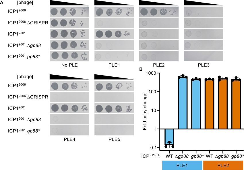
Research article                                                                                        Microbiology and Infectious Disease

           '

                                                                   !"#$

                                                                             "()             "'

                                                      #%&

           (
                                                !"#$!""$    *+23    *+22     ./01     *+,-    *+,'

           !"#$!""#                                            !"#$%"&(
               *+23              !"#$    !"#%        !"#&   !"#'                 !("&)%                  !("'       /5()6789     *+234'    *+,'
                      !"#$%"&'

                         ()*

           )
         /#00
                                                                                                                     !
          1"(2
       !*/34+25
          67,8
         9:;&79

                 !"#$!%$&"'(      )&*!%+*,!-.

Figure 1. Some ICP1 isolates encode a free-standing nuclease in place of CRISPR-Cas. (A) A model of ICP1 interference of phage-inducible
chromosomal island-like elements (PLEs) via CRISPR. When ICP1 infects a PLE(+) V. cholerae cell, ICP1 is able to overcome PLE restriction and
reproduce if it possesses a CRISPR-Cas system with complementary spacers to the PLE. Cas and CR refer to the CRISPR associated genes and CRISPR
array respectively. (B) Schematics of the region between gp87 and gp91 as it appears in ICP12001 (top) and ICP12006 (bottom). Genes represented by
black arrows are conserved in all ICP1 isolates, while genes represented with gray arrows covary with gp88 or CRISPR-Cas. (C) An alignment between
the T5orf172 domain of Gp88 and the GIY-YIG domains of several structurally resolved endonucleases. Secondary structure for Gp88 was predicted
using HHPRED (Zimmermann et al., 2018). Alpha helices are shown in yellow shading, and beta strands are shown in blue shading. Key residues of the
GIY-YIG motif are bolded. We included an atypical GIY-YIG endonuclease domain from a chloroplast-encoded glutoredoxin atGRXs16 to demonstrate
the potential for alternative residues at core motif positions. A conserved glutamate that was previously found to be required for catalysis in I-TevI is
denoted by an asterisk (and corresponds to the E180A mutation in Gp88 in subsequent experiments).
The online version of this article includes the following figure supplement(s) for figure 1:
Figure supplement 1. ICP12005 has an atypical CRISPR-Cas arrangement.

                                        isolates that lack CRISPR-Cas. Surprisingly, we found that all natural ICP1 isolates that do not encode
                                        CRISPR-Cas instead encode an endonuclease in the same genomic locus that is necessary for propa-
                                        gation on V. cholerae strains containing PLEs 1, 4, or 5. Lending further support to the ‘guns for
                                        hire’ model, we find that this anti-PLE nuclease is of chimeric origin, being partially derived from a
                                        PLE-encoded DNA-binding domain while its nucleolytic domain appears to be derived from an
                                        ICP1-encoded family of putative HEGs. Harnessing the rich evolutionary interplay of PLE and ICP1,
                                        this work shows that domain shuffling between hostile genomes can allow for new forms of

Barth et al. eLife 2021;10:e68339. DOI: https://doi.org/10.7554/eLife.68339                                                                       3 of 21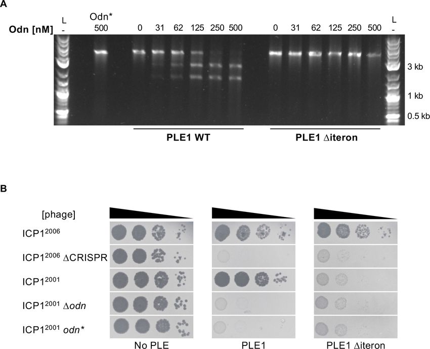
Research article                                                                              Microbiology and Infectious Disease

                                    antagonism, and that phage-encoded HEGs can be repurposed for antiparasite functions. Addition-
                                    ally, this work reveals key mediators of ICP1-PLE host range that inform observed patterns of PLE
                                    temporal succession and modularity, broadening our understanding of subcellular host-parasite
                                    coevolution.

                                    Results
                                    A subset of ICP1 isolates deploy a stand-alone nuclease instead of
                                    CRISPR-Cas to counter PLE
                                    We set out to identify which gene(s) determined host range in ICP1 isolates that lack CRISPR-Cas. It
                                    has long been recognized that phages are mosaic entities composed of functional gene neighbor-
                                    hoods, and syntenic neighborhoods of divergent sequence may fulfill analogous functions
                                    (Brüssow and Hendrix, 2002). Previous work suggests that ICP1 conforms to these general patterns
                                    of phage genome structure. Transcriptomics and bioinformatic predictions show that ICP1 genes
                                    with related biological functions are organized together in the genome and expressed at the same
                                    time, demonstrating the presence of gene neighborhoods (Barth et al., 2020a). Additionally, while
                                    the ICP1 genome is highly conserved between isolates and does not display large-scale rearrange-
                                    ments (Angermeyer et al., 2018), there is indication that nonhomologous sequence can serve anal-
                                    ogous functions. ICP1 isolates encode one of two alternative SF1B-type helicases thought to be of
                                    shared function (McKitterick et al., 2019a), suggesting that ICP1 isolates can use alternative genes
                                    to fulfill the same adaptational requirement. We reasoned that such genome organization and mosa-
                                    icism warranted a ‘guilt by location’ approach to investigating gene function, and that the locus syn-
                                    tenic to CRISPR-Cas in those ICP1 isolates that lack CRISPR might hold clues as to how they
                                    overcome PLEs.
                                        In isolates without CRISPR-Cas, the locus is replaced with a single open reading frame, desig-
                                    nated gp88 for its location in ICP12001, the original sequenced ICP1 isolate (Seed et al., 2011;
                                    Figure 1B). The two coding sequences immediately upstream of the CRISPR-Cas system and ori-
                                    ented divergently from the system are also replaced in the phage with gp88. These genes generally
                                    covary with the presence of the CRISPR-Cas system or gp88. Most ICP1 CRISPR-Cas systems are
                                    adjacent to a phage regulatory protein Rha domain (pfam09669) encoding gene. A gene encoding a
                                    Bro-N domain (pfam02498) and a KilAC domain (pfam03374) occurs adjacent to gp88. Their posi-
                                    tions and putative annotations suggest that these divergently transcribed genes may have a regula-
                                    tory function. In one sequenced ICP1 isolate with a functional CRISPR-Cas system (O’Hara et al.,
                                    2017), the bro_N domain coding gene and its partner are found instead of the rha-like gene con-
                                    taining pair, suggesting that these pairs are redundant in function or not involved in CRISPR-Cas
                                    activity (Figure 1—figure supplement 1).
                                        Further analysis of gp88 revealed that it encodes a T5orf172 domain (pfam10544) containing pro-
                                    tein. This domain is a member of the GIY-YIG endonuclease domain superfamily, suggesting that
                                    Gp88 may be a nuclease. We aligned the Gp88 T5orf172 domain with GIY-YIG domains from endo-
                                    nucleases that had been biochemically characterized and structurally resolved (Liu et al., 2013;
                                    Swapna et al., 2005; Truglio et al., 2005; Van Roey et al., 2002; Figure 1C). Unlike most identified
                                    GIY-YIG nucleases, Gp88 lacks a conserved histidine or tyrosine in the first alpha helix of the GIY-YIG
                                    domain. However, Gp88 retains conservation of the catalytic arginine and glutamate. The relatively
                                    high motif conservation found in Gp88 suggests that the protein possesses endonucleolytic activity.
                                    Given that nucleases are a particularly prominent class of proteins engaged in conflicts between
                                    hosts, mobile elements and viruses (Koonin et al., 2020), and gp88’s syntenic location to ICP1’s
                                    CRISPR-Cas system, we hypothesized that Gp88 interferes with PLE activity to protect ICP1 isolates
                                    that naturally lack CRISPR-Cas.
                                        To test our hypothesis, we generated ICP12001 mutants with either an in-frame deletion of gp88
                                    or harboring a single amino acid substitution (E180A) predicted to abolish Gp88’s nucleolytic activity
                                    (Figure 1C). We also used ICP12006 and a DCRISPR derivative to serve as controls for host suscepti-
                                    bility. As expected, the PLE (-) V. cholerae strain was susceptible to all ICP1 variants, and the
                                    ICP12006 DCRISPR variant was restricted by all PLEs (Figure 2A). CRISPR(+) ICP12006 was able to
                                    propagate on all strains except the one containing PLE3 as ICP12006 does not have a matching
                                    spacer against PLE3. ICP12001 was able to propagate on PLEs 1, 4, and 5 (Figure 2A), but it was

Barth et al. eLife 2021;10:e68339. DOI: https://doi.org/10.7554/eLife.68339                                                          4 of 21
Research article                                                                                         Microbiology and Infectious Disease

Figure 2. The alternative nuclease Gp88 controls ICP1 host range in a natural isolate that lacks CRISPR-Cas. (A) Tenfold dilutions of the phage isolate
or mutant derivative indicated spotted on V. cholerae with the PLE indicated (bacterial lawns in gray, zones of killing are shown in black). Gp88* possess
a single amino acid substitution (E180A) predicted to abolish nuclease activity. Spot assays were performed in biological triplicate, and a single
representative image is shown. Replicate spot assays are shown in Figure 2—figure supplement 1 and Figure 2—figure supplement 2. (B)
Replication of PLE1 and PLE2 in V. cholerae host strains calculated as the fold change in PLE DNA copy 20 minutes post infection with the ICP1 variant
indicated.
The online version of this article includes the following source data and figure supplement(s) for figure 2:
Source data 1. Values for the graph in Figure 2B.
Figure supplement 1. Biological replicate of spot assays.
Figure supplement 2. Biological replicate of spot assays.

                                     restricted on PLEs 2 and 3. Unlike the wild-type (WT) variant, the Dgp88 and gp88* mutants were
                                     unable to propagate on V. cholerae with PLEs 1, 4, or 5 (Figure 2A), indicating that catalytically
                                     active gp88 is necessary for overcoming these PLEs in phages naturally lacking CRISPR-Cas. Unsur-
                                     prisingly, the gp88 mutants retained sensitivity to restriction by PLEs 2 and 3 (Figure 2A).
                                        As CRISPR targeting was previously observed to diminish PLE replication that occurs during ICP1
                                     infection (McKitterick et al., 2019b), we tested whether the presence of Gp88 could impact PLE
                                     replication during infection. We observed that PLE1 is unable to replicate in the presence of Gp88
                                     encoding ICP1, and replication is restored during infection with the Gp88 knockout or catalytically
                                     inactive mutant phages (Figure 2B). Consistent with endonucleolytic activity, the PLE1 copy
                                     decreases following infection by Gp88 encoding phage. This is notable given that previous work has
                                     shown that multiple PLE matched spacers are required for CRISPR-Cas to completely abolish PLE
                                     replication during infection (McKitterick et al., 2019b). PLE2 replication is unaffected by the pres-
                                     ence or absence of Gp88, consistent with Gp88 not providing ICP1 with immunity against PLE2
                                     (Figure 2A).

                                     PLE replicons are modular
                                     Having identified gp88’s role in preventing PLE restriction of ICP1, we next sought to determine
                                     how Gp88 was recognizing PLE, and why it did not confer protection against PLEs 2 and 3. We rea-
                                     soned that PLEs 2 and 3 most likely lacked sequence targeted by Gp88 or encoded an inhibitor of

Barth et al. eLife 2021;10:e68339. DOI: https://doi.org/10.7554/eLife.68339                                                                       5 of 21
Research article Microbiology and Infectious Disease
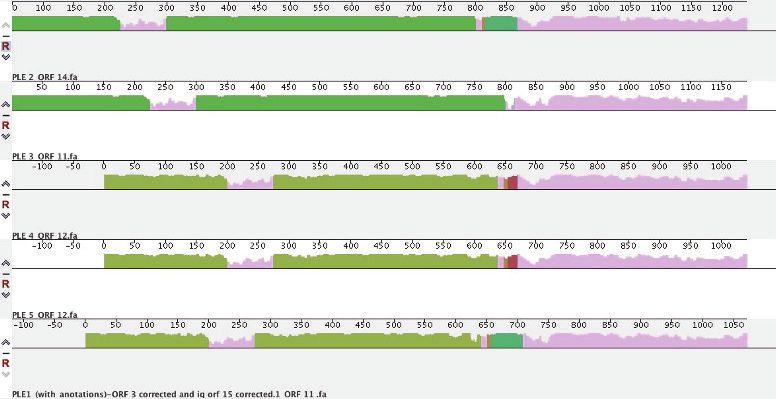
Gp88’s activity. To explore these possibilities, we compared the PLE genomes looking for nucleotide
sequence that was uniquely present or uniquely absent in PLEs 2 and 3.
Strikingly, only two stretches of sequence met these criteria, and both had been previously impli-
cated in PLE replication (Barth et al., 2020b). The repA gene encoding the replication initiation fac-
tor, and the intergenic region containing the PLE origin of replication (ori) to which RepA binds
covaried, with the PLE1, 4, and 5 sequences clustering together as one group, and the PLE2 and 3
sequences clustering as another (Figure 3A). More specifically, it was the DNA-binding RepA_N
'
#%&$
()* -."/ 6961D9C+6,0./,+96 '+", 0(12
#4(M-+6,0NE0
&'( &'($ '/3KQ8 O3PP) 3A(,/
#%&
$ !
: "
= "
< !
; !
2%?%
Research article                                                                              Microbiology and Infectious Disease

                                    domain of RepA that covaried with the origin, while the C-terminal domain, hypothesized to facili-
                                    tate replisome recruitment (Barth et al., 2020b), was conserved across all PLEs (Figure 3A).
                                        Previously, we found that during ICP1 infection, ectopic expression of RepA was sufficient to
                                    drive replication of a synthetic ‘midiPLE’ construct. The midiPLE consists of the PLE attachment sites,
                                    the PLE integrase, and the noncoding region that bears the origin of replication (Barth et al.,
                                    2020b). Additionally, midiPLE replication did not occur without RepA, and the PLE integrase was
                                    shown to be dispensable for PLE replication (Barth et al., 2020b). These data suggest that the mini-
                                    mal components of the PLE replicon are the replication origin and RepA, the two components that
                                    covaried across PLEs. Alignment of the conserved 30 ends of PLE repA genes suggests that RepA
                                    specificity swapping has occurred multiple times (Figure 3A, Figure 3—figure supplement 1).
                                    Despite the PLE1 RepA_N domain clustering with the PLE4 and PLE5 variants, the PLE1 C-terminal
                                    sequence is more similar to PLE2 (98.88% identical over the last 178 bp) than PLE5 (93.25% identical
                                    over the last 178 bp). PLE5 and PLE3 are 99.44% identical over the same region, while the PLE4
                                    C-terminal region is the most diverged from other PLEs (Figure 3—figure supplement 1). This
                                    cross-clustering of repA ends would only be expected to occur after multiple gene recombination
                                    events, suggesting that PLE replisome module swapping occurred at least twice, and may be an
                                    important part of PLE evolution.
                                        The putative modularity of the PLE origins and RepA_N domains covaried with susceptibility to
                                    ICP12001 (Figure 3A), leading us to hypothesize that one of the replicon modules but not the other
                                    was susceptible to Gp88-mediated interference. Before testing this hypothesis directly, we wanted
                                    to confirm that the covariation of PLE replication origins and RepA_N domains truly reflects modu-
                                    larity of the PLE replicon. We first tested whether the putative PLE2 origin and repA gene were nec-
                                    essary for PLE2 replication and found that deletion of either component abolished replication
                                    following infection by ICP12006 DCRISPR (Figure 3—figure supplement 2), as was previously
                                    observed for PLE1 (Barth et al., 2020b).
                                        Having confirmed that the PLE2 variant replicon components are necessary for replication, we
                                    then sought to demonstrate specificity of the RepA variants to their cognate origin of replication.
                                    We generated chimeric ‘origin-swapped’ DrepA PLEs for PLEs 1 and 2 (Figure 3B), and ectopically
                                    expressed each RepA variant in the different PLE backgrounds during phage infection. As expected,
                                    PLE replication only occurred when cognate origins and repA alleles co-occurred, revealing that the
                                    two components of the PLE replicon function together as a module, irrespective of which PLE back-
                                    bone they are encoded in.

                                    Gp88 is an origin-directed nuclease
                                    Having established the specificity between RepA variants and their cognate origins of replication
                                    and recognizing that sensitivity to Gp88 covaried with replicon type, we took a closer look at Gp88
                                    to decipher how it might interface with the PLE replication module. Remarkably, Gp88’s own N-ter-
                                    minal domain is 42% identical and 61% sequence similar across 93% of PLE1’s RepA_N domain
                                    (Figure 4A). This was surprising as Gp88’s T5orf172 domain is similar to those of several putative
                                    HEGs within the ICP1 genome (Figure 4—figure supplement 1). The high similarity of Gp88’s N-ter-
                                    minal portion to some PLE-encoded RepA alleles and the C-terminal portions similarity to putative
                                    HEGs suggest that gp88 may have arisen as a chimeric hybrid of PLE and ICP1 coding sequences.
                                    Additionally, the similarity of Gp88’s N-terminal region to PLEs 1, 4, and 5 RepA DNA-binding
                                    domains suggested that Gp88 might bind to the replication origins of PLEs 1, 4, and 5 and cleave at
                                    or proximal to that site.
                                        To evaluate this hypothesis, we next wanted to test whether the PLE origin of replication was a
                                    necessary component for Gp88 activity. Previously, it was shown that loss of replication partially
                                    attenuated PLE1-mediated restriction of ICP1 but nonreplicating PLE1 mutants were still broadly
                                    restrictive to ICP1 DCRISPR-Cas (Barth et al., 2020b). We hypothesized that PLE could escape Gp88
                                    targeting through deletion of the PLE origin and thus block propagation of Gp88 encoding phage.
                                    We tested this by deleting the entire conserved stretch of sequence that contained the origin of rep-
                                    lication in PLEs 1, 4, and 5. In support of our hypothesis, these mutants regained restrictive activity
                                    against Gp88 encoding phage (Figure 4B). Conversely, cloning a Gp88 recognized replication origin
                                    into PLEs that are insensitive to Gp88 should sensitize them to Gp88 activity. To test this, we
                                    infected our ‘ori-swapped’ PLE2 strain (Figure 3B) with Gp88 encoding phage. We included a PLE2
                                    Dori strain to control for the possibility that loss of replication would abolish PLE2-mediated

Barth et al. eLife 2021;10:e68339. DOI: https://doi.org/10.7554/eLife.68339                                                          7 of 21
Research article                                                                                         Microbiology and Infectious Disease

Figure 4. Gp88 is a PLE replication origin-directed nuclease. (A) Sequence alignment of the N-terminal portion of Gp88 with the RepA_N domain from
PLE1 RepA. Identical residues are denoted with a ‘*.’ Strong residue similarity is denoted by ‘:’, and weak similarity is denoted by ‘..’ (B, C) Tenfold
dilutions of the phage isolate or mutant derivative indicated spotted on V. cholerae with the PLE indicated (bacterial lawns in gray, zones of killing are
shown in black). Spot assays were performed in parallel with those in Figure 2, and images labeled with the same PLE background are the same image.
Spot assays were performed in biological triplicate, and a single representative image is shown. Biological replicates are shown in Figure 2—figure
supplement 1. Gp88* possess a single amino acid substitution (E180A) predicted to abolish nuclease activity. (B) shows phage susceptibility of V.
cholerae with PLE1, PLE4, and PLE5 Dori derivatives as compared to a strain without PLE. (C) shows phage susceptibility for V. cholerae with PLE2 Dori
and PLE2 Dori::oriPLE1. (D) Nuclease assay showing the integrity of a PCR product amplified from the noncoding region containing the ori from the PLE
variant indicated (numbers) treated with (+) and without (–) 500 nM of purified Gp88. Nuclease assays were performed in triplicate, replicates are
presented in Figure 4—figure supplement 3. (E) Nuclease assay showing the integrity of a PCR product amplified from the noncoding region
containing the ori from the PLE variant indicated (numbers) treated with (+) and without (–) 500 nM of purified Gp88*. Nuclease assays were performed
in triplicate, replicates are presented in Figure 4—figure supplement 4.
The online version of this article includes the following source data and figure supplement(s) for figure 4:
Source data 1. Original uncropped gels for Figure 4D (top) and Figure 4E (bottom).
Figure supplement 1. The nuclease domain of Gp88 is similar to those of putative homing endonucleases in ICP1.
Figure supplement 2. Protein preparations of Gp88 (Odn) and Gp88* (Odn*) used for in vitro assays.
Figure supplement 2—source data 1. Original uncropped gels for Figure 4—figure supplement 2.
Figure supplement 3. Replicates of Gp88 nuclease assay.
Figure supplement 3—source data 1. Original uncropped gels for Figure 4—figure supplement 3.
Figure 4 continued on next page

Barth et al. eLife 2021;10:e68339. DOI: https://doi.org/10.7554/eLife.68339                                                                       8 of 21
Research article                                                                                Microbiology and Infectious Disease

Figure 4 continued
Figure supplement 4. Replicates of Gp88* nuclease assay.
Figure supplement 4—source data 1. Original uncropped gels for Figure 4—figure supplement 4.
Figure supplement 5. Cleavage of an altered PLE4 probe.
Figure supplement 5—source data 1. Original uncropped gels for Figure 4—figure supplement 5.

                                    restriction of ICP1. The PLE2 Dori strain retained the ability to restrict all isolates of ICP12001
                                    (Figure 4C). In contrast, the PLE2 strain bearing the PLE1 origin sequence was no longer restrictive
                                    to ICP12001, but still restricted variants that lacked Gp88 activity (Figure 4C), confirming that the
                                    presence of the replication origin sequence mediated sensitivity to Gp88.
                                       We wanted to confirm that Gp88-mediated interference manifested through nucleolytic cleavage
                                    of PLE. To determine if Gp88 was truly acting as a nuclease, we purified Gp88 (Figure 4—figure
                                    supplement 2) and performed in vitro nuclease activity assays. Consistent with the host range of
                                    ICP1 encoding Gp88, we found that the purified Gp88 protein cut PCR products amplified from the
                                    region containing the origin of replication from PLEs 1, 4, and 5, but did not cut those of PLEs 2 and
                                    3 (Figure 4D), confirming that Gp88 disrupts PLE through nuclease activity. Supporting this interpre-
                                    tation, the E180A Gp88* mutant that was inactive against PLEs 1, 4, and 5 in vivo, and predicted to
                                    be catalytically inert, did not cleave PCR products amplified from PLEs 1, 4, and 5 (Figure 4E), fur-
                                    ther linking the in vivo activity of Gp88 to its capacity to cleave in vitro. Curiously, Gp88 produced
                                    only one cleavage product from the PLE4 probe. We reasoned that since the PLE noncoding regions
                                    are diverse and the PLE4 origin of replication was located near to the center of the PLE4 probe,
                                    Gp88 cleavage of the PLE4 probe might produce two bands of indistinguishable size. To check this,
                                    we produced a new PLE4 probe with the origin of replication offset from the middle and found that
                                    Gp88 produced two visible bands (Figure 4—figure supplement 5). In light of these results, we
                                    renamed Gp88 the origin-directed nuclease or Odn.

                                    Odn requires iterons to cleave the PLE origin of replication
                                    Our results so far suggested a model where Odn mimics the specificity of the PLE1, 4, and 5 RepA
                                    proteins to bind and cut at their cognate origins of replication. Previously, PLE1 RepA was found to
                                    bind specifically to a set of iterons, a series of three ~30 bp semi-palindromic repeats in the PLE1 ori-
                                    gin of replication (Barth et al., 2020b). If Odn specificity truly mimicked that of RepA, then it should
                                    require the iteron sequence for cutting. We tested this in vitro by titrating increasing concentrations
                                    of Odn in a nuclease assay with the WT PLE1 origin of replication, as well as the same substrate
                                    except with the iterons deleted. In support of our model, we found that Odn does require the iteron
                                    sequence for cleavage (Figure 5A). Consistent with iterons being necessary for Odn-mediated in
                                    vitro cleavage of the PLE origin, ICP12001 infection was restricted by a PLE1 strain harboring the
                                    same iteron deletion (Figure 5B). Together, these results strongly support that Odn has DNA-bind-
                                    ing specificity that mimics that of the replication initiation factor of some PLEs.

                                    PLE mutations lead to escape from Odn
                                    Odn activity against the origin, as well as the pattern of cross-clustering at the N and C termini of
                                    RepA, suggested that Odn may impose substantial evolutionary pressure on PLE replication mod-
                                    ules. Since swapping the PLE origin and cognate RepA_N domain could abolish Odn targeting of
                                    PLE, it appears likely that Odn selected for the multiple domain shuffling events in PLE RepA
                                    inferred by comparison of PLE genomes (Figure 3—figure supplement 1). Because PLE replication
                                    is necessary for both PLE mobility and complete restriction of ICP1 (Barth et al., 2020b;
                                    McKitterick et al., 2019a), simple deletions of the replication origin would not likely be favored as
                                    long-term solutions to evading recognition and subsequent cleavage by Odn.
                                       While the swapping and diversification of certain sequences can be traced through the five PLEs,
                                    each PLE variant is remarkably conserved. All members of each variant have been found to be 100%
                                    nucleotide identical in previously published data sets (McKitterick et al., 2019b; O’Hara et al.,
                                    2017). However, we found a single instance of diversity in PLE1 within a lineage of V. cholerae iso-
                                    lated from Pakistan. This lineage was represented in five sequenced strains (biosample accession
                                    numbers      SAMN08979118,        SAMN08979175,        SAMN08979185,         SAMN08979188,       and

Barth et al. eLife 2021;10:e68339. DOI: https://doi.org/10.7554/eLife.68339                                                            9 of 21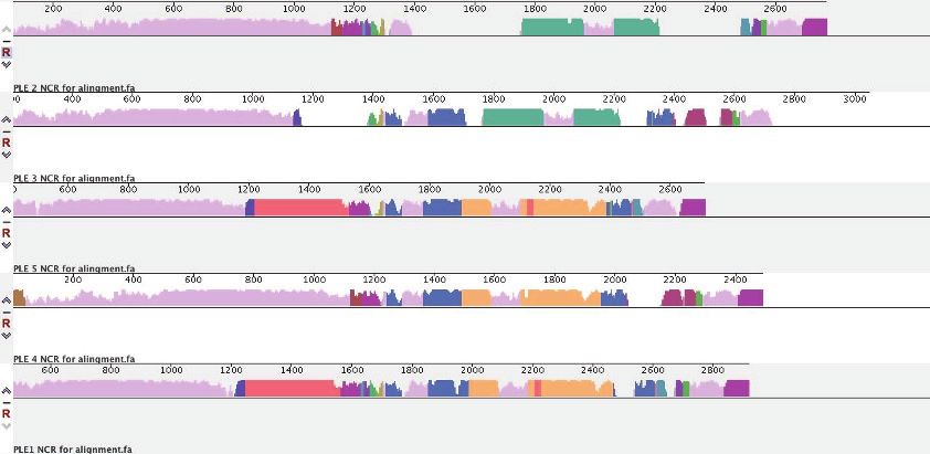
Research article                                                                                      Microbiology and Infectious Disease

                                    Figure 5. ICP1-encoded Odn (Gp88) requires the PLE iterons for cleavage. (A) Nuclease assay showing the
                                    integrity of a PCR product amplified from the noncoding region containing the ori from wild-type (WT) PLE1 and
                                    the Diteron mutant, with purified Odn (31.25–500 nM) titrated in. 500 nM catalytically inactive Odn (Odn*) with a
                                    single amino acid substitution (E180A) with the WT PLE1 sequence was also included (far left). Nuclease assays
                                    were performed in triplicate and replicates are presented in Figure 5—figure supplement 1. (B) Tenfold dilutions
                                    of the phage isolate or mutant derivative indicated spotted on V. cholerae with the PLE indicated (bacterial lawns
                                    in gray, zones of killing are shown in black). Spot assays were performed in parallel with those in Figures 2 and
                                    4, and images labeled with the same PLE background are the same image. Spot assays were performed in
                                    biological triplicate, and a single representative image is shown. Replicate assays are shown in Figure 2—figure
                                    supplement 1.
                                    The online version of this article includes the following source data and figure supplement(s) for figure 5:
                                    Source data 1. Original uncropped gel for Figure 5A.
                                    Figure supplement 1. Replicates of nuclease assays.
                                    Figure supplement 1—source data 1. Original uncropped gels for Figure 5—figure supplement 1.

                                    SAMN08979253). In these five strains, we discovered variation in a 67 bp stretch covering the iterons
                                    that results in several nucleotide changes. Within the first iteron, there is an A to T transversion, and
                                    starting at that transversion, the next 42 bp are duplicated and replace the sequence that is normally
                                    downstream (Figure 6A). This change maintains the presence of the three iterons, and even reverts
                                    a few variant bases to ones in the PLE4 and 5 iterons (Figure 6B). Notably, these changes are the
                                    only sequence differences between these PLE variants and all other PLE1 isolates, aside from a 2 bp
                                    extension of an 11 bp polyA tract that also occurs in these atypical PLE1 variants.
                                       We sought to test if this natural example of PLE1 iteron diversity had any effect on susceptibility
                                    to Odn. We generated a DNA probe covering this variant region (denoted PLE1Mut) and tested its
                                    sensitivity to Odn in comparison to the WT PLE1 ori sequence. The PLE1Mut sequence was notably
                                    less susceptible to cleavage by Odn than the WT allele, but some cutting of the PLE1Mut probe at
                                    the highest concentration of Odn was apparent (Figure 6C). This raised the question of whether this

Barth et al. eLife 2021;10:e68339. DOI: https://doi.org/10.7554/eLife.68339                                                                   10 of 21
Research article Microbiology and Infectious Disease
Figure 6. Mutations in PLE1 present in V. cholerae isolates from Pakistan render the PLE1 ori resistant to Odn-mediated cleavage. (A) The PLE1Mut
iteron region. Iterons are bolded and sub-repeats are denoted with arrows. The underlined sequence is identical to the sequence in red. An asterisk (*)
denotes the location of an A to T substitution. (B) An alignment of the iterons from PLE5, PLE4, PLE1, and PLE1Mut. Iterons are in bold with sub-repeats
indicated with arrows. Sequence deviating from a consensus is shown in red. Regions with 100% conservation are denoted with an asterisk. (C)
Nuclease assay showing the integrity of a PCR product amplified from the noncoding region containing the ori from wild-type (WT) PLE1 or PLE1Mut
with purified Odn (31.25–500 nM) titrated in. Nuclease assays were performed in triplicate, and replicates are presented in Figure 6—figure
supplement 1. (D) Replication of PLE1 WT and PLE1Mut in V. cholerae calculated as the fold change in PLE DNA copy 20 minutes post infection with
the ICP1 variant indicated. (E) Tenfold dilutions of the phage isolate or mutant derivative indicated spotted on V. cholerae with the PLE indicated
(bacterial lawns in gray, zones of killing are shown in black). Biological replicates of the spot assays are presented in Figure 6—figure supplement 2.
The online version of this article includes the following source data and figure supplement(s) for figure 6:
Source data 1. Original uncropped gel for Figure 6C.
Source data 2. Values for the graph in Figure 6D.
Figure supplement 1. Replicates of nuclease assays.
Figure supplement 1—source data 1. Original uncropped gels for Figure 6—figure supplement 1.
Figure supplement 2. Biological replicates of PLE1Mut spot assays.
PLE1 variant could resist attack by Odn in vivo. To investigate this possibility, we cloned the PLE1Mut
origin of replication into our PLE1 strain and challenged this mutant with WT, Dodn and odn*
ICP12001. The PLE1Mut origin of replication was able to drive replication to levels comparable to that
of the WT origin, and robust replication was maintained even in the presence of Odn (Figure 6D). In
agreement with our replication data, we also found that our PLE1Mut strain restricted production of
WT ICP12001 (Figure 6E). These data demonstrate that PLE can escape Odn activity through subtle
Barth et al. eLife 2021;10:e68339. DOI: https://doi.org/10.7554/eLife.68339 11 of 21Research article Microbiology and Infectious Disease
restructuring of the iterons, in addition to more extensive replication module exchange (Figure 3A),
and suggest that ICP1 counter-defenses like Odn may select for diversification of the PLE replication
machinery.
Discussion
Here, we have identified a new anti-PLE effector present in some ICP1 isolates and identified its
mode of anti-PLE activity. The anti-PLE effector Odn possess two domains, an endonuclease domain
and a DNA-binding domain, which has sequence similarity to the DNA-binding domain of the RepA
initiation factor from PLEs 1, 4, and 5. Mimicking RepA’s specificity, Odn cuts the origin of replica-
tion in those PLEs. PLEs are able to escape Odn antagonism through mutation of their origin of rep-
lication, and there is compelling evidence suggesting that PLEs have exchanged replicon modules
for an alternative replication origin and initiation factor origin-binding domain on at least two sepa-
rate occasions. While sampling of ICP1 isolates and V. cholerae strains is far from complete, we do
see the oldest ICP1 strains encode Odn and the oldest PLE variants encode the sensitive origin of
replication. It is possible that Odn selected for alternative replicon modules, leading to the decline
of PLE4 and succession of PLE2 that occurred in the early 2000s (O’Hara et al., 2017). This may in
turn have caused selection for the presence of CRISPR-Cas, a decrease in prominence of Odn, and
the reemergence of the sensitive replicon module in PLE1 matching the trends of PLE and ICP1
occurrence observed thus far (McKitterick et al., 2019b; O’Hara et al., 2017). This model of PLE
and ICP1 succession is consistent with an antagonistic frequency-dependent selection (aFDS) mecha-
nism of host-pathogen coevolution where some level of genetic diversity is maintained among
antagonistic genes and rare alleles are selected for, leading to oscillation of the dominant genotype
(Papkou et al., 2019). In some experimental evolution studies using bacteriophages, aFDS dynamics
are found to eventually develop (Gómez and Buckling, 2011; Hall et al., 2011; Lopez Pascua
et al., 2014). This seems a likely possibility for PLEs and ICP1 whose specific adaptations and
counter adaptations suggest that the two genomes have been coevolving for a long period of time.
It is interesting to consider why a specialized anti-PLE mechanism might persist within some ICP1
isolates when adaptive CRISPR-Cas immunity exists as an alternative. An obvious benefit of Odn is
its small size; odn is less than 700 bp and more than 10 times smaller than ICP1’s CRISPR-Cas sys-
tem. Given that ICP1’s genome size is limited by what it can package into its capsid, the extra
sequence taken up by CRISPR-Cas could be better spent on other auxiliary genes if it is not needed
to overcome PLE. Additionally, Odn may provide more complete restriction of certain PLEs. While a
single CRISPR spacer is sufficient to enable ICP1 plaque formation on PLE(+) cells, it was previously
found that multiple spacers were needed to completely abolish PLE replication, and PLE transduc-
tion could still be detected from single-spacer ICP1 infections (McKitterick et al., 2019b;
O’Hara et al., 2017). Additionally, different CRISPR spacers had different outcomes in terms of PLE
transduction and ICP1 reproduction (McKitterick et al., 2019b), suggesting that where PLE is tar-
geted may be important. Position-dependent outcomes for cleavage may explain why Odn is suffi-
cient to provide robust interference against the PLEs it targets. By destroying the origin of
replication, Odn-mediated PLE degradation might not have to compete with PLE replication and
could head off pathways for PLE escape through recombinational repair. It should also be consid-
ered that certain environmental and physiological conditions as well as some genetic factors might
render ICP1’s CRISPR-Cas system less effective. While no PLEs appear to encode anti-CRISPRs,
numerous anti-CRISPRs have been found in Gammaproteobacteria (Pawluk et al., 2016), and co-
occurrence of these genes with PLE could neutralize ICP1’s CRISPR-Cas as an anti-PLE strategy.
Overall, it appears that Odn lacks the flexibility provided by CRISPR adaptation but provides more
reliable interference against a subset of PLE variants.
In addition to ICP1 and Odn, at least one other anti-PLE mechanism has evolved as some CRISPR
(+) ICP1 isolates are able to plaque on PLE2 when CRISPR is deleted (O’Hara et al., 2017). What-
ever this anti-PLE mechanism may be, it may have helped select against PLE2, leading to the emer-
gence of PLE1 as another possible example of an aFDS dynamic in the ICP1-PLE arms race. It seems
unlikely that this anti-PLE2 mechanism is specific to the alternative replicon module as PLE3, which
has a similar replicon to PLE2, maintains restriction of those particular ICP1 isolates (O’Hara et al.,
2017). In any case, the co-occurrence of a separate anti-PLE mechanism in the same phage as a
Barth et al. eLife 2021;10:e68339. DOI: https://doi.org/10.7554/eLife.68339 12 of 21Research article Microbiology and Infectious Disease
CRISPR-Cas system further suggests that CRISPR-Cas may have weak spots in terms of PLE
inhibition.
It is somewhat surprising that the PLE1Mut variant is able to escape Odn activity, given that the
new iteron sequences are largely similar to those that exist in PLEs 1, 4, and 5. The most notable dif-
ferences are an extra T/A base between the inverted sub-repeats in both the second and third iter-
ons, and a reduction of space between the second and third iterons (now 5 bp instead of 14 or 16
bp) (Figure 6B). These changes appear more consistent with some sort of steric effect on catalysis
from improper spacing rather than a loss of sequence recognition; however, the molecular details of
Odn binding and catalysis remain to be elucidated. The T4-encoded GIY-YIG HEG I-TevI is known to
have two separate target specificities, one for DNA binding and one for cleavage, and cleavage is
only efficient if the two recognition sites are properly spaced (Liu et al., 2006). It is possible that
Odn nucleolytic activity also has some sequence specificity, and closer spacing of the iterons is suffi-
cient to block binding and cleavage by Odn. Based on the data presented here, it is not possible to
infer if Odn nucleolytic activity has sequence specificity, and what that specificity might be. For
I-TevI, the sequence requirements for cleavage are somewhat loose (Roy et al., 2016), making cleav-
age sites harder to predict from sequence alone.
Alternatively, Odn cleavage might require recruitment of multiple Odn proteins, spaced a certain
distance apart. RepA is thought to bind as dimer (Barth et al., 2020b), but at least some GIY-YIG
endonucleases, including I-TevI, function as monomers (Van Roey et al., 2002). It is conceivable that
Odn could require cooperative binding for catalysis or alternatively bind and cut at multiple sub-
repeats independently. While our results show robust double-stranded cleavage by Odn, this could
be achieved through multiple single-strand nicks from appropriately spaced iterons. Odn and RepA
provide an interesting example of proteins with overlapping sequence specificity. Working out the
intricacies of their binding specificity and activity may prove fruitful for understanding how DNA-
binding proteins evolve new targets and functions.
One of the most compelling aspects of Odn is its evolutionary relationship to a family of putative
HEGs. HEGs are usually considered selfish genetic elements and are common in phage genomes
(Edgell, 2009; Edgell et al., 2010; Stoddard, 2011). This adds a layer of symmetry to the V. chol-
erae-ICP1-PLE arms race. To defend itself against ICP1, V. cholerae makes use of the PLE, a selfish
MGE. To protect against PLE, ICP1 has repurposed a nuclease domain from its own HEG parasites.
While this recruitment of MGEs for antagonistic functions fits nicely within the ‘guns for hire’ model
of MGE evolution, we see an interesting twist where it seems the horizontal gene transfer of DNA-
binding domains is a central mediator of the PLE-ICP1 conflict. Recently, it was found that PLEs
reduce ICP1 capsid gene expression through use of a regulator that resembles the DNA-binding
domains of ICP1-encoded HEGs (Netter et al., 2021), the very same family of genes from which
Odn’s nuclease domain is derived (Figure 4—figure supplement 1). While viral ‘capture’ of host
genes by genetic parasites for the purpose of manipulating gene expression is well described in
both phage and eukaryotic viruses (Alcami, 2003; Bryan et al., 2008; Zeng and Chisholm, 2012),
to our knowledge Odn is the first example of an anti-MGE gene where a sequence-specific DNA-
binding protein has been ‘captured’ and fused to an endonuclease domain as a means of destroying
the very genome that the sequence originated from. This highlights how horizontal gene transfer
can serve as a shortcut to acquiring new sequence-specific antagonists during antagonistic coevolu-
tion and provides a slower evolving parallel to adaptive immunity through spacer acquisition.
It is striking that two examples of horizontal transfer between ICP1 and PLE relate to the same
family of HEGs. Domain shuffling has been described for HEGs previously (Landthaler and Shub,
2003) and is likely especially adaptive for HEGs, which are thought to amplify within host genomes
by acquiring new sequence specificities (Gogarten and Hilario, 2006; Roy et al., 2016). The domain
architecture of some phage-encoded HEGs has been likened to ‘beads on a string’: with indepen-
dent functional domains connected by linker sequences (Van Roey and Derbyshire, 2005). It is con-
ceivable that proteins with this architecture might be more amenable to domain shuffling, and given
the selfish nature of HEGs, they may have adaptations to tolerate or promote this shuffling as a
means of diversification and dispersal. While HEG domestication has been much discussed
(Coughlan et al., 2020; Stoddard, 2011) and the connection between genome antagonism and
nucleases is well established in bacteria (Koonin et al., 2020), a specific link between HEGs and
MGE antagonism had not been established prior to characterization of Odn. It is not clear if Odn
has any intrinsic homing activity, but Odn may be able to cleave and replace CRISPR-Cas loci that
Barth et al. eLife 2021;10:e68339. DOI: https://doi.org/10.7554/eLife.68339 13 of 21Research article Microbiology and Infectious Disease
have acquired spacers matching the Odn recognition sequence. This could layer a Odn vs. CRISPR-
Cas genetic conflict on top of the ICP1-PLE arms race. Because of their selfish nature, ability to
mobilize, nucleolytic activity, and enrichment within viral and mobile genomes, HEGs are poised to
be at the forefront of antagonistic coevolution between viruses and other MGEs, and we anticipate
that other putative HEGs have unrecognized antiparasite activities.
Materials and methods
Key resources table
Reagent type
(species) or Source or Additional
resource Designation reference Identifiers information
Gene RepAPLE1 Barth et al., 2020b WP_002040284.1
(Vibrio cholerae) (PLE1 ORF11)
Gene RepAPLE2 This paper AGG36643.1
(Vibrio cholerae) (PLE2 ORF14)
Gene Odn (ICP1_2001_ This paper YP_004251029
(Bacteriophage Dha_0 gp88)
ICP1)
Gene Odn* (ICP1_2001_ This paper The E180A
(Bacteriophage Dha_0 gp88E180A) mutation is
ICP1) predicted to
abolish catalytic
activity
Recombinant Ptac-repAPLE1 Barth et al., 2020b pZKB129 Inducible RepA
DNA reagent (plasmid) from PLE1
Recombinant Ptac-repAPLE2 This paper pKS2159 Inducible RepA
DNA reagent (plasmid) from PLE2
Recombinant pE-SUMO-Odn This paper pKS2187 Vector to
DNA reagent (plasmid) express 6xHisSumo-
fusion protein,
fused to N-
terminus
of Odn (Gp88)
Recombinant pE-SUMO-Odn* This paper pKS2189 Vector to
DNA reagent (plasmid) express 6xHisSumo-
fusion protein,
fused to N-
terminus
of Odn*
(Gp88E180A)
Strain, strain PLE V. cholerae Levine et al., 1982 KDS6
background (E7946)
(Vibrio cholerae)
Strain, strain PLE1 V. cholerae O’Hara et al., 2017 KDS36
background (PLE1 E7946)
(Vibrio cholerae)
Strain, strain PLE2 V. cholerae O’Hara et al., 2017 KDS37
background (PLE2 E7946)
(Vibrio cholerae)
Strain, strain PLE3 V. cholerae O’Hara et al., 2017 KDS38
background (PLE3 E7946)
(Vibrio cholerae)
Strain, strain PLE4 V. cholerae O’Hara et al., 2017 KDS39
background (PLE4 E7946)
(Vibrio cholerae)
Strain, strain PLE5 V. cholerae O’Hara et al., 2017 KDS40
background (PLE5 E7946)
(Vibrio cholerae)
Continued on next page
Barth et al. eLife 2021;10:e68339. DOI: https://doi.org/10.7554/eLife.68339 14 of 21Research article Microbiology and Infectious Disease
Continued
Reagent type
(species) or Source or Additional
resource Designation reference Identifiers information
Strain, strain PLE1 Dori This paper KDS297 Used for all
background V. cholerae spot assays
(Vibrio cholerae) (PLE1 E7946)
Strain, strain PLE2 Dori V. cholerae This paper KDS298 Figure 3—figure
background (PLE2 E7946) supplement 2
(Vibrio cholerae)
Strain, strain PLE2 DrepA V. cholerae This paper KDS299 Figure 3—figure
background (PLE2 E7946) supplement 2
(Vibrio cholerae)
Strain, strain PLE1 DrepA Dori::oriPLE2; This paper KDS300 Figure 3B
background Ptac-repAPLE1 V. cholerae E7946
(Vibrio cholerae)
Strain, strain PLE1 DrepA Dori::oriPLE2; This paper KDS301 Figure 3B
background Ptac-repAPLE2 V. cholerae E7946
(Vibrio cholerae)
Strain, strain PLE2 DrepA Dori::oriPLE1; This paper KDS302 Figure 3B
background Ptac-repAPLE1 V. cholerae E7946
(Vibrio cholerae)
Strain, strain PLE2 DrepA Dori::oriPLE1; This paper KDS303 Figure 3B
background Ptac-repAPLE2 V. cholerae E7946
(Vibrio cholerae)
Strain, strain PLE4 Dori V. cholerae This paper KDS304 Used for all
background (PLE4 E7946) spot assays
(Vibrio cholerae)
Strain, strain PLE5 Dori V. cholerae This paper KDS305 Used for all
background (PLE5 E7946) spot assays
(Vibrio cholerae)
Strain, strain PLE1 Diterons Barth et al., 2020b KDS263 Used for all
background V. cholerae spot assays
(Vibrio cholerae) (PLE1 E7946)
PLE1
Strain, strain PLE2 Dori::ori This paper KDS306 Used for
background V. cholerae all spot assays
(Vibrio cholerae) (PLE2 E7946)
Strain, strain PLE1Dori::oriMut This paper KDS319 Ori engineered
background DlacZ::KanR V. cholerae to match what
(Vibrio cholerae) E7946 (referred to as PLE1Mut) is observed in
PLE1(+) strains
from Pakistan:
biosample
accession
numbers SAMN08979118,
SAMN08979175,
SAMN08979185,
SAMN08979188,
and SAMN08979253
Strain, strain pE-SUMO- This paper KDS307 Expression strain for
background Odn E. coli BL21 Gp88/Odn
(Escherichia coli)
Strain, strain pE-SUMO- This paper KDS308 Expression strain for
background Odn* E. coli BL21 Gp88*/Odn*
(Escherichia coli)
Strain, strain 2006 WT O’Hara et al., 2017 MH310934
background (ICP1_2006_Dha_E)
(Bacteriophage ICP1)
Strain, strain 2006 DCR; DCas2_3 McKitterick and Seed, 2018
background (ICP1_2006_Dha_E)
(Bacteriophage ICP1)
Continued on next page
Barth et al. eLife 2021;10:e68339. DOI: https://doi.org/10.7554/eLife.68339 15 of 21Research article Microbiology and Infectious Disease
Continued
Reagent type
(species) or Source or Additional
resource Designation reference Identifiers information
Strain, strain 2001 WT (ICP1_ Seed et al., 2011 HQ641347
background 2001_Dha_0)
(Bacteriophage ICP1)
Strain, strain 2001 Dodn (ICP1_ This paper KSf93 odn is gp88
background 2001_Dha_0)
(Bacteriophage ICP1)
Strain, strain 2001 odn* (ICP1_ This paper KSf134 odn* is
background 2001_Dha_0) gp88E180A
(Bacteriophage ICP1)
Sequence- 5’-AGGGTTTGAGT O’Hara et al., 2017 zac14 qPCR primer
based reagent GCGATTACG-3’ targeting a
conserved portion of the
PLE noncoding
region
Sequence- 5’-TGAGGTTTTACC O’Hara et al., 2017 zac15 qPCR primer
based reagent ACCTTTTGC-3’ targeting a
conserved portion of the
PLE noncoding
region
Sequence- 5’-GTCATTTAACGCA This paper KS459 F-primer used
based reagent TCTTATCACC-3’ to amplify
noncoding region probes
for PLE1 and PLE5
Sequence- 5’-GGCTTAGCAACTG This paper zac267 F-primer used
based reagent TCTACGG-3’ to amplify
noncoding region probes
for PLE2,
PLE3,
and PLE4
Sequence- 5’-GTTACGTCTGAT This paper KS321 R-primer used
based reagent TGCTGACG-3’ to amplify
noncoding region
probes for
PLE1
Sequence- 5’-CCGCTTATATCAAT This paper zac269 R-primer used
based reagent TTCACTAATATCT-3’ to amplify
noncoding region probes
for PLE2 and PLE3
Sequence- 5’-GGACGGCTAA This paper KS323 R-primer used
based reagent ACCATTCTCG-3’ to amplify
noncoding region probes
for PLE4 and PLE5
Sequence- 5’-CATAAGGTTGGC This paper KS458 R-primer used
based reagent TCCTCAATG-3’ to amplify
noncoding region probe
for PLE4
in Figure 4—
figure supplement 5
Strains and culture conditions
V. cholerae strains used in this study are derived from E7946. Bacteria were routinely grown on LB
agar plates and in LB broth with aeration at 37˚C. Antibiotics were supplemented as appropriate at
the following concentrations: 75 mg/ml kanamycin, 100 mg/ml spectinomycin, 1.25 or 2.5 mg/ml chlor-
amphenicol (V. cholerae for broth or plate conditions, respectively), 25 mg/ml chloramphenicol
(Escherichia coli), and 100 mg/ml streptomycin. A detailed list of all strains used throughout this
study can be found in the Key resources table.
Phage titers were determined using a soft agar overlay method wherein ICP1 was allowed to
adsorb to V. cholerae for 10 minutes at room temperature before the mixture was added to molten
Barth et al. eLife 2021;10:e68339. DOI: https://doi.org/10.7554/eLife.68339 16 of 21Research article Microbiology and Infectious Disease
LB soft agar (0.5%) and poured onto 100 mm 15 mm LB agar plates. Plaques were counted after
overnight incubation at 37˚C. Prior to phage infection for purposes of quantification or qPCR or spot
assay analysis, V. cholerae was grown on plates overnight and then inoculated into 2 ml LB liquid cul-
tures. Liquid cultures were grown to an OD > 1, then back diluted in fresh media to OD600 = 0.05,
and then grown to OD600 = 0.3, at which point they were infected.
Generation of mutant strains and constructs
V. cholerae mutants were generated through natural transformation as described previously
(Dalia et al., 2014). For gene knockouts, splicing by overlap extension (SOE) PCR was used to gen-
erate deletion constructs with a spectinomycin resistance cassette flanked by frt recombination sites.
Following selection of spectinomycin-resistant mutants, a plasmid bearing an isopropyl b-d-1-
thiogalactopyranoside (IPTG)-inducible Flp recombinase was mated into transformants and Flp
expression was induced to generate in-frame deletions. The plasmid was cured by growing mutants
under inducing conditions with 300 mg/ml streptomycin. For unmarked replication origin-swapped
constructs, mutants were generated through natural transformation by cotransformation
(Dalia et al., 2014). For plasmid expression constructs, a derivative of the pMMB67EH vector with a
theophylline-inducible riboswitch was used as previously described (McKitterick and Seed, 2018).
All constructs were confirmed with DNA sequencing over the region of interest, and primer sequen-
ces and construct designs are available on DRYAD at https://datadryad.org/stash/share/HSB-
bM3fCu3gSdF_yMQpCqyYuT4wW6_2IsZAkY0P5Ho.
Phage infection spot assays
V. cholerae was added to molten 0.5% LB top agar and poured over LB plates. Following solidifica-
tion of the top agar, 3 ml of serially 10-fold diluted phage were spotted onto the plate. Once phage
spots dried, plates were incubated for at 37˚C for 2 hr and then overnight at room temperature
before visualization.
Real-time quantitative PCR
qPCR experiments were performed as previously described (Barth et al., 2020b; O’Hara et al.,
2017). Briefly, liquid cultures were infected with ICP1 at a multiplicity of infection (MOI) of 2.5 at
OD600 = 0.3. Samples were taken at 0 and 20 minutes post infection and boiled before serving as
templates for IQ SYBR (Bio-Rad) qPCR reactions. For assays involving induction of repA, 2 ml cul-
tures were grown with 1.25 mg/ml chloramphenicol for plasmid maintenance and induced for 20
minutes prior to infection using a final concentration of 1.5 mM theophylline and 1 mM IPTG starting
at OD600 = 0.17. All conditions were tested in biological triplicate, and each reported data point is
the mean of two technical replicates. A single primer set (Key resources table) that amplifies a con-
served region in all PLEs was used to detect PLE replication by qPCR.
Protein purification
E. coli BL21 cells containing a His6-SUMO fusion to WT or E185A Gp88 were grown to OD600 = 0.5
at 37˚C and induced with IPTG to a final concentration of 0.5 mM. The culture was grown for 2 hr
and harvested by centrifugation at 4000g for 20 minutes. The pellet was resuspended in lysis buffer
(50 mM Tris–HCl pH 8, 200 mM NaCl, 1 mM BME, 0.5% Triton-X 50 mM imidazole, 1 Pierce Prote-
ase Inhibitor Mini Tablet [Thermo Scientific]) and sonicated. Cell debris was removed by centrifuga-
tion (29,097g for 40 minutes). The lysate was applied to a HisTrap HP column (Cytiva). The column
was washed with wash buffer (50 mM Tris–HCl pH 8, 200 mM NaCl, 1 mM BME, 50 mM imidazole),
and a high salt wash (50 mM Tris–HCl pH 8, 2 M NaCl, 1 mM BME, 50 mM imidazole) was used to
remove residual DNA. The protein was eluted using an elution buffer (50 mM Tris–HCl pH 8, 200
mM NaCl, 1 mM BME, 300 mM imidazole), and then the eluate was applied to a HiTrap Heparin HP
column (Cytivia) for further purification. Following elution from the HiTrap Heparin column, the pro-
tein was dialyzed using a 10k Slide-A-Lyzer Dialysis cassette (Thermo Fisher) in 50 mM Tris–HCl pH
7.5, 150 mM NaCl, 1 mM dithiothreitol (DTT). Concomitant with dialysis, the His6-SUMO tag was
cleaved using SUMO protease. The SUMO tag was removed using Dynabeads (Invitrogen).
Barth et al. eLife 2021;10:e68339. DOI: https://doi.org/10.7554/eLife.68339 17 of 21You can also read