Outer Membrane Vesicle Induction and Isolation for Vaccine Development - Frontiers
←
→
Page content transcription
If your browser does not render page correctly, please read the page content below
REVIEW
published: 04 February 2021
doi: 10.3389/fmicb.2021.629090
Outer Membrane Vesicle Induction
and Isolation for Vaccine
Development
Melanie D. Balhuizen , Edwin J. A. Veldhuizen * and Henk P. Haagsman
Division of Infectious Diseases and Immunology, Department of Biomolecular Health Sciences, Faculty of Veterinary
Medicine, Utrecht University, Utrecht, Netherlands
Gram-negative bacteria release vesicular structures from their outer membrane, so called
outer membrane vesicles (OMVs). OMVs have a variety of functions such as waste
disposal, communication, and antigen or toxin delivery. These vesicles are the promising
structures for vaccine development since OMVs carry many surface antigens that are
identical to the bacterial surface. However, isolation is often difficult and results in low
Edited by: yields. Several methods to enhance OMV yield exist, but these do affect the resulting
Mickaël Desvaux,
OMVs. In this review, our current knowledge about OMVs will be presented. Different
Institut National de la Recherche
Agronomique (INRA), France methods to induce OMVs will be reviewed and their advantages and disadvantages will
Reviewed by: be discussed. The effects of the induction and isolation methods used in several
Mariagrazia Pizza, immunological studies on OMVs will be compared. Finally, the challenges for OMV-based
GlaxoSmithKline, Italy
Denice C. Bay, vaccine development will be examined and one example of a successful OMV-based
University of Manitoba, Canada vaccine will be presented.
Maricarmen Rojas Lopez,
GlaxoSmithKline, Italy Keywords: outer membrane vesicles, isolation, induction, vaccine development, Bordetella pertussis,
Roland Lloubes, Neisseria meningitidis, host defense peptides
UMR7255 Laboratoire d’ingénierie
des systèmes macromoléculaires
(LISM), France
INTRODUCTION ON OUTER MEMBRANE VESICLES
*Correspondence:
Edwin J. A. Veldhuizen Gram-negative bacteria have two membranes, the inner membrane (IM) and the outer membrane
e.j.a.veldhuizen@uu.nl
(OM) with a network of peptidoglycan (PG) and the periplasmic space in between. Both the
IM and OM consist of phospholipids and membrane proteins, with only the outer leaflet of
Specialty section:
This article was submitted to the OM containing lipopolysaccharide (LPS). From the OM, small protrusions can form that
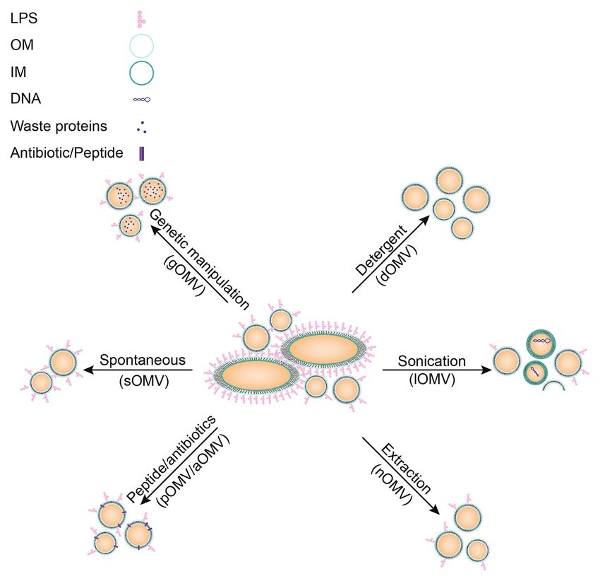
Microbial Physiology and Metabolism, pinch off and become extracellular vesicles, called outer membrane vesicles (OMVs; Figure 1;
a section of the journal Beveridge, 1999). Resulting OMVs are between 20 and 300 nm in diameter. They consist of
Frontiers in Microbiology a single lipid bilayer containing LPS, phospholipids, and various outer membrane proteins
Received: 13 November 2020 (OMPs), which represents the OM of the originating bacteria. Formation of OMVs has been
Accepted: 08 January 2021 the subject of much debate, since the driving force of OMV formation was long unknown
Published: 04 February 2021 (Zhou et al., 1998; Haurat et al., 2011). The formation of OMVs was long thought to be an
Citation: arbitrary stress response from bacterial cells (McBroom and Kuehn, 2007), but OMVs were
Balhuizen MD, Veldhuizen EJA and later proven to have many more functions, which will be discussed below.
Haagsman HP (2021) Outer
Membrane Vesicle Induction and
Isolation for Vaccine Development. Formation of OMVs
Front. Microbiol. 12:629090. For OMV formation, it is necessary to detach the OM from the PG layer and the IM. These
doi: 10.3389/fmicb.2021.629090 layers are stably linked by many different lipoproteins. A local decrease in the number of
Frontiers in Microbiology | www.frontiersin.org 1 February 2021 | Volume 12 | Article 629090
Balhuizen et al.                                                                                                                         OMV Induction and Isolation

  FIGURE 1 | Gram-negative bacterial membrane during OMV formation and functions of resulting OMVs. OMVs have been implicated in many different processes.
  Depicted here are the different functions OMVs have been shown to be involved in such as transport of toxins, waste removal, or communication between bacteria.
  LPS: lipopolysaccharide, PL: phospholipid, OM: outer membrane, PG: peptidoglycan, IM: inner membrane.

lipoproteins, and therefore the number of crosslinks, has been                       in Salmonella and E. coli (Bernadac et al., 1998; Deatherage
implicated in OMV formation. For example, the deletion of                            et al., 2009). Furthermore, alterations to the PG structure can
lipoprotein (Lpp) in combination with magnesium starvation                           prevent proper attachment of lipoproteins, which in turn
or the deletion of outer membrane protein A (OmpA) in                                decreases the number of crosslinks between the IM and OM.
Escherichia coli that results in hypervesiculating mutants (Sonntag                  This indirectly causes an increase in OMV formation due to
et al., 1978; Yem and Wu, 1978). Similarly, the Tol-Pal system                       outer-membrane instability. For instance, a PG hydrolase mutant
consists of several proteins connecting the IM with the OM                           of E. coli, defective in peptide crosslinks of the PG, prevented
and disruption of the Tol-Pal system resulted in hypervesiculation                   attachment of Lpp in the PG layer (Schwechheimer et al., 2014).

Frontiers in Microbiology | www.frontiersin.org                                  2                                         February 2021 | Volume 12 | Article 629090
Balhuizen et al.                                                                                                           OMV Induction and Isolation

   An increase in membrane turgor, the force of internal fluids              Staphylococcus aureus membranes, thereby eradicating competing
pressing outward, also results in an increase in OMV release.                bacterial species (Kadurugamuwa and Beveridge, 1996).
For example, the accumulation of misfolded periplasmic proteins                 Besides bacterial interactions, OMVs are involved in
in a periplasmic serine endoprotease (degP) mutant, resulting                pathogen-host interactions (Jones et al., 2020; Zingl et al.,
in loss of a periplasmic chaperone, resulted in hypervesiculation            2020). OMVs are used by many bacterial species to deliver
(Schwechheimer and Kuehn, 2013). In a mutant defective in                    toxins and other virulence factors (Kadurugamuwa and
PG recycling, PG fragments accumulated and increased                         Beveridge, 1995; Ellis and Kuehn, 2010; Yoon, 2016; Gasperini
membrane turgor, leading to membrane pressure and increased                  et al., 2017; Jha et al., 2017). For example, P. aeruginosa
OMV release. It was shown that Lpp-based crosslinks between                  was shown to package small RNAs in OMVs that silenced
the PG and IM in this mutant remained at a similar level as                  host RNA involved in the innate immune response (Koeppen
in the wild-type strain (Schwechheimer et al., 2014). This                   et al., 2016). Sorting of OMV cargo must therefore be a
suggests that this mechanism is independent of crosslink                     selective process and might be regulated by LPS microdomains,
formation and therefore increases OMV formation through a                    but the exact sorting mechanism has yet to be elucidated
distinct mechanism (Schwechheimer and Kuehn, 2013;                           (Haurat et al., 2011; Bonnington and Kuehn, 2014).
Schwechheimer et al., 2014).                                                    Furthermore, OMVs are beneficial to bacterial growth in
   The most recent hypothesis for OMV formation is the                       several ways. Despite the fact that OMV release seems to
induction of curvature in the OM due to an increase in                       be a one-way process, OMVs also have been shown to fuse
phospholipid (PL) content. An increase in OMV formation                      with bacterial membranes, for instance to aid in nutrient
was shown for mutants missing components of a retrograde                     acquisition. For Neisseria meningitidis, it was shown that
PL transporter system (Roier et al., 2016). This was shown                   OMVs are enriched in proteins involved in iron and zinc
for Haemophilus influenzae, Vibrio cholerae, and E. coli, indicating         acquisition (Lappann et al., 2013). Similarly, for Bordetella
that it is a conserved mechanism in several species. Altogether,             pertussis, the process of iron retrieval by OMVs from medium
many different mechanisms for OMV formation have been                        was demonstrated. When OMVs from an iron-rich culture
described in the past decades and most likely all these mechanisms           were supplemented to a culture growing in iron-limited
are simultaneously at play in bacteria (McBroom et al., 2006;                conditions, they were able to transfer iron to bacterial cells
Kulp and Kuehn, 2010; Kulkarni and Jagannadham, 2014;                        and boost bacterial growth (Gasperini et al., 2017).
Schwechheimer and Kuehn, 2015).                                                 Another function related to OMV production is protection,
                                                                             both from exogenous and endogenous molecules. For instance,
Functions of OMVs                                                            OMVs are used to dispose of bacterial waste, such as
OMVs exert many different functions, all beneficial to the                   misfolded proteins, to prevent bacteria from collapsing under
bacterium. Mostly, OMVs act as a transportation system not                   the pressure (Manning and Kuehn, 2013; Schwechheimer
only for proteins, but also for DNA and RNA (Dorward et al.,                 et al., 2013). This is regulated by stress responses, such as
1989; Koeppen et al., 2016; Bitto et al., 2017). Vesicles provide            the sigma E pathway (Kulp and Kuehn, 2010) or independent
a protected environment for bacterial molecules and delivery                 of envelope stress responses (McBroom and Kuehn, 2007),
by OMVs may act as a long-distance delivery system (Bomberger                as a protection mechanism. Many exogenous molecules can
et al., 2009; Jones et al., 2020). Additionally, transport by OMVs           also threaten bacteria such as antimicrobial peptides (AMPs)
prevents dilution of cargo. OMV cargo has been shown to                      and bacteriophages. Addition of OMVs to an E. coli or
be involved in inter-cellular communication. For example, OMVs               Helicobacter pylori culture increased bacterial resistance to
of Pseudomonas aeruginosa contain the Pseudomonas quinolone                  AMPs and bacteriophages (Manning and Kuehn, 2011;
signal (PQS) and removal of OMVs from the bacterial culture                  Roszkowiak et al., 2019; Murray et al., 2020), presumably
inhibits cell-cell communication (Mashburn and Whiteley, 2005).              by acting as a decoy for these substances to attach to, instead
Furthermore, antibiotic resistance genes are often transported               of targeting the bacterial membrane.
via OMVs. OMVs from Neisseria gonorrhoeae were shown to                         The functions of OMVs in biofilms have been described
contain circular DNA and supplementation with these OMVs                     in all stages of biofilm formation, being a common component
provided penicillin resistance in susceptible bacterial strains              of the biofilm matrix (Schooling and Beveridge, 2006). Addition
(Dorward et al., 1989). Additionally, Acinetobacter baumannii                of OMVs to H. pylori cultures was shown to correlate with
was shown to transfer carbapenem resistance genes in their                   increased biofilm forming ability (Yonezawa et al., 2009). OMVs
OMVs (Rumbo et al., 2011). However, OMVs are not only                        of P. aeruginosa have been shown to aid in attachment and
used for communication within one bacterial species, but also                aggregation of bacterial cells in early stages of biofilm formation
for inter-species communication. When E. coli or Salmonella                  and carry molecules to protect the biofilm later on such as
species were incubated with OMVs derived from P. aeruginosa                  β-lactamases (Ciofu, 2000). The most well-known functions
or Shigella flexneri, antigens of the latter two were readily detected       of OMVs are schematically depicted in Figure 1.
on the surface of the first two bacterial species, suggesting inter-            Despite the many physiological functions of OMV, their
species communication by OMVs (Kadurugamuwat and Beveridge,                  release is often insignificant and insufficient for industrial
1999). Furthermore, E. coli OMVs were shown to package Shiga                 purposes (van der Pol et al., 2015). Several methods exist to
toxins (Kolling and Matthews, 1999) and P. aeruginosa OMVs                   induce OMV release in bacterial cultures and increase OMV
were shown to contain PG hydrolases and fuse with E. coli and                yields (Klimentová and Stulík, 2015). However, these induced

Frontiers in Microbiology | www.frontiersin.org                          3                                   February 2021 | Volume 12 | Article 629090
Balhuizen et al. OMV Induction and Isolation
OMVs may have different properties compared to OMVs by deletion of the tolR gene, which is part of the Tol-Pal
that are spontaneously released from bacteria (Collins, 2011; system discussed above (Micoli et al., 2020). The Tol-Pal system
Schwechheimer and Kuehn, 2013; Michel et al., 2020; Balhuizen has often been a target for creation of hypervesiculating mutants
et al., 2021). In this review, we describe the different methods (Henry et al., 2004; Chen et al., 2016; Stevenson et al., 2018).
used to induce OMVs and to compare the properties of the Deletion of lipoproteins connecting the OM and the PG layer,
resulting vesicles. Additionally, a standard nomenclature is such as Lpp for E. coli, has been shown to increase OMV
introduced to prevent confusion between different types of production (Sonntag et al., 1978). Another example is the
OMVs. The potential of OMV-based vaccines is illustrated using knock-out of chaperones to increase stress due to the presence
N. meningitidis as an example, since it is the only licensed of misfolded proteins, which in turn increases vesicle formation,
OMV-based vaccine to date. Furthermore, we compared as shown for a degP mutant of E. coli (McBroom et al., 2006).
immunological properties of differently induced OMVs from Deletion of a lytic transglycosylase, which resulted in
B. pertussis, a pathogen for which an OMV-based vaccine exhibits hypervesiculating N. meningitidis strain, is another example of
great potential. Future challenges for OMV-based vaccines are a long list of deletion mutants (Adu-Bobie et al., 2004; Ferrari
discussed, as well as different applications for use of OMVs. et al., 2006). Deletion of genes is not the only modification
that resulted in hypervesiculating bacteria. For example,
overexpression of the outer membrane protease OmpT resulted
INDUCTION AND ISOLATION OF OMVs in hypervesiculation in E. coli (Premjani et al., 2014). Additionally,
FOR THERAPEUTIC PURPOSES expression of the deacylase PagL resulted in hypervesiculation,
due to increased curvature of the bacterial outer membrane,
OMVs have many potential therapeutic applications, which caused by an inverted cone-shaped LPS (Elhenawy et al., 2016).
will be described later, but often their release is insignificant, This list is not exhaustive and research is still performed to
resulting in low harvested yields from bacterial cultures. identify additional hypervesiculating mutations. These
Spontaneous OMVs (sOMVs) are naturally released by Gram- modifications result in spontaneously formed vesicles, but a
negative bacteria and considered most similar to OMVs formed disadvantage is that these gOMVs can differ from in vivo
in vivo based on protein and lipid content (Gasperini et al., formed vesicles since the bacterium has been genetically altered.
2017, 2018; Valguarnera and Feldman, 2017). These OMVs For example, cargo in gOMVs resulting from a degP mutant
can be obtained by growing bacteria until end-logarithmic is substantially different from cargo in sOMVs, with an increased
phase and harvested without the addition of any foreign presence of periplasmic proteins, which are suggested to
molecules. Therefore, all OMVs have been formed spontaneously be misfolded (Schwechheimer and Kuehn, 2013). Analysis of
and resemble the composition of in vivo formed OMVs by gOMVs produced by a ΔtolB mutant in Buttiauxella agrestis
unstressed bacteria. The low yield of sOMVs makes them not even revealed multilamellar vesicles (Takaki et al., 2020) and
easily feasible for vaccine production, yet these vesicles are E. coli ΔtolR gOMVs were shown to have reduced entry into
most desirable for vaccine development due to their natural epithelial Caco-2 cells (Pérez-Cruz et al., 2016). Another
composition resembling the outer membrane of the bacterium. disadvantage of genetic modification is that one mutation may
not work in all Gram-negative bacteria, requiring research to
Induction Methods of OMVs find distinct mutations for different bacteria. To facilitate this,
Several methods exist to increase release of OMVs, all with publishing data on genetic mutations not resulting in
their own advantages and disadvantages (Figure 2). For instance, hypervesiculating bacterial strains would prevent other research
vesicles can be induced by disruption of the membrane with groups from trying similar strategies.
either addition of a detergent or by sonication. OMVs can A second method, using detergent for the extraction of OMVs
also be induced by an extracting agent, such as (dOMVs), has been used for decades and is a widespread method
ethylenediaminetetraacetic acid (EDTA), or with sub-lethal in industry. Neisseria meningitidis OMV vaccines used to
concentrations of antibiotics (Maredia et al., 2012; Duperthuy be prepared based on detergent extraction (Acevedo et al., 2014).
et al., 2013). Furthermore, OMVs might also be induced by With this method OMVs are induced with detergent-like molecules
genetic modifications, which will be discussed in more detail such as deoxycholate or sodium dodecyl sulfate. These molecules
below. These different methods can all distinctly affect the interact with the bacterial membrane to increase vesicle formation
resulting OMVs in size, proteolytic or thermal stability, or and additionally remove LPS from the outer membrane, creating
composition, which may influence the immune responses evoked LPS-containing micelles. The resulting dOMVs lack LPS (van
by the OMVs (Collins, 2011). der Pol et al., 2015), which will decrease the undesired LPS-based
In the next paragraphs, different methods to induce OMVs innate immune response. However, the loss of LPS results in
will be discussed in more detail, starting with genetic loss of many antigens, which are loosely attached to the membrane.
modifications that are applied to increase yields of OMVs Additionally, the intrinsic adjuvant activity of OMVs is likewise
(gOMVs; Ojima et al., 2020). Included in the term gOMVs lost upon LPS removal (van der Pol et al., 2015). This shows
are generalized modules for membrane antigens (GMMA), that there is a fine balance between potential beneficial and
since this term likewise refers to OMVs from bacteria in which detrimental effects of LPS in OMVs.
mutations induce hypervesiculation (Gerke et al., 2015). These Furthermore, OMVs can be induced by membrane
gOMVs can be produced by various mutations, for example, destabilization using sonication, which does not remove LPS
Frontiers in Microbiology | www.frontiersin.org 4 February 2021 | Volume 12 | Article 629090Balhuizen et al. OMV Induction and Isolation FIGURE 2 | Schematic overview of different induction methods for OMVs including characteristics of resulting OMVs. No stimulation: these vesicles are most similar to spontaneous vesicles released in vivo (sOMVs). Genetic manipulation may alter OMV cargo (gOMVs). Detergent isolation of OMVs results in OMVs lacking LPS (dOMVs), an important immunogenic molecule. Sonication of bacteria disrupts the entire membrane, resulting in impurities in the vesicles’ fraction due to cell lysis (lOMVs). Extraction with membrane destabilizing molecules may alter vesicle composition (nOMVs), but they are more representative of the OM. OMV induction by peptides or antibiotics may alter membrane stability and may result in the peptide or antibiotic being present in the resulting OMV (pOMVs/aOMVs). A new technique researched to induce OMVs is heat-shock, resulting in hOMVs, but this technique is not yet established and therefore not included in this figure. from the membrane (Roberts et al., 2008; Asensio et al., 2011; One such a molecule is EDTA, which is a chelating agent Bottero et al., 2018). These vesicles are prepared by sonication that removes calcium ions from the environment (Hart, 2000). of the bacterial pellet, thereby forming membrane fragments, Calcium ions stabilize bacterial membranes by neutralization which fuse to form lysis OMVs (lOMVs). These lOMVs are of repelling negative charges of LPS and other anionic lipids not prepared from the bacterial supernatant, where the sOMVs (Thomas and Rice, 2014). Removal of calcium ions causes the can be found, and therefore likely contain cargo not natively negative charges of LPS to repel each other, and thereby it present in OMVs (Roberts et al., 2008). So, despite high OMV destabilizes the membrane (Clifton et al., 2015). Therefore, yields obtained through sonication of bacteria, these vesicles yields of OMVs are increased using EDTA, but LPS remains do not represent in vivo protein compositions of sOMVs and present. These vesicles are better suited for vaccine development therefore are not always suitable for vaccine development. but might be less stable due to the lack of calcium ions. Another method to induce OMVs is the use of extraction Yet another method to increase OMV release is the induction molecules such as EDTA (van de Waterbeemd et al., 2010). of membrane stress by supplementation of external molecules, These extraction molecules aim to destabilize the bacterial as was shown for naturally occurring antimicrobial peptides membrane similar to the two methods described above (dOMVs (AMPs; Manning and Kuehn, 2011; Balhuizen et al., 2021). and lOMVs) but are relatively mild and thus retain LPS and These OMVs have been named peptide induced OMVs native cargo in the OMVs (van de Waterbeemd et al., 2013a). (pOMVs). AMPs are part of the innate immune system and Therefore, they are named native OMVs (nOMVs). are expressed by different cell types, such as granulocytes or Frontiers in Microbiology | www.frontiersin.org 5 February 2021 | Volume 12 | Article 629090
Balhuizen et al.                                                                                                                 OMV Induction and Isolation

epithelial cells, in response to bacterial signals, such as LPS,           literature for an identical OMV type while, vice versa, one
or cytokines, such as interleukin 1-beta (IL-1β; Zasloff, 2002;            abbreviation is sometimes used for two different OMV types.
van Harten et al., 2018; Mookherjee et al., 2020; Scheenstra               Different induction methods will result in different OMVs.
et al., 2020). These AMPs often have high affinity for bacterial           Therefore, OMVs should be extensively studied before being
membranes, which is part of their antibacterial mechanism of               used in immunization studies. In order to compare results
action (Zanetti, 2004; Hazlett and Wu, 2011; Schneider et al.,             from different studies, a common nomenclature is useful.
2016, 2017). As discussed above, increased OMV production                  A suggested nomenclature is summarized in Table 1 for all
may be a means of the bacterium to protect from induced                    OMV types currently described.
stress. Bordetella bronchiseptica pOMVs resulting from induction
by the porcine myeloid antimicrobial peptide 36 (PMAP-36)                  Isolation of Secreted OMVs
were indeed shown to contain PMAP-36 (Balhuizen et al.,                    Isolation of OMVs is independent of the induction method
2021). Furthermore, pOMVs contained relatively more                        used and literature shows very similar procedures with small
phosphatidylglycerol compared to sOMVs, a negatively charged               differences between studies. First, OMVs are separated from
lipid, which might interact with the positively charged AMP.               bacteria by centrifugation (Klimentová and Stulík, 2015). Next,
Bordetella bronchiseptica pOMVs were also shown to have                    contaminations are removed by filtration. In literature, the
decreased thermal stability compared to sOMVs, possibly due                use of both 0.22 and 0.45 μm filters have been described,
to the presence of PMAP-36 in the membrane (Balhuizen et al.,              being the first discrepancy between methods. The use of a
2021). As for peptide-based antibiotics, such as polymyxin B,              0.22 μm filter could decrease yields by preventing passage
also these molecules were shown to induce OMV release                      of the larger vesicles, since vesicle sizes range between 20
(Kulkarni et al., 2015). The mechanism of OMV induction                    and 300 nm (Kulp and Kuehn, 2010; Schwechheimer and
might be similar to AMPs mechanism and is based on membrane                Kuehn, 2015). After filtration, OMVs can be concentrated
disruption resulting in stress for the bacterium and subsequent            by precipitation or ultrafiltration (Klimentová and Stulík,
OMV production.                                                            2015). Vesicles are eventually collected by ultracentrifugation,
    Antibiotics targeting intracellular processes were also shown          ranging from 40,000 up to 175,000xg, depending on the
to induce OMV release. OMV formation might be a response                   bacterial species studied. Unfortunately, rotor type and
to antibiotics, since in P. aeruginosa it was shown that antibiotics       centrifugation times are not specified in most papers, although
induce PQS secretion (Bru et al., 2019) and PQS was shown                  these parameters are critical for yields of OMVs (Kohl et al.,
to induce OMV formation (Schertzer and Whiteley, 2012).                    2018). Furthermore, ultracentrifugation alone may leave
However, antibiotic-induced OMVs (aOMVs) are mostly                        contaminants still present in the isolated OMV fraction.
characterized based on protein content (Godlewska, 2019), so               Sucrose density gradient ultracentrifugation will result in the
a good comparison to pOMVs cannot be made yet. aOMVs                       purest fraction of OMVs and therefore also in the most
of extra-intestinal pathogenic E. coli were characterized after            consistent results between labs (Lee et al., 2016). When
induction by gentamicin and particle sizes were not altered                isolation methods are not described in detail, results obtained
(Chan et al., 2017). Remarkably, when the cargo of these                   in immunization studies are not relevant for industrial
aOMVs was assessed using mass-spectrometry, mostly                         application. To ensure possibilities to replicate experiments,
cytoplasmic and periplasmic proteins were enriched, relative               transparency and detailed description of methods is critical.
to sOMVs. Likely these are misfolded proteins formed after                 This will aid the scientific community and increase the relevance
gentamicin’s interference with the ribosome machinery.                     and comparability of described results, which could eventually
Escherichia coli aOMVs induced by ampicillin were shown to                 accelerate OMV-based therapeutic applications.
have an increased amount of the OMP Pal, further demonstrating
that antibiotics can alter OMV cargo (Michel et al., 2020). In
another study, Acinetobacter baumannii, was stimulated with                TABLE 1 | Summary of used abbreviations for OMVs based on their induction
tetracycline, imipenem, and eravacycline, and the resulting                method, as described in the text.
OMVs were quantified (Yun et al., 2018; Kesavan et al., 2020).
                                                                           Method                     Abbreviation    Yield          Remarks
Whereas tetracycline did not induce OMV release, imipenem
did induce release of aOMVs, which showed a relative increase              No induction               sOMV            Low            -
in OMPs and proteases (Yun et al., 2018). Eravacycline-induced             Genetically induced OMVs   gOMV            Variable       Possible change in
aOMVs likewise contained not only relatively more OMPs but                                                                           cargo
also resistance-associated proteins such as ATP-binding cassette           Detergent induced OMVs     dOMV            High           Loss of LPS and
                                                                                                                                     lipoproteins
(ABC) and other transporter proteins (Kesavan et al., 2020).               Sonication induced OMVs    lOMV            High           Contamination with IM
This demonstrates the possible risks of this induction method,             Extraction molecule        nOMV            High           Potential loss of
as sub-lethal concentrations of antibiotics may result in                  induced OMVs                                              membrane stability
development of antibiotic resistance.                                      Peptide induced OMVs       pOMV            Low            Potential loss of
    Since these different methods all result in slightly different                                                                   membrane stability
                                                                           Antibiotic induced OMVs    aOMV            Variable       Antibiotic presence or
vesicles, nomenclature to distinguish between different categories                                                                   resistance
is important. However, the current use of abbreviations in                 Heat induced OMVs          hOMV            High           Possible change in
literature is not consistent. Different abbreviations are used in                                                                    lipid composition

Frontiers in Microbiology | www.frontiersin.org                        6                                        February 2021 | Volume 12 | Article 629090
Balhuizen et al. OMV Induction and Isolation
Applications of OMVs showed promising inductions of cytokines and chemokines in
The use of OMVs as a vaccine for their originating bacterium macrophages and other cell types. sOMVs isolated from Brucella
will be elaborated on below, but OMVs have many more melitensis were used to stimulate bone marrow-derived
therapeutic purposes. For instance, OMVs could also be suitable macrophages and showed induction of interleukin (IL)-6, IL-10,
as a carrier system for proteins, glycans, and other molecules IL-12, or tumor necrosis factor (TNF) α, depending on the
(Gerritzen et al., 2017; Gnopo et al., 2017). OMVs may LPS structure of the strain used (Avila-Caldern et al., 2012).
be decorated with proteins, for instance, by coupling heterologous sOMVs from E. coli were shown to induce CXCL1 expression
antigens to endogenous autotransporters in a hypervesiculating in mouse endothelia, leading to an increased influx of neutrophils
bacterial strain. This technique is developed for the hemoglobin- (Lee et al., 2018). Escherichia coli gOMVs, loaded with a
binding protease (Hbp) of E. coli in a hypervesiculating Chlamydia muridarum antigen, elicited a neutralizing antibody
Salmonella enterica serovar typhimurium SL3261, using not response, in contrast to recombinant antigen (Bartolini et al.,
only genetic engineering but also click chemistry to ensure 2013). This was confirmed for several other heterologous antigens
display of larger antigens (Jong et al., 2014; van Ulsen et al., loaded in E. coli gOMVs (Fantappiè et al., 2014), showing the
2018). This technique can provide a robust system using well- benefit of retaining native conformation of antigens in OMVs.
defined OMVs as carrier that can be decorated with antigens For some bacteria, studies on immunization with OMVs
of any bacterium of interest. The principle was demonstrated in mice have been performed and showed protection against
for antigens of Mycobacterium tuberculosis and Chlamydia subsequent infection. For example, immunization with sOMVs
trachomatis, where the antigens were shown to be processed from Vibrio cholerae in mice induced immunoglobulin production
and recognized (Daleke-Schermerhorn et al., 2014). Not only and demonstrated a protective effect toward this bacterium in
can protein antigens be displayed on the OMV surface, but their offspring (Schild et al., 2008). Studies on E. coli sOMVs
also heterologous glycans can be displayed. Delivery of Salmonella in mice revealed that immunization with sOMVs protected
O-antigen by gOMVs induced high levels of IgG antibodies against sepsis and mainly induced the protective effect via T
in mice (De Benedetto et al., 2017). Glycosylated OMVs have cell immunity (Kim et al., 2013). For Shigella flexneri, merged
also been proven to protect against subsequent bacterial sOMVs were used to immunize mice and also this provided
challenges and may be another route of immunization with protection against a subsequent lethal bacterial Shigella challenge
the use of OMVs (Gnopo et al., 2017). Thus, OMVs are useful (Camacho et al., 2011). An sOMV-based vaccine against
as carrier system, and they also have useful intrinsic adjuvant Burkholderia pseudomallei provided protection in a mouse
properties (Tan et al., 2018). The presence of LPS can activate model and even induced humoral immunity in a nonhuman
the innate immune system, thereby enhancing a subsequent primate immunization model (Petersen et al., 2014). In chicken,
immune response. an sOMV-based vaccine against Salmonella enterica protected
Besides using OMVs as carrier for the delivery of antigens, against a subsequent challenge and induced high expression
they could also be loaded with therapeutic molecules. Escherichia of interferon γ (Li et al., 2020a). All together the potential of
coli OMVs decorated with human epidermal growth factor OMVs for the use as a vaccine component seems promising.
receptor 2 (HER2) specific antibodies and loaded with siRNAs Induction and isolation methods will have consequences for
were shown to target HER2-tumor cells and exert cytotoxic immune properties of OMVs, which was shown for Acinetobacter
effects (Gujrati et al., 2014). The advantage of using natural baumannii. sOMVs and two types of vesicular structures prepared
OMVs over synthetic liposomes is their enhanced fusion from the bacterial pellet were tested and while immunization
capability with target cells (Wang et al., 2018). These examples with both types elicited protection against subsequent challenge,
altogether show the versatile applications of OMVs and the antibody profiles differed substantially (Li et al., 2020b). However,
exciting progress made over the last decades. two types of OMV-based vaccine against Neisseria meningitidis
are currently the only OMV-based vaccines licensed, MeNZB
and Bexsero, and research into these will be discussed in more
OMVs IN VACCINATION detail below (Oster et al., 2005; Acevedo et al., 2014; FDA, 2015).
OMVs have been implicated in many different carrier functions, The Success Story of Neisseria
as described above. However, OMVs also have a great potential meningitidis
as endogenous vaccine. The presence of several antigens on One Gram-negative bacterium for which a safe and effective
OMVs limits the possibilities for pathogens to mutate all the OMV-based vaccine has been in use since 1990 is N. meningitidis
target antigens present in the vaccine and thereby limits the (Holst et al., 2013). This capsule forming bacterium has several
possibility to generate vaccine escape variants. Furthermore, serogroups and for most serogroups vaccines have been
OMV isolation is relatively low-cost, compared to manufacturing developed, except for serogroup B, which is estimated to be the
of synthetic molecules for instance. This makes OMVs of great cause of 65% of all meningitis cases in children under 5 years
interest for vaccine development (van der Pol et al., 2015). of age in the United States and 51% of total cases in Europe
In vivo, OMVs have a wide variety of interactions with (Centers for Disease Control and Prevention, 2017; European
immune cells showing their potential to be used for immunization Centre for Disease Prevention and Control, 2019). The vaccines
(Kaparakis-Liaskos and Ferrero, 2015; Cai et al., 2018). The for other N. meningitidis serogroups rely on recombinant capsular
first studies into immune responses evoked by OMVs already proteins, but for serogroup B the capsular protein resembles
Frontiers in Microbiology | www.frontiersin.org 7 February 2021 | Volume 12 | Article 629090Balhuizen et al. OMV Induction and Isolation
a molecule in the human brain (Finne et al., 1983). This for a peptidoglycan-binding outer membrane protein. Removal
provokes the risk of auto-immunity when used in a vaccine of the rmpM gene results in decreased attachment between
and therefore a different vaccine approach was necessary. the PG and OM, and thereby an increased formation of
The vaccine approach for serogroup B was focused on vesicles. Since OMVs were purified without detergents, no
OM proteins. To maintain stability and native fold of OM LPS was removed. To decrease toxicity of LPS, a second
proteins, it is essential to utilize them in a membranous genetic modification has been implemented, by generating
environment and therefore OMVs were considered as most a knock-out of the lpxL1 gene (van Der Ley et al., 2001).
promising for this approach. The most abundant OM protein Mutants lacking the acyltransferase lpxL1 produce LPS
in N. meningitidis OMVs was shown to be the porin protein containing five acyl chains as opposed to the regular six.
PorA, which is also the most immunogenic protein (Granoff, This altered LPS results in decreased activation of toll like
2010). Unfortunately, variation in PorA is substantial among receptor 4 (TLR4) and is therefore less reactogenic, but it
various serogroup B strains and little cross-protection is does not affect bacterial growth (van Der Ley et al., 2001;
observed (Sacchi et al., 2000). It was suggested that more Fransen et al., 2010). This ngOMV-based vaccine of Neisseria
than 20 different PorA molecules should be included in the has shown promising results in clinical trials and no severe
vaccine to cover all N. meningitidis strains circulating adverse effects have been observed (Keiser et al., 2011).
worldwide (Sacchi et al., 2000). Therefore, no worldwide Research has even shown the possibility of a continuous
vaccine has been developed yet. However, OMV-based vaccines production of N. meningitidis gOMVs, without the use of
have proven to be very effective to control clonal outbreaks. EDTA (Gerritzen et al., 2019). This example shows how
Several outbreaks have occurred in the past, including in OMV-based vaccines could be a promising strategy for
Cuba, Norway (Holst et al., 2009), New Zealand (Holst et al., combatting diseases caused by Gram-negative bacteria. The
2013), and Normandy (Sevestre et al., 2017). Because these different types of OMVs studied for N. meningitidis are
outbreaks were caused by a single N. meningitidis serogroup summarized in Table 2.
B strain, a dOMV vaccine was employed to prevent further
spread and causalities. Analysis of the immune responses OMV-Vaccine Candidate: Bordetella
elicited by the OMV-based vaccine in Normandy demonstrated pertussis
that it indeed elicited short-lasting responses, but it also Bordetella pertussis is a Gram-negative bacterium for which
elicited larger strain coverage than expected (Sevestre et al., an OMV-based vaccine might be the optimal strategy for disease
2017). Effectiveness of OMV-based vaccines was determined prevention. The bacterium is the causative agent for pertussis,
to be 87% after 10 months for the vaccines used in Cuba or whooping cough, a disease most dangerous for infants
and Norway (Holst et al., 2009), and around 80% in (Cherry, 2016). Upon inhalation or ingestion of the bacterium,
New Zealand (Holst et al., 2013). However, these numbers it adheres to ciliated cells and invades the lungs (Kilgore et al.,
are not based on clinical efficacy trials and therefore have 2016). Because B. pertussis attaches to and immobilizes the
to be assessed critically. Nevertheless, OMV-based vaccines cilia, the infected individual cannot clear debris from the lungs
are a safe and effective measure to control clonal epidemics and develops coughing fits. This results in the risk of suffocation,
of N. meningitidis and might even show cross-protection particularly in infants (Melvin et al., 2014).
(Trzewikoswki de Lima et al., 2019). Due to the severity of B. pertussis infection and the mortality
OMV-based vaccines for N. meningitidis used in clonal caused in infants, vaccines were developed as soon as the
outbreaks were prepared using detergent extraction, and causative agent of pertussis was identified in 1906 by Jules
thus removal of large amounts of LPS, decreasing the Bordet and Octave Gengou. The first pertussis vaccine was
reactogenicity of the vaccine and increasing the necessity licensed in 1914 and consisted of whole-cell inactivated bacteria
of an external adjuvant. Currently, detergent free nOMVs (Ligon, 1998). This whole-cell pertussis vaccine (wPv) provided
from Neisseria are being developed, using EDTA (van de satisfactory efficacy but due to adverse effects of the vaccine,
Waterbeemd et al., 2010, 2012). Additionally, OMV yields like systemic fever, convulsions and even acute encephalopathy,
have been improved by deletion of the rmpM gene in the most countries switched in the 1990s to an acellular pertussis
bacterium (van de Waterbeemd et al., 2010). This gene codes vaccine (aPv). aPv contains 3–5 purified B. pertussis proteins
TABLE 2 | Overview of tested OMV types for Neisseria meningitidis and Bordetella pertussis and their results.
Bacterium OMV type Modifications Results Remarks
dOMVs None 80–87% effectiveness Clonal outbreaks
Neisseria meningitidis ngOMVs ΔrmpM, ΔlpxL1 79% effectiveness 41–82% cross-reactivity
lgOMVs PagL 5-fold decrease in bacterial colonization Compared to naïve mice
nOMVs None 5-fold decrease in bacterial colonization Compared to naïve mice
Bordetella pertussis sOMVs None 5-fold decrease in bacterial colonization Compared to naïve mice at day 63
Neisseria meningitidis OMVs results are obtained in humans (Holst et al., 2009, 2013; Keiser et al., 2011), Bordetella pertussis OMV results are obtained from mice experiments
(Roberts et al., 2008; Asensio et al., 2011; Raeven et al., 2016).
Frontiers in Microbiology | www.frontiersin.org 8 February 2021 | Volume 12 | Article 629090Balhuizen et al. OMV Induction and Isolation and does not elicit adverse effects. However, aPv has shown Immunization with B. pertussis nOMVs, extracted by EDTA, waning immunity, partly because B. pertussis mutates vaccine resulted in a rapid clearance of bacteria after challenge, similar antigens such as pertactin (Barkoff et al., 2019; Jayasundara to immunization with killed whole-cell B. pertussis. et al., 2020). Additionally, the aPV does not evoke the effective Characterization of B. pertussis nOMVs revealed that the T helper 1 cell (Th1)/T helper 17 cell (Th17) response that a presence of pertussis toxin and pertactin in the nOMVs is natural infection evokes in humans, but a T helper 2 cell essential for evoking an effective immune response (Ormazábal (Th2) response (Burdin et al., 2017). Furthermore, the current et al., 2014). Bordetella pertussis nOMVs elicited a long-lasting vaccine can prevent disease but not transmission as shown by protection, for up to 9 months in mice (Gaillard et al., 2014). studies in a baboon model (Pinto and Merkel, 2017). By this, However, it is unsure how this can be translated to humans. B. pertussis can maintain itself in a population, causing disease More recently, B. pertussis sOMVs have been used to in non-vaccinated individuals, such as infants. study the immune response. The adaptive immune responses The incidences of B. pertussis infections are increasing, evoked by these sOMVs have been characterized extensively despite high-vaccination coverage. Worldwide approximately in mice. Both immunization with sOMVs and heat-killed 140,000 cases were reported in 2016, despite the vaccination whole-cell B. pertussis evoked mixed Th1/Th2/Th17 responses coverage of approximately 90% (WHO, 2016; CDC, 2017). but the sOMV-based vaccine seems to induce a different This increase in the number of cases was observed around antibody response. After booster immunization, the antibody the same time the vaccination program for B. pertussis was profile was dominated by IgG3 for the sOMV-based vaccine changed in the 1990s. Therefore, development of an increased and IgG1 for the whole-cell based vaccine (Raeven et al., immunogenic B. pertussis vaccine that can elicit the right 2016). The most prominent antibody response was shown immunological response and maintain increased immunological to be directed against BrkA, Vag8, and LOS, all outer memory has become a priority (Rumbo and Hozbor, 2014; membrane components (Raeven et al., 2015). Most importantly, Brennan, 2017; Debrie et al., 2019). Recently, the optimal the sOMV-based vaccine showed less pro-inflammatory administration of a B. pertussis vaccine was investigated in cytokine production compared to the whole-cell vaccine mouse experiments and was found to be intranasal, which (Raeven et al., 2016). This suggests that a sOMV-based vaccine might increase effectiveness of new vaccines (Raeven et al., could resolve any reactogenicity problems encountered by 2018). However, experiments in baboons will give more relevant the whole-cell vaccine. All types of studied B. pertussis OMVs information, since their immune system is more representative are summarized in Table 2. of a human immune system. Bordetella pertussis OMVs have been extensively studied as an alternative strategy for vaccine development, since wPv has FUTURE PROSPECTS shown adverse effects and aPv has shown waning immunity (Hozbor, 2017, 2019). Bordetella pertussis lOMVs have been OMV-based vaccines have great potential for next generation studied first and induced using sonication methods. Additionally, vaccine development. Several challenges remain, such as the pagL gene was introduced in this bacterial strain, which yields of OMVs, after isolation and the composition and removes one acyl chain of the LPS, to decrease LPS toxicity thereby immunogenicity and toxicity of the vesicles (van (therefore resulting in lgOMVs; Asensio et al., 2011). der Pol et al., 2015). While OMVs are a natural product Immunization with these vesicles showed faster clearance of and beneficial to the bacterium, no large quantities are bacteria in the lungs of infected mice compared to produced during bacterial growth but there might be a rather non-immunized mice. Furthermore, immunization of mice with simple solution to increase OMV yields. OMV release has lgOMVs showed decreased gene expression of inflammatory been shown to increase upon stress, as described above. cytokines compared to immunization with lOMVs. Previous The most trivial stress a bacterium could experience is attempts to detoxify LPS by genetic removal of acyl chains environmental stress, for instance, nutrient depletion, pressure, did not always lead to these results, sometimes endotoxic effects or temperature stress (van de Waterbeemd et al., 2013b). were even increased (Geurtsen et al., 2006). This is probably In Pseudomonas putida, it was shown that a heat shock of due to an increased LPS release upon modification, which 55°C increased OMV release (von Bergen et al., 2012). resulted in increased TLR4 activation (Geurtsen et al., 2006). Similarly, after treatment with higher temperatures B-band Recently, the immune response evoked by B. pertussis lOMVs LPS export in OMVs was increased in P. aeruginosa (Eberlein was studied further and revealed to activate the inflammasome et al., 2018). Recently it was shown that heat treatment in mice and human macrophages (Elizagaray et al., 2020). also increased OMV production in B. pertussis (de Jonge However, since the lOMVs or lgOMVs were extracted using et al., 2021). These heat-induced OMVs (hOMVs) were sonication, which disrupts the entire bacterial membrane, shown to still contain important antigens, which could contamination of the OMV sample by other bacterial products be detected with antibodies. Furthermore, the same treatment could have occurred, or the loss of natural cargo, making the was applied to B. bronchiseptica and the resulting OMVs studied immune responses not relevant to in vivo produced were further characterized to ensure quality of the vesicles. sOMVs (Asensio et al., 2011). hOMVs were stable up to 40°C and sOMVs even up to In later studies, B. pertussis nOMVs have also been used 50°C. Additionally, hOMVs had a large increase in the amount in in vivo mice experiments (Roberts et al., 2008). of lysophospholipids, as was shown by lipidomic analysis. Frontiers in Microbiology | www.frontiersin.org 9 February 2021 | Volume 12 | Article 629090
Balhuizen et al.                                                                                                                                     OMV Induction and Isolation

Despite these differences, hOMVs evoked a comparable                                           CONCLUSION
immune response to spontaneous OMVs in vitro (Balhuizen
et al., 2021). However, the quantities of LPS might still                                      OMVs are a promising tool for vaccine development, especially
pose a problem and molecules to modulate the resulting                                         compared to acellular vaccines. The immunogenicity of OMV
immune response are needed.                                                                    based vaccines is increased compared to acellular vaccines and
   AMPs were originally known for their antimicrobial function,                                the risk of evolutionary escape pathogens is almost diminished
but recently immunomodulatory functions have been described                                    compared to using an acellular vaccine. Especially in cases
for these peptides as well (Hilchie et al., 2013; Hancock et al.,                              where whole-cell approaches are not applicable, OMV-based
2016; van Harten et al., 2018; Mookherjee et al., 2020;                                        vaccines pose a potential solution. However, some challenges
Scheenstra et al., 2020). For example, the human cathelicidin                                  lie ahead of the OMV-based vaccine field such as low yields
antimicrobial peptide LL-37 has been shown to direct dendritic                                 and endotoxic effects due to the presence of LPS. Many solutions
cell (DC) differentiation to promote a Th1 response (Davidson                                  have been created such as extraction to increase vesicle yields
et al., 2004). This could be employed in vaccine development                                   or genetic modifications to both increase yields and decrease
by steering the immune response to a desired Th1/Th17                                          endotoxicity. However, these solutions often alter vesicles as
response. Furthermore, the chicken cathelicidin 2 (CATH-2)                                     such that their representation of the originating bacterium is
was shown to induce several chemokines, suggesting that                                        no longer optimal. The use of spontaneous OMVs would
immunomodulatory mechanisms might be conserved among                                           circumvent this. To increase yields of sOMVs, a simple solution
species (van Dijk et al., 2016; van Harten et al., 2018). On                                   seems to be optimal: heat induction. To reduce LPS endotoxicity,
the other hand, LL-37 has also been shown to inhibit TLR4                                      host defense peptides show great potential. These peptides are
activation on DCs by agonists such as LPS (Kandler et al.,                                     known for their antimicrobial activity but additionally have
2006). Likewise, CATH-2 was shown to neutralize LPS-induced                                    shown to exhibit immunomodulatory activities such as the
TLR4 activation by interacting with LPS. This was shown in                                     neutralization of LPS-induced TLR4 activation. Furthermore,
the context of non-viable bacteria, possibly as a mechanism                                    they can steer immune responses, possibly into an ideal
to prevent an unnecessary immune response (Coorens et al.,                                     Th1/Th17 response. Concluding, induced OMVs are a promising
2017; Scheenstra et al., 2020). Therefore, AMPs could decrease                                 future for bacterial vaccine development, with AMPs being a
LPS-induced TLR4 activation in an OMV-based vaccine, as                                        potential solution to the challenges that lie ahead.
was recently been shown for B. bronchiseptica OMVs. When
the porcine AMP, PMAP-36, was supplemented to isolated
sOMVs and subsequently used to stimulate macrophages,                                          AUTHOR CONTRIBUTIONS
cytokine secretion decreased (Balhuizen et al., 2021).
Furthermore, a synthetic anti-endotoxin (non-AMP) peptide                                      MB designed the study. MB, EV, and HH wrote the manuscript.
was also shown to decrease E. coli OMV-induced activation                                      All authors contributed to the article and approved the
of human macrophages (Pfalzgraff et al., 2019). These results                                  submitted version.
indicate that AMPs are the promising molecules for tailoring
immune responses in vaccines, however, studies on other
pathogens should reveal whether this mechanism is                                              FUNDING
broadly applicable. Furthermore, tailor-made AMPs could
be synthesized with desired immune modulating properties                                       This research was supported in part by NWO-TTW grant
(Haney et al., 2017; Wuerth et al., 2017; Etayash et al., 2020).                               14921 and 14924 to the Bac-Vactory 468 program.

REFERENCES                                                                                         response induced by Bordetella bronchiseptica-derived outer membrane vesicles.
                                                                                                   Curr. Res. Microbial. Sci. 2:100010. doi: 10.1016/j.crmicr.2020.100010
Acevedo, R., Fernández, S., Zayas, C., Acosta, A., Sarmiento, M. E., Ferro, V. A.,             Barkoff, A. M., Mertsola, J., Pierard, D., Dalby, T., Hoegh, S. V., Guillot, S.,
   et al. (2014). Bacterial outer membrane vesicles and vaccine applications.                      et al. (2019). Pertactin-deficient Bordetella pertussis isolates: evidence of
   Front. Immunol. 5:121. doi: 10.3389/fimmu.2014.00121                                            increased circulation in Europe, 1998 to 2015. Eur. Secur. 24:1700832. doi:
Adu-Bobie, J., Lupetti, P., Brunelli, B., Granoff, D., Norais, N., Ferrari, G.,                    10.2807/1560-7917.ES.2019.24.7.1700832
   et al. (2004). GNA33 of Neisseria meningitidis is a lipoprotein required for                Bartolini, E., Ianni, E., Frigimelica, E., Petracca, R., Galli, G., Scorza, F. B.,
   cell separation, membrane architecture, and virulence. Infect. Immun. 72,                       et al. (2013). Recombinant outer membrane vesicles carrying Chlamydia
   1914–1919. doi: 10.1128/IAI.72.4.1914-1919.2004                                                 muridarum HtrA induce antibodies that neutralize chlamydial infection in
Asensio, C. J. A., Gaillard, M. E., Moreno, G., Bottero, D., Zurita, E., Rumbo, M.,                vitro. J. Extracell. Vesicles 2:20181. doi: 10.3402/jev.v2i0.20181
   et al. (2011). Outer membrane vesicles obtained from Bordetella pertussis                   Bernadac, A., Gavioli, M., Lazzaroni, J. C., Raina, S., and Lloubès, R. (1998).
   Tohama expressing the lipid a deacylase PagL as a novel acellular vaccine                       Escherichia coli tol-pal mutants form outer membrane vesicles. J. Bacteriol.
   candidate. Vaccine 29, 1649–1656. doi: 10.1016/j.vaccine.2010.12.068                            180, 4872–4878. doi: 10.1128/jb.180.18.4872-4878.1998
Avila-Caldern, E. D., Lopez-Merino, A., Jain, N., Peralta, H., Lpez-Villegas, E. O.,           Beveridge, T. J. (1999). Structures of gram-negative cell walls and their derived
   Sriranganathan, N., et al. (2012). Characterization of outer membrane vesicles                  membrane vesicles. J. Bacteriol. 181, 4725–4733. doi: 10.1128/JB.181.16.4
   from Brucella melitensis and protection induced in mice. Clin. Dev. Immunol.                    725-4733.1999
   2012:352493. doi: 10.1155/2012/352493                                                       Bitto, N. J., Chapman, R., Pidot, S., Costin, A., Lo, C., Choi, J., et al. (2017).
Balhuizen, M. D., Versluis, C. M., van Harten, R. M., de Jonge, E. F., Brouwers, J. F.,            Bacterial membrane vesicles transport their DNA cargo into host cells. Sci.
   van de Lest, C. H. A., et al. (2021). PMAP-36 reduces the innate immune                         Rep. 7:7072. doi: 10.1038/s41598-017-07288-4

Frontiers in Microbiology | www.frontiersin.org                                           10                                           February 2021 | Volume 12 | Article 629090
Balhuizen et al. OMV Induction and Isolation Bomberger, J. M., MacEachran, D. P., Coutermarsh, B. A., Ye, S., O’Toole, G. A., de Jonge, E. F., Balhuizen, M. D., van Boxtel, R., Wu, J., Haagsman, H. P., and Stanton, B. A. (2009). Long-distance delivery of bacterial virulence and Tommassen, J. (2021). Heat shock enhances outer-membrane vesicle factors by Pseudomonas aeruginosa outer membrane vesicles. PLoS Pathog. release in Bordetella spp. Curr. Res. Microbial. Sci. 2:100009. doi: 10.1016/j. 5:e1000382. doi: 10.1371/journal.ppat.1000382 crmicr.2020.100009 Bonnington, K. E., and Kuehn, M. J. (2014). Protein selection and export via Deatherage, B. L., Lara, J. C., Bergsbaken, T., Barrett, S. L. R., Lara, S., and outer membrane vesicles. Biochim. Biophys. Acta 1843, 1612–1619. doi: Cookson, B. T. (2009). Biogenesis of bacterial membrane vesicles. Mol. 10.1016/j.bbamcr.2013.12.011 Microbiol. 72, 1395–1407. doi: 10.1111/j.1365-2958.2009.06731.x Bottero, D., Zurita, M. E., Gaillard, M. E., Bartel, E., Vercellini, C., and Hozbor, D. Debrie, A. -S., Mielcarek, N., Lecher, S., Roux, X., Sirard, J. -C., and Locht, C. (2018). Membrane vesicles derived from Bordetella bronchiseptica: active (2019). Early protection against pertussis induced by live attenuated Bordetella constituent of a new vaccine against infections caused by this pathogen. pertussis BPZE1 depends on TLR4. J. Immunol. 203, 3293–3300. doi: 10.4049/ Appl. Environ. Microbiol. 84:e01877–17. doi: 10.1128/AEM.01877-17 jimmunol.1901102 Brennan, M. (2017). A new whooping cough vaccine that may prevent colonization Dorward, D. W., Garon, C. F., and Judd, R. C. (1989). Export and intercellular and transmission. Vaccine 5:43. doi: 10.3390/vaccines5040043 transfer of DNA via membrane blebs of Neisseria gonorrhoeae. J. Bacteriol. Bru, J. -L., Rawson, B., Trinh, C., Whiteson, K., Molin Høyland-Kroghsbo, N., 171, 2499–2505. doi: 10.1128/jb.171.5.2499-2505.1989 and Siryaporn, A. (2019). PQS produced by the Pseudomonas aeruginosa Duperthuy, M., Sjöström, A. E., Sabharwal, D., Damghani, F., Uhlin, B. E., stress response repels swarms away from bacteriophage and antibiotics. and Wai, S. N. (2013). Role of the vibrio cholerae matrix protein Bap1 in J. Bacteriol. 201:e00383–19. doi: 10.1128/JB.00383-19 cross-resistance to antimicrobial peptides. PLoS Pathog. 9:e1003620. doi: Burdin, N., Handy, L. K., and Plotkin, S. A. (2017). What is wrong with 10.1371/journal.ppat.1003620 pertussis vaccine immunity? The problem of waning effectiveness of pertussis Eberlein, C., Baumgarten, T., Starke, S., and Heipieper, H. J. (2018). Immediate vaccines. Cold Spring Harb. Perspect. Biol. 9:a029454. doi: 10.1101/cshperspect. response mechanisms of Gram-negative solvent-tolerant bacteria to cope a029454 with environmental stress: cis-trans isomerization of unsaturated fatty acids Cai, W., Kesavan, D. K., Wan, J., Abdelaziz, M. H., Su, Z., and Xu, H. (2018). and outer membrane vesicle secretion. Appl. Microbiol. Biotechnol. 102, Bacterial outer membrane vesicles, a potential vaccine candidate in interactions 2583–2593. doi: 10.1007/s00253-018-8832-9 with host cells based. Diagn. Pathol. 13:95. doi: 10.1186/s13000-018-0768-y Elhenawy, W., Bording-Jorgensen, M., Valguarnera, E., Haurat, M. F., Wine, E., Camacho, A. I., de Souza, J., Sánchez-Gómez, S., Pardo-Ros, M., Irache, J. M., and Feldman, M. F. (2016). LPS remodeling triggers formation of outer and Gamazo, C. (2011). Mucosal immunization with Shigella flexneri outer membrane vesicles in Salmonella. MBio 7:e00940–16. doi: 10.1128/ membrane vesicles induced protection in mice. Vaccine 29, 8222–8229. doi: mBio.00940-16 10.1016/j.vaccine.2011.08.121 Elizagaray, M. L., Gomes, M. T. R., Guimaraes, E. S., Rumbo, M., Hozbor, D. F., CDC (2017). Pertussis | Whooping Cough | Surveillance | Cases by Year | Oliveira, S. C., et al. (2020). Canonical and non-canonical inflammasome CDC. Pertussis (Whooping Cough). Available at: https://www.cdc.gov/pertussis/ activation by outer membrane vesicles derived from Bordetella pertussis. surv-reporting/cases-by-year.html (Accessed March 20, 2020). Front. Immunol. 11:1879. doi: 10.3389/fimmu.2020.01879 Centers for Disease Control and Prevention (2017). Meningococcal | Surveillance Ellis, T. N., and Kuehn, M. J. (2010). Virulence and immunomodulatory roles | CDC. Available at: https://www.cdc.gov/meningococcal/surveillance/index. of bacterial outer membrane vesicles. Microbiol. Mol. Biol. Rev. 74, 81–94. html (Accessed January 5, 2021). doi: 10.1128/MMBR.00031-09 Chan, K. W., Shone, C., and Hesp, J. R. (2017). Antibiotics and iron-limiting Etayash, H., Pletzer, D., Kumar, P., Straus, S. K., and Hancock, R. E. W. (2020). conditions and their effect on the production and composition of outer Cyclic derivative of host-defense peptide IDR-1018 improves proteolytic membrane vesicles secreted from clinical isolates of extraintestinal pathogenic stability, suppresses inflammation, and enhances in vivo activity. J. Med. E. coli. Proteomics Clin. Appl. 11:1600091. doi: 10.1002/prca.201600091 Chem. 63, 9228–9236. doi: 10.1021/acs.jmedchem.0c00303 Chen, L., Valentine, J. L., Huang, C. J., Endicott, C. E., Moeller, T. D., European Centre for Disease Prevention and Control (2019). Invasive Rasmussen, J. A., et al. (2016). Outer membrane vesicles displaying engineered meningococcal disease annual epidemiological report for 2017. Annual glycotopes elicit protective antibodies. Proc. Natl. Acad. Sci. U. S. A. 113, Epidemiological Report on Communicable Diseases in Europe. Available E3609–E3618. doi: 10.1073/pnas.1518311113 at: http://ecdc.europa.eu/sites/portal/files/documents/AER_for_2017-invasive- Cherry, J. D. (2016). Pertussis in young infants throughout the world. Clin. meningococcal-disease.pdf (Accessed January 5, 2021). Infect. Dis. 63, S119–S122. doi: 10.1093/cid/ciw550 Fantappiè, L., de Santis, M., Chiarot, E., Carboni, F., Bensi, G., Jousson, O., Ciofu, O. (2000). Chromosomal beta-lactamase is packaged into membrane et al. (2014). Antibody-mediated immunity induced by engineered Escherichia vesicles and secreted from Pseudomonas aeruginosa. J. Antimicrob. Chemother. coli OMVs carrying heterologous antigens in their lumen. J. Extracell. Vesicles 45, 9–13. doi: 10.1093/jac/45.1.9 3:24015. doi: 10.3402/jev.v3.24015 Clifton, L. A., Skoda, M. W. A., Le Brun, A. P., Ciesielski, F., Kuzmenko, I., FDA (2015). FDA approves a second vaccine to prevent serogroup B meningococcal Holt, S. A., et al. (2015). Effect of divalent cation removal on the structure disease. U.S. Food and Drug Administration. Available at: https://www.fda. of gram-negative bacterial outer membrane models. Langmuir 31, 404–412. gov/news-events/press-announcements/first-vaccine-approved-fda-prevent- doi: 10.1021/la504407v serogroup-b-meningococcal-disease (Accessed December 17, 2020). Collins, B. S. (2011). Gram-negative outer membrane vesicles in vaccine Ferrari, G., Garaguso, I., Adu-Bobie, J., Doro, F., Taddei, A. R., Biolchi, A., development. Discov. Med. 12, 7–15. et al. (2006). Outer membrane vesicles from group B Neisseria meningitidis Coorens, M., Schneider, V. A. F., de Groot, A. M., van Dijk, A., Meijerink, M., Δgna33 mutant: proteomic and immunological comparison with detergent- Wells, J. M., et al. (2017). Cathelicidins inhibit Escherichia coli-induced derived outer membrane vesicles. Proteomics 6, 1856–1866. doi: 10.1002/ TLR2 and TLR4 activation in a viability-dependent manner. J. Immunol. pmic.200500164 199, 1418–1428. doi: 10.4049/jimmunol.1602164 Finne, J., Leinonen, M., and Mäkelä, P. H. (1983). Antigenic similarities between Daleke-Schermerhorn, M. H., Felix, T., Soprova, Z., Ten Hagen-Jongman, C. M., brain components and bacteria causing meningitis. Implications for vaccine Vikström, D., Majlessi, L., et al. (2014). Decoration of outer membrane development and pathogenesis. Lancet 2, 355–357. doi: 10.1016/S0140- vesicles with multiple antigens by using an autotransporter approach. Appl. 6736(83)90340-9 Environ. Microbiol. 80, 5854–5865. doi: 10.1128/AEM.01941-14 Fransen, F., Hamstra, H. J., Boog, C. J., van Putten, J. P., van Den Dobbelsteen, Davidson, D. J., Currie, A. J., Reid, G. S. D., Bowdish, D. M. E., MacDonald, K. L., G. P. J. M., and van Der Ley, P. (2010). The structure of Neisseria meningitidis Ma, R. C., et al. (2004). The cationic antimicrobial peptide LL-37 modulates lipid A determines outcome in experimental meningococcal disease. Infect. dendritic cell differentiation and dendritic cell-induced T cell polarization. Immun. 78, 3177–3186. doi: 10.1128/IAI.01311-09 J. Immunol. 172, 1146–1156. doi: 10.4049/jimmunol.172.2.1146 Gaillard, M. E., Bottero, D., Errea, A., Ormazábal, M., Zurita, M. E., and De Benedetto, G., Alfini, R., Cescutti, P., Caboni, M., Lanzilao, L., Necchi, F., Moreno, G., et al. (2014). Acellular pertussis vaccine based on outer membrane et al. (2017). Characterization of O-antigen delivered by Generalized Modules vesicles capable of conferring both long-lasting immunity and protection for Membrane Antigens (GMMA) vaccine candidates against nontyphoidal against different strain genotypes. Vaccine 32, 931–937. doi: 10.1016/j. Salmonella. Vaccine 35, 419–426. doi: 10.1016/j.vaccine.2016.11.089 vaccine.2013.12.048 Frontiers in Microbiology | www.frontiersin.org 11 February 2021 | Volume 12 | Article 629090
You can also read