The Interplay Between Adipose Tissue and Vasculature: Role of Oxidative Stress in Obesity
←
→
Page content transcription
If your browser does not render page correctly, please read the page content below

REVIEW
                                                                                                                                      published: 04 March 2021
                                                                                                                                doi: 10.3389/fcvm.2021.650214

                                            The Interplay Between Adipose
                                            Tissue and Vasculature: Role of
                                            Oxidative Stress in Obesity
                                            Yawen Zhou, Huige Li and Ning Xia*

                                            Department of Pharmacology, Johannes Gutenberg University Medical Center, Mainz, Germany

                                            Cardiovascular diseases (CVDs) rank the leading cause of morbidity and mortality
                                            globally. Obesity and its related metabolic syndrome are well-established risk factors
                                            for CVDs. Therefore, understanding the pathophysiological role of adipose tissues is of
                                            great importance in maintaining cardiovascular health. Oxidative stress, characterized by
                                            excessive formation of reactive oxygen species, is a common cellular stress shared by
                                            obesity and CVDs. While plenty of literatures have illustrated the vascular oxidative stress,
                                            very few have discussed the impact of oxidative stress in adipose tissues. Adipose tissues
                                            can communicate with vascular systems, in an endocrine and paracrine manner, through
                                            secreting several adipocytokines, which is largely dysregulated in obesity. The aim of
                                            this review is to summarize current understanding of the relationship between oxidative
                                            stress in obesity and vascular endothelial dysfunction. In this review, we briefly describe
                         Edited by:         the possible causes of oxidative stress in obesity, and the impact of obesity-induced
                      Nirmal Parajuli,
           Henry Ford Health System,        oxidative stress on adipose tissue function. We also summarize the crosstalk between
                       United States        adipose tissue and vasculature mediated by adipocytokines in vascular oxidative stress.
                        Reviewed by:        In addition, we highlight the potential target mediating adipose tissue oxidative stress.
                           Gaia Favero,
             University of Brescia, Italy   Keywords: reactive oxygen species, adipokines, antioxidant, perivascular adipose tissue, vascular dysfunction
                    Manfredi Tesauro,
  University of Rome Tor Vergata, Italy

                   *Correspondence:         INTRODUCTION
                             Ning Xia
                 xianing@uni-mainz.de       The prevalence of obesity has been rapidly growing in the past few decades (1). Obesity is defined
                                            as abnormal or excessive accumulation of fat, which may impair health (2). Obesity is a major risk
                    Specialty section:      factor for type 2 diabetes mellitus, cardiovascular diseases (CVDs), and several cancers (3). These
          This article was submitted to     diseases are together known as noncommunicable diseases, accounting for over 70% of the early
           Cardiovascular Metabolism,       deaths globally and ranking the leading cause of mortality and premature disability (4). Obesity is
                a section of the journal
                                            assessed by body mass index (BMI), which is the ratio of body weight and height (kg/m2 ). Persons
  Frontiers in Cardiovascular Medicine
                                            with a BMI equal or over 30 kg/m2 are diagnosed as obesity (2). Elevated BMI accounted for 4
         Received: 06 January 2021          million deaths worldwide in 2015, more than two thirds of which were caused by CVDs (5).
        Accepted: 10 February 2021
                                               The excessive fat accumulation in obesity is primarily caused by overconsumption of calories
         Published: 04 March 2021
                                            and lack of physical activities (6). Adipose tissue, as a caloric reservoir, expands to accommodate
                              Citation:     overnutrition, which leads to the dysfunctional remodeling (7). Bioactive substances secreted from
     Zhou Y, Li H and Xia N (2021) The
                                            adipose tissue, known as adipokines, exert important functions in regulating systemic metabolism
Interplay Between Adipose Tissue and
  Vasculature: Role of Oxidative Stress
                                            and inflammation. The obesity-induced adipose tissue dysfunction affects the adipokines secretion
                             in Obesity.    profiles, which further regulates remote tissues including cardiovascular systems (8). In addition,
   Front. Cardiovasc. Med. 8:650214.        the expansion of adipose tissues also causes ectopic fat deposition in other organs, such as liver,
      doi: 10.3389/fcvm.2021.650214         heart, and kidney, further exacerbating metabolic disorders (7).

Frontiers in Cardiovascular Medicine | www.frontiersin.org                      1                                         March 2021 | Volume 8 | Article 650214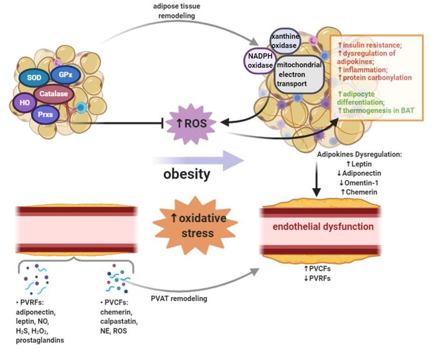
Zhou et al.                                                                                                    Adipose Oxidative Stress in Obesity

    Oxidative stress is accompanied with both obesity and CVDs.           circumference and waist to hip ratio, to indicate central
Oxidative stress occurs when the production of reactive oxygen            obesity (22).
species (ROS) exceeds the antioxidant defense (9). Normally,                  Obesity-induced oxidative stress in adipose tissue is regulated
ROS are involved in homeostatic signaling and are secondary               in a depot-specific manner. Epicardial adipose tissue, adjacent
messengers in various important intracellular signaling pathways          to coronary arteries and myocardium, produced a higher level
(10). The abnormal generation of ROS and oxidative stress                 of ROS than SAT in patients with CVDs (23). Oxidized lipids
in adipose tissue emerge as a potential pathophysiological                and proteins tended to accumulate in VAT compared to SAT
mechanism underlying vascular dysfunction. For example, the               in both obese mice and humans (24, 25). The level of aldehyde
important anticontractile function of perivascular adipose tissue         products of lipid peroxidation was increased in epididymal
(PVAT) is lost after obesity-induced oxidative stress (11).               adipose tissue but decreased in SAT in both high-fat diet-induced
Therefore, oxidative stress in adipose tissues could be targeted          obese mice and ob/ob mice (26). Furthermore, exercise training-
for the prevention and treatment of vascular dysfunction.                 mediated oxidative stress reduction, measured by decreased level
In this review, we summarize current understanding of how                 of lipid peroxidation and NADPH oxidase expression, was more
oxidative stress induced by obesity contributes to adipose                remarkable in VAT than in SAT (27).
tissue dysfunction, and further promotes the development of                   Based on function and gene expression profile, adipose tissue
vascular dysfunction (Figure 1).                                          can be further categorized into white, brown, and beige adipose
                                                                          tissue. White adipose tissue (WAT), including SAT and VAT,
OXIDATIVE STRESS IN OBESITY                                               represents most of the fat mass in humans. Brown adipose
                                                                          tissue (BAT) accounts for around 4.3% of overall fat mass
Oxidative Stress and ROS                                                  in adults and is located predominantly in the interscapular
ROS are generated during aerobic metabolism of oxygen.                    and supraclavicular region (28). BAT is highly vascularized
The primary sources of ROS include the mitochondrial                      and consists mostly brown adipocytes. Morphologically, brown
respiratory chain and oxidase enzymes, such as nicotinamide               adipocytes have numerous small lipid droplets and a much higher
adenine dinucleotide phosphate (NADPH) oxidases, xanthine                 number of mitochondria, which leads to their brown color and
oxidase (XO), lipoxygenases, cyclooxygenases, cytochrome P450             multilocular histological appearance (29). Upon cold exposure
enzymes and uncoupled nitric oxide synthases (12). Under                  or certain pharmacological stimulations, beige adipocytes can
normal conditions, small quantities of ROS, such as superoxide            be induced in WAT depots (30, 31). Beige adipocytes, similar
(O2 − ), hydrogen peroxide (H2 O2 ), hydroxyl radical (· OH) and          to brown adipocytes, exhibit multilocular lipid droplets and
peroxynitrite (ONOO− ), are neutralized by the antioxidant                are packed with a high number of mitochondria (32). BAT
defense systems. Under the pathological conditions, however,              and beige adipose tissue activations are linked to non-shivering
enzymatic production of ROS exceeds the available antioxidant             thermogenesis (28), but more importantly, their activations have
defense systems, leading to the state of oxidative stress (13). The       been shown to promote energy expenditure and improve insulin
accumulated ROS cause cellular dysfunction and tissue damage              sensitivity (33).
by direct oxidative modification of biomolecules, and modulate                In the past years, PVAT has been identified as an important
redox-sensitive signal transduction pathways and transcription            regulator of vascular functions due to its proximity to the
factors (14). For instance, the reaction between O2 − and the             vasculature (34). For a long time, PVAT was thought to
gaseous messenger nitric oxide (NO) leads to the deprivation              be a connective tissue providing mechanical protection to
of NO bioactivity, while the reaction product ONOO− causes                the surrounding vessels (35). However, with the reveal of
endothelial nitric oxide synthase (eNOS) dysfunction and hence            the endocrine and paracrine functions of PVAT, it has been
reduced NO production (13).                                               recognized that PVAT could regulate vascular tone through
                                                                          secreting adipokines, chemokines, and hormone-like factors (36).
Anatomical Features of Adipose Tissue                                     The anticontractile function of PVAT has been demonstrated in
In human body, adipose tissue can be commonly classified as               both rodents and humans (11, 37). Several studies indicated that
subcutaneous adipose tissue (SAT) and visceral adipose tissue             PVAT in different anatomic locations could resemble different
(VAT) by their anatomical localizations (15). It is now widely            adipose tissues (32). In rodents, abdominal PVAT and mesenteric
recognized that expansion of SAT has a minor contribution to              PVAT are morphologically more like WAT, whereas the thoracic
or even a positive impact on cardiometabolic diseases, while              periaortic adipose tissue resembles BAT from a morphological
expansion of VAT is significantly associated with increased               and functional standpoint (38).
cardiometabolic risks (16). Recently, it is suggested that obesity
cannot be evaluated simply by BMI, because the amount of VAT
and ectopic fat also significantly defines the risk of developing         Increased Systemic Oxidative Stress in
CVDs in overweight and moderate obese subjects (17). Although             Obesity
cumulative weight burden tends to aggravate arterial diseases in          It has been shown that obesity contributes to the development of
children and adults (18, 19), visceral fat volume has been shown          systemic oxidative stress in both human and animal studies (39,
to be more associated with systemic endothelial dysfunction               40). Systemic oxidative stress was positively correlated with fat
than subcutaneous fat (20, 21). Therefore, numerous clinical              mass in humans and rodents (39). Mitochondrial oxidative stress
studies have been using adiposity measures, including waist               markers, such as protein carbonyls, lipid peroxidation products,

Frontiers in Cardiovascular Medicine | www.frontiersin.org            2                                     March 2021 | Volume 8 | Article 650214
Zhou et al. Adipose Oxidative Stress in Obesity
FIGURE 1 | Oxidative stress occurs when the production of reactive oxygen species (ROS) exceeds the antioxidant defense. Obesity leads to the increased systemic
oxidative stress. In adipose tissue, ROS can be generated by NADPH oxidase, xanthine oxidase, and mitochondrial oxidative phosphorylation system. On one hand,
the production of ROS in adipose tissue of obese subjects leads to insulin resistance, dysregulated adipokines secretion, inflammation and increased protein
carbonylation. On the other hand, ROS in adipose tissue could promote adipocyte differentiation and thermogenesis in brown adipose tissue (BAT). A variety of
enzymes, including superoxide dismutase (SOD), catalase, glutathione peroxidases (GPx), heme oxygenase (HO), and peroxiredoxins (Prxs), can reduce ROS burden
and act as antioxidant defense in adipose tissue. Adipose tissue exerts direct effects on vascular system through releasing a wide range of bioactive products, which
include circulating adipokines. Perivascular adipose tissue (PVAT) is important adipose tissue that regulates vascular function and remodeling due to its close
proximity. In PVAT, the modulation of vascular contractility is conducted through the secretion of PVAT-derived relaxing factors (PVRFs) and PVAT-derived contracting
factors (PVCFs). In obesity, increased oxidative stress, inflammation and eNOS dysfunction in PVAT may alter the balance between PVRFs and PVCFs.
Obesity-induced PVAT dysfunction leads to the reduction of PVRFs and the production of PVCFs, hence causing enhanced vasocontraction. In addition, chronic
changes in the adipokines profile may result in the pathological vascular remodeling which can further increase the risk of CVDs.
and malondialdehyde (MDA), as well as ROS production were oxidative damage to DNA in adipose tissue (48). Obesity is also
increased in adipose tissues of obese subjects (41). In db/db accompanied with increased circulating free fatty acids (FFA).
mice, ROS production was upregulated in adipocytes, along FFA, such as palmitate, stimulate ROS production via protein
with lipid peroxidation product 4-hydroxynonenal (4-HNE) kinase C (PKC)-dependent activation of NADPH oxidase in
accumulation (42). smooth muscle cells and endothelial cells (49). In addition to
Possible triggers of obesity-induced oxidative stress include FFA, the high cytosolic triglyceride level in obesity correlates
altered nutritional status, hyperglycemia, hyperlipidemia, and to elevated levels of cytosolic long-chain acyl-CoA esters, which
chronic inflammation (43). Consumption of high-fat, high- can inhibit mitochondrial adenine nucleotide translocators and
carbohydrates food induced prolonged oxidative stress and lead to an intramitochondrial adenosine diphosphate (ADP)
inflammation in obese people (44). Moreover, lack of dietary deficiency. The ADP deficiency, in vitro, is a strong promoter of
consumption of protective antioxidant phytochemicals caused mitochondrial ROS production (50).
decreased plasma levels of vitamins and minerals in overweight Low-grade chronic inflammation is an important cause
and obese subjects, hence contributing to increased oxidative of oxidative stress in obesity (51). Although adipocytes
stress (45–47). consist mostly of the adipose tissue volume, adipose tissue
Obesity is closely related to hyperglycemia and insulin contains another heterogeneous cell population, referred to as
resistance. A strong induction of ROS accumulation in stromal vascular fractions (SVFs). SVFs include preadipocytes,
cultured adipocytes was associated with exposure of the fibroblasts, vascular endothelial cells, and immune cells.
cells to hyperglycemia condition, while in vivo study, with Accompanied with adipose tissue expansion, obesity leads to a
streptozotocin-treated hyperglycemic mice, showed an enhanced quantitative and qualitative alteration in cellular composition
Frontiers in Cardiovascular Medicine | www.frontiersin.org 3 March 2021 | Volume 8 | Article 650214Zhou et al. Adipose Oxidative Stress in Obesity
in adipose tissue. Among all the immune cells in adipose tissue, which a portion of oxygen molecules generate ROS. Complex
macrophages are the most abundant immune cells in obese I and III of the electron transport chain (ETC) are the
subjects. The recruitment and proliferation of macrophages main sites of ROS production. In complex I, electrons are
under high-fat diet condition are directly linked to adipose removed from nicotinamide adenine dinucleotide (NADH) or
tissue inflammation. There are two distinct population of the reverse reaction of complex II occurs, which leads to
macrophages, the pro-inflammatory M1 macrophages and the premature electron leakage to oxygen and ROS production in
anti-inflammatory M2 macrophages. The pro-inflammatory mitochondrial matrix. In complex III, ROS are produced in
cytokines, such as tumor necrosis factor-alpha (TNF-α), both sides of mitochondrial inner membrane (59). In obesity,
interleukin (IL)-6, and IL-1β, secreted from activated M1 the excessive nutrients accumulated in adipocytes increase
macrophages increase ROS production in adipose tissue of mitochondrial substrate loading, resulting in an enhanced ROS
obese individuals (52). Treatment of 3T3-L1 adipocytes with generation in mitochondria (60). In vitro studies showed that
TNF-α decreased the expression of mitochondrial antioxidants, high concentration of glucose or FFA increased ROS generation
including glutathione S-transferase A4 (Gsta4), peroxiredoxin 3 in mitochondria (48, 61, 62).
(Prx3), and glutathione peroxidases (GPx), therefore resulting Another enzyme system linked to adipocyte ROS generation
in increased protein carbonylation, ROS generation, and is the xanthine dehydrogenase (XDH)/oxidoreductase (XOD)
mitochondrial dysfunction (26, 53). Mice with a myeloid cell- system. Under physiological conditions, XOD dominates in the
specific NADPH oxidase 2 (Nox2) knockout showed protective form of XDH, whereas under oxidative stress, XOD converts into
effect on high-fat diet induced adipose tissue inflammation and XO, which is considered as the major source of ROS production
improved metabolic functions (54). (63). XO is an oxidant form of XOD, which converts purine
bases to uric acid (63). In a clinical study with overweight
and obese volunteers, obesity was an independent predictor of
THE ROLE OF OXIDATIVE STRESS IN high XO activity (63). Besides, obesity is often accompanied by
ADIPOSE TISSUE FUNCTION hyperuricemia (64). The production of uric acid through XOD
was enhanced in adipose tissue of obese mice (64). The process of
Sources of ROS in Adipose Tissue purine catabolism, conversion of xanthine to uric acid, generates
Several enzyme systems have been identified that can generate ROS, such as O2 − and H2 O2 , which potentially links the de novo
ROS in adipocytes. In adipose tissue, ROS can be produced by lipogenesis with ROS production (52).
NADPH oxidase (Nox), XO, and the mitochondrial oxidative
phosphorylation system. In the time course of obesity, sources Effect of ROS in Adipose Tissue
of ROS in adipose tissue may change from Nox4 primarily in Obesity-induced oxidative stress in adipose tissue is a major
adipocytes at early stage, to Nox2 in macrophages at intermediate contributor to cellular dysfunction and insulin resistance (65).
stage, and finally to mitochondria oxidative phosphorylation at Prolonged exposure of 3T3-L1 adipocytes to H2 O2 resulted in an
later stage (55). impaired insulin-induced activation of glucose transporter type 4
Nox is a multicomponent enzyme, which produces ROS when (GLUT4) (66). In obesity, the increased ROS production is closely
transferring electrons from NADPH across the cell membrane related to the dysregulation of adipokines expression in adipose
to oxygen. The Nox family consists of seven isoforms, including tissues (67). Oxidative stress in adipose tissue activated nuclear
Nox1, Nox2, Nox3, Nox4, Nox5, Duo1, and Duo2 (56). Among factor kappa-light-chain-enhancer of activated B cells (NF-κB)
the Nox family members, Nox4 is the only isoform expressed in and mitogen-activated protein kinase (MAPK), which further
adipocytes (57). During obesity development, induced by a high- downregulated anti-inflammatory adipokines and upregulated
fat and high-sucrose diet, adipocyte Nox4 and pentose phosphate pro-inflammatory cytokines in adipose tissue (67). In cultured
pathway activity were transiently increased in mice (58). Mice adipocytes, increased oxidative stress led to dysregulation of
with adipocyte-specific Nox4 deficiency were protected against adipocytokines production, including adiponectin, IL-6 and
obesity-induced insulin resistance (58). Glucose and palmitate monocyte chemotactic protein 1 (MCP1) (39).
induced ROS production in adipocytes through activation Protein carbonylation, the irreversible modification of
of Nox4 rather than mitochondrial oxidation (57). Primary proteins by reactive lipid aldehydes, is a major result of oxidative
adipocytes isolated from Nox4 knockout mice were resistant stress in adipose tissue. The most widely studied aldehyde
against high glucose or palmitate-induced inflammation (58). In products of lipid peroxidation are 4-HNE and 4-oxononenal
cultured adipocytes, fatty acids treatment promoted oxidative (4-ONE), which are abundantly present in adipose tissue (26).
stress through activation of Nox (39). In obese mice, the In obese mouse models, levels of lipid peroxidation products
expressions of Nox subunits, gp91phox, p47phox, p22phox, were increased 5 to 11-fold in epididymal adipose tissues (26).
p67phox, were upregulated in WAT (39). In addition, treatment Carbonylation of histones by 4-HNE was potentiated in adipose
with Nox inhibitor reduced ROS production in WAT of obese tissue of ob/ob mice and high-fat diet-induced obese mice (68).
mouse models, and improved obesity-induced adipose tissue Although ROS are often correlated with CVDs and negative
dysfunction (39). metabolic outcome, they also play important regulatory roles in
Mitochondrial source of ROS has been regarded as a major adipose tissue biology. 3T3-L1 cell differentiation was accelerated
trigger of oxidative stress in adipose tissue. Mitochondria after H2 O2 treatment, which was mediated by upregulation
produce energy through oxidative phosphorylation, during of peroxisome proliferator-activated receptor gamma (PPARγ)
Frontiers in Cardiovascular Medicine | www.frontiersin.org 4 March 2021 | Volume 8 | Article 650214Zhou et al. Adipose Oxidative Stress in Obesity
expression (61). Acute activation of thermogenesis in BAT antioxidant genes in epididymal adipose tissue, such as Prx3,
induced a substantial increase of mitochondrial ROS, whereas Gsta4, aldehyde dehydrogenase 1 (Aldh1), and GPx-4 (71). Prx3
pharmacological depletion of mitochondrial ROS resulted in knockout mice exhibited adipocyte hypertrophy and increased
hypothermia upon cold exposure and inhibited uncoupling mitochondrial protein carbonylation (79). 3T3-L1 cells with
protein 1 (UCP1)-dependent enhancement in whole body energy Prx3 knockdown showed increased ROS production, decreased
expenditure (69). mitochondrial potential, and lower adiponectin expression (79).
Mice, with adipocytes overexpression of antioxidant enzyme,
Antioxidant Systems in Adipose Tissue catalase and SOD1, showed adipose expansion with decreased
There are a variety of enzymes that can reduce ROS ectopic fat accumulation and improved insulin sensitivity
burden and act as antioxidant defence. These enzymes include (80). On the contrary, mice with glutamate-cysteine ligase
superoxide dismutase (SOD), catalase, heme oxygenase (HO), (Gclc) deleted in adipocytes, exhibited restricted adipose tissue
peroxiredoxins (Prxs), and GPx (13). expansion associated with increased ectopic fat accumulation
Catalase, mainly present in peroxisomes, is a cellular and deteriorated insulin sensitivity (80). In this Gclc adipocyte-
antioxidant enzyme that eliminates excessive H2 O2 . Attenuating specific knockout mice, glutathione synthesis was disabled,
ROS emission, either by treatment of antioxidant or by and ROS production was increased in adipocytes. In vitro
genetically overexpression of catalase, has been shown to studies revealed that oxidative stress resulted in suppression
improve obesity-induced metabolic disorders (70, 71). Catalase- of de novo lipogenesis, possibly through lysine K-specific
knockout mice exhibited more weight gain and higher fat demethylase 1A (KDM1A)-mediated attenuation of sterol-
mass under either normal chow or high-fat diet feeding regulatory element-binding transcription factor 1 (SRBF1)
conditions than control mice (72, 73). This phenotype of catalase- transcriptional activities (80).
knockout mice can be attenuated by concomitant treatment with Whereas, these data suggest that increasing mitochondrial
antioxidant, melatonin or N-acetyl cysteine (73). In vitro study, antioxidants protects against oxidative stress in adipose tissue,
using 3T3-L1-derived adipocytes transfected with catalase-small other studies also reveal different phenotypes. SOD catalyzes the
interfering RNA, revealed an increased lipogenesis and Nox4 dismutation of O2 − into oxygen and H2 O2 , serving as a key
expression in catalase-deficient cells (73). antioxidant. However, mice with an adipocyte-selective knockout
HO is an enzyme catalyzing the degradation of heme of SOD2, the isoform in mitochondrial matrix, exhibited
that is a pro-oxidant and strong inducer of HO1, thereby resistance to obesity induced by high-fat diet and enhanced
producing biliverdin, ferrous iron and carbon monoxide. While energy expenditure (81). The anti-obesity effect of SOD2 deletion
HO1 is the stress inducible isoform, HO2 is the constitutive in adipocytes was attributed to an activated mitochondrial
isoform that is expressed under homeostatic conditions. biogenesis and enhanced mitochondrial fatty acid oxidation (81).
Adipocyte-specific HO1 knockout caused an enhanced fasting GPx is an antioxidant enzyme family with peroxidase activity,
hyperglycemia and insulinemia in female mice, but not male which converts lipid hydroperoxides to their corresponding
mice on both standard diet and high-fat diet, indicating a alcohols and free H2 O2 to water. However, mice lacking GPx-
greater protective role of HO1 in females (74). Lentiviral- 1 were protected from high-fat diet-induced insulin resistance,
mediated adipocyte-specific overexpression of human HO1 in which was due to an improved insulin signaling in muscle cells
mice led to an increased human HO1 expression in adipose (82). Mice with hepatocyte-specific deficiency in GPx-1 showed
tissue without affecting endogenous murine HO1 (75). The an enhanced hepatic insulin sensitivity and were protected from
adipocyte-specific overexpression of HO1 attenuated high-fat diet-induced non-alcoholic steatohepatitis (83). High-fat diet-
diet induced adiposity and vascular dysfunction and improved induced glucose intolerance and hepatic steatosis were improved
insulin sensitivity and adipocyte function through modulating in mice with both GPx-1 and catalase knockout, which was due
adiponectin level and inflammation (75). to attenuated inflammation and enhanced browning in visceral
Prxs, a ubiquitous family of antioxidant enzymes, control adipose tissues (84) (Figure 2).
the cytokine-induced peroxide levels in mammalian cells. Prxs
can be regulated by phosphorylation, oxidation, reduction or
oligomerization (76). Prxs family members are classified by THE INTERPLAY OF ADIPOSE TISSUE AND
their intracellular location. Prx2, present in the cytoplasm and VASCULATURE
cell membranes, is significantly involved in intracellular redox
balance with its ROS scavenging activity (77). Prx2 expression Adipokines in Vascular Oxidative Stress
was upregulated during adipocyte differentiation. Silencing of Adipose tissue exerts direct effects on vascular systems
Prx2 in 3T3-L1 cells increased ROS production and inhibited through releasing a wide range of bioactive products, including
adipogenesis via modulating adipogenic gene expression, which adipokines [leptin (85), adiponectin (86, 87), omentin-1 (88) and
suggested that Prx2 deficiency caused adipocyte dysfunction chemerin (89)], inflammatory cytokines and chemokines (90),
and cell death via promoting ROS production (78). The gaseous messengers such as hydrogen sulfide (H2 S) (91) and NO
mitochondrial antioxidant Prx3 decreased significantly in (92), ROS (93), microRNAs (94), microvesicles (95), and fatty
adipose tissue of obese mice and humans, contributing to a acid metabolites (96).
state of oxidative stress in obesity (79). Inflammation induced by In the vasculature, these bioactive molecules are involved
obesity is associated with decreased expression of mitochondrial in regulation of (1) local redox state through alteration of
Frontiers in Cardiovascular Medicine | www.frontiersin.org 5 March 2021 | Volume 8 | Article 650214Zhou et al. Adipose Oxidative Stress in Obesity
FIGURE 2 | Functions of the antioxidant enzyme systems in adipose tissues. In adipocytes, catalase inhibits lipogenesis and Nox4 expression, which prevents weight
gain and fat mass increase induced by high-fat diet. Overexpression of catalase and superoxide dismutase (SOD)-1 in adipocytes can inhibit ectopic fat accumulation
and improve insulin sensitivity. Heme oxygenase 1 (HO1) can stimulate the expression of adiponectin, therefore preventing hyperglycemia and insulinemia in female
mice, and improving vascular function and insulin sensitivity. Peroxiredoxin (Prx)-2 can promote adipogenesis, whereas Prx3 can stimulate the expression of
adiponectin. Depletion of these antioxidant enzymes in adipocytes has shown detrimental effects in adipocyte functions and promoted cardiometabolic diseases.
Glutamate-cysteine ligase (Gclc) facilitates glutathione synthesis and inhibits ROS production, thereby inhibiting ectopic fat accumulation and insulin resistance. On the
other hand, deletion of either SOD2 or glutathione peroxidases (GPx) has been reported to provide beneficial effect in adipose tissue function. The anti-obesity effect
of SOD2 deletion in adipocytes can be attributed to an activated mitochondrial biogenesis and enhanced mitochondrial fatty acid oxidation, which can promote
energy expenditure. Insulin signaling can be enhanced by knocking down GPx in either muscle cells and hepatocytes. GPx-1 deletion can attenuate inflammation and
enhance browning in visceral adipose tissues.
Nox activity and eNOS coupling; (2) endothelial function pressure when infused chronically in rodents (102). In human
through modulation of eNOS activity and NO production; (3) umbilical vein endothelial cells (HUVEC), leptin increased the
inflammation; (4) neointima formation; (5) vascular smooth production of ROS and the expression of MCP1, indicating
muscle cell migration; (6) local endothelial cell activation that chronic oxidative stress in endothelial cells, induced
(97). On the contrary, changes in vascular biology, such as by hyperleptinemia, may contribute to vascular dysfunction
increased oxidative stress and inflammation, can also affect (103). Another study carried in bovine aortic endothelial cells
the functions of adipose tissue, particularly PVAT, in a showed that leptin increased ROS generation by promoting
bidirectional loop. Lipid peroxidation products, such as 4-HNE, fatty acid oxidation through protein kinase A activation (104).
and inflammatory cytokines produced in vascular wall can diffuse In HUVEC, exposure to leptin induced eNOS expression and
to the surrounding PVAT and activate specific signaling pathways reduced intracellular L-arginine, leading to eNOS uncoupling
in adipocytes (98, 99). accompanied with reduced NO bioavailability and increased
cytotoxic ONOO− (105). In human aortic endothelial cells,
Leptin leptin-induced eNOS activation was mediated through AMP-
Leptin is best known for its interaction with central nervous activated protein kinase (AMPK)/Akt signaling pathways, which
system to decrease food intake and increase energy expenditure could be blunted by the interaction between leptin and C-reactive
(100). However, leptin also plays an important role in endothelial protein (CRP) (106). Besides, leptin may directly mediate
functions. Obesity is associated with the increased circulating vascular tone, since leptin at a high concentration caused
leptin level, which is an independent risk factor for a number of endothelium-dependent NO-mediated vascular relaxation in
vascular diseases (101). Leptin was also shown to increase arterial vessels from control, but not obese rats (101).
Frontiers in Cardiovascular Medicine | www.frontiersin.org 6 March 2021 | Volume 8 | Article 650214Zhou et al. Adipose Oxidative Stress in Obesity
Adiponectin adipocytes, which indicates the linkage between inflammation
Adiponectin, an adipokine involved in diabetes and insulin and chemerin secretion (113).
resistance, appears to be the link between obesity and CVDs
(107). Adiponectin produced in PVAT exerts paracrine effects
on the vascular wall (11). In patients undergoing coronary PVAT Oxidative Stress in Obesity-Related
artery bypass graft surgery, circulating adiponectin level was Vascular Dysfunction
independently associated with NO bioavailability and O2 − The crosstalk between PVAT and the vasculature is important
formation/eNOS uncoupling in saphenous vein segments for vascular function. PVAT exerts its anticontractile function
and internal mammary arteries. However, vascular O2 − through secretion of PVAT-derived relaxing factors (PVRFs),
formation and eNOS uncoupling were positively correlated which include adiponectin, leptin, NO, H2 S, H2 O2 , and
with adiponectin gene expression/release in PVAT (87). prostaglandins (114, 115). In addition to PVRFs, PVAT-derived
Adiponectin improved eNOS coupling in the underlying vessels, contracting factors (PVCFs) have been recently identified. These
via induction of Akt-mediated eNOS phosphorylation and PVCFs, including chemerin, calpastatin, norepinephrine (NE)
increasing tetrahydrobiopterin (BH4 ) bioavailability (87). Ex vivo and ROS, can modulate vasoconstriction (116–118).
experiments showed that peroxidation products, such as 4-HNE, PVRFs regulate vascular tone in both endothelium-dependent
produced in the vascular wall downregulated adiponectin gene and -independent manners (119). In endothelium-dependent
expression in PVAT through a PPARγ-dependent mechanism mechanism, PVRFs modulate NO production and the
(87). Analysis of internal mesenteric artery (MA) and their activation of calcium-dependent potassium channels to facilitate
adjacent PVAT, revealed a significant correlation of type 2 vasorelaxation. On the other hand, the endothelial-independent
diabetes with both hypoadiponectinemia and increased vascular anticontractile property of PVAT may involve the generation of
O2 − production (108). H2 O2 and subsequent guanylyl cyclase activation (119). PVAT
The conserved paralog of adiponectin, complement ROS are continuously generated from the mitochondria in
C1q/tumor necrosis factor-related protein (CTRP) has response to contractile stimuli. ROS, as important mediators of
been shown to exert protective effects in the cardiovascular PVAT anticontractile effects, act directly on VSMC (120).
system (109). In diet-induced obese mice, treatment of CTRP- In obesity, PVAT dysfunction leads to the dysregulation
9 promoted phosphorylation of eNOS and reduced O2 − of PVAT-derived vasoactive factors, which in turn affects the
production and TNF-α levels in PVAT, which further improved vascular function. The anticontractile responses to vasodilators
anticontractile effect of PVAT (110). Adiponectin and CTRP-9 showed no significant differences between obese and lean mice
exert beneficial effects on vascular function through improving if the PVAT were removed from the vessels, suggesting that
eNOS coupling. Although circulating adiponectin level was obesity did not directly impair the intrinsic vascular function,
reduced in type 2 diabetic patients, adiponectin expression in but rather the function of PVAT (121). The shift toward a
PVAT was increased and positively correlated with vascular O2 − proinflammatory and prooxidative state of PVAT may lead to
production (108). Additionally, treatment of human arteries endothelial dysfunction (122). Under normal conditions, the
with recombinant adiponectin directly reduced the production deleterious effects of ROS are neutralized by the antioxidant
of O2 − by Nox (108). In high-fat diet-fed obese mice, increased enzymes in PVAT (123, 124), whereas in obesity, the increased
oxidative stress in PVAT mediated by TNF-α, resulted in loss of oxidative stress and inflammatory response, together with the
PVAT anticontractile effect (111). dysfunction of eNOS and NO result in PVAT dysfunction (125).
In rats, MA with intact PVAT showed a greater contraction
to perivascular nerve stimulation than those without PVAT,
Omentin-1
which was mediated via the production of ROS by Nox in
Omentin-1, also known as intelectin-1, is an anti-inflammatory
PVAT (123). MA incubated with aortic PVAT from high-fat
adipokine which is produced primarily by SVFs within adipose
diet-fed rats showed reduced endothelium-dependent relaxation
tissue. In diabetic Goto-Kakizaki rats fed with or without
compared to those incubated with PVAT from normal chow
high-fat diet, vascular O2 − production was decreased and
diet-fed rats (126). High-fat diet significantly increased the
NO bioavailability was improved by omentin-1 treatment (88).
mass of PVAT and the number of hypertrophic adipocytes,
Besides, omentin-1 ameliorated endothelium dysfunction in
and promoted the shift to a WAT characteristic of PVAT in
the obese diabetic rats by improving endothelium-dependent
rodents (126). In diet-induced obese mice, abdominal aortic
relaxation to acetylcholine (88).
PVAT showed increased formation of ROS, including H2 O2
and O2 − (127). Moreover, high-fat diet-fed mice showed
Chemerin reduced expression of SOD3 and glutathione levels in mesenteric
Whereas, adiponectin and CTRP9, exert beneficial effect on PVAT (128). Abdominal aorta with PVAT displayed impaired
vascular function through improving eNOS coupling, the endothelium-dependent vasodilation, whereas the vasodilation
adipokine chemerin has an opposing effect, that is to promote was restored after inhibition of ROS formation in vascular wall
eNOS uncoupling and reduce NO production in favor of or simply removal of PVAT (127). Obesity-induced oxidative
ROS (89). Chemerin has been identified as an endogenous stress in PVAT was exacerbated in aged mice, leading to
vasoconstrictor, and its expression is upregulated in hypertensive vascular oxidative stress and inflammation (129). Inflammatory
rats (112). TNF-α can stimulate chemerin production from factors secreted from PVAT of obese aged mice significantly
Frontiers in Cardiovascular Medicine | www.frontiersin.org 7 March 2021 | Volume 8 | Article 650214Zhou et al. Adipose Oxidative Stress in Obesity
promoted prooxidative and proinflammatory phenotype in POTENTIAL TARGETS MEDIATING
cultured arteries isolated from young healthy mice (129). OXIDATIVE STRESS IN ADIPOSE TISSUES
Nrf2
BAT Oxidative Stress in Obesity-Related Nuclear factor E2-related factor 2 (Nrf2), a member of the Cap-
Vascular Dysfunction n-Collar subfamily of basic leucine zipper (bZIP) transcription
BAT-specific adipokines, referred to as batokines, exert beneficial factors, regulates the expression of antioxidant proteins that
endocrine, paracrine and autocrine effects on peripheral tissues, protect against oxidative stress. Under normal conditions, Nrf2
including vascular systems (31). In high-fat diet-induced obesity, is kept in the cytoplasm by Kelch like-ECH-associated protein
BAT showed higher levels of ROS generation and enhanced 1 (Keap1) and Cullin 3, which degrade Nrf2 by ubiquitination.
antioxidant enzyme activity compared with lean counterparts Oxidative stress disrupts the Keap1-Cullin 3 ubiquitination
(130). Large amount of ROS, such as O2 − , H2 O2 , and system, leading to Nrf2 translocation to the nucleus. In the
oxidized lipids are generated in BAT upon acute activation of nucleus, Nrf2 binds to the antioxidant response element (ARE)
thermogenesis (69, 131). Mitochondrial ROS accumulated in in the upstream promoter region of many antioxidant genes
BAT converge on UCP1 C253, inducing cysteine sulfenylation and initiates the transcription, including NADPH quinone
(69). UCP1 C253A desensitizes UCP1 to adrenergic activation, oxidoreductase 1 (NQO1) and HO1 (148). In obese mouse
however, it does not impair the thermogenesis in brown models and human subjects, oxidative stress was significantly
adipocytes (69). The anticontractile effect of BAT on vasculature increased in WAT (149). The oxidative stress in WAT
was abolished in the Nox4 knockout mice. BAT could induce induced Nrf2 expression and activity, further exacerbating lipid
the activation of cyclic GMP-dependent protein kinase G accumulation in adipocytes and promoting the development of
type-1α (PKG-1α) through Nox4-derived H2 O2 , leading to a obesity (149). Oxidative stress-induced lipid accumulation in
reduced vascular contractility (93). PVAT from β3 agonist-treated WAT was reduced in Nrf2 knockout mice (149). Mechanistically,
mice exhibited a browning phenotype and an increased anti- Nrf2 mediated lipogenesis through binding to sterol regulatory
contractile effect (93). element binding protein 1 (SREBP1) promoter and inducing
Currently, there are a few well-known strategies that lipogenic gene transcription. Besides, Nrf2 also participated in
can induce browning in adipose tissues, which include cold lipolysis in adipocytes via modulating protein kinase A (PKA)
acclimation and treatment of growth factors such as fibroblast pathway (149).
growth factor 21 (FGF21) (132), atrial natriuretic peptide (ANP)
(133), and bone morphogenetic protein (BMP) (134). Cold BAMBI
acclimation can stimulate triglyceride clearance and glucose Bone morphogenetic protein and activin membrane-bound
uptake in adipose tissues, which contribute to the modulation inhibitor (BAMBI) is a transmembrane glycoprotein which is
of oxidative stress (135, 136). In mice, cold acclimation highly homologous to type I receptors of the transforming
inhibited high-fat diet-induced endothelial dysfunction and growth factor beta (TGF-β) superfamily. Therefore, BAMBI is
atherosclerosis, which were associated with a significant regarded as a pseudoreceptor of the TGF-β-related signaling
reduction in levels of proinflammatory markers (137). In pathway and acts as a negative regulator of the TGF-β signaling
high-fat diet-fed rats, cold acclimation stimulated the browning pathway (150). Altered BAMBI expression was observed during
of abdominal aortic PVAT, which was accompanied by the periods of adipose tissue remodeling (151). BAMBI is a negative
increased expression of phospho-AMPK, UCP1 and peroxisome regulator of adipogenesis through modulating anti- and pro-
proliferator-activated receptor gamma coactivator 1-alpha adipogenic effects of paracrine factors, such as Wnt-3a, TGF-β1,
(PGC-1α), as well as the reduced expression of TNF-α, IL-6, and and BMP (152, 153). A recent study showed that adipocyte-
p65 (138). specific depletion of BAMBI caused an induction of Nox4
Apart from cold exposure, a well-recognized antioxidant expression in adipose tissue, thereby promoting ROS generation
melatonin is also an activator of BAT. Melatonin, a derivative in cytoplasm and mitochondria. The deficiency of BAMBI in
of tryptophan, is secreted primarily during the dark phase adipose tissue resulted in an enhanced DNA-binding activity
of the light/dark cycle by pinealocytes. It is regarded as an of C/EBPβ and promoted adipogenesis (154). These results
important chronobiotic regulating internal biological clock together suggested that BAMBI may be an important mediator of
(139). In addition, melatonin is also involved in energy adipogenesis through regulating ROS. Therefore, manipulation
metabolism (140, 141). Melatonin supplementation prevented of BAMBI may present a new therapeutic approach to improve
rodents against obesity without affecting food intake and adipose tissue function.
physical activities (142–144). In fact, melatonin can increase
energy expenditure through activation of BAT and preserve WWP1
mitochondrial functions (145, 146). Clinically, melatonin WW domain containing E3 ubiquitin protein ligase 1 (WWP1)
also plays an important role in maintaining cardiovascular is a HECT-type ubiquitin E3 ligase that has been implicated
homeostasis through regulating blood pressure and is regarded in many pathologies. Proteome analysis of 3T3-L1 cells with
as a putative antihypertensive treatment (147). Melatonin may WWP1 overexpression revealed an increased abundance of
exert its cardiovascular protective effect through its free radical several antioxidative proteins and a reduced ROS levels, and vice
scavenger activity and indirectly through its activation of BAT. versa (155). In WWP1 knockout mice, oxidative stress markers
Frontiers in Cardiovascular Medicine | www.frontiersin.org 8 March 2021 | Volume 8 | Article 650214Zhou et al. Adipose Oxidative Stress in Obesity
were increased in WAT after high-fat diet feeding, indicating excessive ROS have been shown to be a strong risk factor of
WWP1 might participate in the antioxidative response (156). obesity-related CVDs. Targeting the abnormal ROS production
However, whole-body glucose metabolism was improved in the and oxidative stress in adipose tissues emerges as a potential
obese WWP1 knockout mice compared to wild-type controls strategy to prevent and treat CVDs. The detailed mechanisms
(156). Since the mouse model used in this study were not of the two sides of ROS function in adipose tissues should be
adipocyte-specific knockout, it cannot be ruled out that the fully studied. Antioxidant treatments may have negative effect
deficiency of WWP1 affected insulin sensitivity in other tissues on oxygen consumption and cannot prevent oxidative stress
than WAT or the secretion of insulin from pancreatic β-cells. in adipose tissues (160). Therefore, novel and target-specific
strategies should be further investigated.
Exercise In this review, we have proposed a few potential and
In rat, exercise training can enhance eNOS expression and reduce novel targets in linking obesity-induced oxidative stress and
oxidative stress in adipose tissues, thereby inducing browning functions of adipose tissue. Recent results from the Nrf2,
and thermogenic responses (157). In mice, resistance exercise WWP1, and BAMBI studies are promising, but further studies
training induced a brown-like adipocyte reprogramming in using adipocytes-specific knockout mice would provide a
WAT. The expressions of vascular endothelial growth factor more extensive investigation on their roles in adipose tissue.
(VEGF), cluster of differentiation 31 (CD31), UCP1, and Manipulation of Nrf2, BAMBI and WWP1 may present new
browning-related genes were increased in adipocytes of mice therapeutic approaches to improve adipose tissue function. In
after 8 weeks of training (158). However, a recent study showed addition, promoting browning of adipose tissues during the
that under thermoneutral conditions, exercise training did not development of obesity might be another crucial strategy to
induce browning in obese rats. These rats showed reduced prevent CVDs. The current well-known strategies to trigger
weight gain, but an oxidative signature in the BAT. Proteomics browning of adipose tissues include cold acclimation and
revealed significant changes in 2-oxoglutarate metabolic process, exercise training. However, the detailed mechanism of browning
mitochondrial respiratory chain complex IV, carbon metabolism, remains unclear. Future studies could also investigate whether
and oxidative phosphorylation in adipose tissues of obese rats Nrf2, BAMBI, and WWP1 are involved in the browning or
at thermoneutrality. The author suggested a potential exercise- thermogenic responses in adipose tissues.
induced UCP1-independent pathway which may modulate the
BAT physiology (159). AUTHOR CONTRIBUTIONS
CONCLUSION YZ wrote the initial draft of the manuscript. HL and NX critically
reviewed and edited the manuscript. All authors have read and
Obesity is characterized by the excessive accumulation of agreed to the published version of the manuscript.
fat in adipose tissue, which leads to adipocyte hypertrophy,
hypoxia, as well as the development of systemic oxidative stress. FUNDING
Excess calories intake, overloading of mitochondria, and the
generation of oxidative stress in adipose tissues lead to adipose Original works from the authors’ laboratory contributing
tissue dysfunction and insulin resistance. Since adipose tissues, to this review were supported by grants LI-1042/1-1,
especially PVAT, are responsible for the production of various LI-1042/3-1, LI-1042/5-1, and XI 139/2-1 from the Deutsche
vasoactive adipokines to modulate vascular function, obesity- Forschungsgemeinschaft (DFG), Bonn, Germany. HL and
induced adipose tissue dysfunction significantly contributes to NX were supported by a research grant from the Boehringer
the pathogenesis of CVDs. Although physiological levels of ROS Ingelheim Foundation for the collaborative research consortium
are required for adipogenic differentiation and act as secondary Novel and neglected cardiovascular risk factors: molecular
messengers of the insulin signaling pathway and vasocontraction, mechanisms and therapeutic implications.
REFERENCES 6. Taubes G. The science of obesity: what do we really know about
what makes us fat? An essay by Gary Taubes. BMJ. (2013) 346:f1050.
1. Bluher M. Obesity: global epidemiology and pathogenesis. Nat Rev doi: 10.1136/bmj.f1050
Endocrinol. (2019) 15:288–98. doi: 10.1038/s41574-019-0176-8 7. Rutkowski JM, Stern JH, Scherer PE. The cell biology of fat expansion. J Cell
2. WHO (2020). Obesity and Overweight. Available online at: https://www.who. Biol. (2015) 208:501–12. doi: 10.1083/jcb.201409063
int/news-room/fact-sheets/detail/obesity-and-overweight 8. Nakamura K, Fuster JJ, Walsh K. Adipokines: a link between
3. Fontaine KR, Redden DT, Wang C, Westfall AO, Allison DB. Years of obesity and cardiovascular disease. J Cardiol. (2014) 63:250–9.
life lost due to obesity. JAMA. (2003) 289:187–93. doi: 10.1001/jama. doi: 10.1016/j.jjcc.2013.11.006
289.2.187 9. Sena CM, Leandro A, Azul L, Seiça R, Perry G. Vascular oxidative
4. WHO (2017). Noncommunicable Diseases Progress Monitor. Available online stress: impact and therapeutic approaches. Front Physiol. (2018) 9:1668.
at: https://www.who.int/publications/i/item/WHO-NMH-NVI-17.9 doi: 10.3389/fphys.2018.01668
5. Collaborators GBDO, Afshin A, Forouzanfar MH, Reitsma MB, Sur P, Estep 10. Sies H. Hydrogen peroxide as a central redox signaling molecule in
K, et al. Health effects of overweight and obesity in 195 countries over 25 physiological oxidative stress: oxidative eustress. Redox Biol. (2017)
years. N Engl J Med. (2017) 377:13–27. doi: 10.1056/NEJMoa1614362 11:613–9. doi: 10.1016/j.redox.2016.12.035
Frontiers in Cardiovascular Medicine | www.frontiersin.org 9 March 2021 | Volume 8 | Article 650214Zhou et al. Adipose Oxidative Stress in Obesity
11. Greenstein AS, Khavandi K, Withers SB, Sonoyama K, Clancy O, 31. Villarroya F, Cereijo R, Villarroya J, Giralt M. Brown adipose
Jeziorska M, et al. Local inflammation and hypoxia abolish the protective tissue as a secretory organ. Nat Rev Endocrinol. (2017) 13:26–35.
anticontractile properties of perivascular fat in obese patients. Circulation. doi: 10.1038/nrendo.2016.136
(2009) 119:1661. doi: 10.1161/CIRCULATIONAHA.108.821181 32. Pfeifer A, Hoffmann LS. Brown, beige, and white: the new color code of fat
12. Mueller CF, Laude K, McNally JS, Harrison DG. ATVB in focus: redox and its pharmacological implications. Annu Rev Pharmacol Toxicol. (2015)
mechanisms in blood vessels. Arterioscler Thromb Vasc Biol. (2005) 25:274–8. 55:207–27. doi: 10.1146/annurev-pharmtox-010814-124346
doi: 10.1161/01.ATV.0000149143.04821.eb 33. Stanford KI, Middelbeek RJ, Townsend KL, An D, Nygaard EB, Hitchcox
13. Li H, Horke S, Forstermann U. Oxidative stress in vascular disease and KM, et al. Brown adipose tissue regulates glucose homeostasis and insulin
its pharmacological prevention. Trends Pharmacol Sci. (2013) 34:313–9. sensitivity. J Clin Invest. (2013) 123:215–23. doi: 10.1172/JCI62308
doi: 10.1016/j.tips.2013.03.007 34. Hildebrand S, Stumer J, Pfeifer A. PVAT and its relation to brown, beige, and
14. Vaziri ND, Rodriguez-Iturbe B. Mechanisms of disease: oxidative stress and white adipose tissue in development and function. Front Physiol. (2018) 9:70.
inflammation in the pathogenesis of hypertension. Nat Clin Pract Nephrol. doi: 10.3389/fphys.2018.00070
(2006) 2:582–93. doi: 10.1038/ncpneph0283 35. Szasz T, Webb RC. Perivascular adipose tissue: more than just structural
15. Wajchenberg BL. Subcutaneous and visceral adipose tissue: their support. Clin Sci. (2012) 122:1–12. doi: 10.1042/CS20110151
relation to the metabolic syndrome. Endocr Rev. (2000) 21:697–738. 36. Gollasch M, Dubrovska G. Paracrine role for periadventitial adipose tissue
doi: 10.1210/edrv.21.6.0415 in the regulation of arterial tone. Trends Pharmacol Sci. (2004) 25:647–53.
16. Pou KM, Massaro JM, Hoffmann U, Vasan RS, Maurovich-Horvat P, doi: 10.1016/j.tips.2004.10.005
Larson MG, et al. Visceral and subcutaneous adipose tissue volumes 37. Gao Y-J, Zeng Z-H, Teoh K, Sharma AM, Abouzahr L, Cybulsky I, et al.
are cross-sectionally related to markers of inflammation and oxidative (2005). Perivascular adipose tissue modulates vascular function in the
stress: the Framingham Heart Study. Circulation. (2007) 116:1234–41. human internal thoracic artery. J Thorac Cardiovasc Surg. 130, 1130–6.
doi: 10.1161/CIRCULATIONAHA.107.710509 doi: 10.1016/j.jtcvs.2005.05.028
17. Piche ME, Tchernof A, Despres JP. Obesity phenotypes, diabetes, 38. Brown NK, Zhou Z, Zhang J, Zeng R, Wu J, Eitzman DT, et al. Perivascular
and cardiovascular diseases. Circ Res. (2020) 126:1477–500. adipose tissue in vascular function and disease: a review of current research
doi: 10.1161/CIRCRESAHA.120.316101 and animal models. Arterioscler Thromb Vasc Biol. (2014) 34:1621–30.
18. Woo KS, Chook P, Yu CW, Sung RY, Qiao M, Leung SS, et al. Overweight doi: 10.1161/ATVBAHA.114.303029
in children is associated with arterial endothelial dysfunction and intima- 39. Furukawa S, Fujita T, Shimabukuro M, Iwaki M, Yamada Y, Nakajima Y, et al.
media thickening. Int J Obes Relat Metab Disord. (2004) 28:852–7. Increased oxidative stress in obesity and its impact on metabolic syndrome.
doi: 10.1038/sj.ijo.0802539 J Clin Invest. (2004) 114:1752–61. doi: 10.1172/JCI21625
19. Arkin JM, Alsdorf R, Bigornia S, Palmisano J, Beal R, Istfan N, et al. Relation 40. Wonisch W, Falk A, Sundl I, Winklhofer-Roob BM, Lindschinger
of cumulative weight burden to vascular endothelial dysfunction in obesity. M. Oxidative stress increases continuously with BMI and age
Am J Cardiol. (2008) 101:98–101. doi: 10.1016/j.amjcard.2007.07.055 with unfavourable profiles in males. Aging Male. (2012) 15:159–65.
20. Hashimoto M, Akishita M, Eto M, Kozaki K, Ako J, Sugimoto N, doi: 10.3109/13685538.2012.669436
et al. The impairment of flow-mediated vasodilatation in obese men with 41. Chattopadhyay M, Khemka VK, Chatterjee G, Ganguly A, Mukhopadhyay
visceral fat accumulation. Int J Obes Relat Metab Disord. (1998) 22:477–84. S, Chakrabarti S. Enhanced ROS production and oxidative damage
doi: 10.1038/sj.ijo.0800620 in subcutaneous white adipose tissue mitochondria in obese and
21. Parikh NI, Keyes MJ, Larson MG, Pou KM, Hamburg NM, Vita JA, et al. type 2 diabetes subjects. Mol Cell Biochem. (2015) 399:95–103.
Visceral and subcutaneous adiposity and brachial artery vasodilator function. doi: 10.1007/s11010-014-2236-7
Obesity. (2009) 17:2054–9. doi: 10.1038/oby.2009.60 42. Boyer F, Diotel N, Girard D, Rondeau P, Essop MF, Bourdon E. Enhanced
22. Abraham TM, Pedley A, Massaro JM, Hoffmann U, Fox CS. Association oxidative stress in adipose tissue from diabetic mice, possible contribution
between visceral and subcutaneous adipose depots and incident of glycated albumin. Biochem Biophys Res Commun. (2016) 473:154–60.
cardiovascular disease risk factors. Circulation. (2015) 132:1639–47. doi: 10.1016/j.bbrc.2016.03.068
doi: 10.1161/CIRCULATIONAHA.114.015000 43. Manna P, Jain SK. Obesity, oxidative stress, adipose tissue dysfunction, and
23. Salgado-Somoza A, Teijeira-Fernandez E, Fernandez AL, Gonzalez-Juanatey the associated health risks: causes and therapeutic strategies. Metab Syndr
JR, Eiras S. Proteomic analysis of epicardial and subcutaneous adipose tissue Relat Disord. (2015) 13:423–44. doi: 10.1089/met.2015.0095
reveals differences in proteins involved in oxidative stress. Am J Physiol Heart 44. Patel C, Ghanim H, Ravishankar S, Sia CL, Viswanathan P, Mohanty P, et al.
Circ Physiol. (2010) 299:H202–9. doi: 10.1152/ajpheart.00120.2010 Prolonged reactive oxygen species generation and nuclear factor-kappaB
24. Frohnert BI, Sinaiko AR, Serrot FJ, Foncea RE, Moran A, Ikramuddin S, et al. activation after a high-fat, high-carbohydrate meal in the obese. J Clin
Increased adipose protein carbonylation in human obesity. Obesity. (2011) Endocrinol Metab. (2007) 92:4476–9. doi: 10.1210/jc.2007-0778
19:1735–41. doi: 10.1038/oby.2011.115 45. Reitman A, Friedrich I, Ben-Amotz A, Levy Y. Low plasma antioxidants and
25. Hauck AK, Zhou T, Hahn W, Petegrosso R, Kuang R, Chen Y, et al. normal plasma B vitamins and homocysteine in patients with severe obesity.
Obesity-induced protein carbonylation in murine adipose tissue regulates Isr Med Assoc J. (2002) 4:590–3.
the DNA-binding domain of nuclear zinc finger proteins. J Biol Chem. (2018) 46. Kaidar-Person O, Person B, Szomstein S, Rosenthal RJ. Nutritional
293:13464–76. doi: 10.1074/jbc.RA118.003469 deficiencies in morbidly obese patients: a new form of malnutrition? Part A:
26. Long EK, Olson DM, Bernlohr DA. High-fat diet induces changes vitamins. Obes Surg. (2008) 18:870–6. doi: 10.1007/s11695-007-9349-y
in adipose tissue trans-4-oxo-2-nonenal and trans-4-hydroxy-2-nonenal 47. Kaidar-Person O, Person B, Szomstein S, Rosenthal RJ. Nutritional
levels in a depot-specific manner. Free Radic Biol Med. (2013) 63:390–8. deficiencies in morbidly obese patients: a new form of malnutrition? Part B:
doi: 10.1016/j.freeradbiomed.2013.05.030 minerals. Obes Surg. (2008) 18:1028–34. doi: 10.1007/s11695-007-9350-5
27. Sakurai T, Izawa T, Kizaki T, Ogasawara JE, Shirato K, Imaizumi K, et al. 48. Lin Y, Berg AH, Iyengar P, Lam TK, Giacca A, Combs TP, et al.
Exercise training decreases expression of inflammation-related adipokines The hyperglycemia-induced inflammatory response in adipocytes: the
through reduction of oxidative stress in rat white adipose tissue. Biochem role of reactive oxygen species. J Biol Chem. (2005) 280:4617–26.
Biophys Res Commun. (2009) 379:605–9. doi: 10.1016/j.bbrc.2008.12.127 doi: 10.1074/jbc.M411863200
28. Leitner BP, Huang S, Brychta RJ, Duckworth CJ, Baskin AS, McGehee S, et al. 49. Inoguchi T, Li P, Umeda F, Yu HY, Kakimoto M, Imamura M,
Mapping of human brown adipose tissue in lean and obese young men. Proc et al. High glucose level and free fatty acid stimulate reactive oxygen
Natl Acad Sci USA. (2017) 114:8649–54. doi: 10.1073/pnas.1705287114 species production through protein kinase C–dependent activation of
29. Enerback S. The origins of brown adipose tissue. N Engl J Med. (2009) NAD(P)H oxidase in cultured vascular cells. Diabetes. (2000) 49:1939–45.
360:2021–3. doi: 10.1056/NEJMcibr0809610 doi: 10.2337/diabetes.49.11.1939
30. Harms M, Seale P. Brown and beige fat: development, function and 50. Bakker SJ, RG IJ, Teerlink T, Westerhoff HV, Gans RO, Heine RJ. Cytosolic
therapeutic potential. Nat Med. (2013) 19:1252–63. doi: 10.1038/nm.3361 triglycerides and oxidative stress in central obesity: the missing link between
Frontiers in Cardiovascular Medicine | www.frontiersin.org 10 March 2021 | Volume 8 | Article 650214You can also read