Brainstem dysfunction in critically ill patients - Arca
←
→
Page content transcription
If your browser does not render page correctly, please read the page content below
Benghanem et al. Critical Care (2020) 24:5
https://doi.org/10.1186/s13054-019-2718-9
REVIEW Open Access
Brainstem dysfunction in critically ill
patients
Sarah Benghanem1,2 , Aurélien Mazeraud3,4, Eric Azabou5, Vibol Chhor6, Cassia Righy Shinotsuka7,8, Jan Claassen9,
Benjamin Rohaut1,9,10† and Tarek Sharshar3,4*†
Abstract
The brainstem conveys sensory and motor inputs between the spinal cord and the brain, and contains nuclei of
the cranial nerves. It controls the sleep-wake cycle and vital functions via the ascending reticular activating system
and the autonomic nuclei, respectively. Brainstem dysfunction may lead to sensory and motor deficits, cranial nerve
palsies, impairment of consciousness, dysautonomia, and respiratory failure. The brainstem is prone to various
primary and secondary insults, resulting in acute or chronic dysfunction. Of particular importance for characterizing
brainstem dysfunction and identifying the underlying etiology are a detailed clinical examination, MRI,
neurophysiologic tests such as brainstem auditory evoked potentials, and an analysis of the cerebrospinal fluid.
Detection of brainstem dysfunction is challenging but of utmost importance in comatose and deeply sedated
patients both to guide therapy and to support outcome prediction. In the present review, we summarize the
neuroanatomy, clinical syndromes, and diagnostic techniques of critical illness-associated brainstem dysfunction for
the critical care setting.
Keywords: Brainstem dysfunction, Brain injured patients, Intensive care unit, Sedation, Brainstem reflexes, Disorders
of consciousness, Autonomic nervous system, Neurological respiratory failure, Immune reflex, Auditory and
somatosensory evoked potentials and electroencephalogram
Introduction: the concept of brainstem as structural and non-structural origin. Brainstem dys-
dysfunction function can then contribute to impairment of con-
The brainstem is the caudal portion of the brain that sciousness, cardiocirculatory and respiratory failure, and
connects the diencephalon to the spinal cord and the thus increased mortality [2–5].
cerebellum [1]. The brainstem mediates sensory and In the present review, we describe brainstem func-
motor pathways between the spinal cord and the brain tional neuroanatomy, clinical syndromes, and assessment
and contains nuclei of the cranial nerves, the ascending methods before addressing the concept of critical illness-
reticular activating system (ARAS), and the autonomic associated brainstem dysfunction.
nuclei. It controls the brainstem reflexes and the sleep-
wake cycle and is responsible for the autonomic control
of the cardiocirculatory, respiratory, digestive, and im- Functional neuroanatomy of the brainstem
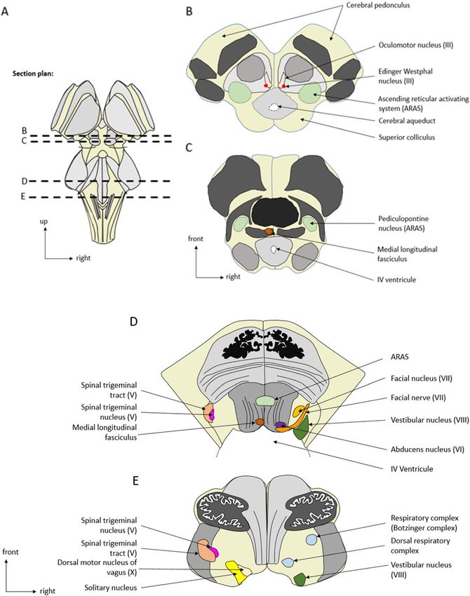
mune systems. Brainstem dysfunction may result from The brainstem can be categorized into three major parts:
various acute or chronic insults, including stroke, infec- midbrain, pons, and medulla oblongata (Figs. 1 and 2).
tious, tumors, inflammatory, and neurodegenerative dis- The brainstem contains both gray and white matter,
eases. In the context of critical illness, the brainstem can with the basilar artery representing the vascular supply.
be susceptible to various insults that can be categorized The gray matter includes the nuclei of the cranial nerves
(anterior part), the ARAS (posterior part), the extra-
pyramidal and the central autonomic nervous system
* Correspondence: t.sharshar@ghu-paris.fr
†
Benjamin Rohaut and Tarek Sharshar contributed equally to this work.
(ANS). This gray matter controls brainstem reflexes,
3
Department of Neuro-ICU, GHU-Paris, Paris-Descartes University, Paris, France arousal, automatic movements, and homeostasis, re-
4
Laboratory of Experimental Neuropathology, Pastuer Institute, Paris, France spectively. The white matter is composed of ascending
Full list of author information is available at the end of the article
© The Author(s). 2019 Open Access This article is distributed under the terms of the Creative Commons Attribution 4.0
International License (http://creativecommons.org/licenses/by/4.0/), which permits unrestricted use, distribution, and
reproduction in any medium, provided you give appropriate credit to the original author(s) and the source, provide a link to
the Creative Commons license, and indicate if changes were made. The Creative Commons Public Domain Dedication waiver
(http://creativecommons.org/publicdomain/zero/1.0/) applies to the data made available in this article, unless otherwise stated.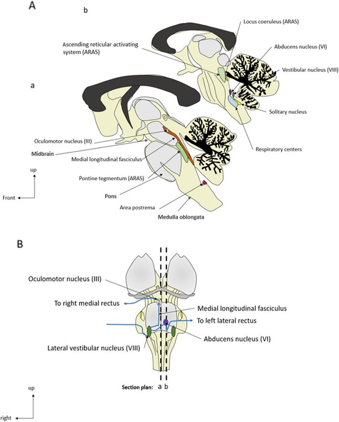
Benghanem et al. Critical Care       (2020) 24:5                                                                                        Page 2 of 14

 Fig. 1 General anatomy of the brainstem and oculocephalic circuit. A Anatomical sagittal sections. B Representation of the sagittal section plans
 and of the oculocephalic circuit (ventral part)

sensory pathways and descending pyramidal and extra-                        or the spinal cord [6]. While the former controls volun-
pyramidal pathways (Table 1).                                               tary movement, the latter is involved in reflexes, motion,
                                                                            complex movements, and postural control (Tables 1
Brainstem syndromes and assessment                                          and 2). Upper motor neuron damage can lead to symp-
Brainstem pathology should be considered in cases of (a)                    toms, ranging from hemiparesis to the locked-in
sensory or motor deficits combined with cranial nerve                       syndrome, which is typically characterized by intact
palsy, (b) impairment of consciousness, (c) dysautono-                      awareness, quadriplegia, anarthria, and absence of eye
mia, or (d) neurological respiratory failure.                               movements except for preserved vertical gaze. It usually
                                                                            results from bilateral pontine white matter lesions [7].
Brainstem motor and sensory deficits and cranial nerve                      Characteristic clinical features of brainstem lesions in-
palsy                                                                       clude ipsilateral cranial nerve palsies or cerebellar signs
The pyramidal and extrapyramidal tracts connect the                         combined with contralateral motor deficits. Brainstem
upper motor neurons and the extrapyramidal nuclei with                      lesions may present with abnormal movements, such as
the lower motor neurons located in either the brainstem                     hemichorea, hemiballism, dystonia, tremor, asterixis,
Benghanem et al. Critical Care (2020) 24:5 Page 3 of 14 Fig. 2 Anatomical axial sections of the brainstem. a Representation of the brainstem (dorsal part) and of the axial sections plans. b, c Midbrain axial sections. d Pons axial sections. e Medulla oblongata axial sections pseudo-athetosis, and non-epileptic myoclonus [8] diagnosis of third nerve lesion (i.e., mydriasis) or (Table 2). Bilateral motor corticobulbar tract lesion may Horner’s syndrome (i.e., myosis, ptosis, enophtalmia, and present with swallowing impairment, dysphagia, dyspho- anhidrosis). Pupillary light, corneal, oculocephalic, and nia, velo-pharyngo-laryngeal impairments, uncontrol- gag reflexes are routinely assessed in the critical care set- lable crying/laughing episodes, and emotional lability ting. The oculovestibular responses and oculocardiac are (i.e., pseudobulbar affect; Table 2). A brainstem lesion of less frequently tested, except to determine brain death. the posterior column-medial lemniscus pathway and the The absence of brainstem reflexes and spontaneous spinothalamic tract results in a contralateral propriocep- breathing is a prerequisite for the diagnosis of brain tive/touch and temperature/pain deficit, respectively. death [9]. Automated pupillometry could improve the The testing of the cranial nerves and brainstem re- assessment of the pupil light reflex and thereby its prog- flexes is described in Table 3. Abnormal spontaneous nostic value [10]. Corneo-mandibular reflexes can be de- eye position and movements may be encountered in pa- tected in acute brain injury, but its prognostic relevance tients with brainstem lesions and can be seen in coma- remains controversial. Finally, assessments of primitive tose patients. Assessment of pupillary size allows the reflexes are less relevant in the ICU context but can
Benghanem et al. Critical Care       (2020) 24:5                                                                                       Page 4 of 14

Table 1 Functional neuroanatomy for the intensivist
          Anatomic structures                                                                      Function
Gray      Nuclei of cranial nerves                                                                 Brainstem reflexes
matter
          Nuclei of ascending reticular activating system (ARAS)                                   Arousal, sleep/wake cycles, and alertness
          Nuclei of the extrapyramidal system                                                      Automatic movements
          Nuclei of the central autonomic system                                                   Vital function regulation and homeostasis
White     Axons of ascending pathways:                                                             Sensory information:
matter    Posterior column-medial lemniscus pathway                                                Fine touch, vibration, two-point discrimination,
          Spinothalamic tract and lateral lemniscus pathway                                        and proprioception
                                                                                                   Pain and temperature
          Axons of descending pathways:                                                            Voluntary motor control
          Pyramidal corticospinal and corticobulbar tracts                                         Reflexes, locomotion, complex movements, and
          Extrapyramidal tract (rubrospinal, pontine and medullary reticulospinal tract, lateral   postural control
          vestibulospinal and tectospinal tracts)

be seen in patients with neurodegenerative disease                            functional disability in ICU survivors, and hospital mor-
(Table 3).                                                                    tality [15]. Brainstem dysfunction could account for
  When suspecting brainstem lesions, MRI will have the                        some features of delirium, such as fluctuations in arousal
highest yield to further localize and characterize brain-                     and attentional impairment that could be related to
stem lesions [6] (Table 4). Evoked potentials may be also                     ARAS and to ponto-mesencephalic tegmentum dysfunc-
useful for detecting a brainstem lesion. EEG [11] may be                      tion, respectively. Other states of acute impairment of
supportive in patients with abnormal movements and                            consciousness include clouding of consciousness and
disorders of consciousness, and cerebrospinal fluid (CSF)                     stupor, but they are less frequently used [14].
analysis for those with suspected inflammatory or infec-                         Subacute or chronic disorders of consciousness in-
tious diseases.                                                               clude the vegetative state (VS, also called Unresponsive
                                                                              Wakefulness Syndrome) defined as state of unrespon-
Impairment of consciousness                                                   siveness in which the patient shows spontaneous eye
The ARAS controls the sleep-wake cycle and includes                           opening without any behavioral evidence of self or envir-
several nuclei mainly located in the pontine and mid-                         onmental awareness [17]. The minimally conscious state
brain tegmentum [12] (Table 2, Figs. 1 and 2): the rostral                    (MCS) is defined as state of severely impaired conscious-
raphe complex, the parabrachial nucleus, the laterodor-                       ness with minimal behavioral evidence of self or envir-
sal tegmental nucleus, the locus coeruleus (LC), the nu-                      onmental awareness, characterized by the presence of
cleus pontis oralis, the basal forebrain, and the thalamus.                   non-reflexive behavior (visual pursuit, appropriate motor
Monoaminergic neurons are directly linked to the cortex                       response to painful stimulus) or even intermittent com-
and are inhibited during deep sleep. Cholinergic pedun-                       mand following indicating a cortical integration [18, 19].
culopontine and laterodorsal tegmental nuclei are indir-                      The VS and MCS are related to a preservation of brain-
ectly connected to the cortex via the thalamus and                            stem arousal functions but with persistent impairment
remain active during rapid eye movement sleep. These                          of supratentorial networks implicated in consciousness
pathways are modulated by hypothalamic neurons [13].                          [20]. Stimulation of the ARAS may improve conscious-
  Disorders of consciousness can be organized between                         ness in vegetative or MCS patients [21]. In addition to
acute and subacute or chronic [14]. Acute impairments                         deep brain stimulation, vagal nerve stimulation, which
of consciousness include coma which is defined as a                           probably modulates the activity of the nucleus of the
“state of unresponsiveness in which the patient lies with                     tractus solitarius and the dorsal raphe, has shown
eyes closed and cannot be aroused to respond appropri-                        promising results [22].
ately to stimuli even with vigorous stimulation” [14].                           In addition to these classical syndromes, other con-
The association of a prolonged non-responsive coma                            sciousness impairments have been described. Peduncular
with a complete cessation of brainstem reflexes and                           lesions can cause hallucinations [23] which may be en-
functions suggests the diagnosis of brain death which is                      countered in ICU patients. More generally speaking, it is
defined as an irreversible loss of all functions of the                       likely that brainstem dysfunctions account for a portion
entire brain. Delirium is defined as an acute and fluctua-                    of the sleep-wake cycle impairments experienced by ICU
ting disturbance of consciousness, including attention                        patients. Brainstem lesions can induce cognitive deficits
and impairment of cognition, associated with motor                            including impaired attention, naming ability, executive
hyperactivity or hypoactivity [15, 16]. Delirium has been                     functioning, and memory impairment [24], ascribed to a
associated with long-term cognitive impairment,                               disruption of interconnection between the frontal-
Benghanem et al. Critical Care (2020) 24:5 Page 5 of 14
Table 2 Functional anatomy of the brainstem
Brainstem structures Functions Centers Symptoms
Midbrain (rostral to the Eye movements Cranial nerve nuclei: Oculomotor signs:
pons and caudal to the III oculomotor nerve (mainly Ptosis (III)
thalamus and the basal motor) Ophthalmoplegia (III, IV)
ganglia) IV trochlear nerve (motor)
Pupillary size: sphincter pupillae and Cranial nerve nuclei: Pupillary anomalies:
muscles of the ciliary body, pupil light III oculomotor nerve Myosis (sympathetic lesion)
reflex Mydriasis (parasympathetic lesion)
Anisocoria
Movement control Substantia nigra Parkinsonian syndrome and movement
disorders (hemichorea, hemiballism,
dystonia, tremor, asterixis, pseudo-
athetosis, non-epileptic myoclonus)
Posture tone Red nucleus Postural tone impairment
Posture/auditory and visual integration Accessory optic tractus Balance disorder
Posture and movement integration Tectum (dorsal part) Balance disorder
Posture and inhibitor motor centers Tegmentum (ventral portion) Involuntary movements
(basal ganglia and thalamus
connections)
Sleep/wake cycles, alertness, and arousal ARAS: composed of almost 100 Sleep disturbance
nuclei, including locus coeruleus- Consciousness disorders
raphe nuclei with neocortex
connections
Central thermic regulation ARAS-hypothalamus connections Hypo/hyperthermia
Pons (between the Facial sensitivity, muscles of mastication Cranial nerve nuclei: Facial symptoms:
medulla and the midbrain) V trigeminal nerve (sensory and Facial dysesthesia
motor) Oculomotor signs:
Corneal/ciliary reflex impairment
Facial muscles and taste from the Cranial nerve nuclei: Facial symptoms:
anterior 2/3 of the tongue (VII) VII facial nerve (sensory and Peripheral facial palsy
motor)
Eye movement (abduction) Cranial nerve nuclei: Oculomotor signs:
VI abducens nerve (motor) Ophthalmoplegia
Posture, sensation of rotation, gravity, Cranial nerve nuclei: Altered audition (VIII)
and sound VIII vestibulocochlear nerve Balance disorders (VIII and cerebellum
(mostly sensory) tract)
Cerebellum tract
Posture Spinocerebellar tracts Cerebellar ataxia
Posture and inhibitor motor center Tegmentum (thalamus and basal Involuntary movement
nuclei connections)
Motor efference integration Tracts carrying signals to the Motor deficit
Sensory efference integration thalamus Sensory deficit
Consciousness, alertness, and sleep Tracts carrying signals to the Sleep disturbance
regulation thalamus Consciousness disorders
Sleep/wake cycles, alertness, and arousal ARAS: composed of almost 100 Sleep disturbance
nuclei, including raphe nuclei and Consciousness disorders
locus coeruleus-raphe nuclei-
neocortex connections
Emotion ARAS: locus coeruleus and Anxiety and post-traumatic stress disorder
amygdala connections (PTSD)
Central thermic regulation ARAS-hypothalamus connections Hypo/hyperthermia
Respiratory drive: respiratory rate and Pedunculopontine tegmentum, Respiratory drive dysfunction:
tidal volume control locus coeruleus, lateral Kölliker-Fuse and parabrachial nuclear:
parabrachial respiratory group, increase tidal volume, decrease respiratory
and Kölliker-Fuse nuclei rate
Lower part/ponto-peduncular injury:
respiratory asynchronism
Medulla (lower half of the Taste from the posterior 1/3 of the Cranial nerve nuclei: Tongue sensory impairmentBenghanem et al. Critical Care (2020) 24:5 Page 6 of 14
Table 2 Functional anatomy of the brainstem (Continued)
Brainstem structures Functions Centers Symptoms
brainstem, connects the tongue IX glossopharyngeal (sensory and
higher levels of the brain motor)
to the spinal cord)
Pharyngo-laryngeal reflex Cranial nerve nuclei: Oro-pharyngo-laryngeal anomalies:
IX glossopharyngeal nerve Dysphagia (swallowing impairment)
X vagus nerve (sensory and Dysphonia
motor) Velo-pharyngo-laryngeal impairment
XI spinal nerve (motor) Absence of pharyngeal/gag reflex
Glossal muscles XII hypoglossal (mainly motor) Tongue motor impairment (fasciculation,
motor deficit)
Cough IX glossopharyngeal nerve Absence of cough reflex (IX, X)
X vagus nerve
Posture Spinocerebellar tracts Cerebellar ataxia
Regulation of autonomic nervous Sympathetic nuclei Autonomic dysfunction
system: Parasympathetic nuclei: vagus
nerve (X) control of the heart,
lung, digestive tracts
Cardiac regulation Sympathetic nuclei Oculocardiac reflex impairment (X)
Parasympathetic nuclei: vagus Dysautonomia: tachycardia
nerve (X) control of the heart, (parasympathetic impairment),
lung, digestive tracts bradycardia (sympathetic impairment),
sudden death
Vasomotor regulation Hemodynamic failure:
Dysautonomia with hypertension
(parasympathetic impairment),
hypotension (sympathetic impairment)
Gastrointestinal motility Gastrointestinal motility anomalies
Respiratory drive: respiratory rate and Respiratory centers: dorsal Respiratory drive dysfunction: respiratory
tidal volume control respiratory complex rate irregularities and ataxic breathing,
hyperventilation, respiratory-ventilator
asynchronism, central apnea
Microbiota gut-brain axis, senses and Vagus nerve (X) Maladaptive immune response, gut-brain
peripheral inflammation modulation axis impairment
Tracts all along the Connection of the oculomotor nerves Medial longitudinal fasciculus Internuclear ophthalmoplegia
brainstem (see Fig. 1)
Motor information from the periphery Corticospinal tract Motor deficit, locked-in syndrome
to supratentorial structures Pyramidal and extrapyramidal Tetrapyramidal and extrapyramidal
tracts syndromes with movement disorders
(tremor)
Non-epileptic myoclonus
Sensory information from the periphery Posterior column-medial lemnis- Sensory deficit
to supratentorial structures cus pathway and spinothalamic
tracts
Oculosympathetic control Centers control of the ciliary Horner’s syndrome (ptosis, myosis,
nerve, superior tarsal muscle, enophtalamos, anhidrosis)
pupillary sphincter/dilator
subcortical system and the brainstem [1]. Finally, deep the corneal, pupil light, and cough reflexes and respira-
sedation is a pharmacologically induced coma, and its tory patterns [26]. In comatose patients, pupil sizes and
mechanisms of action involve the brainstem GABA and reactivity can be suggestive of particular etiologies, such
N-methyl-D-aspartate (NMDA) receptors [25]. as drug overdose (myosis for opioids or mydriasis for
Assessments of consciousness are based on neuro- tricyclic anti-depressants). In comatose brain-injured
logical examination to confirm the diagnosis, determine patients, brainstem reflex assessment is crucial to detect
the underlying cause, and evaluate the prognosis. In a uncal or downward cerebellar (tonsillar) herniation
clinical practice, this assessment most commonly relies [10]. While the absence of corneal and pupillary light
on the Glasgow Coma Scale (GCS) [20]. Focusing on the reflexes is strongly associated with poor outcome in
brainstem in particular, the FOUR (Full Outline of Un- post-anoxia, their prognostic value is less validated in
Responsiveness) score is to be preferred as it includes other causes [27].Benghanem et al. Critical Care (2020) 24:5 Page 7 of 14
Table 3 Brainstem reflexes neuroanatomical and clinical description
Reflex Examination technique Normal response Afferent pathway Brainstem centers Efferent pathway
Physiological reflexes
Pupillary light Response to light Direct and consensual Retina, optic nerve, Pupillo-constrictor: midbrain, pretectal Sympathetic fibers of
reflex myosis followed by chiasma, optic tract olivary nucleus, Edinger-Westphall cranial nerve III
mydriasis nucleus (oculomotor)
Pupillo-dilator: posterior-lateral hypo-
thalamus, cervical ganglion, trigeminal
ganglion, abducens
Cilio-spinal Latero-cervical nociceptive Uni- or bilateral irido- Sensory ascending Midbrain Cranial nerve III
stimulation dilatation pathways to centro-
spinal centers
Fronto-orbicular Glabellar percussion Eyes closing Cranial nerve V Pons Cranial nerve VII
(trigeminal) (facial)
Oculocephalic Turn head from side to side Eyes move conjugately Semicircular canals, Pons, nucleus vestibularus, nucleus Cranial nerves III
in direction opposite to Cranial nerve VIII abducens (oculomotor) and VI
head (oculovestibular) (abducens)
Oculovestibular Irrigate external auditory canal Nystagmus
with 50 ml of cold water
Corneal Stimulation of cornea with Eyelid closure Cranial nerve V Pons, trigeminal and facial nuclei Cranial nerve VII
saline drops (trigeminal) (facial)
Grimace/ Deep pressure on nail bed, Facial grimace and
masseterian supraorbital ridge, or temporo- limb movement
mandibular joint
Cough reflex Stimulation of the carina with a Cough Cranial nerve IX Medulla, nucleus tractus solitarius Cranial nerve IX
suction tube (Glossopharyngeal) (glossopharyngeal)
and X (vagal) and X (vagal)
Gag/pharyngeal Stimulation of the soft palate Symmetrical rise of soft
reflex palate gag reflex
Oculocardiac Ocular globe compression Decrease in heart rate Cranial nerve V Pons, medulla Cranial nerve X
(trigeminal) (vagal)
Primitive reflexes
Palmo-mental Pressure of the thenar Single twitch of the Posterior column- Pons Cranial nerve VII
eminence with a thin stick ipsilateral mentalis medial lemniscus (facial)
muscle pathway
Corneo- Corneal stimulation Contralateral deviation Cranial nerve V Pons Cranial nerve
mandibular of the jaw (trigeminal) VII (facial)
Other syndromes
Internuclear Oculomotricity testing Disconjugate lateral Lesion of the medial Connects the sixth nucleus with the
ophthalmoplegia Can be observed during gaze with a preserved longitudinal fasciculus contralateral third nucleus
(see Fig. 1) oculocephalogyric or convergence
oculovestibular tests
Claude Bernard- Ptosis, myosis, enophtalamos, Sympathetic pathway
Horner’s anhidrosis injury
syndrome
Vertical Midbrain or medulla
nystagmus, skew injury
deviation
Ocular bobbing Pons injury
Patients with severe critical illness may be comatose Coma due to structural brainstem lesions is predomin-
due to sedation, which in clinical practice can be antly related to pedunculo-pontine tegmental lesions,
assessed using the RASS (Richmond Agitation Sedation usually detected on MRI [12] (Table 4). Neurophysio-
Scale) [28]. In deeply sedated patients (i.e., RASS − 4 or logical tests may be useful to assess the neurological
− 5), the Brainstem Reflexes Assessment Sedation prognosis in patients with impairment of consciousness.
Scale (BRASS) might be useful to assess the effect Somatosensory evoked potentials (SSEP) assess conduc-
of sedatives on the brainstem and potentially detect tion from peripheral nerves (N9) to the somatosensory
a brainstem dysfunction [29] (Table 5). The CAM- cortical (N20) regions passing through the brainstem
ICU and ICDSC are appropriate to monitor deli- (P14). Brainstem auditory evoked potentials (BAEP) are
rium [16, 30]. Finally, in VS and MCS patients, the described in Table 6 [11]. Interestingly, sedation in-
Coma Recovery Scale-Revised has also been vali- creases latencies and decreases amplitudes of evoked po-
dated [20]. tentials in a dose-dependent manner but does probablyBenghanem et al. Critical Care (2020) 24:5 Page 8 of 14
Table 4 Acute and chronic diseases involving the brainstem Table 5 Brainstem Reflexes Assessment Sedation Scale (BRASS)
Causes of brainstem dysfunction Variable Score point
Acute primary insult Absence of cough reflex 1
Vascular injury Absence of pupillary light reflex 1
Ischemic: thrombotic or cardio-embolic, lacunar ischemia due to Absence of corneal reflex 2
small vessel disease, vasculitis
Absence of grimacing to pain and absence of OCR 1
Hemorrhage
Absence of grimacing to pain and presence of OCR 3
Inflammatory
OCR: oculocephalic reflex
Multiple sclerosis (MS) BRASS is a clinical score that has been developed for scoring brainstem
dysfunction in deeply sedated, non-brain-injured, mechanically ventilated,
Acute disseminated encephalomyelitis (ADEM) critically ill patients and ranges from 0 to 7
The BRASS has prognostic value, as 28-day mortality proportionally increases
Neuromyelitis optica (NMO) (anti-MOG, anti-AQP4 antibodies, or
with the BRASS score
seronegative types)
Birkenstaff encephalitis (anti-ganglioside GQ1b antibodies)
Behcet disease and rarely other autoimmune disease (lupus, neuro- outcome [36]. Wave I can disappear if the auditory nerve
sarcoidosis) is injured (traumatic or hypoxic injuries) [37].
Langerhans cell histiocytosis Reactivity on EEG to auditory, visual, or nociceptive
Traumatic: direct or indirect injury stimuli is important to assess after cardiac arrest because
its absence is associated with poor outcome [38, 39]. Ab-
Metabolic: central pontine myelinolysis
sent reactivity can result from a thalamus-brainstem
Infectious: rhombencephalitis, abscess, Listeria monocytogenes and
enterovirus 68 and 71, followed by herpes simplex viruses and
loops and ARAS dysfunction [40–43]. The electro-
tuberculosis, Epstein-Barr virus (EBV), and human herpesvirus 6 (HHV6) physiological measurement of the blink reflex (Table 6)
Paraneoplastic (anti-neuronal NMDA, AMPA, GABA, CASPR2, Hu, Ma2, is a way to study the trigemino-facial loop [44], but its
Ri, Yo, CV2, amphiphysin, Lgi1,glycine, mGluR1/5, VGKC/VGCC, GAD prognostic value in comatose patients remains insuffi-
antibodies) ciently supported [45].
Chronic primary insult
Tumoural Autonomic nervous system impairment
Degenerative/atrophic injury The ANS plays a key role in homeostasis and allostasis
MRI magnetic resonance imaging, TDM tomodensitometry, CSF cerebrospinal
by controlling vital functions and the immune system
fluid, ECG electrocardiogram [46] and is composed of sympathetic (e.g., noradrener-
MRI results according to etiologies: gic) and parasympathetic (e.g., cholinergic) systems.
Vascular injury: diffusion and FLAIR-weighted sequence hyperintensity
restricted to a vascular territory Sympathetic effects originate from the spinal cord (D1
Hemorrhage: SWI/T2* sequence hypointensity to L3), while parasympathetic neuronal cell bodies are
Inflammatory: diffuse or multifocal white matter lesions on T2- and FLAIR-
weighted sequences, with or without contrast enhancement
present in the nuclei of cranial nerves III (Edinger West-
Inflammatory NMO (MRI of optical nerve and medullary MRI): extensive and phal nuclei), VII, IX, and X and the sacral spinal cord
confluent myelitis on more than three vertebrae and optical neuritis with (S2 to S4). Activation of the parasympathetic nervous
possible contrast enhancement
Traumatic injury: hyperintensity on diffusion sequence, diffuse axonal injuries system results in a decrease in heart rate (HR) and blood
on DTI (diffusion tensor imaging) sequence, hemorrhage lesions on T2*/SWI pressure (BP), and an increase in gastrointestinal tonus,
Metabolic: T2 hyperintensity specifically involves the central pons
Infectious: abscess/nodes with contrast enhancement vesical detrusor contraction, and myosis. Activation of
Paraneoplastic: limbic encephalitis with temporal diffusion and the sympathetic system results in opposite effects. Cor-
FLAIR hyperintensity
Tumor: mass with possible necrosis, contrast enhancement and oedema
tical input can modulate responses in the ANS [46] as
revealed by a FLAIR hyperintensity around tumor well as various receptors throughout the body, including
Degenerative injury: brain and brainstem atrophy (colibri sign) the baroreceptors [47].
Brainstem injury may cause dysautonomic symptoms,
not change the amplitudes with low to moderate doses which can be life-threatening [48] (Table 2). Cardiac ar-
used in ICU [31]. rhythmias frequently occur after brainstem stroke and
The intracranial conduction time and intrapontine are associated with increased mortality [48]. An intracra-
conduction time are assessed by measures of the P14– nial hypertension-induced midbrain insult can impair
N20 inter-peak latency on SSEP and the III–V inter- parasympathetic control and thereby induce adrenergic
peak latency on BAEP [11]. The prognostic value of storm. In brain death, there is a disappearance of the
BAEP has been explored in various causes of coma [32– vasomotor tone and an impairment of myocardial con-
34]. After cardiac arrest, the predictive value of BAEP tractility [49]. As exhaustive discussions of tests that
for poor outcomes is limited [35]. However, in traumatic allow testing of the ANS are beyond the scope of this
brain injury, preserved BAEP are associated with a good review, we will focus on cardiovascular testsBenghanem et al. Critical Care (2020) 24:5 Page 9 of 14
Table 6 BAEP waves and blink test nerves (motor neurons present in the V, VII, and XII
BAEP waves Anatomic localization nuclei) and contractor/pump muscles (diaphragm, inter-
I Distal portion of the auditory costal, sternocleidomastoid, abdominal muscles) that are
nerve innervated by spinal motor neurons. They are controlled
II Proximal portion of the auditory by bulbospinal (automatic command) and corticospinal
nerve or cochlear nuclear complex, (voluntary command) pathways. The respiratory drive
in the upper part of the medulla,
ipsilateral to the stimulation side
originates from neurons of the latero-rostro-ventral me-
dulla oblongata, which includes the pre-Botzinger com-
III Cochlear nucleus or superior
olivary complex in caudal pontine plex and the parafacial respiratory group that control
tegmentum, ipsilateral to the inspiration and expiration, respectively [52] (Table 2).
stimulation side This center receives various inputs to automatically ad-
IV Superior olivary complex (lateral just the respiratory drive to metabolic and mechanic
lemniscus), contralateral to the changes [53]. Metabolic inputs are mediated by both
stimulation side
peripheral (aortic and carotid) and central (medulla
V Inferior colliculus located in the
midbrain, contralateral to the
oblongata and LC) chemoreceptors [54]. The mechanical
stimulation side inputs are mediated by mechanoreceptors localized in
Blink test Response the pulmonary parenchyma, bronchial wall, and muscle.
At the level of the pons, the pedunculopontine tegmen-
After stimulation of the R1 response generated at the level
supraorbital nerve, three of the pons, R2 responses at the tum, the LC, the lateral parabrachial and Kölliker-Fuse
responses are recorded on level of the trigeminal-spinal tract nuclei are involved in the automatic respiratory control
eyelid orbicular muscles: an early at the pons level, the medulla [55] (Table 2).
ipsilateral (R1) response and the oblongata, and the caudal
two (ipsi- and contralateral) late trigeminal-spinal nucleus Automatic and voluntary control of respiratory motor
responses (R2) neurons can be injured together or separately. For in-
Brainstem lesions can result in absent or delayed peaks III and V, prolonged stance, automatic control is impaired in central congeni-
III–V and I–V inter-peak latency, or a reduced I/V amplitude ratio (< 0.5) tal and acquired hypoventilation syndrome (i.e., Ondine
Delay or absence of R1 indicates a facial /trigeminal nerve injury. R2 can be
delayed in comatose patient and is also bilaterally delayed or absent in syndrome), while voluntary control is preserved [56].
Wallenberg’s syndrome (with a R1 preserved) Acquired hypoventilation syndrome can result from
brainstem tumoral, traumatic, ischemic, and inflamma-
applicable to ICU patients. Standard monitoring tory injuries [57], which implies the need for long-term
allows for the detection of variations in HR and BP mechanical ventilation.
that can be suggestive of dysautonomia. However, the Ventilator management may be significantly affected
lack of apparent changes in cardiovascular signals by brainstem lesions, and importantly, clinical features
does not rule out dysautonomia, which can be then of neurological respiratory dysfunction are related to the
assessed with the HR and BP spectral analysis. High localization of brainstem injury. The more caudal the le-
frequency (HF) band (i.e., 0.15 to 0.4 Hz) variability of sion is, the more it is associated with an impairment of
the HR is thought to predominantly reflect para- the respiratory drive. Midbrain injuries do not usually
sympathetic tone, while low frequency (LF) variability affect the respiratory rate (RR). Injuries to the upper
(i.e., 0.04 to 0.15 Hz) is primarily mediated by sympa- pons increase the tidal volume and decrease the RR,
thetic activity. The LF/HF ratio reflects the sympatho- while injuries of the lower pons are associated with re-
vagal balance. Therefore, spectral analysis allows spiratory asynchrony (e.g., ponto-peduncular injury).
studying the sympathetic, parasympathetic, and baro- Ataxic breathing (irregular pauses and apnea periods)
reflex activities both at rest and during stimulation and central apnea are observed in rostro-ventral medulla
[50]. If the Valsalva maneuver, the cold pressure test, oblongata injuries and associated with poor outcomes.
and the pharmacological tests (with yohimbine or clo- Central neurogenic hyperventilation results from activa-
nidine) allow testing the ANS, their use in ICU is tion of the medullary respiratory center. Finally, yawning
very limited. Conversely, pupillometry is much more or refractory hiccups may be seen with lesions of the
applicable for assessing dysautonomia in ICU. Thus, posterolateral medulla oblongata [58]. Swallowing im-
patients with dysautonomia present a pupil dilatation pairment contributes also to the difficulty of weaning
at resting state and a slow redilatation time [51]. mechanical ventilation and can be an indication for a
tracheostomy.
Neurogenic respiratory failure There are various structural and non-structural causes
There are two types of muscles that play a major role in of neurological respiratory dysfunction, including infra-
the respiratory system, dilatator muscles of the superior tentorial lesions, drug toxicity, heart failure, and sepsis
airway that are innervated by the brainstem via cranial [59–61]. Diagnosis relies on standard assessments ofBenghanem et al. Critical Care (2020) 24:5 Page 10 of 14
respiratory function (e.g., ventilator curves, tidal volumes section, we will discuss evidence for brainstem dysfunc-
(Vt), and RR in mechanically ventilated patients) but also tion encountered in critically ill patients beyond primary
on assessing the ventilatory response to hypercapnia brainstem dysfunction.
(e.g., during a t-piece trial). An electromyogram of the
respiratory muscles, notably the diaphragm, provides Clinical features
relevant information on the central drive. This technique The “brainstem dysfunction” hypothesis originates from
may be helpful in patients that are impossible to wean our study on usefulness of neurological examination in
from mechanical ventilation. As a caveat, it may be at non-brain-injured critically ill patients who required
times difficult to differentiate central respiratory dys- deep sedation. These patients have usually a severe crit-
function from critical illness neuropathy/myopathy. ical illness and therefore a higher risk to develop severe
EMG and nerve conduction studies may help with secondary brain insult [3, 29]. Furthermore, protracted
the distinction, but limited assessments of every re- deep sedation is still required in more than 30% of critic-
spiratory muscle group and available at highly special- ally ill patients [63] and has been reported to be associ-
ized units limit this approach [62]. In mechanically ated with increased mortality [63]. We found that
ventilated patients, spirography can be performed assessment of brainstem reflexes was reproducible in
(with the Vt/inspiration duration (Ti) ratio reflects this population [3, 29]. We also found that routinely
the ventilatory command intensity) as well as the oc- used sedative and analgesic agents such as midazolam
clusion pressure measurement (i.e., P0.1). The latter and fentanyl do not impair pupillary light, corneal, and
reflects the “unconscious”/central respiratory com- cough reflexes in 90% of cases but depress oculocephalic
mand, but variability of its measurements limits rou- response and grimacing to painful stimulation (absent in
tine application. 50 and 70%, respectively) [3, 29, 63]. The cessation of
brainstem reflexes results from the combining effects of
Brainstem dysfunction in critically ill patients critical illness (i.e., secondary brain insult), sedative, and
The leading causes of primary brainstem dysfunction are analgesic agents. It is interesting to note that Guedel ob-
summarized in Table 4 and major differential diagnosis served more than 70 years ago that sedative drugs abol-
of brainstem dysfunction in Table 7. In the following ish brainstem reflexes according to a sequential pattern
Table 7 Differential diagnosis of brainstem dysfunction
Brainstem dysfunction Differential diagnosis
Oculomotor anomalies (III, IV, VI cranial Cranial nerve palsy
nerves nuclei) Myopathy involving oculomotor muscles
Neuromuscular disorders: myasthenia, Lambert-Eaton syndrome and botulism
Pupillary size anomalies Anisocoria: compressive lesion of the III cranial nerve such as herniation/intracranial hypertension
and posterior communicative artery aneurysm
Mydriasis: third nerve lesion
Claude Bernard-Horner’s syndrome (ptosis, myosis, Pancoast tumor
enophtalmia, anhidrosis) Carotid or aortic dissection
Facial sensory anomalies (V cranial nerve nucleus) Contralateral brain injury
Cranial nerve palsy (V)
Facial motor anomalies (VII cranial nerve nucleus) Contralateral brain injury
Cranial nerve palsy (VII)
Myopathy with facial paralysis
Neuro-muscular disorders: myasthenia, Lambert-Eaton syndrome and botulism
Posture and movement anomalies Uni- or bilateral basal ganglia lesions
Motor and/or sensory deficit Contralateral brain injury
Critical illness neuromyopathy
Guillain-Barre syndrome
Motor deficit Myopathy
Neuro-muscular disorders: myasthenia, Lambert-Eaton syndrome and botulism
Autonomic (sympathetic and parasympathetic) Spine injury
dyfunctions Guillain-Barre syndrome
Respiratory control anomalies Cervical spine injury (C3–C5)
Phrenic nerve palsy
Diaphragmatic injury
Critical illness neuromyopathy
Neuromuscular disorders: myasthenia, Lambert-Eaton syndrome and botulismBenghanem et al. Critical Care (2020) 24:5 Page 11 of 14
(the loss of consciousness, followed by the cessation of Critical illness is also associated with decreased vari-
brainstem reflexes in a rostro-caudal way until apnea) [64]. ability in HR and BP, with an impaired sympathetic tone
In deeply sedated non-brain-injured critically ill pa- and baroreflex [2, 50] and also with a reduced tidal vol-
tients, the cessation of brainstem responses follows ume variability [66] that can correlate with weaning fail-
two distinct patterns. The first is characterized by a ure. Since most of these findings concerned sedated
depression of whole brainstem responses (similar to patients, one may argue that sedative agents might be in-
Guedel’s description), and the second is characterized volved as a revealing or aggravating underlying insults.
by a preferential impairment of the corneal reflex, the This hypothesis is further supported by the fact that in-
pupillary light reflex, and to a lesser extent the cough crease in evoked potential latencies cannot be only
reflex, with paradoxical preservation of the oculoce- ascribe to sedation since long-term swallowing disorders
phalic response. The latter profile is associated with [67] and aspiration pneumonia are more frequent in sep-
the severity of critical illness and the depth of sed- sis survivors [68].
ation. Interestingly, this pattern cannot be ascribed to Thus, a multimodal assessment of brainstem dys-
a unique focal brainstem lesion which most likely re- function in critical illness is warranted. The under-
lies on a functional rather than a structural origin. going multicenter PRORETRO study (ClinicalTrials.gov:
This suggests that some neuroanatomical centers are NCT02395861) aims to evaluate a multimodal approach
more sensitive to deep sedation, critical illness, or based on neurological examination and neurophysiological
both. Opioids might also contribute to brainstem dys- tests.
function, as they depress the ARAS, respiratory cen-
ters, and brainstem reflexes (notably pupillary light Mechanisms of brainstem dysfunction
and cough reflexes). However, morphine infusion Neuroimaging and neuropathological studies show that
rates did not differ in our study between the two ces- the brainstem is prone to vascular, inflammatory, and
sation patterns of brainstem reflexes [29]. excitotoxic insults [5]. For instance, sepsis can be associ-
To assess brainstem reactivity in deeply sedated ated with impaired autoregulation of cerebral blood flow
critically ill patients, we developed the BRASS [29] and microcirculatory dysfunction, which may comprom-
(Table 5). The principle of the BRASS development is ise the brainstem perfusion. Second, a multifocal necro-
not in agreement with the traditional paradigm of tizing leukoencephalopathy involving the brainstem can
Jackson, which states that the brainstem reflexes are be secondary to an intense systemic inflammatory re-
abolished in a rostro-caudal way. It thus differs from sponse [69]. Finally, the neuro-inflammatory process can
the FOUR score [65], which conditions the assess- culminate in neuronal apoptosis, which is evidenced in
ment of the cough reflex to the cessation of the brainstem autonomic nuclei in patients who died from
pupillary light and corneal reflexes. Besides improving septic shock or in experimental sepsis [5]. Interestingly,
the prediction of mortality in deeply sedated patients, it has been shown that apoptosis of autonomic nuclei
the assessment of brainstem reflexes, with help of can induce hypotension in septic rat [70].
either the BRASS or the FOUR score, might prompt Both humoral and neural pathways can induce a neuro-
the ICU physician to perform a brainstem imaging. It inflammatory process. The former involves the area post-
is however likely that the processes involved in rema (Fig. 1), which allows the diffusion of circulating in-
critical illness-related brainstem dysfunction are radio- flammatory mediators into the brainstem; the latter
logically assessable. involves mainly the vagal nerve, which mediates the trans-
mission of peripheral inflammatory signals to the brain-
Electrophysiological, autonomic, and respiratory features stem [71, 72]. Autonomic brainstem nuclei are regulated
of brainstem dysfunction by these two pathways, which then play a major role in
Neurophysiological tests provide further arguments for the control of systemic inflammatory response.
brainstem dysfunction in critically ill patients without Finally, metabolic processes can be involved. It is well
primary brainstem injury. For instance, EEG is not react- known that electrolyte disturbances but also renal and
ive in 25% of patients with sepsis [42, 43], knowing that liver failure impair brainstem responses, as illustrated by
absence of reactivity can result from a dysfunction of the centro-pontine myelinolysis or by usefulness of FOUR
ARAS [40–43]. Middle latency BAEP responses and score in hepatic encephalopathy [73].
SSEP latencies were increased in 24% and 45% of deeply
sedated non-brain-injured critically ill patients, respect- Prognostic value of brainstem dysfunction and
ively [34], indicating an impairment of the brainstem therapeutic perspective
conduction. Interestingly, mean values of these latencies The predictive value of the neurological examination
did not differ from those recorded in deeply sedated findings and neurophysiological responses has been
brain-injured patients. assessed in critically ill patients. There is a proportionalBenghanem et al. Critical Care (2020) 24:5 Page 12 of 14
relationship between the BRASS value and mortality. However, we shall remind that rivastigmine, a cholin-
Interestingly, absence of a grimacing response associated esterase inhibitor, is deleterious in critically ill patients.
with preserved oculocephalic responses is the most pre- Vagal nerve stimulation is also proposed in refractory
dictive of mortality [29], suggesting that prediction is status epilepticus [83] and consciousness disorders [22],
better when first based on a combination of signs, and suggesting its potential but not yet demonstrated effect
second, a decoupling process between the upper and in critical illness-related encephalopathy. Beta-blockers
lower part of the brainstem is involved [29]. The absence reduce the mortality in cardiac diseases by attenuating
of EEG reactivity and of SSEP P14 response and the deleterious effects of sympathetic hyperactivation
increased P14–N20 SSEP latencies are associated with and increasing the vagal tone [84]. In sepsis, beta-
increased mortality [34, 42, 43]. Impaired HR variability blockers improve HR control, reduce systemic inflam-
and decreased sympathetic control are associated with mation, and decrease mortality, acknowledging that their
mortality and organ failure [74]. routine use is not yet warranted [85, 86].
There are arguments for a relationship between delir-
ium and brainstem dysfunction. The drugs currently Conclusion
used for treating delirium are involving brainstem recep- Brainstem dysfunction can present with central sensory
tors. Thus, neuroleptics are antagonists of the dopamine and motor deficits, cranial nerve palsies and abnormal
D2 and serotoninergic 5HT2A receptors that are preva- brainstem reflexes, disorders of consciousness, respiratory
lent in the brainstem [75]. Dexmedetomidine is a select- failure, and dysautonomia. Clinical examination is essen-
ive agonist of alpha-2 receptor, notably at the level of tial for detecting a brainstem dysfunction that may be sup-
the LC [76]. The role of the brainstem in patients with ported by neuroimaging, electrophysiological, autonomic,
delirium is supported by these pharmacological data and and respiratory assessments. Brainstem dysfunction
further supported by neuropathological findings that mainly results from secondary insult and might contribute
demonstrate hypoxic and ischemic insults of the pons in to critical illness-related mortality, organ dysfunction, im-
delirious patients [77]. Absent oculocephalic responses mune dysregulation, delayed awakening, and delirium.
and delayed middle-latency BAEP have been associated The assessment of the brainstem should then be included
with delayed awakening or delirium after sedation dis- in the routine neuromonitoring of critically ill patients.
continuation [34]. In neuroanatomical point of view, it is
likely that cessation of the oculocephalic response re- Abbreviations
ADEM: Acute disseminated encephalomyelitis; ANS: Autonomic nervous
flects a dysfunction of the ARAS while cessation of the system; ARAS: Ascending reticular activating system; BAEP: Brainstem
cough reflex reflects a dysfunction of the cardiovascular auditory evoked potentials; BP: Blood pressure; BRASS: Brainstem Reflexes
and respiratory autonomic nuclei. Finally, if conceivable, Assessment Sedation Scale; CAM-ICU: Confusion Assessment Method for the
ICU; CRS-R: Coma Recovery Scale-Revised; CSF: Cerebrospinal fluid;
we do not know to what extent brainstem dysfunction ECG: Electrocardiogram; EEG: Electroencephalogram; EMG: Electromyogram;
contributes to long-term post-ICU mortality and func- FOUR: Full Outline of UnResponsiveness; GABA: Gamma-aminobutyric acid;
tional disability. GCS: Glasgow Coma Scale; GRpF: Parafacial respiratory group; HF: High
frequency; HR: Heart rate; ICCT: Intracranial conduction time; ICDSC: Intensive
Another contributing factor of the brainstem dysfunc- care delirium screening checklist; ICU: Intensive care unit; IPCT: Intrapontine
tion in critical illness course might be the impaired conduction time; IPL: Inter-peak latencies; LC: Locus coeruleus; LF: Low
sympatho-vagal control of the inflammatory response. frequency; MCS: Minimally conscious state; MRI: Magnetic resonance
imaging; MS: Multiple sclerosis; MSA: Multiple systematrophy; NMDA: N-
The vagus nerve first senses and modulates peripheral methyl-D-aspartate; NMO: Neuromyelitis optica; P0.1: Occlusion pressure
inflammation, constituting the so-called cholinergic re- measurement; PCR: Polymerase chain reaction; PreBotC: Pre-Botzinger
flex [78]; second, it senses the microbiota metabolites, complex; PTSD: Post-traumatic stress disorder; RASS: Richmond Agitation
Sedation Scale; RE: Rhombencephalitis; RR: Respiratory rate;
being a major component of the gut-brain axis [79] SSEP: Somatosensory evoked potentials; TDM: Tomodensitometry;
(Table 2). The adrenergic system controls the immune Ti: Inspiration duration; Vt: Tidal volume
system, with alpha and beta-1 receptors being pro-
inflammatory and beta-2 receptors anti-inflammatory Acknowledgements
We thank the reviewers for their comments and suggestions to improve our
[80]. It is therefore conceivable that a brainstem-related manuscript.
neuro-immune impairment can contribute to infection,
organ failure, or death by facilitating a maladapted im- Authors’ contributions
SB, AM, BR, and EA drafted the manuscript. JC, CRS, VB, and TS critically
mune response. The modulation of the cholinergic reflex
revised the manuscript for important intellectual content. All authors read
by α7nAChR agonists and by vagal nerve stimulation and approved the final manuscript.
has been proposed in sepsis and critical illness to im-
prove peripheral immune response and reduce organ Funding
None
dysfunction [81]. In addition to its peripheral immune
effects, cholinergic modulation and vagal stimulation can Availability of data and materials
promote anti-inflammatory microglial polarization [82]. Not applicableBenghanem et al. Critical Care (2020) 24:5 Page 13 of 14
Ethics approval and consent to participate assessment method for the intensive care unit (CAM-ICU). JAMA. 2001;
Not applicable 286(21):2703–10.
17. Laureys S, Celesia GG, Cohadon F, Lavrijsen J, León-Carrión J, Sannita WG,
Consent for publication et al. Unresponsive wakefulness syndrome: a new name for the vegetative
Not applicable state or apallic syndrome. BMC Med. 2010;8:68.
18. Naccache L. Reply: Response to « Minimally conscious state or cortically
Competing interests mediated state? ». Brain J Neurol. 2018;141(4):e27.
The authors declare that they have no competing interests. 19. Giacino JT, Ashwal S, Childs N, Cranford R, Jennett B, Katz DI, et al. The
minimally conscious state: definition and diagnostic criteria. Neurology.
Author details 2002;58(3):349–53.
1
Department of Neurology, Neuro-ICU, Sorbonne University, APHP 20. Giacino JT, Katz DI, Schiff ND, Whyte J, Ashman EJ, Ashwal S, et al. Practice
Pitié-Salpêtrière Hospital, Paris, France. 2Medical ICU, Cochin Hospital, AP-HP, guideline update recommendations summary: disorders of consciousness:
Paris, France. 3Department of Neuro-ICU, GHU-Paris, Paris-Descartes report of the Guideline Development, Dissemination, and Implementation
University, Paris, France. 4Laboratory of Experimental Neuropathology, Subcommittee of the American Academy of Neurology; the American
Pastuer Institute, Paris, France. 5Department of Physiology, Clinical Congress of Rehabilitation Medicine; and the National Institute on Disability,
Neurophysiology Unit, APHP, Raymond Poincaré Hospital, University of Independent Living, and Rehabilitation Research. Arch Phys Med Rehabil.
Versailles Saint Quentin en Yvelines, Garches, France. 6Department of 2018;99(9):1699–709.
Intensive Care Medicine, Saint-Joseph Hospital, Paris, France. 7Intensive Care 21. Bourdillon P, Hermann B, Sitt JD, Naccache L. Electromagnetic brain
Unit and Postgraduate Program, Instituto Nacional de Câncer, Rio de Janeiro, stimulation in patients with disorders of consciousness. Front Neurosci.
Brazil. 8D’Or Institute for Research and Education, Rio de Janeiro, Rio de 2019;13:223.
Janeiro, Brazil. 9Department of Neurology, Neuro-ICU, Columbia University, 22. Corazzol M, Lio G, Lefevre A, Deiana G, Tell L, André-Obadia N, et al.
New York, NY, USA. 10Institut du Cerveau et de la Moelle épinière, ICM, Restoring consciousness with vagus nerve stimulation. Curr Biol CB. 2017;
INSERM UMRS 1127, CNRS UMR 7225, Pitié- Salpêtrière Hospital, Paris 27(18):R994–6.
F-75013, France. 23. Geddes MR, Tie Y, Gabrieli JDE, McGinnis SM, Golby AJ, Whitfield-Gabrieli S.
Altered functional connectivity in lesional peduncular hallucinosis with REM
Received: 30 May 2019 Accepted: 23 December 2019 sleep behavior disorder. Cortex J Devoted Study Nerv Syst Behav janv. 2016;
74:96–106.
24. Fu X, Lu Z, Wang Y, Huang L, Wang X, Zhang H, et al. A clinical research
References study of cognitive dysfunction and affective impairment after isolated
1. Hurley RA, Flashman LA, Chow TW, Taber KH. The brainstem: anatomy, brainstem stroke. Front Aging Neurosci. 2017;9:400.
assessment, and clinical syndromes. J Neuropsychiatry Clin Neurosci. 2010; 25. Brown EN, Lydic R, Schiff ND. General anesthesia, sleep, and coma. N Engl J
22(1):iv 1-7. Med. 2010;363(27):2638–50.
2. Annane D, Trabold F, Sharshar T, Jarrin I, Blanc AS, Raphael JC, et al. 26. Wijdicks EFM, Bamlet WR, Maramattom BV, Manno EM, McClelland RL. Validation
Inappropriate sympathetic activation at onset of septic shock: a spectral of a new coma scale: the FOUR score. Ann Neurol. 2005;58(4):585–93.
analysis approach. Am J Respir Crit Care Med août. 1999;160(2):458–65. 27. Nolan JP, Soar J, Cariou A, Cronberg T, Moulaert VRM, Deakin CD, et al.
3. Sharshar T, Porcher R, Siami S, Rohaut B, Bailly-Salin J, Hopkinson NS, et al. European Resuscitation Council and European Society of Intensive Care
Brainstem responses can predict death and delirium in sedated patients in Medicine guidelines for post-resuscitation care 2015: section 5 of the
intensive care unit. Crit Care Med août. 2011;39(8):1960–7. European Resuscitation Council guidelines for resuscitation 2015.
4. Sharshar T, Gray F, Lorin de la Grandmaison G, Hopkinson NS, Ross E, Resuscitation. 2015;95:202–22.
Dorandeu A, et al. Apoptosis of neurons in cardiovascular autonomic 28. Ely EW, Truman B, Shintani A, Thomason JWW, Wheeler AP, Gordon S, et al.
centres triggered by inducible nitric oxide synthase after death from septic Monitoring sedation status over time in ICU patients: reliability and validity of
shock. Lancet Lond Engl. 2003;362(9398):1799–805. the Richmond Agitation-Sedation Scale (RASS). JAMA. 2003;289(22):2983–91.
5. Mazeraud A, Pascal Q, Verdonk F, Heming N, Chrétien F, Sharshar T. 29. Rohaut B, Porcher R, Hissem T, Heming N, Chillet P, Djedaini K, et al.
Neuroanatomy and physiology of brain dysfunction in sepsis. Clin Chest Brainstem response patterns in deeply-sedated critically-ill patients predict
Med. 2016;37(2):333–45. 28-day mortality. PLoS One. 2017;12(4):e0176012.
6. Quattrocchi CC, Errante Y, Rossi Espagnet MC, Galassi S, Della Sala SW, 30. Bergeron N, Dubois MJ, Dumont M, Dial S, Skrobik Y. Intensive Care
Bernardi B, et al. Magnetic resonance imaging differential diagnosis of Delirium Screening Checklist: evaluation of a new screening tool. Intensive
brainstem lesions in children. World J Radiol. 2016;8(1):1–20. Care Med. 2001;27(5):859–64.
7. Smith E, Delargy M. Locked-in syndrome. BMJ. 2005;330(7488):406–9. 31. Agarwal S, Morris N, Der-Nigoghossian C, May T, Brodie D. The influence of
8. Handley A, Medcalf P, Hellier K, Dutta D. Movement disorders after stroke. therapeutics on prognostication after cardiac arrest. Curr Treat Options
Age Ageing Mai. 2009;38(3):260–6. Neurol. 2019;21(12):60.
9. Citerio G, Murphy PG. Brain death: the European perspective. Semin Neurol. 32. Amantini A, Grippo A, Fossi S, Cesaretti C, Piccioli A, Peris A, et al. Prediction
2015;35(2):139–44. of « awakening » and outcome in prolonged acute coma from severe
10. Payen J-F, Isnardon S, Lavolaine J, Bouzat P, Vinclair M, Francony G. traumatic brain injury: evidence for validity of short latency SEPs. Clin
Pupillometry in anesthesia and critical care. Ann Fr Anesth Reanim. 2012; Neurophysiol Off J Int Fed Clin Neurophysiol. 2005;116(1):229–35.
31(6):e155–9. 33. Haupt WF, Hojer C, Pawlik G. Prognostic value of evoked potentials and
11. André-Obadia N, Zyss J, Gavaret M, Lefaucheur J-P, Azabou E, Boulogne S, clinical grading in primary subarachnoid haemorrhage. Acta Neurochir.
et al. Recommendations for the use of electroencephalography and evoked 1995;137(3–4):146–50 discussion 150.
potentials in comatose patients. Neurophysiol Clin Clin Neurophysiol. 2018; 34. Azabou E, Rohaut B, Heming N, Magalhaes E, Morizot-Koutlidis R,
48(3):143–169. Kandelman S, et al. Early impairment of intracranial conduction time
12. Parvizi J, Damasio AR. Neuroanatomical correlates of brainstem coma. Brain predicts mortality in deeply sedated critically ill patients: a prospective
J Neurol. 2003;126(Pt 7):1524–36. observational pilot study. Ann Intensive Care. 2017;7(1):63.
13. Saper CB, Scammell TE, Lu J. Hypothalamic regulation of sleep and circadian 35. De Santis P, Lamanna I, Mavroudakis N, Legros B, Vincent J-L, Creteur J,
rhythms. Nature. 2005;437(7063):1257–63. et al. The potential role of auditory evoked potentials to assess prognosis in
14. Plum F, Posner JB. The diagnosis of stupor and coma. Contemp Neurol Ser. comatose survivors from cardiac arrest. Resuscitation. 2017;120:119–24.
1972;10:1–286. 36. García-Larrea L, Artru F, Bertrand O, Pernier J, Mauguière F. The combined
15. Girard TD, Jackson JC, Pandharipande PP, Pun BT, Thompson JL, Shintani AK, monitoring of brain stem auditory evoked potentials and intracranial pressure in
et al. Delirium as a predictor of long-term cognitive impairment in survivors coma. A study of 57 patients. J Neurol Neurosurg Psychiatry. 1992;55(9):792–8.
of critical illness. Crit Care Med. 2010;38(7):1513–20. 37. Sohmer H, Freeman S, Gafni M, Goitein K. The depression of the auditory
16. Ely EW, Inouye SK, Bernard GR, Gordon S, Francis J, May L, et al. Delirium in nerve-brain-stem evoked response in hypoxaemia--mechanism and site of
mechanically ventilated patients: validity and reliability of the confusion effect. Electroencephalogr Clin Neurophysiol. 1986;64(4):334–8.You can also read