Involvement of Neuro-Immune Interactions in Pruritus With Special Focus on Receptor Expressions
←
→
Page content transcription
If your browser does not render page correctly, please read the page content below
REVIEW
                                                                                                                                           published: 18 February 2021
                                                                                                                                       doi: 10.3389/fmed.2021.627985

                                              Involvement of Neuro-Immune
                                              Interactions in Pruritus With Special
                                              Focus on Receptor Expressions
                                              Aylin Ruppenstein 1 , Maren M. Limberg 1 , Karin Loser 2 , Andreas E. Kremer 3 ,
                                              Bernhard Homey 4 and Ulrike Raap 1,5*
                                              1
                                               Division of Experimental Allergy and Immunodermatology, Faculty of Medicine and Health Sciences, University of Oldenburg,
                                              Oldenburg, Germany, 2 Division of Immunology, Faculty of Medicine and Health Sciences, University of Oldenburg,
                                              Oldenburg, Germany, 3 Department of Medicine 1, University Hospital Erlangen and Friedrich-Alexander-University
                                              Erlangen-Nürnberg, Erlangen, Germany, 4 Department of Dermatology, Heinrich-Heine-University of Düsseldorf, Düsseldorf,
                                              Germany, 5 University Clinic of Dermatology and Allergy, Oldenburg Clinic, Oldenburg, Germany

                                              Pruritus is a common, but very challenging symptom with a wide diversity of
                                              underlying causes like dermatological, systemic, neurological and psychiatric diseases.
                                              In dermatology, pruritus is the most frequent symptom both in its acute and chronic
                                              form (over 6 weeks in duration). Treatment of chronic pruritus often remains challenging.
                                              Affected patients who suffer from moderate to severe pruritus have a significantly
                          Edited by:          reduced quality of life. The underlying physiology of pruritus is very complex, involving a
                      Sonja Ständer,          diverse network of components in the skin including resident cells such as keratinocytes
University Hospital Münster, Germany
                                              and sensory neurons as well as transiently infiltrating cells such as certain immune
                       Reviewed by:
                         Ethan Lerner,        cells. Previous research has established that there is a significant crosstalk among the
     Massachusetts General Hospital,          stratum corneum, nerve fibers and various immune cells, such as keratinocytes, T cells,
                         United States
                       Laurent Misery,
                                              basophils, eosinophils and mast cells. In this regard, interactions between receptors on
               Université de Bretagne         cutaneous and spinal neurons or on different immune cells play an important role in
                 Occidentale, France          the processing of signals which are important for the transmission of pruritus. In this
                  Simone Garcovich,
     Catholic University of the Sacred        review, we discuss the role of various receptors involved in pruritus and inflammation,
                            Heart, Italy      such as TRPV1 and TRPA1, IL-31RA and OSMR, TSLPR, PAR-2, NK1R, H1R and
                   *Correspondence:           H4R, MRGPRs as well as TrkA, with a focus on interaction between nerve fibers
                           Ulrike Raap
    raap.ulrike@klinikum-oldenburg.de
                                              and different immune cells. Emerging evidence shows that neuro-immune interactions
                                              play a pivotal role in mediating pruritus-associated inflammatory skin diseases such as
                    Specialty section:        atopic dermatitis, psoriasis or chronic spontaneous urticaria. Targeting these bidirectional
          This article was submitted to
                                              neuro-immune interactions and the involved pruritus-specific receptors is likely to
                           Dermatology,
                a section of the journal      contribute to novel insights into the underlying pathogenesis and targeted treatment
                   Frontiers in Medicine      options of pruritus.
       Received: 10 November 2020
                                              Keywords: pruritus, inflammation, neuro-immune, sensory neurons, skin disease, atopic dermatitis (AD), psoriasis,
        Accepted: 27 January 2021
                                              chronic spontaneous urticaria (CSU)
       Published: 18 February 2021
                               Citation:
Ruppenstein A, Limberg MM, Loser K,
     Kremer AE, Homey B and Raap U
                                              INTRODUCTION
 (2021) Involvement of Neuro-Immune
   Interactions in Pruritus With Special
                                              The complex symptom of pruritus shows up in several diseases which ranges from numerous
      Focus on Receptor Expressions.          inflammatory skin diseases, metabolic disorders, liver and kidney diseases, or lymphoproliferative
                 Front. Med. 8:627985.        and myeloproliferative disorders (1). The most common chronic inflammatory skin diseases
      doi: 10.3389/fmed.2021.627985           include atopic dermatitis (AD), psoriasis and chronic spontaneous urticaria (CSU). These patients

Frontiers in Medicine | www.frontiersin.org                                         1                                         February 2021 | Volume 8 | Article 627985
Ruppenstein et al. Neuro-Immune Interactions in Pruritus
often suffer from moderate to severe pruritus and experience categorized in six related protein families including TRPA,
a reduced quality of life (2, 3). Chronic pruritus in these TRPC, TRPM, TRPML, TRPP, and TRPV (18, 19). TRP
patients remains a challenge regarding effective anti-pruritic channels are involved in various sensory functions, such
treatments (4). The physiology of pruritus is transmitted by as mechanosensation, olfaction, osmolarity, pain, taste and
a complex interaction network of cutaneous and neuronal thermoception (20–22). Several studies presented evidence
cells (5–7). Thus, it is very important to understand this showing that TRPV1 and TRPA1 play crucial roles in pruritus
network and dynamic processes to identify novel signaling transmission (23–27). TRPA1 is essential in the signaling
pathways and pruritus mediators. Particularly, immune and pathways that promote histamine-independent pruritus (22,
neuronal systems are not acting separately, but interact rather 24), whereas TRPV1 is presumed to be required for both
closely with each other. Neurons modulate the function of histaminergic and non-histaminergic pruritus (23, 28–31).
immune cells by releasing neurotransmitters and neuropeptides These recent studies used knockout (KO) mice models and
leading to the transmission of pruritus and inflammation. corresponding inhibitors and led to the conclusion that TRP
In turn, activation of immune cells leads to the production channels are necessary in the pruritus pathways initiated by
and release of proinflammatory mediators including several GPCR agonists like chloroquine and histamine (24, 29, 31). On
cytokines, chemokines and neuropeptides that trigger neuronal the other hand, Ru et al. (32) demonstrated that TRPV1 and
pruritus response and inflammation in the skin (8, 9). These TRPA1 channels are not required for chloroquine activation
neuro-immune interactions arise not only from an intense of nerves by using dorsal skin-nerve preparation of healthy
biochemical crosstalk between immune cells and neurons, mice. This indicated that these TRP channels could affect
but also from sharing many properties, including receptor other than primary afferent terminals (32). However, both
and ligand expression, which enables efficient communication ion channels are well-expressed in primary afferent sensory
between these two systems (10, 11). Thus, linking immune neurons, but also in non-neuronal cells like keratinocytes
and neuronal systems provides a powerful way to gain insight (33, 34), monocytes and macrophages (35–38), mast cells (39,
into complex interactions associated with the neuro-immune 40), neutrophils (41, 42) and T cells (25, 43–45). TRPV1 is
interaction mechanism in pruritus. In this review, we highlight additionally expressed in dendritic cells (46, 47) and eosinophils
recent discoveries and approaches concerning interaction of (48). Besides neuro-immune interactions, crosstalk between
pruritus receptors and channels in a neuro-immune manner in the channels TRPV1 and TRPA1 and other receptors has
the field of pruritus research. We set our focus on transient been established in previous studies (5, 24, 47, 49, 50). An
receptor potential (TRP) channels, such as TRP vanilloid experimental study of Oh et al. (40) has described a case
1 (TRPV1) and ankyrin 1 (TRPA1), the heterodimer IL-31 of complex interactions among nerve fibers and mast cells
receptor A (IL-31RA) and oncostatin-M receptor (OSMR), with the TRPA1 channel. The study demonstrated a neural
thymic stromal lymphopoietin receptor (TSLPR) and different TRPA1 dependent mechanism comprising interactions between
G protein-coupled receptors (GPCR). These GPCRs comprise TRPA1+ dermal mast cells and TRPA1+ dermal afferent
protease-activated receptor-2 (PAR-2), neurokinin-1 receptor nerves in a TH 2-dominated inflammatory environment, which
(NK1R), histamine receptors H1 and H4 (H1R/H4R) and is responsible for the pruritogenesis of chronic pruritus in
mas-related G-protein coupled receptors (MRGPRs) as well AD (40). Another example of neuro-immune interactions
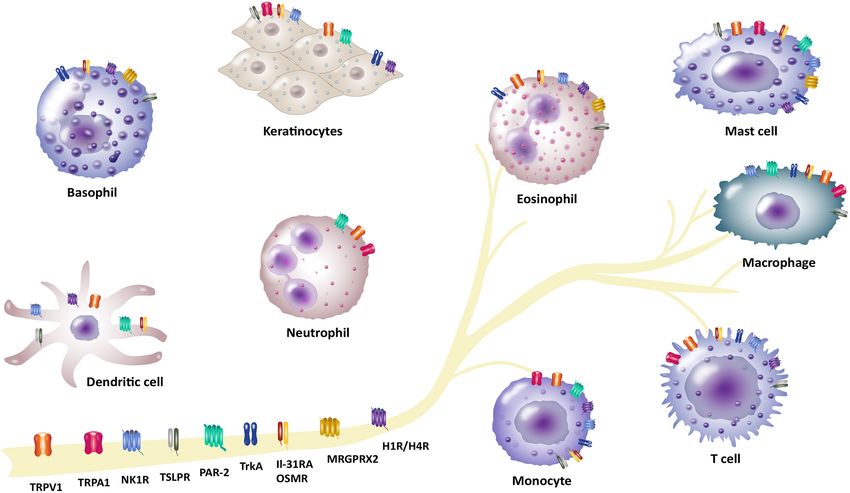
as tropomyosin receptor kinase A (TrkA) receptor (Figure 1, highlights the involvement of TRPV1 in the crosstalk between
Table 1). These receptors and channels have been found on neurons and T-lymphocytes (25). Experiments revealed that IL-
sensory neurons and play a crucial role in pruritus and neuro- 31 induces pruritus by binding to IL-31RA that is exclusively
immune pathways as well as pruritus associated inflammatory expressed on TRPV1+/TRPA1+ DRG neurons indicating TRP
skin diseases (4, 12, 13). Here, we put emphasis on inflammatory channels as key mediators of T-cell mediated IL-31-induced
skin diseases including AD, psoriasis and CSU that are highly in pruritus. Interestingly, only around 4% of DRG neurons were
context with symptom of pruritus. In previous research, several observed to be IL-31RA+ (25, 51). Surprisingly, IL-31 was
treatment options for patients suffering from these pruritus- shown to be a potential pruritogen, since injection of IL-31
associated disorders were described (14–17). Additionally, we into the cheek of mice induced profound pruritus, but not
outline current therapeutic options in correspondence with these pain. This implicates that pruritus and pain may be induced
pruritus-associated receptors and channels (Table 2). Targeting by different subsets of unmyelinated afferents and pruritus
neuro-immune pathways may open up new perspectives in terms specific afferents might exist (25). Interestingly, several studies
of the development of more effective pharmacological treatment reported a delayed pruritus after IL-31 injection in mice (52)
options for patients suffering from chronic pruritus. as well as in patients with AD and healthy volunteers (53).
These studies have led to great interest in targeting TRPV1
and developing potential drugs to treat pruritus, especially
RECEPTORS IN NEURO-IMMUNE in AD. In that regard, a topical TRPV1 antagonist termed
INTERACTIONS asivatrep, has shown to significantly improve symptoms (e.g.,
pruritus, sleep disturbance) of patients with mild-to-moderate
Transient Receptor Potential Channels AD (54, 55). Further investigations will be needed to unravel
TRPV1 and TRPA1 the neuro-immune axis involving TRP channels TRPA1 and
TRP channels are non-selective calcium-permeable cation TRPV1, neurons and different immune cells for new anti-pruritic
channels comprising 28 members in mammals that can be therapeutic options.
Frontiers in Medicine | www.frontiersin.org 2 February 2021 | Volume 8 | Article 627985Ruppenstein et al. Neuro-Immune Interactions in Pruritus FIGURE 1 | Involvement of different receptors/channels in neuro-immune interactions in pruritus. There is a complex interplay between neurons and immune cells in transmission of pruritus and inflammation. Several receptors act as a bridge between the neuronal and immune network and function as pruritus mediators. These receptors are located on neurons, but also expressed by different non-neuronal cells (e.g., basophils, dendritic cells, eosinophils, keratinocytes, mast cells, macrophages and monocytes, neutrophils or T cells): Transient receptor potential vanilloid 1 (TRPV1) and ankyrin 1 (TRPA1), IL-31 receptor A (IL-31RA) and the oncostatin-M receptor (OSMR), thymic stromal lymphopoietin receptor (TSLPR), protease-activated receptor 2 (PAR-2), neurokinin-1 receptor (NK1R), histamine receptors H1/H4 (H1R/H4R), mas-related G-protein coupled receptor X2 (MRGPRX2), tropomyosin receptor kinase A (TrkA). IL-31 Receptor A and Oncostatin-M using omalizumab led to decreased serum levels of IL-31 (80). Receptor Previous research has established a neuro-immune crosstalk The novel cytokine IL-31 signals through a heterodimeric between IL-31 receptor, T cells and sensory nerves in pruritus receptor composed of IL-31RA and the OSMR. IL-31 is a (25). Cevikbas et al. (25) have shown that TH 2-derived IL-31 is TH 2-cell-derived cytokine and the only known ligand for IL- able to activate IL-31RA on TRPV1+/TRPA1+ sensory nerves in 31RA (56–58). It has previously been observed by Cevikbas the skin causing the pruritus associated with AD. Furthermore, et al. (25) that TH 2 cells are main producers of IL-31. Besides it was shown that the TH 2-related and atopy-associated cytokine TH 2 cells, other immune cells like basophils, eosinophils or IL-31 directly induces nerve fiber elongation in vitro and in vivo mast cells can produce and release IL-31 (59–62). The IL- in mice, suggesting that IL-31-associated nerve fiber elongation 31 receptor complex is not only expressed by DRG neurons could be involved in skin hypersensitivity of AD patients (57). (63), but also located on non-neuronal cells, such as basophils In this regard, IL-31 has been shown to correlate with disease (62), eosinophils (61, 64), monocytes and macrophages (65– severity and pruritus in AD patients (75). More recent findings 67), mast cells (13), dendritic cells (68), keratinocytes (69, 70), have demonstrated that nemolizumab, an anti-IL-31RA antibody and T cells (25, 57, 71). IL-31/IL-31RA interaction activates that binds to IL-31RA with subsequent inhibition of IL-31 signal transduction pathways leading to expression and release signaling effectively relieves AD-associated pruritus (81, 82). of various chemokines, proinflammatory cytokines, regulation The first clinical study revealed a statistically significant reduced of cell proliferation and stimulation of DRG neurons that play pruritus visual analog scale (VAS) score to about 50% at week 4 important role in pruritus induction and inflammatory diseases compared with 20% with placebo in patients with AD (81). In (72–74). A number of researchers observed an association a subsequent phase II study 264 adults with moderate to severe between IL-31 and inflammatory skin diseases with severe AD were treated every 4 weeks with nemolizumab in doses of pruritus including AD (61, 75), bullous pemphigoid (76), 0.1, 0.5, or 2.0 mg/kg. Treatment led to decrease of pruritus cutaneous T-cell lymphoma (77), CSU (78) and psoriasis (79). VAS by 43.7% to 63.1% in a dose-dependent manner over a Regarding treatment approaches, a successful therapy of urticaria 12-week period compared with a 20.9% decrease with placebo Frontiers in Medicine | www.frontiersin.org 3 February 2021 | Volume 8 | Article 627985
Ruppenstein et al.                                                                                                      Neuro-Immune Interactions in Pruritus

TABLE 1 | Expression of receptors/channels on various non-neuronal cells.            human monoclonal antibody KPL-716, which specifically targets
                                                                                     the OSMRβ chain and simultaneously inhibits both IL-31 and
Receptors/channels            Non-neuronal cells                  References
                                                                                     OSM signaling. Therefore, blocking OSMRβ with KPL-716 may
TRPV1/TRPA1                   Dendritic cells (only TRPV1)          (46, 47)         be a potential treatment option of inflammatory skin diseases
                              Eosinophils (only TRPV1)                (48)           (e.g., AD) and needs to be clarified in further experiments (84).
                              Keratinocytes                         (33, 34)         These studies indicate that IL-31 is an important cytokine for
                              Macrophages and monocytes             (35–37)          regulating pruritus and AD disease activity.
                              Mast cells                            (39, 40)
                              Neutrophils                           (41, 42)
                              T cells                             (25, 43–45)        Thymic Stromal Lymphopoietin Receptor
IL-31RA/OSMR                  Basophils                               (62)           TSLP is a four-helix bundle, IL-7-like cytokine, and a member
                              Dendritic cells                         (68)           of the IL-2 cytokine family that contributes to the initiation
                              Eosinophils                           (61, 64)         of type-2 inflammation. It is primarily produced by epithelial
                              Keratinocytes                         (69, 70)         cells including keratinocytes, fibroblasts and stromal cells, but
                              Macrophages and monocytes             (65–67)          also by dendritic cells and mast cells (85, 86). TSLP signaling
                              Mast cells                              (13)           requires a heterodimeric receptor complex that consists of the
                              T cells                             (25, 57, 71)       IL-7 receptor α-chain (IL-7Rα) and the TSLP receptor chain
TSLPR                         Basophils                               (89)           (TSLPR) (87, 88). TSLP receptor is expressed by a variety of
                              Eosinophils                             (90)           cell populations including non-neuronal cells, such as basophils
                              Dendritic cells                         (91)           (89), eosinophils (90), dendritic cells (91), keratinocytes (92),
                              Keratinocytes                           (92)           mast cells (93), macrophages and monocytes (94, 95), B and
                              Macrophages and monocytes             (94, 95)         T cells (96, 97), but also by neurons (11, 98). The expression
                              Mast cells                              (93)           of TSLP from these different target cells can be triggered by
                              T and B cells                         (96, 97)         various stimuli comprising respiratory viruses (99), cigarette
PAR-2                         Dendritic cells                      (125, 126)        smoke extracts (100) as well as several cytokines, such as TNF-
                              Keratinocytes                          (124)           α and IL-1β (101). TSLP is known to be involved in various
                              Macrophages and monocytes            (125–127)
                                                                                     allergic diseases such as AD (102, 103), bronchial asthma (104)
                              Mast cells                           (128, 129)
                                                                                     and eosinophilic esophagitis (105). There is a growing evidence
                              Neutrophils                            (130)
                                                                                     indicating that TSLP may also play role in other diseases
NK1R                          Dendritic cells                        (148)
                                                                                     including autoimmune, chronic inflammatory disorders and
                                                                                     cancer (86, 106, 107). In terms of AD several studies show
                              Eosinophils                            (149)
                                                                                     that TSLP serum level as well as TSLP level in the skin of AD
                              Keratinocytes                        (154, 155)
                                                                                     patients is elevated (102, 103, 108). An overexpression of TSLP
                              Macrophages and monocytes              (151)
                                                                                     in mice models resulted in the development of AD (109, 110).
                              Mast cells                             (150)
                                                                                     Wilson et al. (98) have demonstrated that intradermal injection
                              T and B cells                        (152, 153)
                                                                                     of TSLP led to scratching behavior in mice. Additionally, their
H1R/H4R                       Basophils                              (185)
                                                                                     data confirmed that TSLP released from keratinocytes acts
                              Dendritic cells                      (186, 187)
                                                                                     directly on sensory neurons to induce itch-evoked scratching
                              Eosinophils                             (13)
                                                                                     that was depended on TSLPR. Further it was evidenced that
                              Keratinocytes                        (183, 184)
                                                                                     both functional TSLPRs and TRPA1 channels are required for
                              Monocytes                              (187)
                                                                                     TSLP-induced pruritus. A crosstalk between TSLP and PAR-
                              Mast cells                             (188)
                                                                                     2 was also observed. PAR-2 activation by its agonists SLIGLR
                              T cells                            (28, 189–191)
                                                                                     and tryptase induced scratching behavior and Ca2+ -dependent
MRGPRX2                       Basophils                              (206)           release of TSLP (98). However, the mechanism behind the
                              Eosinophils                            (206)           TSLP-induced pruritus remains to be elucidated in further
                              Mast cells                             (205)           experimental studies. Targeting TSLP-TSLPR signaling via anti-
TrkA                          Basophils                              (238)           TSLP therapy like with tezepelumab, a human monoclonal
                              Eosinophils                            (233)           antibody targeting circulating TSLP, might be a promising tool
                              Keratinocytes                          (228)           to prevent and treat several diseases associated with elevated
                              Macrophages and monocytes              (239)           TSLP such as AD (111, 112). Contrarily, a phase II clinical
                              Mast cells                           (231, 232)        trial tezepelumab treatment of patient with moderate to severe
                              T and B cells                        (240, 241)        AD showed limited efficacy and insignificant pruritus reduction
                                                                                     (111). More recently, Fitoussi et al. (113) demonstrated that a
                                                                                     topical spray containing Tambourissa trichophylla leaf extract
(82). Further, a long-term extension study based on phase II trial                   (TTLE) and 18β-glycyrrhetinic acid (GA), which inhibits TSLP
resulted in reduced pruritus up to 90%, whereby it was limited                       secretion, efficiently decreases pruritus in AD patients and
by a placebo group (83). Another approach is provided by a                           improves their quality of life.

Frontiers in Medicine | www.frontiersin.org                                      4                                  February 2021 | Volume 8 | Article 627985
Ruppenstein et al. Neuro-Immune Interactions in Pruritus
TABLE 2 | Emerging therapeutic targets for treatment of pruritus associated inflammatory skin diseases in humans.
Receptors/channels Therapeutic agents Indications References
TRPV1/TRPA1 Asivatrep/PAC-14028 (TRPV1 antagonist) Atopic dermatitis (54, 55)
IL-31RA/OSMR Nemolizumab/CIM331 (IL-31RA antagonist) Atopic dermatitis (83)
Vixarelimab/KPL-716 (OSMR antagonist) Atopic dermatitis (84)
TSLPR Tezepelumab/AMG-157/MEDI9929 (anti-TSLP antibody) Atopic dermatitis (111)
Topical spray containing TTLE and GA (TSLP inhibitor) Atopic dermatitis (113)
PAR-2 currently not available in humans – –
NK1R Aprepitant (NK1R antagonist) Atopic dermatitis (168, 169)
Serlopitant/VPD-737 (NK1R antagonist) Atopic dermatitis, Psoriasis (173)
Tradipitant/VLY-688 (NK1R antagonist) Atopic dermatitis (174)
H1R/H4R Bilastine (H1R antagonist) Chronic spontaneous urticaria (195)
Adriforant/ZPL-3893787 (H4R antagonist) Atopic dermatitis (193)
JNJ-39758979 (H4R antagonist) Atopic dermatitis (202)
MRGPRs currently not available – –
TrkA Pegcantratinib/CT327 (TrkA inhibitor) Psoriasis (255)
Protease-Activated Receptor-2 pruritus in acute and chronic models of AD. For this, MA-
The PAR family consists of four members, PAR-1, PAR-2, 1, a mast cell-degranulating peptide from wasp venom, was
PAR-3, and PAR-4. All together they belong to G-protein utilized to induce severe scratching in mice. Subsequent PZ-
coupled receptors activated by proteolytic cleavage of amino- 235 treatment significantly reduced scratching behavior in mice
terminal exodomain (114–117). Furthermore, an activation by up to 50%. Further results demonstrated that targeting PAR-2
different proteases generated by endogenous (e.g., proteases via PZ-235 application attenuated production of inflammatory
from endothelium, epithelium, fibroblast or immune cells) or factors, leukocyte infiltration, skin thickening as well as severity
exogenous sources (e.g., allergens, dust mite and various plants) of skin lesions. Therefore, PZ-235 may have potential in the
is possible (118, 119). Existing research recognizes the critical effective treatment of patients with AD (142). More studies and
role played by PAR-2 in skin neurogenic inflammation and in clinical trials in humans are currently lacking and needs to
pruritic skin diseases such as AD (119–123). PAR-2 is expressed be investigated.
by various cell types including endothelial cells and keratinocytes
(124), dendritic cells, monocytes and macrophages (125–127), Neurokinin-1 Receptor
mast cells (128, 129), neutrophils (130) and sensory nerve fibers Neurokinin receptors belong to G protein-coupled receptors
(123, 131, 132). Steinhoff et al. (120) reported an increased and consists of three members, neurokinin-1-3 receptors (NK1-
signaling through PAR-2 that comprises an increased release 3R) that are implicated in afferent neuronal signal transduction.
of endogenous PAR-2 agonist mast cell tryptase followed by There are various ligands for these receptors like neurokinin
a higher occurrence of PAR-2+ nerve fibers in AD patients A (NKA), neurokinin B (NKB), neuropeptide K (NPK),
(120, 133). In addition to the crosstalk between nerve fibers, neuropeptide-γ (NKγ), endokinin, hemokinin 1 as well as
mast cells and PAR-2, it was shown that PAR-2 synergistically substance P (SP), belonging to tachykinin family, whereas SP
interact with TRPV1 channel resulting in pruritus sensation binds with high affinity to the NK1R (143–146). Especially,
(134, 135). A key role of TRPV1 channel in PAR2-evoked NK1R is known to mainly contribute to transmission of pruritus
Ca2+ release in differentiated human primary keratinocytes (4, 12, 147). NK1R is widely expressed by different immune
was shown by Gouin et al. (136). They demonstrated that cells, such as dendritic cells (148), eosinophils (149), mast cells
TRPV1 independently regulate the production of inflammatory (150), macrophages and monocytes (151) and T and B cells
mediators, such as IL-1β, TNF-α, and TSLP via Ca2+ and (152, 153), but also by keratinocytes (154, 155) and sensory
NF-kB signaling (136). Overexpression of these inflammatory nerve endings (11, 156, 157). Activation of NK1R via SP leads to
mediators is in connection with inflammatory skin diseases, such multiple signaling cascades involving mast cell degranulation and
as AD or psoriasis (137–141). In a very recent follow-up study release of proinflammatory mediators, such as histamine, nerve
Buhl et al. (119) found that PAR-2 regulates neuro-epidermal growth factor expression and leukotriene B4 in keratinocytes and
communication in AD using a mouse model with epidermal neurogenic inflammation resulting in induction of inflammation
overexpression of PAR-2. The research results indicate that PAR- and pruritus (145, 146, 158). Several studies investigated the
2 signaling in keratinocytes causes epidermal responses leading role of SP and NK1R in the pathogenesis of pruritus in various
to neuronal sensory and inflammatory responses in their AD diseases like AD, psoriasis and CSU (7, 159–163). Recently, it
model (119). A promising therapeutic approach presents a PAR-2 was reported that SP and its receptor NK1R are overexpressed
pepducin, termed PZ-235. Barr et al. (142) examined the capacity in pruritic AD and psoriatic lesional skin (164). A previous study
of PZ-235 to suppress skin lesion thickening, inflammation, and demonstrated that increased serum levels of SP in AD patients
Frontiers in Medicine | www.frontiersin.org 5 February 2021 | Volume 8 | Article 627985Ruppenstein et al. Neuro-Immune Interactions in Pruritus
correlate with pruritus intensity (165, 166). Interestingly, oral clinical study presented a switch to bilastine, a H1R antagonist,
treatment with the NK1R antagonist aprepitant led to reduced as an optional treatment for patients with CSU, who are
serum levels of immunoglobulin E (IgE) and SP levels in tissue unresponsive to H1R antihistamines at the licensed doses (195).
as well as decreased cutaneous infiltration of regulatory T cells Although H1R antihistamines demonstrated convincing anti-
in an NC/Nga mouse model (167). In contrast, clinical studies pruritic effects in urticaria, they show limited efficiency in other
revealed no significant differences between aprepitant treatment pruritic skin diseases such as AD (11, 197, 198). In the study
and placebo concerning reduction in pruritus, improvement in of Gutzmer et al. (190), it was demonstrated that AD patients
pruriginous lesions or quality of life (168, 169). However, another express increased levels of H4R on T cells. Upon stimulation of
clinical study has shown that the NK1R antagonist serlopitant the H4 receptor pruritogenic IL-31 is up-regulated leading to
has potential as a therapeutic agent for the treatment of patients pruritic response (190). H4R antagonists were shown to reduce
with chronic pruritus by significantly reducing the pruritus TH 2 cytokine production, pruritus and skin inflammation in
symptom (170–172). A phase II clinical study concluded that AD-associated animal models (199, 200). Therefore, new clinical
serlopitant reduced pruritus in patients with mild to moderate trials using novel H4R antagonists might a promising treatment
psoriasis (173). Another NK1R antagonist, tradipitant, was for patients with AD such as the H4R antagonist JNJ-39758979,
examined in terms of reduction of pruritus associated with AD which led to an improvement of inflammatory skin lesions
through inhibition of SP-mediated itch signaling. Tradipitant in AD patients (193, 201). In addition, marked effects against
treatment improved pruritus and sleep in mild AD (174). Several pruritus in Japanese patients with AD could be observed in a
NK1R antagonists that potentially reduce pruritus activity in phase II clinical trial, but the development of agranulocytosis by
dermatological diseases are reviewed by Pojawa-Goła et al. (146) 2 subjects resulted in early trial termination (202). More recently,
and Reszke et al. (172). Thus, targeting SP and/or NK1R with H4R antagonist adriforant was shown to improve inflammatory
regard to neuro-immune crosstalk seems to be a promising skin lesions in patients with AD. Although adriforant treatment
approach in the treatment of pruritus. In a previous research cause a 3-point reduction (scale, 1–10) in pruritus, there was no
it was established that Mas-related GPCR X2, which is also significant difference in comparison to reduced pruritus with
activated by SP, induced inflammation (175). Further, it was placebo (193). Interestingly, a combined treatment of both H1R
suggested that SP-induced pruritus may be mediated by MRGPRs and H4R antagonists demonstrated an anti-inflammatory effect
rather than NK1R, since SP-induced pruritus was not decreased in an AD mice model that might be a good strategy to treat
in Nk1r KO mice. Co-injection of QWF and SP in both Nk1r patients with AD (203).
KO and wild-type mice led to significantly decreased SP-induced
pruritus. Interestingly, an NK1R antagonist termed QWF was Mas-Related G-Protein Coupled Receptors
shown to have a dual action on MRGPRX2 (176). However, not MRGPRs are G-protein coupled receptors that comprise at least
only the crosstalk between different immune cells, neurons and 50 family members in mice, divided into subgroups MRGPRA-
NK1R, but also the interaction of NK1R with other receptors H and 8 members in humans named MRGPRX1-X4, D, E, F,
is an interesting approach for a better understanding of the and G. Several members of MRGPRs have emerged as critically
pathogenesis of pruritic diseases. important receptors in histamine-independent pruritus. They are
mainly expressed by sensory neurons and some also by mast
Histamine Receptors H1 and H4 cells (5, 8, 204, 205). Recently, human basophils and eosinophils
One of the well-characterized pruritogens is histamine. were reported to express MRGPRX2 (206). However, the
Histamine is released from mast cells and basophils via MRGPRs can be activated by various endogenous and exogenous
activation of histamine receptors, which belong to the G protein- peptides or molecules, such as antimicrobial host defense or
coupled receptor superfamily. While four histamine receptor opioid peptides, SP or eosinophilic granules, but also by drugs
subtypes (H1–H4) exist, notably histamine receptors H1 and H4 like vancomycin or chloroquine (CQ) (12, 207). Particularly,
are known to modulate pruritus (13, 177–181). Both histamine MRGPRA3 and MRGPRC11 in mice as well as the human
receptors (H1R and H4R) are extensively expressed in a wide ortholog MRGPRX1 got into the focus of pruritus researchers
range of cell types involving sensory neurons (182), epithelial over the past decade (12, 208). It was shown that CQ activated
cells like keratinocytes (183, 184), but also immune cells, such MRGPRA3 leading to a pruritus signal via the activation of
as basophils (185), dendritic cells (186, 187), eosinophils (13), TRPA1 (24). Furthermore, it was demonstrated that expression
monocytes (187), mast cells (188) and T cells (28, 189–191). of MRGPRA3 establishes a subset of nociceptors that specifically
Especially, the H4R is predominantly expressed by immune mediate pruritus, but not pain in a mouse model. In addition,
cells and is in conjunction with lots of functional histamine- a deletion of MRGPRA3+ sensory neurons significantly inhibits
mediated inflammatory responses like modulation of cytokine scratching behavior (209, 210). A recent study by Lee et al.
and chemokine release, chemotaxis and cell recruitment as well (211) determined that Korean Red Ginseng water extract (KRGE)
as upregulation of adhesion molecule expression (192, 193). inhibits CQ-induced pruritus by blocking the MRGPRA3/TRPA1
However, both the H1 and the H4 histamine receptors play pathway. Interestingly, KRGE has also anti-pruritic effects on
pivotal roles in various pruritic skin diseases, such as AD or the histamine-dependent H1R/TRPV1 pathway, which might
CSU (188, 194–196). Various H1R antihistamines like ebastine, provide a dual anti-pruritic candidate agent for the treatment
cetirizine, and levocetirizine were shown to decrease pruritus of pruritus patients (211, 212). Moreover, KRGE treatment
symptom of patients with CSU by 60–70% (197). A very recent significantly decreased hyperplasia and hyperkeratosis in the
Frontiers in Medicine | www.frontiersin.org 6 February 2021 | Volume 8 | Article 627985Ruppenstein et al. Neuro-Immune Interactions in Pruritus
epidermis, infiltration of inflammatory cells and suppressed the (NT-3) and neurotrophin-4 (NT-4) (223–226). The main source
overexpression of cytokines in the AD-like skin lesions of AD of NGF are keratinocytes in the skin (227, 228), but it is also
mice model (167). MRGPRC11 is in addition to MRGPRA3 expressed and secreted by other immune cells, such as basophils
co-localized and expressed in a subset of TRPV1+ afferents (229), monocytes and macrophages (230), mast cells (231, 232)
and mediates pruritus induced by BAM8-22 (24, 209, 213). Liu and eosinophils (233) as well as by neurons (234, 235) during
et al. (204) has proven that activation through MRGPRC11- inflammation. NGF binds with high affinity to its receptor TrkA
specific agonist BAM8-22 induces scratching in murine models. as well as the low-affinity neurotrophin receptor p75NTR. TrkA
In a following clinical study, BAM8-22 triggered pruritus and is widely expressed across the airway smooth muscles, the lung
nociceptive sensations in humans in a histamine-independent epithelium and sensory neurons (236, 237), but also located
manner as topical antihistamine-containing cream did not on various non-neuronal cells like basophils (238), eosinophils
attenuate scratching behavior (214). This indicates BAM8-22 as (233), keratinocytes (228), monocytes and macrophages (239),
an endogenous pruritus mediator and MRGPRX1 antagonists mast cells (231, 232) as well as B and T cells (240, 241). Both
may present potential anti-pruritic therapies. The synthetic NGF and its receptor TrkA are suggested to play important
peptide SLIGRL was long believed to mediate scratching behavior roles in pruritus and allergic inflammation. Several studies
via the PAR-2. However, intradermal injected SLIGRL caused reported that NGF in the skin and NGF serum levels of AD
scratching behavior in PAR-2 KO mice similar to that of wild-type and psoriatic patients as well as serum levels of patients with
mice. Liu and colleagues (215) proved that the pruritus induction asthma are increased (227, 242–245). Additionally, an increased
of SLIGRL was mediated by MRGPRC11 while its hyperalgesic TrkA expression in keratinocytes of patients with AD has been
mode of action was derived from PAR-2 (2, 215). Furthermore, observed during inflammation (228). In AD it was shown, that
MRGPRX1 is responsible for neuronal activation and scratching increased peripheral serum levels of BDNF significantly correlate
behavior induced by both CQ and BAM8-22 (204, 213, 216). with disease severity and pruritus (246, 247). Also scratching
To date, there is a lack of knowledge about the involvement activities were significantly correlated to increased levels of
of MRGPRX1 in the pathology in chronic pruritic diseases BDNF as shown by Hon and colleagues (248) which used a
such as AD and the potential role of MRGPRX1 antagonists in DigiTrac model to assess scratching activities in children with
affected patients. Recently, researchers have shown an increased AD. In this regard, it has been shown that eosinophils are a
interest in MRGPRX2 in terms of pruriceptive receptor and source of BDNF and release BDNF and are functionally activated
its involvement in pruritic diseases like AD or psoriasis (164, by BDNF with induction of chemotaxis (246–248). Thus, the
176, 217, 218). MRGPRX2 is expressed in mast cells and an question arises if BDNF which is released by eosinophils of AD
activation of MRGPRX2 by peptides such as SP results in patients is also capable to stimulate nerves. This has recently
mast cell degranulation leading to release of proinflammatory been shown in a study by us in which we could see that BDNF
factors as well as modulation of neurogenic inflammation and released by peripheral blood eosinophils of patients with AD
pruritus (219, 220). Previous research has established that both led to a significant sprouting of peripheral nerves derived from
the percentage of MRGPRX2+ mast cells and MRGPRX2+ spinal neurons of mice (247). Thus, also BDNF seems to have
skin mast cells of patients with CSU were significantly higher an important impact in neuro-immune interaction mechanisms
in comparison to non-chronic urticaria subjects. It was further and pruritus. However, NGF affects neurite outgrowth and
shown that SP-induced histamine release from human skin neuronal survival (236, 249). Interestingly, sprouting of itch-
mast cells through MRGPRX2 contributing to neurogenic sensitive nerve fibers, promoted by increased NGF levels, has
inflammation (221). Interestingly, Green et al. (222) found out been observed in the skin of patients with AD (242) and in
that SP-mediated inflammatory responses were independent of AD-associated mice models (250, 251). Since NGF is known
its canonical receptor NK1R and identified MRGPRX2 and its to increase cutaneous innervation in AD models and might
mouse homolog MRGPRB2 as an important neuro-immune contribute to the development of chronic pruritus, NGF and
modulator and a potential target for treating inflammatory pain. its receptor TrkA could be targets for future treatment of
Involvement in pruritus transmission and anti-pruritic treatment pruritus and allergic inflammation in pruritic diseases like AD or
therapies remain elusive and needs to be clarified in further psoriasis. A clinical study demonstrated a promising treatment
studies (222). In a recent study, increased MRGPRX2 mRNA of AD by neutralizing antibodies against NGF that inhibited
expression in pruritic skin of patients with AD and psoriasis was the development of skin lesions and epidermal innervation
demonstrated as well (164). However, research has consistently as well as scratching behavior in AD mice model (252). In
shown that only few endogenous agonists for most of these human sensory neurons, NGF up-regulated the expression and
receptors are known so far and their role in the pathogenesis sensitivity of TRPV1 channels by activating TrkA (253, 254).
chronic pruritus diseases such as AD remains still unclear. Interestingly, TrkA inhibitor CT327 was shown to significantly
reduce chronic pruritus in patients with psoriasis as measured
Tropomyosin Receptor Kinase A by VAS in a phase II clinical study. The results demonstrated
Trk receptors were firstly described in 1986 and three members that 62, 46, and 61% of patients treated with CT327 0.05, 0.1,
of the tyrosine kinase receptor family, TrkA, TrkB, and TrkC, and 0.5%, respectively, had at least a 50% decrease in pruritus
have been identified so far. Trk receptors are activated by VAS in comparison to 32% on vehicle (255). Further trials are
various neurotrophins including nerve-growth factor (NGF), necessary to prove the anti-pruritic effects of CT327 in AD.
brain-derived neurotrophic factor (BDNF), neurotrophin-3 There is growing evidence that cutaneous NGF-TrkA-TRPV1
Frontiers in Medicine | www.frontiersin.org 7 February 2021 | Volume 8 | Article 627985Ruppenstein et al. Neuro-Immune Interactions in Pruritus
signaling might be a key mechanism contributing to neurogenic receptor therapeutic targets in the skin as well as in peripheral
inflammation and pruritus in different dermatological nerves comprises TRPV1, TRPA1, IL-31RA, TSLPR, PAR-2,
diseases (147, 255). NK1R, H1R and H4R, MRGPRs and TrkA, which are highlighted
in this review (Figure 1, Tables 1, 2). Future studies targeting
CONCLUSION neuro-immune interactions will help to unravel the underlying
mechanisms of pruritus and to develop specific therapies.
Our understanding of the pathogenesis of pruritus has
significantly evolved in recent years. There is a growing AUTHOR CONTRIBUTIONS
body of literature on the complex crosstalk between neuronal
and immune cells that are involved in the development of acute AR has performed the literature research and wrote
and chronic pruritus. Neurons directly communicate with and the manuscript including the tables. MML designed
regulate the function of various immune cells, such as mast cells, the figure and revised the manuscript. UR, KL,
dendritic cells, eosinophils and T cells in pruritus transmission AEK, and BH critically revised the manuscript. All
and inflammation. Immune cells release proinflammatory authors read and approved the final version of the
mediators including cytokines, chemokines, neurotrophins, submitted manuscript.
and neuropeptides that activate sensory neurons to mediate
pruritus. Activation of these neurons leads to a release of FUNDING
neurotransmitters and neuropeptides that vice versa have
a direct impact on the functional activity of immune cells. This work was kindly supported by the Deutsche
The literature on neuro-immune crosstalk has emphasized Forschungsgemeinschaft-funded consortium FOR 2690:
several key mediators and neuronal pathways involved in the Translationale Pruritusforschung (RA-1026/3-1, KR-3618/3-1,
transmission of pruritus. Potential mediator and promising HO 2092/7-1).
REFERENCES 13. Nakashima C, Ishida Y, Kitoh A, Otsuka A, Kabashima K.
Interaction of peripheral nerves and mast cells, eosinophils, and
1. Raap U, Ständer S, Metz M. Pathophysiology of itch and basophils in the development of pruritus. Exp Dermatol. (2019)
new treatments. Curr Opin Allergy Clin Immunol. (2011) 28:1405–11. doi: 10.1111/exd.14014
11:420–7. doi: 10.1097/ACI.0b013e32834a41c2 14. Misery L, Huet F, Gouin O, Ständer S, Deleuran M. Current pharmaceutical
2. Han L, Dong X. Itch mechanisms and circuits. Annu Rev Biophys. (2014) developments in atopic dermatitis. Curr Opin Pharmacol. (2019) 46:7–
43:331–55. doi: 10.1146/annurev-biophys-051013-022826 13. doi: 10.1016/j.coph.2018.12.003
3. Silverberg JI, Hinami K, Trick WE, Cella D. Itch in the 15. McEwen MW, Fite EM, Yosipovitch G, Patel T. Drugs on the
general internal medicine setting: a cross-sectional study of horizon for chronic pruritus. Dermatol Clin. (2018) 36:335–
prevalence and quality-of-life effects. Am J Clin Dermatol. (2016) 44. doi: 10.1016/j.det.2018.02.016
17:681–90. doi: 10.1007/s40257-016-0215-3 16. Pereira MP, Ständer S. Chronic pruritus: current and emerging treatment
4. Mack MR, Kim BS. The itch-scratch cycle: a neuroimmune perspective. options. Drugs. (2017) 77:999–1007. doi: 10.1007/s40265-017-0746-9
Trends Immunol. (2018) 39:980–91. doi: 10.1016/j.it.2018.10.001 17. Golpanian RS, Yosipovitch G. Current and emerging systemic treatments
5. Mollanazar NK, Smith PK, Yosipovitch G. Mediators of chronic pruritus targeting the neural system for chronic pruritus. Expert Opin Pharmacother.
in atopic dermatitis: getting the itch out? Clin Rev Allergy Immunol. (2016) (2020) 21:1629–36. doi: 10.1080/14656566.2020.1775815
51:263–92. doi: 10.1007/s12016-015-8488-5 18. Clapham DE. TRP channels as cellular sensors. Nature. (2003) 426:517–
6. Pereira MP, Agelopoulos K, Kremer AE, Schmelz M. [New 24. doi: 10.1038/nature02196
findings regarding the neurobiology of pruritus]. Hautarzt. (2018) 19. Montell C. The TRP superfamily of cation channels. Sci STKE. (2005)
69:620–5. doi: 10.1007/s00105-018-4210-x 2005:re3. doi: 10.1126/stke.2722005re3
7. Yosipovitch G, Berger T, Fassett MS. Neuroimmune interactions in chronic 20. Sun S, Dong X. Trp channels and itch. Semin Immunopathol. (2016) 38:293–
itch of atopic dermatitis. J Eur Acad Dermatol Venereol. (2020) 34:239– 307. doi: 10.1007/s00281-015-0530-4
50. doi: 10.1111/jdv.15973 21. Kittaka H, Tominaga M. The molecular and cellular mechanisms of itch and
8. Voisin T, Bouvier A, Chiu IM. Neuro-immune interactions in allergic the involvement of TRP channels in the peripheral sensory nervous system
diseases: novel targets for therapeutics. Int Immunol. (2017) 29:247– and skin. Allergol Int. (2017) 66:22–30. doi: 10.1016/j.alit.2016.10.003
61. doi: 10.1093/intimm/dxx040 22. Xie Z, Hu H. TRP channels as drug targets to relieve itch. Pharmaceuticals.
9. Kabata H, Artis D. Neuro-immune crosstalk and allergic inflammation. J Clin (2018) 11:4. doi: 10.3390/ph11040100
Invest. (2019) 129:1475–82. doi: 10.1172/JCI124609 23. Shim WS, Tak MH, Lee MH, Kim M, Kim M, Koo JY, et al.
10. Lopez-Requena A, Boonen B, Van Gerven L, Hellings PW, TRPV1 mediates histamine-induced itching via the activation
Alpizar YA, Talavera K. Roles of neuronal TRP channels in of phospholipase A2 and 12-lipoxygenase. J Neurosci. (2007)
neuroimmune interactions. In: Emir TLR, editor. Neurobiology of 27:2331–7. doi: 10.1523/JNEUROSCI.4643-06.2007
TRP Channels. Boca Raton, FL: Frontiers in Neuroscience. (2017). p. 24. Wilson SR, Gerhold KA, Bifolck-Fisher A, Liu Q, Patel KN, Dong
277–94. doi: 10.4324/9781315152837-15 X, et al. TRPA1 is required for histamine-independent, Mas-related G
11. Oetjen LK, Kim BS. Interactions of the immune and sensory nervous protein-coupled receptor-mediated itch. Nat Neurosci. (2011) 14:595–
systems in atopy. FEBS J. (2018) 285:3138–51. doi: 10.1111/febs. 602. doi: 10.1038/nn.2789
14465 25. Cevikbas F, Wang X, Akiyama T, Kempkes C, Savinko T, Antal A, et al. A
12. Ständer S, Raap U, Weisshaar E, Schmelz M, Mettang T, Handwerker sensory neuron-expressed IL-31 receptor mediates T helper cell-dependent
H, et al. Pathogenesis of pruritus. J Dtsch Dermatol Ges. (2011) 9:456– itch: Involvement of TRPV1 and TRPA1. J Allergy Clin Immunol. (2014)
63. doi: 10.1111/j.1610-0387.2011.07585.x 133:448–60. doi: 10.1016/j.jaci.2013.10.048
Frontiers in Medicine | www.frontiersin.org 8 February 2021 | Volume 8 | Article 627985Ruppenstein et al. Neuro-Immune Interactions in Pruritus
26. Hojland CR, Andersen HH, Poulsen JN, Arendt-Nielsen L, 46. Toth BI, Benko S, Szollosi AG, Kovacs L, Rajnavolgyi E, Biro T.
Gazerani P. A human surrogate model of itch utilizing the Transient receptor potential vanilloid-1 signaling inhibits differentiation
TRPA1 agonist trans-cinnamaldehyde. Acta Derm Venereol. (2015) and activation of human dendritic cells. FEBS Lett. (2009) 583:1619–
95:798–803. doi: 10.2340/00015555-2103 24. doi: 10.1016/j.febslet.2009.04.031
27. Jian T, Yang N, Yang Y, Zhu C, Yuan X, Yu G, et al. TRPV1 and PLC 47. Gouin O, L’Herondelle K, Lebonvallet N, Le Gall-Ianotto C, Sakka M, Buhe V,
participate in histamine H4 receptor-induced itch. Neural Plast. (2016) et al. TRPV1 and TRPA1 in cutaneous neurogenic and chronic inflammation:
2016:1682972. doi: 10.1155/2016/1682972 pro-inflammatory response induced by their activation and their
28. Brennan F. The pathophysiology of pruritus – a review for clinicians. Progress sensitization. Protein Cell. (2017) 8:644–61. doi: 10.1007/s13238-017-0395-5
in Palliative Care. (2016) 24:133–46. doi: 10.1179/1743291X14Y.0000000110 48. Zhu X, Learoyd J, Butt S, Zhu L, Usatyuk PV, Natarajan V, et al.
29. Imamachi N, Park GH, Lee H, Anderson DJ, Simon MI, Basbaum AI, Regulation of eosinophil adhesion by lysophosphatidylcholine via a non-
et al. TRPV1-expressing primary afferents generate behavioral responses store-operated Ca2+ channel. Am J Respir Cell Mol Biol. (2007) 36:585–
to pruritogens via multiple mechanisms. Proc Natl Acad Sci USA. (2009) 93. doi: 10.1165/rcmb.2006-0391OC
106:11330–5. doi: 10.1073/pnas.0905605106 49. Patricio ES, Costa R, Figueiredo CP, Gers-Barlag K, Bicca MA, Manjavachi
30. Kim SJ, Park GH, Kim D, Lee J, Min H, Wall E, et al. Analysis of cellular and MN, et al. Mechanisms underlying the scratching behavior induced by the
behavioral responses to imiquimod reveals a unique itch pathway in transient activation of proteinase-activated receptor-4 in mice. J Invest Dermatol.
receptor potential vanilloid 1 (TRPV1)-expressing neurons. Proc Natl Acad (2015) 135:2484–91. doi: 10.1038/jid.2015.183
Sci USA. (2011) 108:3371–6. doi: 10.1073/pnas.1019755108 50. Zhang X. Targeting TRP ion channels for itch relief. Naunyn Schmiedebergs
31. Wilson SR, Bautista DM. Role of transient receptor potential channels in Arch Pharmacol. (2015) 388:389–99. doi: 10.1007/s00210-014-1068-z
acute and chronic itch. In: Carstens E, Akiyama T, editors. Itch: Mechanisms 51. Li YR, Gupta P. Immune aspects of the bi-directional
and Treatment. Boca Raton, FL: Frontiers in Neuroscience (2014). neuroimmune facilitator TRPV1. Mol Biol Rep. (2019) 46:1499–
32. Ru F, Sun H, Jurcakova D, Herbstsomer RA, Meixong J, Dong X, et al. 510. doi: 10.1007/s11033-018-4560-6
Mechanisms of pruritogen-induced activation of itch nerves in isolated 52. Kasutani K, Fujii E, Ohyama S, Adachi H, Hasegawa M, Kitamura H,
mouse skin. J Physiol. (2017) 595:3651–66. doi: 10.1113/JP273795 et al. Anti-IL-31 receptor antibody is shown to be a potential therapeutic
33. Denda M, Fuziwara S, Inoue K, Denda S, Akamatsu H, Tomitaka A, et al. option for treating itch and dermatitis in mice. Br J Pharmacol. (2014)
Immunoreactivity of VR1 on epidermal keratinocyte of human skin. Biochem 171:5049–58. doi: 10.1111/bph.12823
Biophys Res Commun. (2001) 285:1250–2. doi: 10.1006/bbrc.2001.5299 53. Hawro T, Saluja R, Weller K, Altrichter S, Metz M, Maurer M. Interleukin-
34. Biro T, Kovacs L. An “ice-cold” TR(i)P to skin biology: the role of TRPA1 31 does not induce immediate itch in atopic dermatitis patients and healthy
in human epidermal keratinocytes. J Invest Dermatol. (2009) 129:2096– controls after skin challenge. Allergy. (2014) 69:113–7. doi: 10.1111/all.12316
9. doi: 10.1038/jid.2009.179 54. Lim KM, Park YH. Development of PAC-14028, a novel transient
35. Billeter AT, Galbraith N, Walker S, Lawson C, Gardner SA, Sarojini H, et al. receptor potential vanilloid type 1 (TRPV1) channel antagonist as a
TRPA1 mediates the effects of hypothermia on the monocyte inflammatory new drug for refractory skin diseases. Arch Pharm Res. (2012) 35:393–
response. Surgery. (2015) 158:646–54. doi: 10.1016/j.surg.2015.03.065 6. doi: 10.1007/s12272-012-0321-6
36. Parenti A, De Logu F, Geppetti P, Benemei S. What is the evidence for the role 55. Lee YW, Won CH, Jung K, Nam HJ, Choi G, Park YH, et al. Efficacy and
of TRP channels in inflammatory and immune cells? Br J Pharmacol. (2016) safety of PAC-14028 cream - a novel, topical, nonsteroidal, selective TRPV1
173:953–69. doi: 10.1111/bph.13392 antagonist in patients with mild-to-moderate atopic dermatitis: a phase IIb
37. Omari SA, Adams MJ, Geraghty DP. TRPV1 channels in immune randomized trial. Br J Dermatol. (2019) 180:1030–8. doi: 10.1111/bjd.17455
cells and hematological malignancies. Adv Pharmacol. (2017) 79:173– 56. Dillon SR, Sprecher C, Hammond A, Bilsborough J, Rosenfeld-Franklin
98. doi: 10.1016/bs.apha.2017.01.002 M, Presnell SR, et al. Interleukin 31, a cytokine produced by activated
38. Khalil M, Alliger K, Weidinger C, Yerinde C, Wirtz S, Becker C, et al. T cells, induces dermatitis in mice. Nat Immunol. (2004) 5:752–
Functional role of transient receptor potential channels in immune cells and 60. doi: 10.1038/ni1084
epithelia. Front Immunol. (2018) 9:174. doi: 10.3389/fimmu.2018.00174 57. Feld M, Garcia R, Buddenkotte J, Katayama S, Lewis K, Muirhead
39. Ständer S, Moormann C, Schumacher M, Buddenkotte J, Artuc M, G, et al. The pruritus- and TH2-associated cytokine IL-31 promotes
Shpacovitch V, et al. Expression of vanilloid receptor subtype 1 in cutaneous growth of sensory nerves. J Allergy Clin Immunol. (2016) 138:500–
sensory nerve fibers, mast cells, and epithelial cells of appendage structures. 8.e24. doi: 10.1016/j.jaci.2016.02.020
Exp Dermatol. (2004) 13:129–39. doi: 10.1111/j.0906-6705.2004.0178.x 58. Nakashima C, Otsuka A, Kabashima K. Interleukin-31 and interleukin-31
40. Oh MH, Oh SY, Lu J, Lou H, Myers AC, Zhu Z, et al. TRPA1-dependent receptor: New therapeutic targets for atopic dermatitis. Exp Dermatol. (2018)
pruritus in IL-13-induced chronic atopic dermatitis. J Immunol. (2013) 27:327–31. doi: 10.1111/exd.13533
191:5371–82. doi: 10.4049/jimmunol.1300300 59. Ishii T, Wang J, Zhang W, Mascarenhas J, Hoffman R, Dai Y, et al. Pivotal role
41. Heiner I, Eisfeld J, Luckhoff A. Role and regulation of TRP of mast cells in pruritogenesis in patients with myeloproliferative disorders.
channels in neutrophil granulocytes. Cell Calcium. (2003) Blood. (2009) 113:5942–50. doi: 10.1182/blood-2008-09-179416
33:533–40. doi: 10.1016/S0143-4160(03)00058-7 60. Niyonsaba F, Ushio H, Hara M, Yokoi H, Tominaga M, Takamori K, et al.
42. Zhou Y, Han D, Follansbee T, Wu X, Yu S, Wang B, et al. Transient Antimicrobial peptides human beta-defensins and cathelicidin LL-37 induce
receptor potential ankyrin 1 (TRPA1) positively regulates imiquimod- the secretion of a pruritogenic cytokine IL-31 by human mast cells. J
induced, psoriasiform dermal inflammation in mice. J Cell Mol Med. (2019) Immunol. (2010) 184:3526–34. doi: 10.4049/jimmunol.0900712
23:4819–28. doi: 10.1111/jcmm.14392 61. Kunsleben N, Rudrich U, Gehring M, Novak N, Kapp A, Raap U. IL-31
43. Bertin S, Aoki-Nonaka Y, de Jong PR, Nohara LL, Xu H, Stanwood Induces Chemotaxis, Calcium Mobilization, Release of Reactive Oxygen
SR, et al. The ion channel TRPV1 regulates the activation and Species, and CCL26 in Eosinophils, Which Are Capable to Release IL-31. J
proinflammatory properties of CD4(+) T cells. Nat Immunol. (2014) Invest Dermatol. (2015) 135:1908–11. doi: 10.1038/jid.2015.106
15:1055–63. doi: 10.1038/ni.3009 62. Raap U, Gehring M, Kleiner S, Rudrich U, Eiz-Vesper B, Haas H,
44. Bertin S, Aoki-Nonaka Y, Lee J, de Jong PR, Kim P, Han T, et al. et al. Human basophils are a source of - and are differentially activated
The TRPA1 ion channel is expressed in CD4+ T cells and restrains T- by - IL-31. Clin Exp Allergy. (2017) 47:499–508. doi: 10.1111/cea.
cell-mediated colitis through inhibition of TRPV1. Gut. (2017) 66:1584– 12875
96. doi: 10.1136/gutjnl-2015-310710 63. Bando T, Morikawa Y, Komori T, Senba E. Complete overlap of interleukin-
45. Sahoo SS, Majhi RK, Tiwari A, Acharya T, Kumar PS, Saha 31 receptor A and oncostatin M receptor beta in the adult dorsal root
S, et al. Transient receptor potential ankyrin1 channel is ganglia with distinct developmental expression patterns. Neuroscience.
endogenously expressed in T cells and is involved in immune (2006) 142:1263–71. doi: 10.1016/j.neuroscience.2006.07.009
functions. Biosci Rep. (2019) 39:BSR20191437. doi: 10.1042/BSR201 64. Cheung PF, Wong CK, Ho AW, Hu S, Chen DP, Lam CW. Activation
91437 of human eosinophils and epidermal keratinocytes by Th2 cytokine IL-31:
Frontiers in Medicine | www.frontiersin.org 9 February 2021 | Volume 8 | Article 627985Ruppenstein et al. Neuro-Immune Interactions in Pruritus
implication for the immunopathogenesis of atopic dermatitis. Int Immunol. 83. Kabashima K, Furue M, Hanifin JM, Pulka G, Wollenberg A, Galus R,
(2010) 22:453–67. doi: 10.1093/intimm/dxq027 et al. Nemolizumab in patients with moderate-to-severe atopic dermatitis:
65. Bilsborough J, Leung DY, Maurer M, Howell M, Boguniewicz M, Yao L, et al. randomized, phase II, long-term extension study. J Allergy Clin Immunol.
IL-31 is associated with cutaneous lymphocyte antigen-positive skin homing (2018) 142:1121–30.e7. doi: 10.1016/j.jaci.2018.03.018
T cells in patients with atopic dermatitis. J Allergy Clin Immunol. (2006) 84. Richards CD, Gandhi R, Botelho F, Ho L, Paolini JF. Oncostatin M induction
117:418–25. doi: 10.1016/j.jaci.2005.10.046 of monocyte chemoattractant protein 1 is inhibited by anti-oncostatin M
66. Kasraie S, Niebuhr M, Werfel T. Interleukin (IL)-31 induces pro- receptor beta monoclonal antibody KPL-716. Acta Derm Venereol. (2020)
inflammatory cytokines in human monocytes and macrophages 100:adv00197. doi: 10.2340/00015555-3505
following stimulation with staphylococcal exotoxins. Allergy. (2010) 85. He R, Geha RS. Thymic stromal lymphopoietin. Ann N Y Acad Sci. (2010)
65:712–21. doi: 10.1111/j.1398-9995.2009.02255.x 1183:13–24. doi: 10.1111/j.1749-6632.2009.05128.x
67. Edukulla R, Singh B, Jegga AG, Sontake V, Dillon SR, Madala 86. Ziegler SF, Roan F, Bell BD, Stoklasek TA, Kitajima M, Han H. The biology
SK. Th2 cytokines augment IL-31/IL-31RA Interactions via of thymic stromal lymphopoietin (TSLP). Adv Pharmacol. (2013) 66:129–
STAT6-dependent IL-31RA expression. J Biol Chem. (2015) 55. doi: 10.1016/B978-0-12-404717-4.00004-4
290:13510–20. doi: 10.1074/jbc.M114.622126 87. Zhong J, Sharma J, Raju R, Palapetta SM, Prasad TS, Huang TC, et al. TSLP
68. Horejs-Hoeck J, Schwarz H, Lamprecht S, Maier E, Hainzl S, Schmittner M, signaling pathway map: a platform for analysis of TSLP-mediated signaling.
et al. Dendritic cells activated by IFN-gamma/STAT1 express IL-31 receptor Database. (2014) 2014:bau007. doi: 10.1093/database/bau007
and release proinflammatory mediators upon IL-31 treatment. J Immunol. 88. Park LS, Martin U, Garka K, Gliniak B, Di Santo JP, Muller W, et al. Cloning
(2012) 188:5319–26. doi: 10.4049/jimmunol.1101044 of the murine thymic stromal lymphopoietin (TSLP) receptor: Formation of
69. Heise R, Neis MM, Marquardt Y, Joussen S, Heinrich PC, Merk HF, et al. a functional heteromeric complex requires interleukin 7 receptor. J Exp Med.
IL-31 receptor alpha expression in epidermal keratinocytes is modulated (2000) 192:659–70. doi: 10.1084/jem.192.5.659
by cell differentiation and interferon gamma. J Invest Dermatol. (2009) 89. Sokol CL, Barton GM, Farr AG, Medzhitov R. A mechanism for the initiation
129:240–3. doi: 10.1038/jid.2008.183 of allergen-induced T helper type 2 responses. Nat Immunol. (2008) 9:310–
70. Kasraie S, Niebuhr M, Baumert K, Werfel T. Functional effects of 8. doi: 10.1038/ni1558
interleukin 31 in human primary keratinocytes. Allergy. (2011) 66:845– 90. Cook EB, Stahl JL, Schwantes EA, Fox KE, Mathur SK. IL-3 and TNFalpha
52. doi: 10.1111/j.1398-9995.2011.02545.x increase thymic stromal lymphopoietin receptor (TSLPR) expression on
71. Cevikbas F, Kempkes C, Buhl T, Mess C, Buddenkotte J, Steinhoff M. Role of eosinophils and enhance TSLP-stimulated degranulation. Clin Mol Allergy.
interleukin-31 and oncostatin M in itch and neuroimmune communication. (2012) 10:8. doi: 10.1186/1476-7961-10-8
In: Carstens E, Akiyama T, editors. Itch: Mechanisms and Treatment. Boca 91. Kashyap M, Rochman Y, Spolski R, Samsel L, Leonard WJ. Thymic stromal
Raton, FL: Frontiers in Neuroscience (2014). lymphopoietin is produced by dendritic cells. J Immunol. (2011) 187:1207–
72. Sonkoly E, Muller A, Lauerma AI, Pivarcsi A, Soto H, Kemeny L, et al. IL-31: 11. doi: 10.4049/jimmunol.1100355
a new link between T cells and pruritus in atopic skin inflammation. J Allergy 92. Kubo T, Kamekura R, Kumagai A, Kawata K, Yamashita K, Mitsuhashi Y,
Clin Immunol. (2006) 117:411–7. doi: 10.1016/j.jaci.2005.10.033 et al. DeltaNp63 controls a TLR3-mediated mechanism that abundantly
73. Stott B, Lavender P, Lehmann S, Pennino D, Durham S, Schmidt-Weber CB. provides thymic stromal lymphopoietin in atopic dermatitis. PLoS ONE.
Human IL-31 is induced by IL-4 and promotes TH2-driven inflammation. J (2014) 9:e105498. doi: 10.1371/journal.pone.0105498
Allergy Clin Immunol. (2013) 132:446–54.e5. doi: 10.1016/j.jaci.2013.03.050 93. Allakhverdi Z, Comeau MR, Jessup HK, Yoon BR, Brewer A, Chartier S,
74. Gibbs BF, Patsinakidis N, Raap U. Role of the pruritic cytokine et al. Thymic stromal lymphopoietin is released by human epithelial cells in
IL-31 in autoimmune skin diseases. Front Immunol. (2019) response to microbes, trauma, or inflammation and potently activates mast
10:1383. doi: 10.3389/fimmu.2019.01383 cells. J Exp Med. (2007) 204:253–8. doi: 10.1084/jem.20062211
75. Raap U, Wichmann K, Bruder M, Ständer S, Wedi B, Kapp A, et al. 94. Hirano R, Hasegawa S, Hashimoto K, Haneda Y, Ohsaki A, Ichiyama
Correlation of IL-31 serum levels with severity of atopic dermatitis. J Allergy T. Human thymic stromal lymphopoietin enhances expression of CD80
Clin Immunol. (2008) 122:421–3. doi: 10.1016/j.jaci.2008.05.047 in human CD14+ monocytes/macrophages. Inflamm Res. (2011) 60:605–
76. Salz M, Haeberle S, Hoffmann J, Enk AH, Hadaschik EN. Elevated IL- 10. doi: 10.1007/s00011-011-0310-0
31 serum levels in bullous pemphigoid patients correlate with eosinophil 95. Ying S, O’Connor B, Ratoff J, Meng Q, Fang C, Cousins D, et al. Expression
numbers and are associated with BP180-IgE. J Dermatol Sci. (2017) 87:309– and cellular provenance of thymic stromal lymphopoietin and chemokines
11. doi: 10.1016/j.jdermsci.2017.07.019 in patients with severe asthma and chronic obstructive pulmonary disease. J
77. Ohmatsu H, Sugaya M, Suga H, Morimura S, Miyagaki T, Kai H, et al. Serum Immunol. (2008) 181:2790–8. doi: 10.4049/jimmunol.181.4.2790
IL-31 levels are increased in patients with cutaneous T-cell lymphoma. Acta 96. Tatsuno K, Fujiyama T, Yamaguchi H, Waki M, Tokura Y. TSLP directly
Derm Venereol. (2012) 92:282–3. doi: 10.2340/00015555-1345 interacts with skin-homing Th2 cells highly expressing its receptor to
78. Raap U, Wieczorek D, Gehring M, Pauls I, Ständer S, Kapp A, et al. Increased enhance IL-4 production in atopic dermatitis. J Invest Dermatol. (2015)
levels of serum IL-31 in chronic spontaneous urticaria. Exp Dermatol. (2010) 135:3017–24. doi: 10.1038/jid.2015.318
19:464–6. doi: 10.1111/j.1600-0625.2010.01067.x 97. Astrakhan A, Omori M, Nguyen T, Becker-Herman S, Iseki M, Aye T, et al.
79. Bodoor K, Al-Qarqaz F, Heis LA, Alfaqih MA, Oweis AO, Almomani R, et al. Local increase in thymic stromal lymphopoietin induces systemic alterations
IL-33/13 axis and IL-4/31 axis play distinct roles in inflammatory process and in B cell development. Nat Immunol. (2007) 8:522–31. doi: 10.1038/ni1452
itch in psoriasis and atopic dermatitis. Clin Cosmet Investig Dermatol. (2020) 98. Wilson SR, The L, Batia LM, Beattie K, Katibah GE, McClain SP, et al. The
13:419–24. doi: 10.2147/CCID.S257647 epithelial cell-derived atopic dermatitis cytokine TSLP activates neurons to
80. Altrichter S, Hawro T, Hanel K, Czaja K, Luscher B, Maurer M, induce itch. Cell. (2013) 155:285–95. doi: 10.1016/j.cell.2013.08.057
et al. Successful omalizumab treatment in chronic spontaneous 99. Tu HY, Chen X, Li J. [Signal transduction in respiratory syncytial virus
urticaria is associated with lowering of serum IL-31 levels. J infection-induced thymic stromal lymphopoietin expression in human
Eur Acad Dermatol Venereol. (2016) 30:454–5. doi: 10.1111/jdv. epithelial cells]. Nan Fang Yi Ke Da Xue Xue Bao. (2007) 27:1581–3.
12831 100. Nakamura Y, Miyata M, Ohba T, Ando T, Hatsushika K, Suenaga F, et al.
81. Nemoto O, Furue M, Nakagawa H, Shiramoto M, Hanada R, Matsuki S, et al. Cigarette smoke extract induces thymic stromal lymphopoietin expression,
The first trial of CIM331, a humanized antihuman interleukin-31 receptor leading to T(H)2-type immune responses and airway inflammation. J Allergy
A antibody, in healthy volunteers and patients with atopic dermatitis to Clin Immunol. (2008) 122:1208–14. doi: 10.1016/j.jaci.2008.09.022
evaluate safety, tolerability and pharmacokinetics of a single dose in a 101. Lee HC, Ziegler SF. Inducible expression of the proallergic cytokine thymic
randomized, double-blind, placebo-controlled study. Br J Dermatol. (2016) stromal lymphopoietin in airway epithelial cells is controlled by NFkappaB.
174:296–304. doi: 10.1111/bjd.14207 Proc Natl Acad Sci USA. (2007) 104:914–9. doi: 10.1073/pnas.0607305104
82. Ruzicka T, Mihara R. Anti-interleukin-31 receptor A antibody for atopic 102. Lee EB, Kim KW, Hong JY, Jee HM, Sohn MH, Kim KE. Increased serum
dermatitis. N Engl J Med. (2017) 376:2093. doi: 10.1056/NEJMoa1606490 thymic stromal lymphopoietin in children with atopic dermatitis. Pediatr
Frontiers in Medicine | www.frontiersin.org 10 February 2021 | Volume 8 | Article 627985You can also read