A Snapshot of Transdermal and Topical Drug Delivery Research in Canada - MDPI
←
→
Page content transcription
If your browser does not render page correctly, please read the page content below
pharmaceutics

Review
A Snapshot of Transdermal and Topical Drug
Delivery Research in Canada
Mahdi Roohnikan, Elise Laszlo, Samuel Babity and Davide Brambilla *
 Faculty of Pharmacy, University of Montreal, Montreal, QC H3T 1J4, Canada;
 mahdi.roohnikan@umontreal.ca (M.R.); elise.laszlo@umontreal.ca (E.L.); samuel.babity@umontreal.ca (S.B.)
 * Correspondence: davide.brambilla@umontreal.ca; Tel.: +1-514-343-6111

 Received: 1 May 2019; Accepted: 30 May 2019; Published: 1 June 2019

 Abstract: The minimally- or non-invasive delivery of therapeutic agents through the skin has several
 advantages compared to other delivery routes and plays an important role in medical care routines.
 The development and refinement of new technologies is leading to a drastic expansion of the arsenal
 of drugs that can benefit from this delivery strategy and is further intensifying its impact in medicine.
 Within Canada, as well, a few research groups have worked on the development of state-of-the-art
 transdermal delivery technologies. Within this short review, we aim to provide a critical overview of
 the development of these technologies in the Canadian environment.

 Keywords: transdermal drug delivery; Canada; skin; permeation enhancers

1. Introduction
      The skin is the most accessible organ of the body, stretching over a surface area of 1.7 m2 and
making up roughly 10–16% of the total mass of the body [1,2]. Its primary function is to act as a
protective layer against environmental hazards such as chemicals, heat, and toxins, as well as to defend
the body against invading microorganisms and allergens. Moreover, the skin plays a key role in
homeostasis and body temperature regulation, and acts as a sensory organ exposed to the environment,
detecting external stimuli such as temperature, pressure, and pain [2].
      While the skin might appear to be an ideal route of administration for local and systemic
therapeutics, it actually represents a challenging barrier against the penetration of most compounds [3].
It is composed of three main layers—the epidermis, the dermis, and the hypodermis—with a total
thickness ranging from 0.5 to 4 mm, dictated by various factors such as body region, age, and
sex [1,3]. The epidermis is a non-vascularized, multilayered stratum whose cells receive nutrients
via diffusion from the lower layers, with the outermost portion, the stratum corneum, acting as
the main barrier against the passage of drugs [4]. The stratum corneum has a wall-like structure
with corneocytes—non-nucleated keratinocytes composed of 70–80% keratin and 20% lipids—acting
as “bricks” in a network of intercellular lipids, while desmosomes act as structural links between
the “bricks” [5]. Beneath the epidermis is the dermis, which contains a network of blood vessels,
which provide nutrients, regulate body temperature, and remove waste products; as well as a
network of lymphatic vessels, which are important in regulating interstitial pressure and clearing large
molecules [5]. These networks are critical for the distribution of molecules crossing the epidermis into
the systemic circulation. When a skin-permeable chemical is applied to the surface of the skin, this
process creates a concentration gradient between the surface and the dermis which helps to drive the
drug into the skin over time. Thus, the capillary network embedded in the dermis is the main target for
transdermal delivery strategies. Finally, the innermost layer, the hypodermis, is mostly composed of
adipose tissue and its primary roles are to provide thermal insulation, protect against physical shock,
and serve as an energy reserve.

Pharmaceutics 2019, 11, 256; doi:10.3390/pharmaceutics11060256           www.mdpi.com/journal/pharmaceutics
Pharmaceutics 2019, 11, 256 2 of 15
Though transdermal and topical drug delivery has important advantages, including bypassing
the harsh conditions of the gastrointestinal tract, as well as the pain and requirement for trained
personnel associated with parenteral administration, the skin represents a significant physical barrier
and only a very limited number of drugs are compatible with this route of administration without
the use of permeation promoters. An important effort has thus been focused on the design of more
efficient strategies to facilitate the permeation of drugs across the skin, while avoiding tissue damage.
These strategies have drastically improved small molecule delivery for cosmetic, dermatological, and
other localized applications, and have allowed the delivery of macromolecules and vaccines in clinical
trials, along with a few other systemic applications [6–8].
The industrial sector for topical and transdermal drug delivery has grown as the global market
continues to prosper. In addition to established transdermal formulations developed by larger
manufacturing companies, there are a number of initiatives focusing on new systems, and numerous
start-ups have been successfully seeded in this expanding market [9]. The general acceptance of
transdermal products by patients is very high, which is also evident in their increasing market share,
worth $20.5 billion in 2010 and currently estimated at over $32 billion [9,10]. Canada is the world’s
10th largest pharmaceutical market with a 1.9% share of the global market, and annual domestic
pharmaceutical manufacturing was valued at $9.6 billion in 2017 [11]. Hence, research on topical
and transdermal drug delivery systems is of great importance in Canada. In this brief review, we
present and discuss the works of Canadian research groups in the field, critically comparing them
to similar research performed outside the country, as well as highlighting instances where research
has translated to the private sector. We categorized the proposed systems based on their nominal
nature and/or performance mechanisms, namely: chemically enhanced, physically enhanced, and
nanoparticle-based delivery systems (Table 1). We believe that this short review suitably fits the overall
topic of this Special Issue, entitled “Drug Delivery Technology Development in Canada”, and will be
useful for the community in positioning Canada within this important research field.
2. Chemical Permeation Enhancer-Based Systems
Chemical permeation enhancers (CPEs)—generally defined as molecular compounds able to
destabilize the stratum corneum and facilitate the passage of drugs while, ideally, limiting or avoiding
deeper tissue damage—have long been used in transdermal and topical formulations [12]. In the
past, several compounds, such as alcohols, surfactants, terpenes, and fatty acids, have been shown
to enhance the permeation of therapeutics, however, only a few have been adopted in commercial
transdermal and topical products [13,14].
A remarkable example of the use of CPEs to help deliver larger molecules across the stratum
corneum has been proposed by a group at the University of Waterloo. The system, initially described
in a patent in 1998 [15], consists of multilamellar biphasic lipid vesicles (BPVs) ranging in size from
0.1–10 µm. These vesicles were proposed to contain a lipophilic dispersion within an aqueous core,
surrounded by over 15 concentric phospholipid bilayers separated by additional aqueous phases. This
allows molecules of interest (otherwise too large to cross the stratum corneum) to be loaded throughout
the vesicle, enabling local or systemic delivery, with permeation enhanced by the incorporation of
solvents and surfactants within the vesicle structure. Importantly, the works demonstrated that
the relative proportions of lipids and permeation enhancers used in these formulations can vary
significantly, possibly as a function of the molecule being delivered. Two of the most abundant
enhancers, namely propylene glycol and oleic acid, were included in a study of the mechanisms
of permeation enhancement by CPEs [16]. Though both were defined as solvents, oleic acid was
demonstrated to increase fluidity and disorder in the structural lipids of the stratum corneum,
whereas propylene glycol was speculated to primarily enhance permeation through solubilization
of the molecule being delivered. Early uses of this technology were described in the context of the
transdermal delivery of insulin and vaccines [17,18], highlighting the initial promise of the delivery
method, though neither application has thus far led to clinical or commercial translation. Nonetheless,Pharmaceutics 2019, 11, 256 3 of 15 research into this topic appears to be ongoing, with more recent articles describing mechanistic studies of vesicle delivery and the use of BPVs for the topical (rather than transdermal) delivery of interferon (IFN)-α in the context of human papilloma virus (HPV) treatment. It was demonstrated in a guinea pig model that, when applied topically, the treatment effectively delivered IFN-α to the dermis, reaching a maximum local concentration of ~100,000 IU/100 cm2 within 6 h, and remaining at a therapeutically effective level for up to 72 h [19]. However, more comprehensive studies of the half-life of IFN-α in the skin were not conducted. Additionally, when compared to an intradermal injection (the standard delivery method for IFN-α), the vesicle-based topical application never generated elevated systemic concentrations (
Pharmaceutics 2019, 11, 256                                                                         4 of 15

liquid lipids and surfactants included within, resulting in a disordered internal structure, and allowing
increased drug loading and improved skin permeation. While many groups have been studying this
delivery route [27–29], none are based in Canada, highlighting a potential topic of interest for groups
studying topical or transdermal delivery. In particular, since the systemic delivery of insulin, vaccines,
and other larger therapeutic compounds through BPVs never reached clinical stages, NLCs could
present another avenue for investigation, especially as they have been primarily studied in the context
of small molecule delivery.
      Another important class of CPEs is ionic liquids (ILs); low melting point salts that have drawn
interest for their uses in green chemistry, but are also being investigated based on their potential
for transdermal drug delivery [30–32]. Indeed, ILs have shown the ability to facilitate transdermal
transport, bypassing the physical barrier of the stratum corneum through disruption of cellular
structure, lipid bilayer fluidization, and generation of permeation routes, all of which facilitate the
diffusion of drugs to the dermis [33–36]. For instance, Zakrewsky et al. have demonstrated that
choline-based ILs can enhance the transdermal delivery of mannitol, a small hydrophilic molecule
with low skin permeability [35]. Importantly, this interest in ILs has also extended to the design of
ILs based on active pharmaceutical ingredients (API-ILs); the combination of an API with an optimal
counterion results in a new chemical entity with improved pharmaceutical properties, including better
solubility and ADME characteristics [37]. Although ionized forms of a drug typically cross biological
membranes to a lesser extent (owing to their charged nature), limiting their transport through lipid
membranes, recent reports have demonstrated that additional interactions (e.g., hydrogen bonding)
with a counterion can promote their co-transport to a greater extent than the free ionic drug [38–42].
However, despite the advantages they offer, research into the transdermal applications of API-ILs
remains in early stages. Recently, a group at McGill University described the development of ILs to
improve the delivery of poorly soluble drugs through the skin. Zavgorodnya et al. studied the effects
of various counterions on the membrane permeability of salicylate-based APIs-ILs [43]. Specifically,
they paired three counterions (choline, tributylammonium, and triethylene glycol monomethyl ether
tributylammonium) with a salicylate anion to generate three different API-ILs and assessed their
impact on transdermal diffusion using a silicone membrane as a skin mimic. Remarkably, each of the
ILs showed increased transmembrane diffusion relative to sodium salicylate dissolved in triethylene
glycol monomethyl ether, suggesting that the counterions play an important role in the permeation of
salicylate, beyond simple solubilization. In particular, the polyethylene glycol (PEG)-functionalized
counterion (triethylene glycol monomethyl ether tributylammonium) enhanced transdermal transport
up to 2.5-fold relative to the non-PEGylated tributylammonium. The improved permeation observed
with the PEGylated counterion was potentially due to capacity of triethylene glycol to act as a CPE
while also improving the dissolution of the whole IL complex [44,45]. Although further evaluations,
both ex vivo and in vivo, are needed to confirm the actual potentials of these salicylate formulations,
the same group proceeded to evaluate the in vivo transdermal delivery of lidocaine—a common local
anesthetic often selected as a model compound due to its limited transdermal permeability—using a
similar strategy [46,47]. For this study, the authors prepared two API-IL pairs: lidocainium docusate
([Lid][Doc]) and Lidocaine·Ibuprofen (Lid·Ibu)—which have previously been reported to generate
strong hydrogen bonds, promoting transport across a synthetic membrane [39]—and compared them
to lidocainium chloride ([Lid]Cl). To perform in the in vivo tests, each form was dissolved in a
commercially-available moisturizing vehicle cream (LUBRIDERM® ), topically applied to shaved rats,
and the plasma profile of concentration vs. time was assessed by ELISA assay. Among the different
forms, Lid·Ibu demonstrated the greatest and most rapid systemic exposure of lidocaine (4 h AUC
of 12, 602, and 1763 µM·h for [Lid][Doc], [Lid]Cl, and Lid·Ibu). Interestingly, the [Lid][Doc] API-IL
displayed a drastically lower plasma concentration compared to the salt API ([Lid]Cl), which could be
due to the strong hydrophobicity of the ionic salt and the high molecular weight of the counterion.
Importantly, after application of Lid·Ibu, the authors also measured the plasma profile of ibuprofen and
Pharmaceutics 2019, 11, 256 5 of 15
observed a higher plasma concentration relative to lidocaine, in contrast with previous observations in
synthetic membranes, where the two drugs showed the same kinetics [47].
This phenomenon requires more investigation; a possible explanation could be that the two
compounds permeate the stratum corneum in paired form, and become dissociated within the complex
skin matrix, leading to different plasma absorption kinetics. Despite the relative novelty of the API-IL
strategy for transdermal and topical delivery, early works appear to indicate that it is a promising
method for the enhancement of transdermal and topical delivery, which could be applied to a vast
range of low molecular-weight drugs [47,48]. Nonetheless, more systematic studies are needed, to
investigate different counterions and their effects on drug absorption, cytotoxicity, and skin irritation
in order to better understand their mechanism of action, and to compare them with other transdermal
and topical delivery systems.
3. Physical Permeation Enhancer-Based Systems
Physical permeation enhancers use a physical process to promote the passage of drugs through the
superficial layers of the skin, avoiding damage to the deeper layers. While some of these methods permit
the delivery of drugs across the stratum corneum without damage—for instance iontophoresis, which
uses an electrical field to promote the electrophoretic mobility of a drug—others cause only superficial
physical disruption to the skin. Indeed, one conceptually simple and effective way to bypass the
stratum corneum without the use of chemical compounds is to physically pierce the superficial layers
of the skin and inject the active compound. However, classical hypodermic needles are usually too
large to do this without damaging the deeper layers, potentially causing pain and tissue damage. Thus,
microneedles (MNs), pointed microstructures with a submillimeter length, have been developed as an
alternative technique to deliver vaccines and drugs. Their potential clinical use presents the substantial
advantage of being painless (as the MNs are not long enough to reach skin nociceptors) and could
potentially be self-administered, similar to other topical formulations [49]. Designs for transdermal
drug delivery include solid, dissolving, and hollow MNs, which can be arranged as in-plane or
out-of-plane arrays. Among these, hollow MNs have the primary advantage of being able to deliver
large doses (comparable to hypodermic needles) directly to the dermis and can be used with any drug
without the need for optimization of the formulation, or post-manufacturing sterilization. Although
different methods have been proposed for the manufacture of hollow MNs, including femtosecond
laser two-photon polymerization [50] and microinjection molding [51], their commercial use has been
curbed by their high costs of fabrication [52]. One important player in this field is the Stoeber group,
located at the University of British Columbia in Vancouver. They introduced a new, allegedly more
cost-effective process based on solvent casting, to manufacture hollow out-of-plane clay-reinforced
polyimide MNs with lengths up to 250 µm [53]. To do so, they used photolithography to manufacture
re-usable micromolds containing pillar-shaped MNs using an epoxy-based photoresist. The mold was
then spin-coated with polydimethylsiloxane and treated with O2 plasma to improve its surface wetting.
Using these molds, they optimized the manufacture of hollow MNs by casting a montmorillonite
nanoclay powder mixed with N-methyl-2-pyrrolidone (NMP) dispersed in a solution of polyimide
PI-2611 (85–95% N-methyl-2-pyrrolidone, 10–20% S-biphenyldianhydride/p-phenylendiamine) onto
the mold structures. Following a 2 h evaporation of NMP at 65 ◦ C, 250 µm MNs were formed and
removed from the molds. The tips of the MNs were then opened at an aperture of 50 µm using 3
µm aluminium oxide polishing film. Mechanical tests indicated that the MNs were robust enough to
penetrate rabbit skin (an in vivo model generally recognized as a suitable mimic of the thicknesses
and elasticity of human skin) and efficiently deliver a 0.0025 wt % suspension of 0.21 µm fluorescent
polystyrene beads when attached to a standard syringe. The same group adjusted the manufacturing
process to allow the preparation of metallic MNs with the same hollow, out-of-plane geometry [54].
To do so, they used a MN mold coated with a layer of poly(methyl methacrylate) seeded with carbon
black (a conductive polymer) as a cathode, and a pure nickel anode, both immersed in an electroplatingPharmaceutics 2019, 11, 256 6 of 15
solution consisting of nickel sulfate, nickel chloride, and boric acid. After the application of a 2 mA
current for 150 min, a 70 µm thick nickel backing layer was obtained.
Following this electroplating process, the tips of the MNs were opened using O2 /CF4 plasma
etching. Subsequently, the outer surface of the MNs was covered with a 20 nm thick gold layer to
avoid any dermal allergic reaction that could be caused by the nickel. Mechanical compression tests
indicated that the MNs were strong enough to pierce human skin without breaking, with a measured
fracture force of 4.2 ± 0.61 N. Moreover, the delivery of 2.28 µm fluorescent beads into pig skin was
demonstrated, using 500 µm hollow metallic MNs with a tip lumen diameter of 40 µm. The significant
advantage of this new hollow out-of-plane MN preparation process is that MNs with a wide range of
heights, spacing, and lumen sizes could be prepared. Importantly, this group has funded the start-up
company Microdermics® , and has begun clinical testing, with the primary goal of evaluating the safety
of these MNs in humans [55]. Their aim was to evaluate the biocompatibility and inertness of gold-
and silver-coated MNs, relative to uncoated MNs when applied to the skin, as nickel is known to cause
skin irritation. Though this clinical trial was completed in 2015, no results have yet been disclosed.
4. Nanoparticle-Based Systems
The last decade has witnessed a remarkable rise of nanomaterials in drug delivery research, which
has also translated into promising results in the field of transdermal delivery [56]. Indeed, micro- and
nano-carriers are among the most sought-after methods that have been extensively studied as potential
delivery systems for the transport of non-permeable molecules across the stratum corneum [57].
Specifically, microemulsions are thermodynamically stable colloidal systems containing oil and water,
stabilized by a combination of surfactants and co-surfactants, that have attracted significant attention
for topical and transdermal delivery purposes [58]. These systems have been studied and developed
for the delivery of a vast range of compounds to and across the skin for dermatological, cosmetic,
and systemic applications. In comparison with conventional emulsions, microemulsions have been
claimed to enhance skin delivery primarily by virtue of their reduced droplet size and the disruption
of the stratum corneum by their constituents [58]. For instance, microemulsion-based formulations
for lidocaine delivery generally have a longer-lasting effect than emulsion-based ones and result in
1.5–2 times greater permeation of lidocaine than the emulsion-based EMLA® cream [59,60].
At the University of Toronto, the Acosta group has investigated the design and optimization of
microemulsion-based systems for transdermal drug delivery applications [61]. To do this, they used a
donor-skin-receiver mass balance model to study the effects of the concentration of surfactant used
to generate the microemulsions on the transdermal delivery of lidocaine. Among different classes of
components, lecithin-based microemulsions have attracted attention thanks to the generally recognized
as safe status of their main constituent [62]. However, lecithin (a mixture of amphiphilic substances)
tends to form lamellar and other liquid-crystal phases, and the addition of co-surfactants—generally
medium-chain alcohols (e.g., butanol and pentanol)—is thus necessary for the formation of stable
microemulsions [63].
While presence of these co-surfactants results in the low interfacial tension and small particle
size observed in the emulsions [63], they are known to have skin-irritation properties [64]. To solve
this problem, the group investigated other classes of additives as co-surfactants, which led to the
selection of sorbitan monooleate (Span 80) as a lipophilic linker, and a mixture of caprylic acid (CA)
and sodium caprylate (SC) as a hydrophilic linker for the fabrication of stable oil-in-water (type I),
water-in-oil (type II), and bicontinuous (type III or IV) microemulsions (classified by studying their
phase behavior), based on an isopropyl myristate oil phase [64,65]. The ratio of Span 80 to lecithin was
kept constant at 3:1, while the ratio of CA to lecithin was maintained at 0.75:1. Using these emulsions,
transdermal delivery performance was assessed as a function of lecithin concentration. The droplet size
(radius) was measured by dynamic light scattering and was found to be constant at 6 nm regardless of
surfactant concentration. It was shown that in lecithin-linker microemulsions, an increase in surfactant
concentration was associated with an increased quantity of lidocaine delivered across porcine earPharmaceutics 2019, 11, 256 7 of 15
skin using a MatTek permeation device [61,66]. In addition, the authors demonstrated the superior
lidocaine delivery of their lecithin-linker-based formulations relative to a pentanol-based formulation,
with the type II microemulsions being the most effective, with a flux of up to 0.4 mg/(cm2 ·h) compared
to the maximum of 0.12 mg/(cm2 ·h) for type I microemulsions.
In vitro cytotoxicity studies using a (3-(4,5-dimethylthiazol-2-yl)-2,5-diphenyltetrazolium
bromide) tetrazolium cell viability assay on human reconstructed skin showed that these
lecithin-linker microemulsions had a reduced toxicity profile compared to medium-chain alcohol-based
microemulsions [64]. Despite having been extensively explored, the actual mechanism by which
nano- and micro-formulations can promote the delivery of compounds through the epidermis remains
controversial [57]. To explain the observed permeation results with their optimized formulations, the
group proposed a dominant transport mechanism: due to their small size, the microemulsion droplets
migrate to the lower epidermis and upper dermis, creating a depot-like effect and release the drug into
the deeper layers of the skin. However, the observed increase in permeation as a function of surfactant
concentration might suggest a combined mechanism in which the surfactants destabilize the lipid
structure within the stratum corneum (acting in a CPE-like manner), leading to the diffusion of the
nano-droplets into the deeper layers. Given this possible mechanism of action, this technology could
also be considered as a CPE-based system.
It should be noted that the proposed system is likely to require further investigation. Indeed, it
has been shown that when a lecithin-related, naturally-derived monoacyl phosphatidycholine (MAPL)
surfactant was used, crystal-like structures formed at the surface of the skin, acting as an additional
barrier and further limiting drug diffusion. This observation, combined with the limited permeation
enhancement of lecithin surfactants, might raise concerns regarding the overall efficacy of this strategy
for the topical or transdermal delivery of drugs [67].
Regardless of the mechanism, after establishing the transdermal delivery potential of their
lecithin-based microemulsion system, the same group went on to tackle a classical problem associated
with topical formulations, namely that the low viscosity of microemulsions makes them challenging
to apply and localize on a designated area of skin [65–68]. As a result, a longer-releasing formula
using gelatin was developed to enhance the viscosity of the system. The selected formulation
containing 20% gelatin had a zero-shear viscosity close to 3 Pa·s, an order of magnitude higher than
the original microemulsions and within the range of commercial topical creams (1–10 Pa·s) [69]. The
authors reported that their microemulsion-based gels (MBGs) performed similarly to their lecithin
microemulsions, though with a slightly lower loading and release of lidocaine.
To make these lecithin-linker microemulsions, the authors modified their previous formulation,
replacing the hydrophilic linkers sodium octanoate and octanoic acid with a milder combination of
PEG-6-caprylic/capric glycerides and decaglycerol monocaprylate/caprate, less irritating to human
skin [70]. Permeability experiments studying passage through a synthetic membrane made of silicone,
as well as transdermal delivery to and through pig ear skin, showed a comparable efficiency for these
newly-formulated MBGs and the parent microemulsions (permeability coefficients = 6 ± 1 × 10⁻³ and
6.3 ± 0.4 × 10⁻³ cm/h, respectively). Though the addition of a gelling agent improves the rheological
behavior of the formulation for clinical use, previous studies by other groups have suggested that
the addition of a gelling agent reduces transdermal delivery (by roughly 1.5-fold) for hydrophobic
nonsteroidal anti-inflammatory drugs, highlighting a potential limitation of the formulation which
would need more evaluation [71]. Compared to commercial microemulsion-based formulations such
as Topicaine® (ESBA Laboratories Inc., Jupiter, FL, USA, 30–60 mg/(cm2 ·h)), these lecithin-based
formulations, with the same loading of 4% w/w, presented a much slower release profile (3 mg/cm2 in
18 h) for the local delivery of lidocaine, potentially beneficial for a longer-release formulation [72,73].
In addition to the reported works, studies of transdermal delivery in vivo and biocompatibility will be
necessary to determine the clinical potential of this system.
Recently, the use of archaeosomes, liposome-like structures composed of archaeal lipids, have
generated interest in drug delivery applications, with the Krishnan group at the National ResearchPharmaceutics 2019, 11, 256 8 of 15
Council of Canada being a very active player in the field. Although most of their research is focused on
the design of archaeosomes as immune adjuvants and delivery systems for parenteral administration,
in 2017, Jia et al. investigated the transdermal permeability potentials of these structures. The authors
screened the capacity of a pool of archaeosomes composed of archaeal total polar lipids, as well
as semi-synthetic glycosylarchaeol, to diffuse through the skin and deliver ovalbumin (a common
reference protein for vaccination experiments) and compared it to a standard DPPC/DPPG liposome
formulation with similar size (100–300 nm) and comparable ovalbumin loading capacities. Using pig
ear skin, the authors showed that all the tested particles generated from the total archaeal lipids had
remarkably improved (up to 5 times) their capacity to cross the SC and deliver ovalbumin to the dermal
layer compared to the liposomes composed of standard lipids or semi-synthetic glycosylarchaeol.
While the authors observed that this improved permeability at least partially correlated with the fluid
character of these archaeal vesicles, as well as with their negative surface charge, no other insights
on the actual mechanism behind these activities has yet been provided [74]. While the small particle
size is self-explanatory when it comes to permeation-enhancing properties of this system, a few
previous works investigated similar systems for transdermal drug delivery applications and showed
that their physical deformability is an important factor behind their significant SC permeability [75].
Nevertheless, further systematic structure-activity investigations will be needed to fully understand by
which mechanism these compounds are able to cross the SC and promote the transdermal delivery
of large hydrophilic molecules, which might lead to the design of ideal synthetic delivery methods.
Aside from the mechanistic investigation, in vivo studies will be necessary to confirm the observed
results and to assess the safety of such archaea-derived compounds.
During the last decade, carbon nanotubes (CNTs) have gained popularity as potential drug and gene
delivery vehicles for two main reasons: (1) their large inner volume, which allows the loading of either
large pharmaceutical molecules, or larger quantities of smaller drugs, and (2) their observed capacity to
operate as “nano-needles” which are able to effectively cross biological membranes, via a diffusion-like
mechanism [76,77]. CNTs have thus been described for the delivery of several chemotherapeutic and
antifungal agents such as cisplatin, doxorubicin, methotrexate, taxol, and amphotericin B, by parenteral
administration [78]. However, their intrinsic hydrophobicity strongly limits their medical applications
and consequently, different strategies have been described to overcome this limitation. Among them,
surface decoration with polar or charged groups (such as the cationic polymer polyethylenimine)
has been widely used to functionalize CNTs, leading to increased solubility and permitting the
effective delivery of therapeutically active compounds [79]. Moreover, this functionalization with
cationic residues has been shown to drastically improve the loading of nucleic acid-based therapeutics
such as siRNA [80]. By virtue of their ability to cross biological membranes, a work from Western
University in London, Ontario in 2014, investigated for the first time the use of single-walled carbon
nanotubes (swCNTs) non-covalently functionalized with succinated polyethylenimine to topically
deliver pharmaceutically active siRNA for the management of melanoma [81]. Functionalized swCNTs
were loaded with an siRNA targeting Braf (a kinase involved in tumor growth via the MAPK pathway)
at a remarkable w/w ratio of 2:1. These swCNTs demonstrated the selective downregulation of the
targeted gene in melanoma cells (B16-F10), although no comparison with standard transfecting agents
(i.e., cationic liposomes) was performed by the authors. Following these promising in vitro results, and
based on a previous work demonstrating the ability of swCNTs to deliver low molecular-weight drugs
transdermally, in association with an iontophoresis system [82]; the authors investigated the capacity of
the particles to deliver siRNA across the epidermal layer, and to transfect melanoma cells in vivo after
topical administration. Fluorescent microscopy of frozen sections of skin following the administration
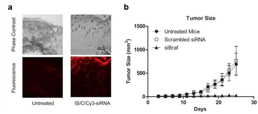
of swCNTs loaded with Cy3-labelled siRNA demonstrated the capacity of the nucleic acid sequence
to cross the epidermis and reach the dermal layer (Figure 1a), while the same formulation without
swCNTs did not cross the stratum corneum. Though these experiments highlighted the key role
played by swCNTs in helping the siRNA to cross the epidermis, is should be noted that 10% DMSO,
a well-known permeation enhancer, was added to the formulation. The authors then investigated thePharmaceutics 2019, 11, 256 9 of 15
actual pharmaceutical potential of the swCNTs loaded with an anti-Braf siRNA in a mouse model of
melanoma (intradermal inoculation of B16-F10 cells). Down-regulation experiments in the tumor cells
indicated a remarkable 70% knockdown of Braf when delivering siRNA loaded in the functionalized
swCNTs, 24 h after a single topical administration. Moreover, multiple administrations every 2 days
for 25 days resulted in drastic inhibition of tumor growth (Figure 1b). Overall, these experiments by
Siu et al. highlighted for the first time the potential of swCNTs to deliver pharmaceutics, even relatively
large and hydrophilic molecules, across the epidermis, though the actual role of DMSO should be
clarified. Nevertheless, prior to any actual clinical translation, the reported experiments will need to be
validated in a more quantitative manner and in a more relevant animal model for transdermal studies,
such as newborn pigs. Above all, while the described protocol used a functionalization step to increase
the solubility of the CNTs, which has been reported to reduce their toxic accumulation and retention
within the body, the actual fate of the CNTs as well as the non-degradable functionalization polymer
will have to be carefully investigated [83,84].
Table 1. Transdermal drug delivery systems covered in the review.
In Vivo Clinical
Category Technology Pros Cons Ref.
Evaluation Trial
Sustained release
Yes (guinea Control of the
Biphasic vesicles Phase II Versatility (small and [15–24]
pig) delivered dose
large molecules)
Chemical permeation
enhancer Ionic liquids (ILs) APIs with enhanced skin Requires specific
(CPE)-based systems and active permeation properties of choice of
[42,43,
pharmaceutical Yes (rats) N/A ionic liquids counter-ions
47]
ingredient-ionic Properties can be Limited to small
liquids (API-ILs) fine-tuned molecules
Physical Ex vivo: Manufacturing cost
Hollow Large doses
enhancer-based rabbit ear Completed Potential clogging [53,54]
microneedles Versatility
systems skin Skilled personnel
Low skin irritation
Sustained release and Lecithin could lead
Lecithin-based [61,64,
N/A N/A higher permeation to skin permeation
microemulsions 65,69]
compared to standard complications
emulsions
Nanoparticle-based
Biocompatibility
systems
Versatility unclear
Archaeosomes
Pharmaceutics 2019, 11, x N/A N/A [74]
Sustained release Permeation 9 of 16
mechanism unclear
animal model for transdermal studies, such as newborn pigs.Effective
Above skin
all, while the described protocol
used a functionalization step to increase the solubility of the CNTs, which Complexity
has been reported to reduce
Carbon nanotubes permeation without
Yes (mice) N/A Biocompatibility [81]
(CNTs)
their toxic accumulation CPEs fate of the CNTs as well as the non-
and retention within the body, the actual unclear
degradable functionalization polymer will have to be carefully High drug loading[83,84].
investigated
Figure 1. Figure
Carbon 1. Carbon nanotube
nanotube (CNT)-mediated delivery
(CNT)-mediated delivery of of
siRNA in mice.
siRNA (a) Representative
in mice. images of images of
(a) Representative
tumor-bearing mouse skin treated with Cy3-labeled siRNA loaded CNTs; (b) tumor size evolution
tumor-bearing mouse skin treated with Cy3-labeled siRNA loaded CNTs; (b) tumor size evolution after
after topical administration of siRNA-loaded CNTs. Three days after the mice were injected with
topical administration of siRNA-loaded CNTs. Three days after the mice were injected with tumor
tumor cells, siRNA-loaded CNT solution was applied every 2 days for 25 days. Adapted with
cells, siRNA-loaded
permission from [81]; solution
CNT was
published by applied
Elsevier, 2014.every 2 days for 25 days. Adapted with permission
from [81]; published by Elsevier, 2014.
Table 1. Transdermal drug delivery systems covered in the review.
In Vivo Clinical
Category Technology Pros Cons Ref.
Evaluation Trial
Sustained
release
Yes Control of the
Biphasic Versatility
(guinea Phase II delivered [15–24]
vesicles (small andPharmaceutics 2019, 11, 256 10 of 15
5. Conclusions
In this brief review, we summarized and discussed the main topical and transdermal drug delivery
technologies that have been developed in recent years in Canada. Overall, although the number of
works describing new technologies appears limited at first glance, it is interesting to note that they
cover a wide range of strategies, from nanotechnology to CPEs, to hollow MNs. Furthermore, the
research spans from the development of innovative technologies based on new materials that have
shown a remarkable capacity to enhance the transdermal delivery of high molecular weight drugs
(e.g., functionalized CNTs), but require intensive and more quantitative studies to better identify their
clinical potentials and biocompatibility profiles; to more fundamental studies with the broader goal
of identifying superior enhancement compounds based on well-known materials. Finally, advanced
strategies have been developed over the past decade, and are currently being further studied by private
companies in clinical settings (e.g., the BiPhasixTM technology) and could ideally soon reach the market,
highlighting the favorable environment for the development of new medical and pharmaceutical
technologies in the country.
Nonetheless, it should be noted that some transdermal drug delivery technologies undergoing
rapid development are not currently represented in Canada. A notable example of this can be seen
in polymeric MNs, two varieties of which have seen increasing use for drug delivery. The first of
these are dissolving MNs made of soluble polymers, where a drug is loaded within the soluble
polymeric matrix of the MNs, allowing release across the skin upon dissolution of the tips following
application [85]. The Prausnitz group at Georgia Tech helped pioneer this technology in the context of
non-invasive vaccination and drug delivery [86–88] and continue to work on this topic [89]. While
research is also active globally, particularly in the context of delivering peptide-based drugs and
macromolecules [90–92], no Canadian groups are currently investigating this class of MNs.
The other class of polymeric MNs being studied for transdermal drug delivery is swellable
hydrogel MNs, typically made of crosslinked hydrophilic polymers able to swell by absorbing fluid
from the skin. While these have also been used for sampling biological fluids from the skin [93], the
Donnelly group at Queen’s University Belfast have primarily studied them for drug delivery purposes,
as their swelling properties also allow drug molecules contained within the MNs to flow into the
skin after application [94,95]. This research appears to have progressed significantly, with recent
studies focusing on optimizing the system for clinical applications [96–98]. Iontophoresis is another
transdermal drug delivery strategy currently experiencing worldwide growth [99]. By passing an
electrical current through the skin, this technique serves to enhance skin permeability, as well as allow
positively charged compounds to be transported into the skin by the resulting electric field. Though
various groups are developing iontophoresis-based transdermal delivery methods for both small
molecule- and peptide-based drugs [100,101], the topic is seemingly not undergoing active research
within Canada.
These methods, alongside the ones discussed previously, serve to reveal the current state of
transdermal drug delivery technology worldwide. Though Canada has generated meaningful
contributions within the past decade, it remains clear that many opportunities for further work exist, if
groups within Canada wish to further the progression of this field.
Author Contributions: M.R., E.L., and S.B. performed the primary literature search and wrote the manuscript.
D.B. corrected and implemented the manuscript.
Funding: The authors acknowledge financial support from the Fonds de Recherche du Québec, the Bourses
d’excellence TransMedTech (M.R.), and the Canadian Generic Pharmaceutical Association and Biosimilars
Canada (D.B.).
Conflicts of Interest: The authors declare no conflict of interest.Pharmaceutics 2019, 11, 256 11 of 15
References
1. Babity, S.; Roohnikan, M.; Brambilla, D. Advances in the design of transdermal microneedles for diagnostic
and monitoring applications. Small 2018, 14, 1803186. [CrossRef] [PubMed]
2. Benson, H.A.E. Transdermal and Topical Drug Delivery: Principles and Practice; Benson, H.A.E., Watkinson, A.C.,
Eds.; Wiley: Hoboken, NJ, USA, 2012; Chapter 1, pp. 1–22.
3. Menon, G.K.; Cleary, G.W.; Lane, M.E. The structure and function of the stratum corneum. Int. J. Pharm.
2012, 435, 3–9. [CrossRef] [PubMed]
4. Eckhart, L.; Lippens, S.; Tschachler, E.; Declercq, W. Cell death by cornification. Biochim. Biophys. Acta Mol.
Cell Res. 2013, 1833, 3471–3480. [CrossRef]
5. Michaels, A.S.; Chandrasekaran, S.K.; Shaw, J.E. Drug permeation through human skin: Theory and in vitro
experimental measurement. AIChE J. 1975, 21, 985–996. [CrossRef]
6. Prausnitz, M.R.; Mitragotri, S.; Langer, R. Current status and future potential of transdermal drug delivery.
Nat. Rev. Drug Discov. 2004, 3, 115–124. [CrossRef]
7. Arora, A.; Prausnitz, M.R.; Mitragotri, S. Micro-scale devices for transdermal drug delivery. Int. J. Pharm.
2008, 364, 227–236. [CrossRef]
8. Prausnitz, M.R.; Langer, R. Transdermal drug delivery. Nat. Biotechnol. 2008, 26, 1261–1268. [CrossRef]
[PubMed]
9. Watkinson, A.C.; Kearney, M.C.; Quinn, H.L.; Courtenay, A.J.; Donnelly, R.F. Future of the transdermal
drug delivery market—Have we barely touched the surface? Expert Opin. Drug Deliv. 2016, 13, 523–532.
[CrossRef]
10. Paudel, K.S.; Milewski, M.; Swadley, C.L.; Brogden, N.K.; Ghosh, P.; Stinchcomb, A.L. Challenges and
opportunities in dermal/transdermal delivery. Ther. Deliv. 2010, 1, 109–131. [CrossRef]
11. Pharmaceutical Industry Profile. Available online: http://www.webcitation.org/query?url=https%3A%2F%
2Fwww.ic.gc.ca%2Feic%2Fsite%2Flsg-pdsv.nsf%2Feng%2Fh_hn01703.html&date=2019-02-02 (accessed on
2 February 2019).
12. Chen, Y.; Quan, P.; Liu, X.; Wang, M.; Fang, L. Novel chemical permeation enhancers for transdermal drug
delivery. Asian J. Pharm. 2014, 9, 51–64. [CrossRef]
13. Karande, P.; Jain, A.; Ergun, K.; Kipersky, V.; Mitragotri, S. Design principles of chemical penetration
enhancers for transdermal drug delivery. Proc. Natl. Acad. Sci. USA 2005, 102, 4688–4693. [CrossRef]
14. Williams, A.C.; Barry, B.W. Penetration enhancers. Adv. Drug Deliv. Rev. 2004, 56, 603–618. [CrossRef]
[PubMed]
15. Foldvari, M. Biphasic Multilamellar Lipid Vesicles. U.S. Patent 5,853,755, 29 December 1998.
16. Moghadam, S.H.; Saliaj, E.; Wettig, S.D.; Dong, C.; Ivanova, M.V.; Huzil, J.T.; Foldvari, M. Effect of chemical
permeation enhancers on stratum corneum barrier lipid organizational structure and interferon alpha
permeability. Mol. Pharm. 2013, 10, 2248–2260. [CrossRef] [PubMed]
17. King, M.J.; Badea, I.; Solomon, J.; Kumar, P.; Gaspar, K.J.; Foldvari, M. Transdermal delivery of insulin from a
novel biphasic lipid system in diabetic rats. Diabetes Technol. Ther. 2002, 4, 479–488. [CrossRef]
18. Baca-Estrada, M.E.; Foldvari, M.; Ewen, C.; Badea, I.; Babiuk, L.A. Effects of IL-12 on immune responses
induced by transcutaneous immunization with antigens formulated in a novel lipid-based biphasic delivery
system. Vaccine 2000, 18, 1847–1854. [CrossRef]
19. King, M.; Kumar, P.; Michel, D.; Batta, R.; Foldvari, M. In vivo sustained dermal delivery and pharmacokinetics
of interferon alpha in biphasic vesicles after topical application. Eur. J. Pharm. Biopharm. 2014, 84, 532–539.
[CrossRef]
20. Foldvari, M.; Badea, I.; Wettig, S.; Baboolal, D.; Kumar, P.; Creagh, A.L.; Haynes, C.A. Topical delivery of
interferon alpha by biphasic vesicles: Evidence for a novel nanopathway across the stratum corneum. Mol.
Pharm. 2010, 7, 751–762. [CrossRef]
21. Foldvari, M.; Badea, I.; Kumar, P.; Wettig, S.; Batta, R.; King, M.; Shear, N. Biphasic vesicles for topical
delivery of interferon alpha in human volunteers and treatment of patients with human papillomavirus
infections. Curr. Drug Deliv. 2011, 8, 307–319. [CrossRef]
22. Rougier, A.; Lotte, C. Predictive approaches I: The stripping technique. In Topical Drug Bioavailability,
Bioequivalence, and Penetration; Shah, V.P., Maibach, H.I., Eds.; Springer: Boston, MA, USA, 1993; pp. 163–181.Pharmaceutics 2019, 11, 256 12 of 15
23. Kurzeja, R.; Böhmer, G.; Schneider, A. Clinical outcome of topical interferon Alpha-2b cream in phase II trial
for LSIL/CIN 1 patients. J. Cancer Ther. 2011, 2, 203–208. [CrossRef]
24. Altum Pharmaceuticals Pipeline. Available online: http://www.webcitation.org/query?url=https%3A%2F%
2Fwww.altumpharma.com%2Fpipeline%2F&date=2019-02-02 (accessed on 2 February 2019).
25. Ashtikar, A.; Nagarsekar, K.; Fahr, A. Transdermal delivery from liposomal formulations—Evolution of the
technology over the last three decades. J. Control. Release 2016, 242, 126–140. [CrossRef] [PubMed]
26. Beloqui, A.; Solinís, M.Á.; Rodríguez-Gascón, A.; Almeida, A.J.; Préat, V. Nanostructured lipid carriers:
Promising drug delivery systems for future clinics. Nanomedicine 2016, 12, 143–161. [CrossRef]
27. Mennini, N.; Cirri, M.; Maestrelli, F.; Mura, P. Comparison of liposomal and NLC (nanostructured lipid carrier)
formulations for improving the transdermal delivery of oxaprozin: Effect of cyclodextrin complexation. Int.
J. Pharm. 2016, 515, 684–691. [CrossRef] [PubMed]
28. Pinto, M.F.; Moura, C.C.; Nunes, C.; Segundo, M.A.; Costa Lima, S.A.; Reis, S. A new topical formulation
for psoriasis: Development of methotrexate-loaded nanostructured lipid carriers. Int. J. Pharm. 2014, 477,
519–526. [CrossRef]
29. Puglia, C.; Sarpietro, M.G.; Bonina, F.; Castelli, F.; Zammataro, M.; Chiechio, S. Development, characterization,
and in vitro and in vivo evaluation of benzocaine- and lidocaine-loaded nanostructured lipid carriers. J.
Pharm. Sci. 2011, 100, 1892–1899. [CrossRef]
30. Das, R.N.; Roy, K. Advances in QSPR/QSTR models of ionic liquids for the design of greener solvents of the
future. Mol. Divers. 2013, 17, 151–196. [CrossRef]
31. Gadilohar, B.L.; Shankarling, G.S. Choline based ionic liquids and their applications in organic transformation.
J. Mol. Liquids 2017, 227, 234–261. [CrossRef]
32. Egorova, K.S.; Gordeev, E.G.; Ananikov, V.P. Biological activity of ionic liquids and their application in
pharmaceutics and medicine. Chem. Rev. 2017, 117, 7132–7189. [CrossRef] [PubMed]
33. Kundu, N.; Roy, S.; Mukherjee, D.; Maiti, T.K.; Sarkar, N. Unveiling the interaction between fatty-acid-modified
membrane and hydrophilic imidazolium-based ionic liquid: Understanding the mechanism of ionic liquid
cytotoxicity. J. Phys. Chem. B 2017, 121, 8162–8170. [CrossRef]
34. Lim, G.S.; Jaenicke, S.; Klähn, M. How the spontaneous insertion of amphiphilic imidazolium-based
cations changes biological membranes: A molecular simulation study. Phys. Chem. Chem. Phys. 2015, 17,
29171–29183. [CrossRef] [PubMed]
35. Zakrewsky, M.; Lovejoy, K.S.; Kern, T.L.; Miller, T.E.; Le, V.; Nagy, A.; Goumas, A.M.; Iyer, R.S.; Del Sesto, R.E.;
Koppisch, A.T.; et al. Ionic liquids as a class of materials for transdermal delivery and pathogen neutralization.
Proc. Natl. Acad. Sci. USA 2014, 111, 13313–13318. [CrossRef]
36. Lane, M.E. Skin penetration enhancers. Int. J. Pharm. 2013, 447, 12–21. [CrossRef]
37. Sidat, Z.; Marimuthu, T.; Kumar, P.; du Toit, L.C.; Kondiah, P.P.D.; Choonara, Y.E.; Pillay, V. Ionic liquids as
potential and synergistic permeation enhancers for transdermal drug delivery. Pharmaceutics 2019, 11, 96.
[CrossRef]
38. Kelley, S.P.; Narita, A.; Holbrey, J.D.; Green, K.D.; Reichert, W.M.; Rogers, R.D. Understanding the effects of
ionicity in salts, solvates, co-crystals, ionic co-crystals, and ionic liquids, rather than nomenclature, is critical
to understanding their behavior. Cryst. Growth Des. 2013, 13, 965–975. [CrossRef]
39. Wang, H.; Gurau, G.; Shamshina, J.L.; Cojocaru, O.A.; Janikowski, J.; MacFarlane, D.R.; Davis, J.H.J.;
Rogers, R.D. Simultaneous membrane transport of two active pharmaceutical ingredients by charge assisted
hydrogen bond complex formation. Chem. Sci. 2014, 5, 3449–3456. [CrossRef]
40. Raza, K.; Kumar, P.; Ratan, S.; Malik, R.; Arora, S. Polymorphism: The phenomenon affecting the performance
of drugs. SOJ Pharm. Pharm. Sci. 2014, 1, 10. [CrossRef]
41. Balk, A.; Holzgrabe, U.; Meinel, L. ‘Pro et contra’ ionic liquid drugs—Challenges and opportunities for
pharmaceutical translation. Eur. J. Pharm. Biopharm. 2015, 94, 291–304. [CrossRef]
42. Hough, W.L.; Smiglak, M.; Rodríguez, H.; Swatloski, R.P.; Spear, S.K.; Daly, D.T.; Pernak, J.; Grisel, J.E.;
Carliss, R.D.; Soutullo, M.D.; et al. The third evolution of ionic liquids: Active pharmaceutical ingredients.
New J. Chem. 2007, 31, 1429–1436. [CrossRef]
43. Zavgorodnya, O.; Shamshina, J.L.; Mittenthal, M.; McCrary, P.D.; Rachiero, G.P.; Titi, H.M.; Rogers, R.D.
Polyethylene glycol derivatization of the non-active ion in active pharmaceutical ingredient ionic liquids
enhances transdermal delivery. New J. Chem. 2017, 41, 1499–1508. [CrossRef]Pharmaceutics 2019, 11, 256 13 of 15
44. Stoimenovski, J.; MacFarlane, D.R. Enhanced membrane transport of pharmaceutically active protic ionic
liquids. Chem. Commun. 2011, 47, 11429–11431. [CrossRef]
45. Thong, H.Y.; Zhai, H.; Maibach, H.I. Percutaneous penetration enhancers: An overview. Skin Pharmacol.
Physiol. 2007, 20, 272–282. [CrossRef]
46. Lloyd, R.S.; Blythe, J.O.J. Clinical experiences with lidocaine as a local anesthetic. J. Am. Dent. Assoc. 1949,
39, 296–298. [CrossRef]
47. Berton, P.; Di Bona, K.R.; Yancey, D.; Rizvi, S.A.A.; Gray, M.; Gurau, G.; Shamshina, J.L.; Rasco, J.F.;
Rogers, R.D. Transdermal bioavailability in rats of lidocaine in the forms of ionic liquids, salts, and deep
eutectic. ACS Med. Chem. Lett. 2017, 8, 498–503. [CrossRef]
48. Park, H.J.; Prausnitz, M.R. Lidocaine-ibuprofen ionic liquid for dermal anesthesia. AIChE J. 2015, 61,
2732–2738. [CrossRef]
49. Van der Maaden, K.; Jiskoot, W.; Bouwstra, J. Microneedle technologies for (trans)dermal drug and vaccine
delivery. J. Control. Release 2012, 161, 645–655. [CrossRef]
50. Ovsianikov, A.; Chichkov, B.; Mente, P.; Monteiro-Riviere, N.A.; Doraiswamy, A.; Narayan, R.J. Two photon
polymerization of polymer-ceramic hybrid materials for transdermal drug delivery. Int. J. Appl. Ceram.
Technol. 2007, 4, 22–29. [CrossRef]
51. Yung, K.L.; Xu, Y.; Kang, C.; Liu, H.; Tam, K.F.; Ko, S.M.; Kwan, F.Y.; Lee, T.M.H. Sharp tipped plastic hollow
microneedle array by microinjection moulding. J. Micromech. Microeng. 2012, 22, 015016. [CrossRef]
52. Lhernould, M.S.; Deleers, M.; Delchambre, A. Hollow polymer microneedles array resistance and insertion
tests. Int. J. Pharm. 2015, 480, 152–157. [CrossRef]
53. Mansoor, I.; Häfeli, U.O.; Stoeber, B. Hollow out-of-plane polymer microneedles made by solvent casting for
transdermal drug delivery. J. Microelectromech. Syst. 2012, 21, 44–52. [CrossRef]
54. Mansoor, I.; Liu, Y.; Häfeli, U.O.; Stoeber, B. Arrays of hollow out-of-plane microneedles made by metal
electrodeposition onto solvent cast conductive polymer structures. J. Micromech. Microeng. 2013, 23, 085011.
[CrossRef]
55. University of British Columbia. Safety Demonstration of Microneedle Insertion, NCT02995057. Available
online: https://clinicaltrials.gov/ct2/show/NCT02995057 (accessed on 31 May 2019).
56. Takeuchi, I.; Shimamura, Y.; Kakami, Y.; Kameda, T.; Hattori, K.; Miura, S.; Okumura, M.; Inagi, T.; Terada, H.;
Makino, K. Transdermal delivery of 40-nm silk fibroin nanoparticles. Colloids Surf. B Biointerfaces 2018, 175,
564–568. [CrossRef] [PubMed]
57. Nastiti, C.; Ponto, T.; Abd, E.; Grice, J.E.; Benson, H.A.E.; Roberts, M.S. Topical nano and microemulsions for
skin delivery. Pharmaceutics 2017, 9, 37. [CrossRef] [PubMed]
58. Kogan, A.; Garti, N. Microemulsions as transdermal drug delivery vehicles. Adv. Colloid Interface Sci. 2006,
123–126, 369–385. [CrossRef]
59. Kreilgaard, M.; Pedersen, E.J.; Jaroszewski, J.W. NMR characterisation and transdermal drug delivery
potential of microemulsion systems. J. Control. Release 2000, 69, 421–433. [CrossRef]
60. Lee, M.-W.C. Topical triple-anesthetic gel compared with 3 topical anesthetics. Cosmet. Dermatol. 2003, 16,
35–37.
61. Yuan, J.S.; Yip, A.; Nguyen, N.; Chu, J.; Wen, X.Y.; Acosta, E.J. Effect of surfactant concentration on transdermal
lidocaine delivery with linker microemulsions. Int. J. Pharm. 2010, 392, 274–284. [CrossRef] [PubMed]
62. Van Nieuwenhuyzen, W.; Tomás, M.C. Update on vegetable lecithin and phospholipid technologies. Eur. J.
Lipid Sci. Technol. 2008, 110, 472–486. [CrossRef]
63. Heuschkel, S.; Goebel, A.; Neubert, R.H. Microemulsions—Modern colloidal carrier for dermal and
transdermal drug delivery. J. Pharm. Sci. 2008, 97, 603–631. [CrossRef]
64. Yuan, J.S.; Ansari, M.; Samaan, M.; Acosta, E.J. Linker-based lecithin microemulsions for transdermal delivery
of lidocaine. Int. J. Pharm. 2008, 349, 130–143. [CrossRef]
65. Xuan, X.Y.; Cheng, Y.L.; Acosta, E.J. Lecithin-linker microemulsion gelatin gels for extended drug delivery.
Pharmaceutics 2012, 4, 104–129. [CrossRef] [PubMed]
66. Jacobi, U.; Kaiser, M.; Toll, R.; Mangelsdorf, S.; Audring, H.; Otberg, N.; Sterry, W.; Lademann, J. Porcine ear
skin: An in vitro model for human skin. Skin Res. Technol. 2007, 13, 19–24. [CrossRef]
67. Hoppel, M.; Juric, S.; Ettl, H.; Valenta, C. Effect of monoacyl phosphatidylcholine content on the formation
of microemulsions and the dermal delivery of flufenamic acid. Int. J. Pharm. 2015, 479, 70–76. [CrossRef]
[PubMed]Pharmaceutics 2019, 11, 256 14 of 15
68. Hajjar, B.; Zier, K.I.; Khalid, N.; Azarmi, S.; Löbenberg, R. Evaluation of a microemulsion-based gel
formulation for topical drug delivery of diclofenac sodium. J. Pharm. Investig. 2018, 48, 351–362. [CrossRef]
69. Adeyeye, M.C.; Jain, A.C.; Ghorab, M.K.; Reilly, W.J.J. Viscoelastic evaluation of topical creams containing
microcrystalline cellulose/sodium carboxymethyl cellulose as stabilizer. AAPS Pharm. Sci. Technol. 2002, 3,
16–25. [CrossRef]
70. Acosta, E.; Chung, O.; Xuan, X.Y. Lecithin-linker microemulsions in transdermal delivery. J. Drug Deliv. Sci.
Technol. 2011, 21, 77–87. [CrossRef]
71. Kriwet, K.; Müller-Goymann, C.C. Diclofenac release from phospholipid drug systems and permeation
through excised human stratum corneum. Int. J. Pharm. 1995, 135, 231–242. [CrossRef]
72. Young, K.D. What’s new in topical anesthesia. Clin. Pediatr. Emerg. Med. 2007, 8, 232–239. [CrossRef]
73. Friedman, P.M.; Mafong, E.A.; Friedman, E.S.; Geronemus, R.G. Topical anesthetics update: EMLA and
beyond. Dermatol. Surg. 2001, 27, 1019–1026. [CrossRef] [PubMed]
74. Jia, Y.; McCluskie, M.J.; Zhang, D.; Monette, R.; Iqbal, U.; Moreno, M.; Sauvageau, J.; Williams, D.;
Deschatelets, L.; Jakubek, Z.J.; et al. In vitro evaluation of archaeosome vehicles for transdermal vaccine
delivery. J. Liposome Res. 2018, 28, 305–314. [CrossRef] [PubMed]
75. Higa, L.H.; Arnal, L.; Vermeulen, M.; Perez, A.P.; Schilrreff, P.; Mundiña-Weilenmann, C.; Yantorno, O.;
Vela, M.E.; Morilla, M.J.; Romero, E.L. Ultradeformable archaeosomes for needle free nanovaccination with
Leishmania braziliensis antigens. PLoS ONE 2016, 11, e0150185. [CrossRef] [PubMed]
76. Jain, K.K. Advances in use of functionalized carbon nanotubes for drug design and discovery. Expert Opin.
Drug Discov. 2012, 7, 1029–1037. [CrossRef]
77. Mehra, N.K.; Jain, K.; Jain, N.K. Pharmaceutical and biomedical applications of surface engineered carbon
nanotubes. Drug Discov. Today 2015, 20, 750–759. [CrossRef]
78. Karimi, M.; Solati, N.; Ghasemi, A.; Estiar, M.A.; Hashemkhani, M.; Kiani, P.; Mohamed, E.; Saeidi, A.;
Taheri, M.; Avci, P.; et al. Carbon nanotubes part II: A remarkable carrier for drug and gene delivery. Expert
Opin. Drug Deliv. 2015, 12, 1089–1105. [CrossRef]
79. Zintchenko, A.; Philipp, A.; Dehshahri, A.; Wagner, E. Simple modifications of branched PEI lead to highly
efficient siRNA carriers with low toxicity. Bioconj. Chem. 2008, 19, 1448–1455. [CrossRef] [PubMed]
80. Wang, L.; Shi, J.; Zhang, H.; Li, H.; Gao, Y.; Wang, Z.; Wang, H.; Li, L.; Zhang, C.; Chen, C.; et al. Synergistic
anticancer effect of RNAi and photothermal therapy mediated by functionalized single-walled carbon
nanotubes. Biomaterials 2013, 34, 262–274. [CrossRef]
81. Siu, K.S.; Chen, D.; Zheng, X.; Zhang, X.; Johnston, N.; Liu, Y.; Yuan, K.; Koropatnick, J.; Gillies, E.R.; Min, W.P.
Non-covalently functionalized single-walled carbon nanotube for topical siRNA delivery into melanoma.
Biomaterials 2014, 35, 3435–3442. [CrossRef]
82. Degim, I.T.; Burgess, D.J.; Papadimitrakopoulos, F. Carbon nanotubes for transdermal drug delivery. J.
Microencapsul. 2010, 27, 669–681. [CrossRef] [PubMed]
83. Kesharwani, P.; Gajbhiye, V.; Jain, N.K. A review of nanocarriers for the delivery of small interfering RNA.
Biomaterials 2012, 33, 7138–7150. [CrossRef]
84. Principi, E.; Girardello, R.; Bruno, A.; Manni, I.; Gini, E.; Pagani, A.; Grimaldi, A.; Ivaldi, F.; Congiu, T.; De
Stefano, D.; et al. Systemic distribution of single-walled carbon nanotubes in a novel model: Alteration of
biochemical parameters, metabolic functions, liver accumulation, and inflammation in vivo. Int. J. Nanomed.
2016, 11, 4299–4316. [CrossRef]
85. Lee, J.W.; Park, J.-H.; Prausnitz, M.R. Dissolving microneedles for transdermal drug delivery. Biomaterials
2008, 29, 2113–2124. [CrossRef] [PubMed]
86. Henry, S.; McAllister, D.V.; Allen, M.G.; Prausnitz, M.R. Microfabricated microneedles: A novel approach to
transdermal drug delivery. J. Pharm. Sci. 1998, 87, 922–925. [CrossRef]
87. Prausnitz, M.R.; Mikszta, J.A.; Cormier, M.; Andrianov, A.K. Microneedle-based vaccines. Curr. Top. Microbiol.
Immunol. 2009, 333, 369–393. [CrossRef]
88. Sullivan, S.P.; Koutsonanos, D.G.; del Pilar Martin, M.; Lee, J.-W.; Zarnitsyn, V.; Murthy, N.; Compans, R.W.;
Skountzou, I.; Prausnitz, M.R. Dissolving polymer microneedle patches for influenza vaccination. Nat. Med.
2010, 16, 915–920. [CrossRef] [PubMed]You can also read