Age-Associated TET2 Mutations: Common Drivers of Myeloid Dysfunction, Cancer and Cardiovascular Disease - MDPI
←
→
Page content transcription
If your browser does not render page correctly, please read the page content below
International Journal of
            Molecular Sciences

Review
Age-Associated TET2 Mutations: Common Drivers of
Myeloid Dysfunction, Cancer and
Cardiovascular Disease
Christina K. Ferrone, Mackenzie Blydt-Hansen and Michael J. Rauh *
 Department of Pathology and Molecular Medicine, Queen’s University, Kingston, ON K7L 3N6, Canada;
 christina.ferrone@queensu.ca (C.K.F.); 15mbh4@queensu.ca (M.B.-H.)
 * Correspondence: rauhm@queensu.ca

 Received: 17 December 2019; Accepted: 13 January 2020; Published: 17 January 2020

 Abstract: Acquired, inactivating mutations in Tet methylcytosine dioxygenase 2 (TET2) are detected
 in peripheral blood cells of a remarkable 5%–10% of adults greater than 65 years of age. They impart
 a hematopoietic stem cell advantage and resultant clonal hematopoiesis of indeterminate potential
 (CHIP) with skewed myelomonocytic differentiation. CHIP is associated with an overall increased risk
 of transformation to a hematological malignancy, especially myeloproliferative and myelodysplastic
 neoplasms (MPN, MDS) and acute myeloid leukemia (AML), of approximately 0.5% to 1% per
 year. However, it is becoming increasingly possible to identify individuals at greatest risk, based
 on CHIP mutational characteristics. CHIP, and particularly TET2-mutant CHIP, is also a novel,
 significant risk factor for cardiovascular diseases, related in part to hyper-inflammatory, progeny
 macrophages carrying TET2 mutations. Therefore, somatic TET2 mutations contribute to myeloid
 expansion and innate immune dysregulation with age and contribute to prevalent diseases in the
 developed world—cancer and cardiovascular disease. Herein, we describe the impact of detecting
 TET2 mutations in the clinical setting. We also present the rationale and promise for targeting
 TET2-mutant and other CHIP clones, and their inflammatory environment, as potential means of
 lessening risk of myeloid cancer development and dampening CHIP-comorbid inflammatory diseases.

 Keywords: clonal hematopoiesis; TET2; driver mutations; NGS; clinical detection; inflammation;
 cancer progression; comorbid disease; aging; targeting TET2 therapeutically

1. Introduction
     Recent technological advancements have permitted the identification of a myriad of genes
mutated in myeloid cancers [1–7]. TET2 is one of the most commonly mutated genes in these
diseases, being highly involved in epigenetic regulation, including cytosine demethylation [5,8].
TET2-inactivating mutations also frequently occur in clonal hematopoiesis of indeterminate potential
(CHIP), a “pre-leukemic” condition involving aberrant clonal expansion of hematopoietic stem and
progenitor cells (HSPCs) in the bone marrow [9,10]. Herein, we explore the importance of detecting
mutations such as those observed in TET2 and its partner DNA-methyltransferase 3A (DNMT3A;
another key epigenetic regulator, frequently mutated in CHIP and myeloid disease), and some of the
associations of these genetic variations with CHIP and myeloid disease progression. We further discuss
recent findings regarding CHIP, TET2 mutations and their connections to increased inflammation and
comorbid disease. Finally, we briefly describe recent attempts to therapeutically increase wildtype
TET2 protein levels or activity in the presence of heterozygous loss-of-function mutations.

Int. J. Mol. Sci. 2020, 21, 626; doi:10.3390/ijms21020626                            www.mdpi.com/journal/ijms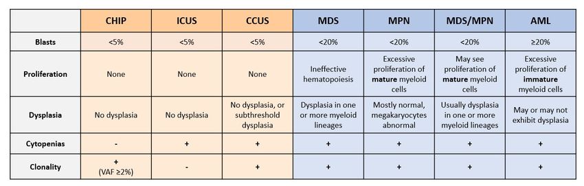
Int. J. Mol. Sci. 2020, 21, 626 2 of 16
Int. J. Mol. Sci. 2020, 21, x FOR PEER REVIEW 2 of 17
2.
2. Hematopoiesis
Hematopoiesis and
and Myeloid
Myeloid Malignancies
Malignancies
Hematopoiesis
Hematopoiesis is is the
the production
production of of mature
mature blood
blood cells
cells from
from HSPCs.
HSPCs. This
This process
process is
is polyclonal,
polyclonal,
with
with a range of approximately 50,000–200,000 HSPCs each giving rise to a fraction of mature blood
a range of approximately 50,000–200,000 HSPCs each giving rise to a fraction of mature blood
cells
cells [11]. Myeloid malignancies occur when individual HSPC mutant clones proliferate, expand and
[11]. Myeloid malignancies occur when individual HSPC mutant clones proliferate, expand and
skew towards
skew towards myeloid
myeloid lineages.lineages. Thesemyelodysplastic
These include include myelodysplastic
syndromes (MDS), syndromes (MDS),
myeloproliferative
myeloproliferative
neoplasms (MPN), neoplasms (MPN), myelodysplastic/myeloproliferative
myelodysplastic/myeloproliferative neoplasms (MDS/MPN), neoplasms (MDS/MPN),
and acute myeloid
and acute myeloid leukemia (AML) (Figure 1) [1]. The WHO describes MDS
leukemia (AML) (Figure 1) [1]. The WHO describes MDS as having “ineffective hematopoiesis”, as having “ineffective
hematopoiesis”,
characterized bycharacterized by abnormalcell
abnormal hematopoietic hematopoietic cell shapes dysplasia)
shapes (morphological (morphological dysplasia)
and peripheral
and peripheral cytopenia (low blood cell counts) [1]. MPN are characterized by
cytopenia (low blood cell counts) [1]. MPN are characterized by the proliferation of mature myeloidthe proliferation of
mature myeloid
cells, usually cells,morphological
lacking usually lacking morphological
dysplasia, dysplasia,
while myeloid while myeloid
neoplasms neoplasms
with clinical, with
laboratory,
clinical, laboratory, and morphologic features overlapping between MDS
and morphologic features overlapping between MDS and MPN are classified as MDS/MPN [12]. and MPN are classified as
MDS/MPN [12]. AML involves the proliferation of immature myeloid cells (“blasts”)
AML involves the proliferation of immature myeloid cells (“blasts”) related to mutations that block related to
mutations
normal HSPC thatdifferentiation
block normal and, HSPC differentiation
like and, like
MPN, can involve MPN,mutations
recurrent can involve recurrent
associated mutations
with cellular
associated with cellular
proliferation [1,12]. proliferation [1,12].
Figure 1.
Figure Summary of
1. Summary of current
current pre-neoplastic
pre-neoplastic states
states and
and myeloid
myeloid neoplasms. Pre-neoplastic states
neoplasms. Pre-neoplastic states (in
(in
orange: clonal hematopoiesis of indeterminate potential, CHIP; idiopathic cytopenia
orange: clonal hematopoiesis of indeterminate potential, CHIP; idiopathic cytopenia of of undetermined
significance, ICUS;
undetermined and clonalICUS;
significance, cytopenias of undetermined
and clonal cytopenias significance; CCUS)
of undetermined are classified
significance; primarily
CCUS) are
based on the presence of clonality (acquired DNA variants or chromosomal aberrations),
classified primarily based on the presence of clonality (acquired DNA variants or chromosomal and on the
presence of peripheral cytopenias [13,14]. The main categories of myeloid neoplasm (blue) are primarily
aberrations), and on the presence of peripheral cytopenias [13,14]. The main categories of myeloid
classified based on blast percentage (morphologically primitive cells, including HSPCs), by the type
neoplasm (blue) are primarily classified based on blast percentage (morphologically primitive cells,
of cells that proliferate aberrantly, and by the morphology of the myeloid cells [15]. VAF, variant
including HSPCs), by the type of cells that proliferate aberrantly, and by the morphology of the
allele frequency; MDS, myelodysplastic syndromes; MPN, myeloproliferative neoplasms; MDS/MPN,
myeloid cells [15]. VAF, variant allele frequency; MDS, myelodysplastic syndromes; MPN,
overlapping MDS and MPN; AML, acute myeloid leukemia.
myeloproliferative neoplasms; MDS/MPN, overlapping MDS and MPN; AML, acute myeloid
leukemia. Cytopenias and Shortcomings of Current Diagnostic Techniques
3. Unexplained
Unexplained
3. Unexplained blood cytopenia
Cytopenias is another condition,
and Shortcomings of Current in Diagnostic
which individuals are deficient in one or
Techniques
more types of blood cell (red cells, white cells, or platelets), where the origin of this deficiency is not
Unexplained
attributable to anyblood cytopenia
identifiable causeis another condition,
or associated in [16].
disease whichTheindividuals are deficient
current diagnostic in onefor
approach or
more types of blood cell (red cells, white cells, or platelets), where the origin
MDS relies primarily on morphological studies of peripheral blood and bone marrow aspirates to of this deficiency is not
attributable to any and
identify dysplasia, identifiable cause or of
on the presence associated
an abnormaldisease [16]. The
karyotype current
[17]; diagnostic
however, approachwith
many patients for
MDS relies primarily
unexplained cytopenias onlack
morphological
characteristic studies
featuresof of
peripheral
MDS and/orblood and bone
display normalmarrow aspirates
karyotypes. to
Thus,
identify dysplasia, and on the presence of an abnormal karyotype [17]; however,
it is difficult to reach a definitive diagnosis of MDS in patients with cytopenias due to the reliancemany patients with
unexplained
on subjectivecytopenias
morphologicallack characteristic
assessment. The features of MDS and/or
classification displaycytopenia
of idiopathic normal karyotypes. Thus,
of undetermined
it is difficult to
significance reachwas
(ICUS) a definitive
adopteddiagnosis
to define of MDS in patients
a condition with cytopenias
of unexplained due to thethat
blood cytopenia reliance on
doesn’t
subjective morphological assessment. The classification of idiopathic cytopenia
coincide with the diagnostic criteria of MDS [18]. Conversely, clonal cytopenia of undetermined of undetermined
significance
significance (ICUS)
(CCUS)was is aadopted
new term to define
used toa condition of unexplained
classify patients with ICUS blood
thatcytopenia
are foundthat doesn’t
to possess
coincide with the diagnostic criteria of MDS [18]. Conversely, clonal cytopenia
one or more somatic mutations [19]. This classification was introduced because recent studies have of undetermined
significance
shown that many (CCUS) is a new
patients term used
diagnosed withto classify
ICUS alsopatients with
possess ICUS that are somatic
MDS-associated found tomutations,
possess one in
or more to
addition somatic mutations
potentially sharing[19]. Thisand
clinical classification was introduced
genetic characteristics with MDS because recent
patients studies
(Figure have
1) [19,20].
shown that many patients diagnosed with ICUS also possess MDS-associated somatic mutations, in
addition to potentially sharing clinical and genetic characteristics with MDS patients (Figure 1)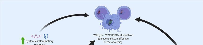
Int. J. Mol. Sci. 2020, 21, 626                                                                     3 of 16

Given the subjective nature of morphological assessment, more accurate and precise diagnostic tools
are necessary for early stage and existing myeloid neoplasms.

4. Promise of NGS in Myeloid Malignancy Diagnosis
     Next-generation sequencing (NGS) has become increasingly important in the detection of genetic
variants in myeloid malignancies [3,21,22]. Hematological variants identified through NGS impact
diagnosis, in addition to revealing new prognostic, predictive, and therapeutic biomarkers in myeloid
malignancies [21–23]. Several genes have been identified as recurrently mutated in these disorders,
including epigenetic regulators (TET2, ASXL1, EZH2, DNMT3A, IDH1/IDH2), splicing factors (SF3B1,
U2AF1, ZRSR2), genes involved in signal transduction (JAK2, KRAS, NRAS, CBL), among others [5,24,25].
For example, mutations in CALR or JAK2 are indicative of MPN [26], while FLT3, NPM1, and CEBPA
are associated with AML [27]. Such mutations can also be prognostic indicators; e.g., the presence
of TP53 or FLT3 mutations are associated with worse outcomes in AML [28,29]. Recent updates to
the WHO classification of myeloid neoplasms have also begun to incorporate such mutations into
diagnostic criteria. Notably, the previous MDS diagnosis of refractory anemia with ring sideroblasts
(RARS) required a minimum of 15% ring sideroblasts to confirm a RARS diagnosis [12], while the
updated diagnosis of MDS with ring sideroblasts (MDS-RS) requires only 5% ring sideroblasts when
accompanied by an SF3B1 mutation (associated with mis-splicing of a mitochondrial iron transporter
leading to abnormal accumulation of iron in mitochondria ringing the nucleus) [1].

5. CHIP and Risk of Hematological Malignancy
      Hematopoietic cells tend to accumulate somatic mutations over time. It has been estimated
that there are normally 50,000–200,000 HSPCs, and that they have a fidelity rate of approximately
0.78 × 10⁻⁹ mutations per genomic base pair per cell division, resulting in random mutations occurring
at a rate of approximately 14 base substitutions and 0.13 coding mutations per year of life (i.e., approx.
one mutation every 7–8 years) [7,11,30]. The prevalence of such mutations thus increases with age, but
they generally do not impact normal HSPC function. However, some of these mutations can occur
in genes that predispose them to malignant disease [7]. CHIP is a term used to describe individuals
with hematological malignancy-associated somatic mutation(s) in the absence of other hematological
malignancy diagnostic criteria [13,31]. Due to this accumulation of mutations over time, CHIP is
especially common in the elderly population (>10% in those over 65 years of age), and therefore often
referred to as age-related clonal hematopoiesis (ARCH) [9,13,32]. The minimum variant allele frequency
(VAF) for genetic variants in individuals to meet the criteria for CHIP is ≥2% [13]. Furthermore, while
CHIP is a clinical entity with specific criteria, including mutations in recognized cancer driver genes
like TET2 and DNMT3A, the more general term clonal hematopoiesis (CH) describes a state in which a
single hematopoietic stem cell clone gives rise to a disproportionate number of an individual’s mature
blood cells. CH can include mutations at VAF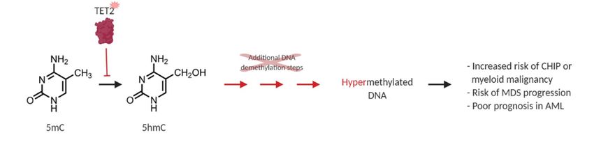
Int.
Int. J.J. Mol.
Mol. Sci.
Sci.2020,
2020, 21,
21, x626
FOR PEER REVIEW 4 4of
of 17
16
6. TET2 and DNMT3A Mutations and Their Impact
6. TET2 and DNMT3A Mutations and Their Impact
TET2 is widely affected by mutations in myeloid neoplasms, and is one of the most commonly
TET2genes
mutated is widely affected
in CHIP by mutations
[9,32,34]. SomaticinTET2
myeloid neoplasms,
mutations and is one
are present of the most commonly
in approximately 50% of
genes in CHIP [9,32,34]. Somatic TET2
chronic myelomonocytic leukemia (CMML; an MDS/MPN) cases, ~30% of MDS, and ~10% of50%
mutated mutations are present in approximately AMLof
chronic
[8]. Suchmyelomonocytic leukemia
TET2 loss-of-function (CMML;are
mutations an associated
MDS/MPN)with cases, ~30% hypermethylation
a DNA of MDS, and ~10% phenotype,
of AML [8].
Such TET2
tumour loss-of-function
progression, mutations
and poor patientare associated
outcome with DNA
[35,36]. a DNA hypermethylation
methylation plays aphenotype,
key role in tumour
proper
progression, and poor patient outcome [35,36]. DNA methylation plays a key
HSPC self-renewal and lineage differentiation, and its dysregulation can lead to aberrant stem role in proper HSPC
cell
self-renewal
function andand lineage
cellular differentiation,
transformation andTET2
[37]. its dysregulation can role
plays a crucial lead in
to aberrant
epigenetic stem cell function
modulation by
and cellular DNA
promoting transformation [37]. TET2
demethylation [38].plays
DNA a crucial role in epigenetic
hypermethylation modulation
resulting from by TET2promoting
mutation DNA is
demethylation [38]. DNA hypermethylation resulting from TET2 mutation
associated with CHIP, increased risk of MDS progression, and poor prognosis in AML [35]. Theis associated with CHIP,
increased
TET2 risk of itself
protein MDS progression,
is one of and the poor prognosis in AML [35].(TET1-3)
ten-eleven-translocation The TET2proteins,
protein itself is oneare
which of
the ten-eleven-translocation (TET1-3) proteins, which are alpha-ketoglutarate- and Fe 2+ -dependent
alpha-ketoglutarate- and Fe -dependent dioxygenases (α-KGDDs). These α-KGDDs primarily
2+
dioxygenases
catalyze (α-KGDDs).
the oxidation These α-KGDDs(5mC)
of 5-methylcytosine primarily catalyze the oxidation of(5hmC)
to 5-hydroxymethylcytosine 5-methylcytosine
(Figure 2).
(5mC) represent
These to 5-hydroxymethylcytosine
key intermediates in (5hmC) (Figure 2). These
DNA demethylation represent
through key intermediates dilution
replication-dependent in DNA
demethylation through
or base excision repair [39].replication-dependent dilution or base excision repair [39].
Figure 2.
2. Loss-of-function
Loss-of-function mutations
mutations in in TET2
TET2 result in in DNA
DNA hypermethylation.
hypermethylation. TET2 TET2 is
is an
alpha-ketoglutarate-
alpha-ketoglutarate- and 2+
and Fe -dependent
2+
-dependent dioxygenase
dioxygenase (α-KGDD)
(α-KGDD) that that catalyzes
catalyzes the
the oxidation
oxidation of
5-methylcytosine
5-methylcytosine (5mC) to 5-hydroxymethylcytosine
5-hydroxymethylcytosine (5hmC).
(5hmC). This
This is
is aa required
required step in proper DNA
repair and DNA demethylation (green). Loss-of-function
Loss-of-function mutations
mutations in in TET2
TET2 in CHIP and myeloid
malignancies disrupt this oxidation step
malignancies disrupt this oxidation step and result in a general DNA hypermethylation phenotype
general DNA hypermethylation
(red) and aberrant HSPC self-renewal, which
which is associated
associated with
with an
an increased
increased risk
risk of
of CHIP,
CHIP, myeloid
malignancy, MDS progression,
progression, and
and poor
poor prognosis
prognosis inin AML
AML [35].
[35].
Several studies
Several studies have
have assessed
assessed the
the structure
structure of the TET2 protein and the context of somatic
mutations in myeloid malignancies, including the domains in which they occur. Whether mutations
occur prior to or within the C-terminal catalytic domain of TET2, they generally generally result in loss of
enzymatic function.
enzymatic function. Studies
Studies have
have shown
shown that
that most
most commonly,
commonly, nonsense
nonsense or frameshift mutations
occur before (and occasionally within) the catalytic domain, while missense mutations and in-frame
deletions occur within the catalytic domain [8,40,41] (Figure 3). To To our knowledge, a comprehensive
comparison of ofTET2
TET2mutations
mutationsin CHIP
in CHIPversus myeloid
versus malignancies
myeloid has yethas
malignancies to be performed;
yet however,
to be performed;
the naturethe
however, TET2 CHIP
of nature mutations
of TET2 are also inare
CHIP mutations keeping
also inwith TET2
keeping catalytic
with TET2 inactivation. Overall,
catalytic inactivation.
somatic mutations
Overall, in TET2 have
somatic mutations been
in TET2 found
have beentofound
occur to
in approximately 15% of patients
occur in approximately 15% of with myeloid
patients with
malignancies [8]. This gene therefore represents a promising molecular feature that
myeloid malignancies [8]. This gene therefore represents a promising molecular feature that can can impact the
clinical the
impact careclinical
of thesecare
patients. Forpatients.
of these example, theexample,
For presencethe absence oforTET2
or presence can of
absence be TET2
indicative of
can be
treatment outcome
indicative [42].outcome [42].
of treatment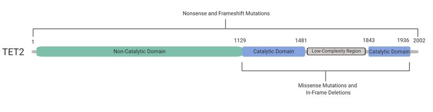
Int. J. Mol. Sci. 2020, 21, 626 5 of 16
Int. J. Mol. Sci. 2020, 21, x FOR PEER REVIEW 5 of 17
Figure3.3.Inactivating
Figure InactivatingTET2
TET2 mutations
mutations occur
occur throughout
throughoutthethecoding
codingregion.
region.The
TheC-terminal
C-terminal catalytic
catalytic
domain of TET2 occurs approximately between amino acid residues 1129
domain of TET2 occurs approximately between amino acid residues 1129 and 1936, including and 1936, including a a
double-strandedβ βhelix
double-stranded helixand
andcysteine-rich
cysteine-richdomain,
domain,binding
bindingsites
sitesfor
forFe(II)
Fe(II)
andand -ketoglutarate,asas
α-ketoglutarate, well
well as a low-complexity linker region. Together, these regions allow the catalytic domain
as a low-complexity linker region. Together, these regions allow the catalytic domain of TET2 to bind toof TET2 to
bind allowing
DNA, to DNA,the allowing the of
oxidation oxidation of 5mC
5mC to 5hmC to Missense
[40]. 5hmC [40]. Missenseand
mutations mutations
in-frameand in-frame
deletions occur
deletions occur in the catalytic domain of the protein (blue). Nonsense and frameshift mutations can
in the catalytic domain of the protein (blue). Nonsense and frameshift mutations can occur throughout
occur throughout the entire coding region. However, they occur most frequently in the non-catalytic
the entire coding region. However, they occur most frequently in the non-catalytic domain (green).
domain (green). Mutations in TET2 are generally inactivating regardless of their nature or location in
Mutations in TET2 are generally inactivating regardless of their nature or location in the coding region
the coding region of the gene [8,40,41].
of the gene [8,40,41].
In 2014, Bejar et al. found that response to hypomethylating agents (HMA) in MDS patients is
In 2014, Bejar et al. found that response to hypomethylating agents (HMA) in MDS patients
enhanced in those that exhibit clonal TET2 mutations [43]. They also found that the presence of
is enhanced in those that exhibit clonal TET2 mutations [43]. They also found that the presence of
additional mutations such as in ASXL1 indicate poorer prognosis [43], while another group found
additional mutations such as in ASXL1 indicate poorer prognosis [43], while another group found
that overall survival is greater in patients with clonal TET2 mutations in the absence of ASXL1
that overall survival is greater in patients with clonal TET2 mutations in the absence of ASXL1
mutations [44]. However, success of HMA treatment in MDS patients has been found to be
mutations [44]. However, success of HMA treatment in MDS patients has been found to be inconsistent
inconsistent and dependent on particular combinations of mutations, though MDS patients are not
and dependent
currently denied onHMA
particular
therapy combinations of mutations,
based on the presence though
of certain MDS [45,46].
mutations patients are not currently
Additionally, the
denied
presence of TET2 mutations has been associated with reduced overall survival in patients with of
HMA therapy based on the presence of certain mutations [45,46]. Additionally, the presence
TET2 mutations hasAML
intermediate-risk been[21].
associated with reduced
Other studies overallthat
have shown survival in patients
co-mutations with intermediate-risk
in DNMT3A and TET2
AML [21]. Other studies have shown that co-mutations in DNMT3A
increase the risk of malignant transformation compared to either mutation alone increaseand TET2 the risk
[47,48], which is of
malignant
concerning transformation
given that these compared
genes aretofrequently
either mutation alone
co-mutated in[47,48],
MDS [3].which is concerning given that
these genes are frequently
The DNMT3A gene isco-mutated
affected byinloss-of-function
MDS [3]. mutations in myeloid malignancies. It is an
The DNMT3A
epigenetic regulator,gene is affected
where by loss-of-function
its associated protein, DNMT3A, mutations in myeloid
catalyzes malignancies.
the methylation of CpGIt is
andinucleotides in genomic
epigenetic regulator, DNA its
where [49]. Specifically,
associated it catalyzes
protein, the covalent
DNMT3A, addition
catalyzes of a methyl group
the methylation of CpG
to the C5 position
dinucleotides in genomicof cytosine
DNA [49]. (generating 5mC)it [49,50].
Specifically, catalyzesDNMT3A is important
the covalent addition in of aHSPC
methyl
differentiation and helps regulate the function of stem cells. Loss-of-function
group to the C5 position of cytosine (generating 5mC) [49,50]. DNMT3A is important in HSPC in murine Dnmt3a
leads to HSPC
differentiation expansion,
and helps regulateclonal dominance,
the function of stemaberrant DNA methylation,
cells. Loss-of-function and Dnmt3a
in murine eventuallyleads
tohematological
HSPC expansion, malignancies [51]. The presence
clonal dominance, aberrantof aDNA
DNMT3A mutation and
methylation, in myeloid malignancies
eventually has
hematological
also been shown
malignancies to presence
[51]. The predict aofhigher likelihood
a DNMT3A of responding
mutation in myeloidto an HMA [52].
malignancies Furthermore,
has also been shown
DNMT3A mutations are associated with a worse prognosis in MDS when they occur in the presence
to predict a higher likelihood of responding to an HMA [52]. Furthermore, DNMT3A mutations are
of some other mutations, especially mutations in SF3B1 [53–55]. Mutations in DNMT3A also predict
associated with a worse prognosis in MDS when they occur in the presence of some other mutations,
poor prognosis in AML [55].
especially mutations in SF3B1 [53–55]. Mutations in DNMT3A also predict poor prognosis in AML [55].
Overall, the use of NGS to detect TET2, DNMT3A, and other mutations in patients with myeloid
Overall, the use of NGS to detect TET2, DNMT3A, and other mutations in patients with myeloid
malignancies is imperative. These molecular features can be used as prognostic and diagnostic
malignancies is imperative. These molecular features can be used as prognostic and diagnostic
indicators for these patients, in addition to informing clinicians regarding the probability of disease
indicators
progressionfor and
these patients,options
treatment in addition to informing
to improve clinicians regarding the probability of disease
patient care.
progression and treatment options to improve patient care.
7. CHIP and Comorbid Diseases
7. CHIP and Comorbid Diseases
While TET2 and DNMT3A mutations are two of most common genetic variations in CHIP and
canWhile TET2
precede and DNMT3A
myeloid mutations
malignancies, are two
recent studies haveof most common
also revealed genetic variations
associations between in CHIP
CHIP andand
can precede
other myeloid
comorbid malignancies,
diseases recent
[9,32]. Such studies
studies have have also revealed
approached associations
the identification of between CHIP and
CHIP-associated
other comorbid diseases [9,32]. Such studies have approached the identification of
diseases by genetically sequencing populations of elderly individuals who are more likely to CHIP-associated
diseases
harbourbypathogenic
geneticallysomatic
sequencing populations
variants for CHIP, of revealing
elderly individuals
that somaticwho are more
changes in likely
specifictogenes
harbour
pathogenic somatic variants for CHIP, revealing that somatic changes in specific genes (especially TET2
Int. J. Mol. Sci. 2020, 21, 626                                                                      6 of 16

and DNMT3A) are associated with increased risk of developing non-neoplastic diseases [9,32,33,56,57].
In a study in The New England Journal of Medicine in 2014, Jaiswal et al. observed an increase in all-cause
mortality for individuals with CHIP, as well as a modestly elevated risk of type 2 diabetes. Individuals
with CHIP also had strong associations with cardiovascular conditions. Notably, CHIP was associated
with elevated risk of coronary heart disease and ischemic stroke [9].
      In a more recent study, compared to individuals without CHIP, carriers of CHIP variants were
found to be 1.9 times as likely to have coronary heart disease, 4.0 times as likely to experience
early-onset myocardial infarction, and had increased coronary artery calcification [57]. In another
study, somatic TET2 or DNMT3A variants in individuals with chronic ischemic heart disease appeared
to associate with worse long-term clinical outcome due to heart failure. The VAF of clonal populations
was associated with the severity of the clinical outcome, suggesting a dose-response relationship [58].
Similarly, aortic valve stenosis patients with DNMT3A or TET2 variants who underwent aortic valve
implantation surgery were observed to have heightened medium-term all-cause mortality compared
to those without CHIP mutations [59]. These results were supported by the finding that mice with
hematopoietic or myeloid Tet2-deficiency had greater cardiac dysfunction and worsened remodeling
following induced heart failure [60]. Hypercholesterolemic mice engrafted with either homozygous
or heterozygous Tet2-knockout bone marrow developed larger atherosclerotic lesions than controls,
suggesting that TET2 dysfunction contributes to the development of atherosclerotic plaques [57,61].
The increased lesion size in mice that received heterozygous Tet2-knockout bone marrow provides
evidence for haploinsufficiency as a potential mechanism for atherogenesis. This is especially important
considering that most individuals with TET2 somatic variants have only a single defective allele.
      The involvement of hematological TET2-deficiency in the pathogenesis of atherosclerosis has been
investigated at the cellular level [57,61]. Tet2-deficiency in myeloid cells was found to independently
accelerate atherogenesis in mice and highlighted a potential role of Tet2-deficient macrophages as agents
of atherosclerosis. Fuster et al. (2017) identified that Tet2-deficiency in murine macrophages facilitates
nod-like receptor family pyrin domain containing 3 (NLRP3) inflammasome-dependent production of
the inflammatory cytokine interleukin 1 beta (IL-1b). IL-1b increases aortic expression of endothelial
adhesion markers that recruit monocytes. Importantly, Tet2-deficient mice that were given an NLRP3
inhibitor had reduced size of atherosclerotic lesions and reduced expression of the endothelial adhesion
marker [61]. A separate study arrived at similar conclusions, showing that Tet2-deficient mice treated
with an NLRP3 inhibitor were protected against accelerated cardiac dysfunction [60]. These findings
suggest that IL-1b or NLRP3 inflammasome inhibitors may be useful therapeutic approaches in the
treatment or prevention of cardiovascular disease in people with TET2 mutations.
      While somatic TET2 changes are common in myeloid malignancy, TET2 mutations have also
been documented in the germline [62]. Interestingly, a family heterozygous for a truncating TET2
mutation had multiple cases of lymphoma among affected individuals but no abnormal cardiovascular
phenotype [63]. Monocyte-derived macrophages that were isolated from TET2-mutation carriers
showed a hypermethylation phenotype but did not have altered cytokine or chemokine secretion.
This trend was affirmed in three unrelated individuals harbouring different germline TET2 mutations.
Members of another family that were affected with a different heterozygous TET2 germline frameshift
mutation all developed myeloid malignancy, possessed thyroid abnormalities, and had no history
of cardiovascular disease [64]. The apparent phenotypic contrast between those with germline vs.
somatic TET2 mutations merits further investigation with larger cohorts.
      In addition to being associated with increased all-cause mortality, NGS has revealed that CHIP is
significantly associated with chronic pulmonary disease [33,56]. Individuals with somatic variants
in TET2 were found to have an elevated prevalence of self-reported asthma or chronic obstructive
pulmonary disease (COPD), though the nature of this relationship is unclear [65]. Future studies
will need to determine whether the relationship between TET2 mutations and COPD is mediated
by smoking, or whether loss of function of TET2 plays a contributing role in the exacerbation of
pulmonary diseases.
Int. J. Mol. Sci. 2020, 21, 626 7 of 16
Regarding DNMT3A, variants in this gene in CHIP appear to be associated with the development
of gastroesophageal reflux disease (GERD), whereas CHIP variants in TET2 or with VAF > 0.1 are
potentially associated with elevated levels of circulating thyroid-stimulating hormone [56], although
these require further validation. In addition, our unpublished data shows that Tet2-deficient mice
spontaneously develop pulmonary arterial hypertension associated with increased lung inflammation.
It is likely that subsequent studies will reveal further comorbidities associated with CHIP.
8. CHIP, Inflammation, and the Connection to the Pathogenesis of Myeloid Cancers
Many of the comorbid diseases associated with CHIP also have a shared inflammatory basis.
Understanding the impact of inactivating TET2 and DNMT3A mutations on myeloid cells is critical for
elucidating the connection between CHIP, inflammation, and the pathogenesis of comorbid disease,
as well as myeloid malignancies. The TET2 protein plays a key role in myeloid cell function as an
epigenetic regulator for cell differentiation and the inflammatory response. TET2 has been identified
as a mediator of transcriptional regulation for inflammatory cytokines, notably interleukin 6 (IL6).
During the resolution of inflammation, TET2 normally recruits histone deacetylase 2 (HDAC2) to
deacetylate IL6, thereby repressing its transcription and IL-6 levels [66]. This epigenetic change is an
important regulatory step for the termination of the inflammatory state in myeloid cells, especially
macrophages and dendritic cells. In vitro, murine macrophages with Tet2 knocked out were observed
to have upregulated Il6 expression, as well as upregulation of Il1b and arginase 1 (Arg1), during the
late-phase response to lipopolysaccharides (LPS) exposure [67]. Tet2-knockout mice had elevated IL-6
production and were more prone to developing endotoxin shock and dextran-sulfate-sodium-induced
colitis. Moreover, TET2 loss may alter the immune environment by interfering with other types of
leukocytes, as Tet2-deficient murine T-cells showed aberrant cytokine signaling [68]. This suggests that
the loss of Tet2 resulted in an exacerbated inflammatory phenotype, especially in myeloid cells.
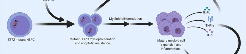
It is well recognized that the prevalence of TET2 somatic variants increases with age [9,32,33].
However, the mechanism for the emergence and subsequent dominance of TET2-mutated clonal
populations in elderly people has not been well studied. A recent study by Abegunde et al. (2018)
showed that Tet2-knockout murine and TET2-deficient human HPSCs had a proliferative advantage
when chronically exposed to tumour necrosis factor alpha (TNF-α) in vitro. Upon prolonged plating
with TNF-α, HSPCs with inactivating TET2 mutations developed a resistance to apoptosis and a
propensity for myeloid differentiation [69]. Similarly, upregulated Il6 expression from HSPCs in murine
Tet2-knockout mice (in response to acute inflammatory stress) led to apoptotic resistance via increased
expression of pro-survival genes and decreased expression of pro-apoptotic genes [70]. These results
suggest that the expansion of TET2-mutated clones may be facilitated by a resistance to an inflammatory
environment that these clones help propagate (Figure 4). Furthermore, another group found that
microbial signals from the intestine appear to trigger the expansion of myeloid Tet2-deficient clones,
where bacterial translocation in Tet2-knockout mice led to increased IL-6 production and myeloid
expansion [71].
Regarding malignant disease, mutant HSPC clones have been found to alter the inflammatory
microenvironment associated with the bone marrow, which can facilitate clonal expansion that leads to
myeloid malignancies such as MDS [72]. This modified microenvironment becomes conducive to MDS
development, often resulting from increased expression of proinflammatory genes such as IL1B, TNF
and IL6, alterations in related immune cell subsets, and susceptibility to apoptosis within the bone
marrow [72]. MDS can then progress to AML; in fact, approximately 40% of MDS cases progress [73].
Increased proinflammatory mediators can be produced by the mutant HSPC clone, macrophages,
dendritic cells, and T-cells, among others. Mutations in TET2 (and other genes) can further promote
proinflammatory gene expression. The resulting increase in inflammation can facilitate clonal expansion
of mutant HSPCs and corresponding MDS pathogenesis or progression [72]. Macrophages, especially,
have been shown to contribute to immune dysregulation and myeloid cancer pathogenesis in certain
cases [74–76]. Other studies suggest that an abnormal immune environment in the bone marrow ofInt. J. Mol. Sci. 2020, 21, x FOR PEER REVIEW 8 of 17
facilitate clonal expansion of mutant HSPCs and corresponding MDS pathogenesis or progression
Int.[72]. Macrophages,
J. Mol. especially, have been shown to contribute to immune dysregulation and myeloid
Sci. 2020, 21, 626 8 of 16
cancer pathogenesis in certain cases [74–76]. Other studies suggest that an abnormal immune
environment in the bone marrow of patients with myeloid malignancies promotes genotoxic stress
patients with myeloid
and pyroptosis, malignancies
with the latter beingpromotes genotoxic
mediated through stress and
activation pyroptosis,
of the with the latter[77].
NLRP3 inflammasome being
mediated through activation of the NLRP3 inflammasome [77]. This in turn contributes to
This in turn contributes to disease pathophysiology and clonal evolution that can lead to HSPC disease
pathophysiology and clonal evolution that can lead to HSPC dysfunction [78–80].
dysfunction [78–80].
Figure
Figure4. 4.Molecular pathogenesis of TET2-mutated
Molecularpathogenesis TET2-mutated HSPC. HSPC.Exposure
Exposuretotoupregulated
upregulated inflammatory
inflammatory
cytokines
cytokines (due(due to systemic
to systemic inflammation
inflammation or local
or local alterations)
alterations) advantages
advantages TET2-mutant
TET2-mutant HSPCs,HSPCs,
fostering
fostering a phenotype characterized by increased proliferation and a resistance to apoptosis
a phenotype characterized by increased proliferation and a resistance to apoptosis [69–71]. TET2-mutant [69–71].
TET2-mutant
HSPCs and the HSPCs
abnormal andimmune
the abnormal immune microenvironment
microenvironment of the can
of the bone marrow bone marrow
cause can cause
premature death
orpremature
quiescencedeath or quiescence
of wildtype of wildtype
(i.e., normal) HSPCs, (i.e., normal) HSPCs,
consequently consequently
manifesting manifesting
as ineffective as
and clonal
ineffective and
hematopoiesis. Theclonal hematopoiesis.
increased proliferationThe increased
associated proliferation
with TET2-mutant associated with to
HSPCs leads TET2-mutant
the expansion
ofHSPCs leads to
TET2-clonal the expansion
populations of TET2-clonal
in the bone marrow populations in the boneblood.
and/or peripheral marrow and/ormyeloid
Mature peripheral cells
blood. Mature myeloid cells (e.g., macrophages) derived from TET2-mutant
(e.g., macrophages) derived from TET2-mutant HSPCs demonstrate a hyper-inflammatory phenotype HSPCs demonstrate a
hyper-inflammatory phenotype that contributes to the pathogenesis of comorbidities,
that contributes to the pathogenesis of comorbidities, with a notable causal role in cardiovascular with a notable
causal role in cardiovascular disease [57,61,67].
disease [57,61,67].
9. 9. Promisesand
Promises andPotential
PotentialPitfalls
Pitfalls of
of Surveillance
Surveillance for
forCHIP
CHIPininHuman
HumanAging
Aging
While
While CHIP
CHIP isiswidely
widelyprevalent
prevalentininelderly
elderlypeople
peopleand
andcan
canpose
poseserious
serious health
health concerns
concerns through its
its associated
associated risks, risks, the question
the question of howof to how to effectively
effectively approachapproach
the clinicalthemanagement
clinical management and
and surveillance
surveillance of CHIP is less clear. An important clinical objective for addressing CHIP
of CHIP is less clear. An important clinical objective for addressing CHIP should be the minimization should be the of
minimization of clonal expansions that contribute to disease states. Some have suggested
clonal expansions that contribute to disease states. Some have suggested that the selective destruction that the
of selective destruction
mutant HSPCs of mutant
is a valid HSPCs
strategy for theis atreatment
valid strategy for the originating
of disorders treatment offrom
disorders originating
such HSPCs [10,13].
from such HSPCs [10,13]. While this could prevent uncontrolled proliferation
While this could prevent uncontrolled proliferation and subsequent disease, older individuals may and subsequent
disease, older individuals may rely on overly proliferative HSPCs to maintain normal blood cell
rely on overly proliferative HSPCs to maintain normal blood cell levels. For such treatment to be
levels. For such treatment to be considered, the benefit of reducing disease risk would have to be
considered, the benefit of reducing disease risk would have to be balanced with the potential health
balanced with the potential health implications of treatment-induced cytopenias. Future
implications of treatment-induced cytopenias. Future management strategies should also address
management strategies should also address the effect of the CHIP inflammatory phenotype on
the effect of the CHIP inflammatory phenotype on broader comorbidities. Tet2-deficiency facilitates
the upregulation of inflammatory cytokines (notably IL-1b) that contribute to the pathogenesis of
atherosclerosis in mice [61]. Complementary to these findings, an IL-1b inhibitor has been shown
to reduce the risk of recurrent cardiovascular events [81]. However, despite blocking IL-1b, residual
IL-6 and interleukin 18 (IL-18) contribute to increased incidence of future cardiovascular events [82].Int. J. Mol. Sci. 2020, 21, 626 9 of 16
Another recent study showed that the presence of the IL-6 receptor (IL6R) p.Asp358Ala variant abolishes
the cardiovascular risk in people with CHIP [83]. It appears that targeting the inflammatory outputs
associated with dysregulated immune function may be a promising strategy for mitigating the risk of
certain comorbidities in CHIP. To better understand this potential avenue of treatment, future research
should seek to elucidate the mechanisms that underpin the pathogenesis of CHIP-associated diseases.
A diagnosis of CHIP can provide opportunities for management and preventative action.
Hundreds of mutations in over fifty genes contribute to CHIP development [9]. These diverse mutational
profiles may provide unique avenues to disease pathogenesis and an opportunity for personalized
treatments. It is worth re-stating that the absolute risk for hematological cancer development in CHIP
is low: 0.5% to 1.0% of individuals with CHIP develop myeloid cancer annually [9,32]. In addition,
variants in TET2 and DNMT3A do not significantly lower the 10-year survival rate for elderly
individuals (>85-year old) [84]. However, mutations in other CHIP-related genes have been found
to associate with increased risk of and accelerated time to AML development [48,85]. Importantly,
models have been constructed that can predict the development of AML either months or years in
advance of disease onset by analyzing the somatic molecular profiles or clinical parameters (notably
red blood cell distribution width) of people with CHIP [48]. Despite this recent progress, the risk
associations between CHIP variants and other comorbid diseases remain poorly understood.
For CHIP surveillance to be implemented, it is important to consider the actionability of a
CHIP diagnosis (i.e. its impact on clinical management). Identification of a pathogenic somatic
variant in an otherwise hematological malignancy-free individual could warrant elevated follow-up
frequencies to monitor potential progression. Individuals with other comorbidities that undergo
testing for CHIP could benefit from a more informed prognosis based on the presence (or lack of)
known pathogenic variants. Treatment plans for specific comorbidities, such as cardiovascular disease,
could be individualized based on the calculated risk associated with testing results. However, studies
concerning best practice for the treatment and prevention of CHIP-associated comorbidities have not
yet been conducted, so the responsiveness of unique CHIP mutational profiles to various treatment
approaches is not presently known. With CHIP clinics and related initiatives currently underway, we
call for prospective studies at the involved institutions to help establish best practice recommendations.
The benefits of a CHIP diagnosis will also have to be balanced against the ramifications of
screening healthy individuals. For example, using blood tests to screen for disease is invasive and
risks provoking anxiety in patients. One should also consider potential ethical, legal, and financial
issues (i.e. insurance eligibility) related to screening. An informed decision regarding the merit of
CHIP surveillance in the general population will optimally account for all these factors.
10. Promise of Targeting the “Good Copy” of TET2
While several studies have assessed Tet2 loss-of-function in mice and reported associated HSPC
aberrant self-renewal and myeloid lineage expansion [35], and though screening techniques have
improved greatly in recent years to detect mutations in TET2 and other genes, therapeutic options
to restore TET2 activity are currently lacking. In a recent study, Cimmino et al. (2017) modeled
restoration of TET2 expression using reversible transgenic RNAi mice to confirm its reversal of aberrant
HSPC self-renewal. Their study suggests that indirect restoration of TET2 function may offer a new
therapeutic strategy for CHIP, MDS, and AML [39]. They found that Tet2 knockdown led to aberrant
HSPC self-renewal, while Tet2 restoration reversed this effect in vitro and in vivo. Additionally, they
found that Tet2-restored cells exhibited increased cell death, decreased proliferation, and priming
toward myeloid differentiation [39]. These findings indicate that sustained TET2 deficiency is required
for disease maintenance, and that restoration of TET2 may offer a viable therapeutic option for patients
with myeloid malignancies possessing TET2 mutations.
Vitamin C is an α-KGDD co-factor that has been shown to promote activity of TET enzymes [86].
Administration of vitamin C has been tested, and demonstrated some efficacy, in the treatment of solid
tumours [87]. To investigate the potential use of vitamin C in treating hematological malignancies,Int. J. Mol. Sci. 2020, 21, 626 10 of 16
Cimmino and colleagues treated mouse HSPCs and human leukemia cells with vitamin C. They found
that vitamin C treatment mimics TET2 restoration in Tet2-deficient mouse HSPCs by increasing 5hmC
formation, as seen in other studies [88]. Similarly, Agathocleous et al. (2017) demonstrated that
ascorbate (vitamin C) regulates TET2 function in hematopoietic stem cells (HSCs). They also found
that feeding ascorbate to leukemogenic mice significantly extended survival, in addition to reducing
myeloblasts in the blood, spleen cellularity and HPC frequency in the spleen [89]. While these findings
support the use of vitamin C to treat TET2-deficient myeloid malignancies and potentially CHIP
patients with TET2 mutations, the quantity of vitamin C required to achieve adequate TET2 level
increases remains a barrier to effectively utilizing vitamin C as a treatment option for these patients.
However, some more recent studies have begun to explore the use of ascorbate in a clinical setting
in TET2-mutant patients [90,91], with one report demonstrating a clinical response of a patient with
TET2-mutant AML to ascorbate, despite the TET2-mutant clone remerging at relapse [90].
Regarding targeting DNMT3A, few studies have explored the protein as a therapeutic target.
While HMA treatment holds some promise for those mutations in DNMT3A, their toxicities remain too
severe to justify use in patients with CHIP. However, some groups have begun to consider possible
methods of targeting this protein. Rau et al. (2016) present a study in Blood that suggests that the
histone lysine methyltransferase, disruptor of telomeric silencing 1-like (DOT1L) may represent a
promising target in cases of DNMT3A-mutant AML. They demonstrated that pharmacologic inhibition
of DOT1L in DNMT3A-mutant cell lines resulted in cell-cycle arrest and terminal differentiation, in
addition to reducing cellular proliferation and inducing apoptosis in vitro [92]. Furthermore, Adema
et al. (2017) published an abstract in Blood that demonstrated that S-adenosyl-l-methionine (SAM)
supplementation in cases of monoallelic DNMT3A loss results in up-modulation of the wild-type
DNMT3A allele in vitro [93]. These studies indicate promise in targeting levels of TET2 and DNMT3A
in cases of heterozygous loss-of-function mutations, though further studies are needed before these
therapeutic strategies could be considered from a clinical perspective.
11. Summary
Recent advances in genomic sequencing technologies have helped to characterize the phenotypic
associations of acquired loss-of-function mutations in TET2, a gene that encodes an epigenetic regulator
highly important for myeloid cell function. These mutations (along with variants in the DNMT3A gene)
are associated with an increased risk of hematological malignancy and are especially prevalent in the
elderly population. In addition, deficiency in TET2 provokes an inflamed immune phenotype (largely
via aberrant cytokine secretion) and associates with several inflammation-based diseases, with a notable
causal role in cardiovascular disease. Recently, mutational status and routine clinical information
has been shown to predict future development of AML in individuals with CHIP. As a common and
significant risk factor, the justification for screening at-risk individuals for TET2 mutation status must
be balanced against potential ramifications of testing. While anti-inflammatory drugs can attenuate the
cardiovascular risk, there are currently no viable therapeutic options for rescuing TET2 insufficiency
in myeloid malignancies. Some approaches (notably vitamin C administration) have demonstrated
restorative properties for TET2. However, barriers remain to these being effective treatments. Recent
studies provide hope for future therapeutic strategies that may address loss-of-function mutations in
both the TET2 and DNMT3A genes in cases of myeloid malignancies, and potentially even CHIP and
comorbid diseases.
Author Contributions: Conceptualization, C.K.F. and M.J.R.; Writing—Original Draft Preparation and
Writing—Review and Editing, C.K.F., M.B.-H., M.J.R.; Project Administration and Funding Acquisition, M.J.R.
All authors have read and agreed to the published version of the manuscript.
Funding: Publication costs were supported by funding from the Southeastern Ontario Academic Medical
Organization (S.E.A.M.O.) and the Innovation Fund, a joint initiative of the Ontario Ministry of Health and
Long-Term Care (M.O.H.L.T.C.) and the Ontario Medical Association (O.M.A.) resulting from the Academic
Health Science Centre Alternative Funding Plan.Int. J. Mol. Sci. 2020, 21, 626 11 of 16
Acknowledgments: Some figures were created using BioRender.
Conflicts of Interest: The authors declare no conflict of interest.
Abbreviations
α-KGDDs Alpha-ketoglutarate- and Fe2+ -dependent dioxygenases
ARCH Age-related clonal hematopoiesis
Arg1 Arginase 1
AML Acute myeloid leukemia
CCUS Clonal cytopenia of undetermined significance
CH Clonal hematopoiesis
CHIP Clonal hematopoiesis of indeterminant potential
CMML Chronic myelomonocytic leukemia
COPD Chronic obstructive pulmonary disease
DNMT3A DNA methyltransferase 3 alpha
DOT1L Disruptor of telomeric silencing 1-like
GERD Gastro-esophageal reflux disease
HDAC2 Histone deacetylase 2
HMA Hypomethylating agents
HSC Hematopoietic stem cells
HSPC Hematopoietic stem/progenitor cells
ICUS Idiopathic cytopenia of undetermined significance
IL-1β Interleukin 1 beta
IL-18 Interleukin 18
IL-6 Interleukin 6
IL6R Interleukin 6 receptor
LPS Lipopolysaccharides
MDS Myelodysplastic syndromes
MDS-RS MDS with ring sideroblasts
MDS/MPN Myelodysplastic syndromes/myeloproliferative neoplasms
MPN Myeloproliferative neoplasm
NGS Next-generation sequencing
NLRP3 Nod-like receptor family pyrin domain containing 3
RARS Refractory anemia with ring sideroblasts
SAM S-adenosyl-l-methionine
TET2 Tet methylcytosine dioxygenase 2
TNF-α Tumour necrosis factor alpha
VAF Variant allele frequency
5mC 5-methylcytosine
5hmC 5-hydroxymethylcytosine
References
1. Arber, D.A.; Orazi, A.; Hasserjian, R.; Thiele, J.; Borowitz, M.J.; Le Beau, M.M.; Bloomfield, C.D.; Cazzola, M.;
Vardiman, J.W. The 2016 revision to the World Health Organization classification of myeloid neoplasms and
acute leukemia. Blood 2016, 127, 2391–2406. [CrossRef] [PubMed]
2. Barbui, T.; Thiele, J.; Gisslinger, H.; Kvasnicka, H.M.; Vannucchi, A.M.; Guglielmelli, P.; Orazi, A.; Tefferi, A.
The 2016 WHO classification and diagnostic criteria for myeloproliferative neoplasms: Document summary
and in-depth discussion. Blood Cancer J. 2018, 8, 15. [CrossRef] [PubMed]
3. Haferlach, T.; Nagata, Y.; Grossmann, V.; Okuno, Y.; Bacher, U.; Nagae, G.; Schnittger, S.; Sanada, M.; Kon, A.;
Alpermann, T.; et al. Landscape of genetic lesions in 944 patients with myelodysplastic syndromes. Leukemia
2014, 28, 241–247. [CrossRef] [PubMed]
4. Mardis, E.R.; Ding, L.; Dooling, D.J.; Larson, D.E.; McLellan, M.D.; Chen, K.; Koboldt, D.C.; Fulton, R.S.;
Delehaunty, K.D.; McGrath, S.D.; et al. Recurring mutations found by sequencing an acute myeloid leukemia
genome. N. Engl. J. Med. 2009, 361, 1058–1066. [CrossRef] [PubMed]Int. J. Mol. Sci. 2020, 21, 626 12 of 16
5. Papaemmanuil, E.; Gerstung, M.; Malcovati, L.; Tauro, S.; Gundem, G.; Loo, P.V.; Yoon, C.J.; Ellis, P.;
Wedge, D.C.; Pellagatti, A.; et al. Clinical and biological implications of driver mutations in myelodysplastic
syndromes. Blood 2013, 122, 3616–3627. [CrossRef]
6. The Cancer Genome Atlas Research Network; Ley, T.J.; Miller, C.; Ding, L.; Raphael, B.J.; Mungall, A.J.;
Robertson, G.; Hoadley, K.; Triche, T.J.; Laird, P.W.; et al. Genomic and epigenomic landscapes of adult de
novo acute myeloid leukemia. N. Engl. J. Med. 2013, 368, 2059–2074. [CrossRef]
7. Welch, J.S.; Ley, T.J.; Link, D.C.; Miller, C.A.; Larson, D.E.; Koboldt, D.C.; Wartman, L.D.; Lamprecht, T.L.;
Liu, F.; Xia, J.; et al. The origin and evolution of mutations in acute myeloid leukemia. Cell 2012, 150, 264–278.
[CrossRef]
8. Delhommeau, F.; Dupont, S.; Della Valle, V.; James, C.; Trannoy, S.; Massé, A.; Kosmider, O.; Le Couedic, J.-P.;
Robert, F.; Alberdi, A.; et al. Mutations in TET2 in cancer. N. Engl. J. Med. 2009, 360, 2289–2301. [CrossRef]
9. Jaiswal, S.; Fontanillas, P.; Flannick, J.; Manning, A.; Grauman, P.V.; Mar, B.G.; Lindsley, R.C.; Mermel, C.H.;
Burtt, N.; Chavez, A.; et al. Age-related clonal hematopoiesis associated with adverse outcomes. N. Engl.
J. Med. 2014, 371, 2488–2498. [CrossRef]
10. Shlush, L.I.; Zandi, S.; Mitchell, A.; Chen, W.C.; Brandwein, J.M.; Gupta, V.; Kennedy, J.A.; Schimmer, A.D.;
Schuh, A.C.; Yee, K.W.; et al. Identification of pre-leukaemic haematopoietic stem cells in acute leukaemia.
Nature 2014, 506, 328–333. [CrossRef]
11. Lee-Six, H.; Øbro, N.F.; Shepherd, M.S.; Grossmann, S.; Dawson, K.; Belmonte, M.; Osborne, R.J.; Huntly, B.J.P.;
Martincorena, I.; Anderson, E.; et al. Population dynamics of normal human blood inferred from somatic
mutations. Nature 2018, 561, 473–478. [CrossRef]
12. Vardiman, J.W.; Thiele, J.; Arber, D.A.; Brunning, R.D.; Borowitz, M.J.; Porwit, A.; Lee Haris, N.; Le Beau, M.M.;
Hellstrom-Lindberg, E.; Tefferi, A.; et al. The 2008 revision of the WHO classification of myeloid neoplasms
and acute leukemia: Rationale and important changes. Blood 2009, 114, 937–952. [CrossRef] [PubMed]
13. Steensma, D.P.; Bejar, R.; Jaiswal, S.; Lindsley, R.C.; Sekeres, M.A.; Hasserjian, R.P.; Ebert, B.L. Clonal
hematopoiesis of indeterminate potential and its distinction from myelodysplastic syndromes. Blood 2015,
126, 9–16. [CrossRef] [PubMed]
14. Bejar, R. CHIP, ICUS, CCUS and other four-letter words. Leukemia 2017, 31, 1869–1871. [CrossRef] [PubMed]
15. Swerdlow, S.H.; Campo, E.; Harris, N.L.; Jaffe, E.S.; Pileri, S.A.; Stein, H.; Thiele, J.;
Vardiman, J.W. (Eds.) WHO Classification of Tumours of Haematopoietic and Lymphoid Tissues, 4th
ed.; International Agency for Research on Cancer: Lyon, France, 2017; Volume 2, Available
online: http://publications.iarc.fr/Book-And-Report-Series/Who-Iarc-Classification-Of-Tumours/Who-Cla
ssification-Of-Tumours-Of-Haematopoietic-And-Lymphoid-Tissues-2017 (accessed on 3 June 2019).
16. Malcovati, L.; Gall, A.; Travaglino, E.; Ambaglio, I.; Rizzo, E.; Molteni, E.; Elena, C.; Ferretti, V.V.; Catrical, S.;
Bono, E.; et al. Clinical significance of somatic mutation in unexplained blood cytopenia. Blood 2017,
129, 3371–3379. [CrossRef] [PubMed]
17. Steensma, D.P. Dysplasia has a differential diagnosis: Distinguishing genuine myelodysplastic syndromes
(MDS) from mimics, imitators, copycats and impostors. Curr. Hematol. Malig. Rep. 2012, 7, 310–320.
[CrossRef]
18. Valent, P.; Horny, H.-P.; Bennett, J.M.; Fonatsch, C.; Germing, U.; Greenberg, P.; Haferlach, T.; Haase, D.;
Kolb, H.-J.; Krieger, O.; et al. Definitions and standards in the diagnosis and treatment of the myelodysplastic
syndromes: Consensus statements and report from a working conference. Leuk. Res. 2007, 31, 727–736.
[CrossRef]
19. Kwok, B.; Hall, J.M.; Witte, J.S.; Xu, Y.; Reddy, P.; Lin, K.; Flamholz, R.; Dabbas, B.; Yung, A.; Al-Hafidh, J.;
et al. MDS-associated somatic mutations and clonal hematopoiesis are common in idiopathic cytopenias of
undetermined significance. Blood 2015, 126, 2355–2361. [CrossRef]
20. Cargo, C.A.; Rowbotham, N.; Evans, P.A.; Barrans, S.L.; Bowen, D.T.; Crouch, S.; Jack, A.S. Targeted sequencing
identifies patients with preclinical MDS at high risk of disease progression. Blood 2015, 126, 2362–2365.
[CrossRef]
21. Patel, J.; Gönen, M.; Figueroa, M.E.; Fernandez, H.; Sun, Z.; Racevskis, J.; Van Vlierberghe, P.; Dolgalev, I.;
Thomas, S.; Aminova, O.; et al. Prognostic relevance of integrated genetic profiling in acute myeloid leukemia.
N. Engl. J. Med. 2012, 366, 1079–1089. [CrossRef]You can also read