Clusters, Assemblies and Aggregates of Tumor Cells in the Blood of Breast Cancer Patients; Composition, Mode of Action, Detection and Impact on ...
←
→
Page content transcription
If your browser does not render page correctly, please read the page content below
Review
Clusters, Assemblies and Aggregates of Tumor Cells in the
Blood of Breast Cancer Patients; Composition, Mode of Action,
Detection and Impact on Metastasis and Survival
Urszula Smietanka, Małgorzata Szostakowska-Rodzos, Sylwia Tabor, Anna Fabisiewicz
and Ewa A. Grzybowska *

                                           Molecular and Translational Oncology, Maria Sklodowska-Curie National Research Institute of Oncology,
                                           Roentgena 5, 02-781 Warsaw, Poland; urszula.smietanka@pib-nio.pl (U.S.);
                                           malgorzata.szostakowska@pib-nio.pl (M.S.-R.); Sylwia.Tabor@pib-nio.pl (S.T.); anna.fabisiewicz@pib-nio.pl (A.F.)
                                           * Correspondence: ewa.grzybowska@pib-nio.pl

                                           Abstract: Circulating tumor cells (CTCs) are gaining momentum as a diagnostic tool and therapeutic
                                           target. CTC clusters are more metastatic, but harder to study and characterize, because they are rare
                                           and the methods of isolation are mostly focused on single CTCs. This review highlights the recent
                                           advances to our understanding of tumor cell clusters with the emphasis on their composition, origin,
                                           biology, methods of detection, and impact on metastasis and survival. New approaches to therapy,
                                           based on cluster characteristics are also described.

                                    Keywords: circulating tumor cells; clusters; breast cancer; metastasis
Citation: Smietanka, U.;
Szostakowska-Rodzos, M.; Tabor, S.;
Fabisiewicz, A.; Grzybowska, E.A.
Clusters, Assemblies and Aggregates        1. Introduction
of Tumor Cells in the Blood of Breast            Breast cancer is the most common invasive cancer in women and remains the second
Cancer Patients; Composition, Mode         leading cause of cancer-related death in women after lung cancer. Significant improvement
of Action, Detection and Impact on         in early diagnostic in recent years followed by new methods of treatment led to a reduction
Metastasis and Survival. Int. J. Transl.
                                           in mortality but still metastatic breast cancer (MBC) remains incurable. This is mainly due
Med. 2021, 1, 55–68. https://doi.org/
                                           to morphological and molecular heterogeneity between tumors as well as within a single
10.3390/ijtm1010005
                                           tumor, which may be responsible for acquisition of the resistance to treatment.
                                                 Breast cancers are subdivided into subtypes based on the gene-expression profiling ac-
Academic Editor: Pier Paolo Claudio
                                           cording to concurrent expression of the three types of receptors: estrogen and progesterone
                                           hormone receptors (HR) and HER2 (human epidermal growth factor receptor) [1,2]. These
Received: 6 May 2021
Accepted: 21 May 2021
                                           subtypes show different clinical behaviors and their characterization makes significant
Published: 4 June 2021
                                           difference in the treatment approach. Luminal tumors (hormone-dependent) comprise
                                           subtype A (high HR expression, low Ki-67 proliferation index, HER2-, good prognosis)
Publisher’s Note: MDPI stays neutral
                                           and subtype B (lower HR expression, high Ki-67, HER2+/−, worse prognosis), both may
with regard to jurisdictional claims in
                                           be treated with hormonal therapies. The other subtypes include HER2+, HR- (respond
published maps and institutional affil-    to HER2-targeted therapy), and triple-negative breast cancer (TNBC), without receptor
iations.                                   expression and the most aggressive, treated with chemotherapy.
                                                 Despite relatively good prognosis, relapse due to metastasis in luminal breast cancer
                                           is larger in numbers than in other subtypes. It is because this subtype constitutes more
                                           than 70% of the cases. Thus, it is of utmost importance to correctly classify luminal tumors
Copyright: © 2021 by the authors.
                                           into those for which the probability of metastasis is high and those which are unlikely-to-
Licensee MDPI, Basel, Switzerland.
                                           metastasize, since it would influence the treatment decisions [3].
This article is an open access article
                                                 The presumptive precursors of distant metastases are circulating tumor cells (CTCs).
distributed under the terms and            CTCs have been detected in blood and described as early as in 1896 by Thomas Ash-
conditions of the Creative Commons         worth [4]. CTCs are defined as circulating cells in the peripheral blood whose antigenic
Attribution (CC BY) license (https://      or genetic characteristics correspond to those of a particular type of tumor. CTCs rep-
creativecommons.org/licenses/by/           resent tumor cells that escaped from the tumor mass and translocated into the blood-
4.0/).                                     stream/lymphatic system; this translocation can be passive (tumor cell shedding into leaky

Int. J. Transl. Med. 2021, 1, 55–68. https://doi.org/10.3390/ijtm1010005                                           https://www.mdpi.com/journal/ijtm
Int. J. Transl. Med. 2021, 1                                                                                               56

                               vessels) or active, in which cells acquire invasive properties and degrade extracellular
                               matrix, clearing the way for intravasation. Either way, these cells have a potential to
                               extravasate and give rise to metastases, albeit not efficiently.
                                    In the blood of patients, CTCs are very rare, occurring in the amount of 1 cell in
                               105 –107 mononuclear cells. In comparison to the classical biopsy of metastatic tissue,
                               the isolation of CTCs is more advantageous in several respects: blood sampling is easier,
                               cheaper, less invasive for the patient, and can be safely repeated while tracing the neoplastic
                               process. Recently circulating tumor cells (CTCs) enumeration and characterization gained
                               recognition as a reliable tool to stratify patients and choose treatment approach. CTC are
                               also supposed to serve as a prognostic marker to monitor efficacy of adjuvant therapy and
                               for early detection of minimal residual disease [5,6].
                                    CTCs may be used to monitor oncogenic changes in disseminating cancers [7,8]. Ana-
                               lyzing the molecular profile of CTCs can therefore facilitate development of personalized
                               treatment for each patient, which will avoid unnecessary or ineffective treatment [5,9]. It
                               seems that in the future analysis of cancer cells in the collected peripheral blood (liquid
                               biopsy) will be included into screening diagnostics, which would enable the detection and
                               monitoring of cancer on the basis of a blood test.
                                    CTCs are usually single, but sometimes they are associated in multicellular groupings
                               (clusters), homo- or heterotypic. It was demonstrated that CTC clusters are much more
                               metastatic than single cells. Different types of CTC clusters, their features, detection
                               methods, and their impact on metastasis and treatment is described in this review.

                               2. Types of CTC Clusters and Cellular Assemblies Present in the Circulation
                               2.1. CTC Clusters
                                     CTCs have been shown to occur in a form of homo- and heterotypic clusters, which
                               are associated with enhanced metastatic potential, compared to single CTCs (Figure 1).
                               CTC clusters are rare, constituting about 7.6% of CTCs. They are defined as nucleated,
                               multicellular (≥3 cells) entities expressing cytokeratins (CK+) and often, but not neces-
                               sarily, EpCAM (epithelial cell adhesion molecule) and not expressing blood cells markers
                               (CD45-). Homotypic clusters comprise only tumor cells and preserve epithelial cell–cell
                               junctions. Heterotypic clusters comprise tumor cells, but also tumor-associated populations
                               of immune cells and stromal cells (fibroblasts). These associated cells do not have carcino-
                               genic mutations, but their phenotype is changed and serves to promote tumor progression.
                               Macrophages and neutrophils undergo polarization to anti-tumoral and pro-tumoral popu-
                               lations, and pro-tumoral population can associate with CTCs [10,11]. Cancer-associated
                               fibroblasts (CAFs) support tumor growth by extracellular matrix (ECM) re-modeling and
                               promoting angiogenesis. CAFs are perpetually activated and express myofibroblastic
                               markers, in particular α-smooth muscle actin. They also have been shown to contribute to
                               immune escape mediated by secretion of cytokines, chemokines, and the crosstalk with
                               immune cells [12]. CAFs are also engaged in the crosstalk with tumor cells, resulting in a
                               deposition of fibrillar ECM proteins like collagens, fibronectin, and laminins and increased
                               ECM stiffness. In effect, CAFs create pro-tumoral microenvironment and enhance tumor
                               progression in situ. Duda et al. [13], have shown that activated fibroblasts are not only a
                               part of the pro-tumorigenic reactive stroma, but also form heterotypic CTC clusters which
                               enhance metastasis by promoting the survival of tumor cells in the circulation and at the
                               metastatic site. Brief schematic of possible types of clusters is presented in Figure 2 and a
                               description of their specific markers in Table 1.
                                     Another component of CTC clusters are platelets. Platelets, non-nucleated cell ele-
                               ments generated by fragmentation of megakaryocytes in bone marrow, are engaged in
                               blood clothing and immune response. They were shown to surround and cloak CTCs [14],
                               so platelet-covered CTCs are sometimes called “microemboli.” Platelet cloaking may confer
                               some protection against shear stress in the circulation and induce changes in tumor cells
                               metabolism toward glycolysis [15]. Akolkar et al. [16] introduced a term “circulating en-
Int. J. Transl. Med. 2021, 1                                                                                                                  57

ransl. Med. 2021, 1, FOR PEER REVIEW          sembles of tumor-associated cells” (C-ETACs), describing heterotypic ensembles
                                                                                                                           3      of tumor
Transl. Med. 2021, 1, FOR PEER REVIEW         emboli, immune cells, and fibroblasts, so, in an essence, a form of heterotypic3 CTC clusters.

                Figure 1. Figure 1. Examples
                           Examples   of singleofCTC
                                                  single CTC
                                                       and    and homotypic
                                                            homotypic        and heterotypic
                                                                      and heterotypic   CTC CTC   clusters.
                                                                                             clusters.      Staining:
                                                                                                        Staining:     tumormarkers (pan-
                                                                                                                    tumor
                           markers (pan-cytokeratin—CK,     EpCAM),  WBC (CD45),   nuclei (DAPI). Luminal   breast cancer,
                cytokeratin—CK, EpCAM), WBC (CD45), nuclei (DAPI). Luminal breast cancer, image generated usingtumor
                            Figure 1. Examples   of single CTC and homotypic  and heterotypic CTC  clusters. Staining:  CytoTrack system
                           image generated
                            markers         using CytoTrackEpCAM),
                                     (pan-cytokeratin—CK,     system (M.
                                                                      WBCSzostakowska-Rodzos).
                                                                            (CD45), nuclei (DAPI). Luminal breast cancer,
                (M. Szostakowska-Rodzos).
                            image generated using CytoTrack system (M. Szostakowska-Rodzos).

                           Figure 2. Types of CTC clusters present in the circulation. Homotypic clusters comprise tumor
                           cells,
                            Figurewhile  heterotypic
                                     2. Types  of CTCcomprise
                                                       clusters also stromal
                                                                present  in thecells (fibroblasts) and immune
                                                                                circulation.                     cells, with a tumor
                                                                                                                                special
                Figure 2. Types   of CTC  clusters present  in the circulation.  HomotypicHomotypic        clusters
                                                                                                clusters comprise    comprise
                                                                                                                    tumor   cells, while heterotypic
                           role of while
                            cells,  neutrophils.  M2 macrophages
                                          heterotypic comprise   alsocan  fuse with
                                                                       stromal  cells CTC   producing
                                                                                       (fibroblasts)   large
                                                                                                     and     hybrid
                                                                                                         immune       cells
                                                                                                                  cells,    expressing
                                                                                                                          with a specialcan fuse with
                comprise also stromal cells (fibroblasts) and immune cells, with a special role of neutrophils. M2 macrophages
                           markers    and surface receptors
                            role of neutrophils.             characteristic
                                                   M2 macrophages            to both
                                                                      can fuse  with types   of cells. large hybrid cells expressing
                                                                                        CTC producing
                CTC producing large hybrid cells expressing markers and surface receptors characteristic to both types of cells.
                            markers and surface receptors characteristic to both types of cells.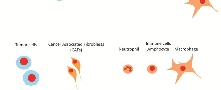
Int. J. Transl. Med. 2021, 1                                                                                                58

                               Table 1. Specific markers used in detection of CTCs, CTC clusters and other circulating, cancer-
                               associated cells. +: expression, −: no expression, +/−: expression in some cells.

                                        Description                                      Markers
                                           CTC                   CK+ (cytokeratins 8+, 18+, 19+), CD45−, EpCAM+, DAPI+
                                                                CK+/− (cytokeratins 8+, 18+, 19+), CD45+/−, EpCAM+/−,
                                  CTC-neutrophil cluster
                                                                    DAPI+, MPO (neutrophil myeloperoxidase)+/−
                                                                     CK+ (cytokeratins 8+, 18+, 19+), CD45+, EpCAM+,
                                  CTC-macrophage fusion
                                                                                      DAPI+, CD14+
                                                                        MPO (neutrophil myeloperoxidase)+, H3Cit
                                           NET
                                                                           (citrullinated histone H3)+, DAPI+

                                   CTCs were also shown to associate with activated, circulating endothelial cells (CECs) [17]
                               which represent yet another form of heterotypic CTC clusters and were suggested to protect
                               CTCs from anoikis.

                               2.2. CTC-Neutrophil Clusters
                                     White blood cells (WBC) include lymphocytes, monocytes, and granulocytes. Neu-
                               trophils, the most abundant granulocytes, constitute the first response in host defense
                               against bacterial infection. Their role in cancer is complex; it is known for some time that
                               neutrophil population can be polarized into anti-tumoral N1 phenotype, which promote
                               tumor cell clearance and pro-tumoral tumor-associated neutrophils (TANs) with N2 pheno-
                               type, which have been shown to promote tumorigenesis [11]. About 3.4% of CTCs found
                               in the bloodstream of breast cancer patients were shown to associate with neutrophils and
                               this association was linked to the worse progression-free survival [18].
                                     CTC-neutrophil clusters are formed via cell–cell adhesion, in particular with vascular
                               cell adhesion molecule 1 (VCAM-1), involved with trans-endothelial cell migration. Thus,
                               association with neutrophils may facilitate intravasation [19].
                                     Szczerba et al. [18] have shown in the mouse model that CTC-neutrophil clusters
                               promote tumor cell proliferation, cell cycle progression, and metastatic seeding. Single-cell
                               RNA-seq analysis of CTC-WBC clusters vs. single CTCs indicated that the IL6 and IL1β
                               signaling pathways are essential in the CTC-neutrophil crosstalk that leads to increased
                               proliferation of cancer cells. The authors also identified several mutations present in these
                               clusters, including the most important mutation in TLE1.

                               2.3. Neutrophil Extracellular Traps (NETs)
                                    Neutrophils also contribute to metastasis via NETs formation. The phenomenon
                               of NET was described in 2004 by Brinkmann et al. [20] as yet another form of innate
                               response designed to entrap and kill bacteria. However, later on it was shown that NETs
                               are also responsible for trapping CTCs and enhancing metastasis [21]. Neutrophils were
                               shown to decondense their chromatin and extrude DNA in a form of fibrils decorated
                               with antimicrobial peptides and enzymes, including neutrophil elastase, cathepsin G,
                               and myeloperoxidase (MPO). NETs are degraded mostly by nucleases but not cleared by
                               phagocytes, so their degradation takes longer. NETosis is triggered by microbe-associated
                               factors, like bacterial LPS, but also by many other endogenous stimuli, including cytokines,
                               platelets, activated endothelial cells, and some metabolites [22–24]. NETs have been shown
                               to sequester CTCs via interactions with β1-integrin [25]. Trapped CTCs are more prone to
                               adhesion to capillaries and subsequent extravasation to target organs [21]. Using intravital
                               imaging Park et al. demonstrated in the mouse model that cancer cells induce metastasis-
                               supporting neutrophil extracellular DNA traps [26].

                               2.4. Macrophage-Tumor Cell Fusion
                                   As with neutrophils, macrophages can have pro- or anti-tumorigenic properties. Tu-
                               mor microenvironment (TME)-infiltrating macrophages undergo M1 or M2 polarization,
Int. J. Transl. Med. 2021, 1 59
which plays a role in cancer progression. M1 phenotype, triggered by bacterial lipopolysac-
charide (LPS) or cytokines is associated with anti-tumor properties, while M2 phenotype
is pro-tumorigenic, enhancing proliferation, survival, invasiveness, and immunosuppres-
sion [10,27]. M2 phenotypes have been observed to fuse with tumor cells, creating a
large (≥30 µm diameter), polymorphic, mono- or polynuclear hybrid cells, with dual,
epithelial, and myeloid features. These hybrids expressing together CK+/EpCAM+ and
CD14/CD45+, are found in the blood of patients with many cancer types, including breast
cancer patients [28–31]. In these fused cells cytokeratin pattern is diffused, resembling
mesenchymal cells, not filamentous, as in epithelial cells [32]. Interestingly, fusion can be
partial, and occurs via tunneling nanotubes (TNTs), cellular projections that allow physical
connection between the cytoplasm of both cells and transport of different cargos [33].
Permanent cell fusion produces a true hybrid cell with enhanced metastatic phenotype and
surface receptors of both types.
Fusion is generally considered to confer migratory and invasive properties, enhancing
metastasis. This was demonstrated by the study of the artificial fusion between breast tumor
cells and macrophages; the fusion was shown to activate the Wnt/β-catenin signaling
pathway via TCF/LEF transcription factor activating downstream targets [34]. Fused
cells have been also described as displaying enhanced expression of the mesenchymal
markers. Moreover, fusion of tumor cells with macrophages may lead to chemoresistance
and immunosuppression [27].
3. The Origin of CTC Clusters
It has been demonstrated in an elegant mouse model experiment that CTC clusters are
poly/oligoclonal, so they do not derive from a single, proliferating cell [35,36]. However,
there are still two controversies surrounding the origin of CTC clusters: (1) do they exit
primary tumor already as a cluster or do they form later as a result of aggregation and (2)
do they derive from the edge or from the center of primary tumor?
3.1. Clusters Shedding vs. Intravascular Aggregation
In a seminal paper by Aceto et al. [35] the authors argue that CTC clusters cannot arise
from intravascular aggregation, because in their mouse model experiments differentially
stained tumor cells do not show any mixed seeding. Indeed, the circulation seems to be
not the best place to aggregate, since it provides very hostile conditions for epithelial cells:
shear stress, oxidative stress, and immune attack. Additionally, it should be mentioned that
CTCs lifespan in the circulation is rather short—hours or minutes, even less in case of CTC
clusters, giving no time for aggregation events. Despite this, using intravital microscopic
imaging Liu et al. [37], demonstrated aggregation of individual tumor cells resulting in
cluster formation, which were not generated by collective migration and cohesive shedding.
The authors have shown that these clusters were enriched in breast cancer stem cell marker
CD44, resulting in homophilic interactions and multicellular aggregation, which further
initiates CD44-PAK2 interaction, resulting in activation of FAK signaling [37]. Observed
clusters were shown to be linked to polyclonal metastasis, providing another explanation
for this phenomenon.
3.2. Edge vs. Center
While it seems logical that molecularly diverse and more oxygenated tumor edge
can shed CTC clusters more easily than hypoxic and partially necrotic inner core, there
are evidences that CTC clusters in breast cancer derive from the hypoxic regions of the
primary tumor and that these regions retain functional blood vessels [38]. Moreover, it
was demonstrated that hypoxia results in cell–cell junction upregulation and intravasation
of CTC clusters [38]. This association has the unexpected implications for breast cancer
therapy; in the same paper it was demonstrated that targeting vascular endothelial growth
factor A (VEGFA) in order to reduce primary tumor size is effective in itself, but increases
CTC clusters and metastasis, via reducing angiogenesis and increasing hypoxia.Int. J. Transl. Med. 2021, 1 60
4. Markers and Phenotypes of CTC Clusters
4.1. Stemness
It is well-known that cancer cells may possess the features characteristic for stem cells,
including cell renewal and formation of heterogeneous cell lineages [39]. Therefore, cells
with these properties are believed to be critical in treatment failure and metastasis forma-
tion. Currently, cancer stem cells (CSCs) are one of the main focuses in cancer research.
There are several markers used for CSCs identification depending on the cancer type (re-
viewed: [40]). Of all markers the combination of CD44, CD24, CD133, and ALDH-1 seems
to be the most universal for CSCs identification in solid tumors. As indicated previously,
newest research highlighted that CD44 might be critical for CTCs cluster formation via
its target PAK2 [37,41], leading to multicellular aggregation. Moreover, in vitro studies
on colon cancer cell line HCT-116 highlighted that post-sphere cultured cells that formed
clusters had higher expression of stem markers than single cells [42]. This suggests that
stemness might play a role in cluster formation. Stem cell properties are associated with
expression of transcription factors characteristic for embryonic cells—SOX2, NANOG,
OCT4. However, in healthy differentiated cells expression of these transcription factors is
silenced. This inhibition of expression occurs at early stages of differentiation and should
be permanent for fully differentiated cells. Recent studies indicated promoter methyla-
tion as possible regulatory system for long-term stem-related genes inhibition in tracheal
cells [43]. Therefore, it is probable that changes in methylation status of stem-associated
transcription factors are crucial for CSCs determination. Accordingly, major differences
in methylation patterns between single CTCs and clustering CTCs were reported: clus-
tering CTCs were characterized by hypomethylation of binding sites for major stemness
regulators—OCT4, NANOG, SOX2, and SIN3A. Interestingly this methylation pattern was
found to be exclusive for CTCs clusters, as dissociation reverts the methylation profile of
CTC clusters and suppresses metastasis [44]. Many studies suggest that stem cell properties
and cluster formation are linked. However, big cohort studies assessing clinical significance
of stemness in CTC clusters are still to come.
4.2. Epithelial-Mesenchymal Plasticity
Epithelial-mesenchymal transition (EMT) is commonly associated with more invasive
carcinoma; however, it seems that the highest tumorigenicity and metastatic potential can
be attributed to the hybrid epithelial-mesenchymal (E/M) phenotype, in which markers of
both types are overexpressed [45–47]. E/M phenotype is not some transitional state occur-
ring during EMT, it is a stable, long-lasting phenotype. Hybrid phenotype is particularly
eminent in CTCs, which display a wide spectrum of epithelial-mesenchymal plasticity
(EMP) (reviewed in [48,49]). For example, it was demonstrated that more than 75% of
CTCs from patients with metastatic breast cancer co-express epithelial cytokeratins with
vimentin, and N-cadherin [50]. Hybrid phenotype traits are associated with increased
migration, invasion, and survival, hence, these cells are considered more metastatic.
Some confusion pertains to epithelial cell marker E-cadherin. Long regarded as tumor
suppressor, its expression was associated with good prognosis and its loss during EMT
with increased invasiveness. However, recent findings undermine this view, showing that
E-cadherin expression, while indeed limiting invasiveness (meant as the ability to degrade
extracellular matrix), is in fact a survival factor, required for metastasis in multiple models
of breast cancer [51].
Crosstalk with the elements of microenvironment inside the vessel has also huge impact on
the EMT status of CTCs; tumor-activated platelets have been demonstrated to release α-granules
containing TGF-β and ATP, promoting EMT in associated CTCs [52]. Indirect interactions with
CAFs were also postulated in producing epithelial-mesenchymal plasticity [53].
4.3. Cell-Cell Junctions
CTC clusters retained at least partially epithelial characteristics, including high ex-
pression of cell–cell junction proteins. The most upregulated are: plakoglobin [35], keratinInt. J. Transl. Med. 2021, 1 61
14 [36], and claudin 11 [54]. Plakoglobin is a desmosomal or, less commonly, adherent
junction protein and its expression was reported as an independent prognostic factor
in breast cancer [55,56]. Keratin 14 is an intermediate filament protein associated with
hemidesmosome; keratin 14 (+) cells were enriched in desmosomal and hemidesmosomal
components [36]. Thus, it seems that desmosomes play an important role in CTC cluster
formation. Claudins are internal membrane proteins and tight junction components, they
form a physical barrier between cells, controlling the flow of solutes. Claudin upregulation
in CTC clusters was demonstrated for squamous cell carcinomas [54]. Stem cell marker
CD44, implicated in cluster formation, is also involved in the regulation of tight junc-
tions [57]. Cell–cell junctions are crucial for cluster formation; knockdown of plakoglobin
or CD44 precluded CTC cluster formation and suppressed metastasis [30,35].
In heterotypic clusters cell–cell junctions are also heterotypic; CAFs association with
CTCs is mediated by E-cadherin-N-cadherin adhesions [58]. This direct interaction was
shown to be crucial for directing clusters migration.
Interestingly, cell–cell junctions in clusters display remarkable plasticity and undergo
dynamic re-modeling when clusters traverse narrow capillaries; it was shown that cells form
a single file (without disintegrating), so they can fit into a vessel without blocking it [59].
5. Methods of Detection; Pros and Cons
The lack of the reliable CTC detection techniques represented for long decades a big
stumbling block to CTC research. Last two decades were pivotal in this aspect and now
we observe a multitude of effective techniques; however, there is still a problem with their
standardization, compatibility, and reproducibility. Below, we present a short recapitulation
of the main available techniques, with respect of their utility for CTC clusters detection.
There are several types of CTC isolation techniques, classified according to the prin-
ciples of the method of their capture/detection: methods based on the enrichment or
depletion of the CTC fraction achieved with specific antibodies (biological properties),
isolation based on physical properties (size, density, cellular charge), microfluidics isolation
which can use both size and specific markers and systems without prior selection, based
on the detection in the whole sample. Mixed strategies are also often employed.
Enrichment with specific antibodies detecting proteins expressed on the surface of
tumor cells is the most popular; the only (so far) FDA-approved method, CellSearch [60],
falls into this category. However, CellSearch is not optimal for cluster analysis: the most
obvious flaw is its dependence on EpCAM expression on tumor cell surface, which may not
be the case for cells shifted toward mesenchymal characteristics. Second, in immunoaffinity-
dependent systems, the enrichment step may not be as efficient for heterotypic clusters,
covered with other cells or cell fragments (platelets), which restrict epitope accessibility.
The other immunoaffinity-based methods (AdnaTest [61], EasySep [62], Dynabeads [62],
MINDEC- Multi-marker Immuno-magnetic Negative Depletion Enrichment of CTCs [63])
use mostly magnetic beads and employ either positive (e.g., EpCAM, cytokeratins) or
negative (e.g., CD45) selection to enrich CTC fraction. These methods offer free selection of
markers, but produce only enriched cellular fraction, thus, analyzing single CTCs or CTC
clusters is impossible.
Filter-based methods (CellSieve [64,65], ISET [66], ScreenCell [67], FMSA [68]) should
provide a good tool to study clusters, since clusters are much bigger than single cells of
any type, but they also have their challenges; whole-blood filtration takes time and often
leads to clogging, while using pressure to improve filtration may result in cell deformation
or cluster disintegration.
The enrichment can be also achieved by density gradient centrifugation (Onco-
Quick [69], Ficoll Paque [70]); these are simple centrifugation devices which enable to
separate different types of blood cells. Naturally, these devices do not provide very specific
enrichment, similarly to dielectric separation, based on differences in membrane area and
charge of tumor cells versus blood cells (DEP). These methods can be used as tools to
improve specificity of some other methods at the beginning or the end of other procedures.Int. J. Transl. Med. 2021, 1 62
The best promise, especially with respect to CTC clusters is probably held by microflu-
idic devices, often coupled with some other method. Microfluidic devices used in CTC
isolation are numerous and, in essence, are based on different size or deformability of CTCs,
which implies their different flow pattern. They can rely only on physical properties, which
has an advantage of being label-free and hence, independent of the specific markers (Parsor-
tix [71], Labyrinth [72], JETTA [73]). Some of them were specially designed to isolate CTC
clusters (deterministic lateral displacement: DLD [74], Straight Microfluidic Chips [75]).
Another type of microfluidic devices combines specific flow pattern with immunoaffinity,
boosting specificity, but—as with any immunoaffinity-based method—limiting sensitivity
(CTC-iChip [76], CMx platform [77], HB-CTC-Chip [78,79], OncoCEE [80]). Microfluidic
approaches are numerous, from very basic to extremely complicated, many of them are
custom-made, but in the selection process some may achieve the status of clinically ap-
proved method (for example, Parsortix is currently waiting for FDA approval). They offer
high specificity, are relatively gentle and enable isolation of viable cells for further analysis.
They seem to hold a big promise for the efficient and reliable CTC isolation, especially
regarding CTC clusters.
Different type of methods is represented by imaging-based approaches, working
without any specific enrichment apart from basic centrifugation, which produces buffy coat
layer containing nucleated blood cells and CTCs (RareCyte: [81], CytoTrack [82]). Material
from this layer is stained with the appropriate antibodies, spread on a disk or a slide, and
examined microscopically. In the CytoTrack system single cells can be picked from the disk
using micromanipulator (Cytopicker) and their genetic material can be examined, but since
they are fixed, no other types of analysis are available. This approach is free from the flaws
of immunoaffinity-dependent enrichment and can be as specific as are the antibodies used
to detect CTCs.
Summary of the CTC detection methods described here is presented in Figure 3 and Table 2.
Table 2. Description of CTC detection methods presented in Figure 2 with the assessment of their usefulness in CTC cluster
analysis. For more detailed description see review [83].
Category Methods (Examples) Key Features Ref.
Immunoaffinity-based (marker-dependent isolation),
sensitivity in a range from 27% (CellSearch) up to
CellSearch, AdnaTest, EasySep, 70% (CellSearch)–73% (AdnaTest), specificity 89–99%
[60–63]
Dynabeads, MINDEC (CellSearch), clinically validated (CellSearch),
Biological properties EpCAM(−) cells may be lost, not optimal
for cluster isolation
Imaging-based approaches (marker-dependent
RareCyto, CytoTrack identification), detection of EpCAM(+) and [81,82]
EpCAM(−) cells, clusters can be observed (Figure 1)
Density-gradient centrifugation,
OncoQuick, Ficoll Paque [69,70]
marker-independent, low purity of CTCs
Mircrofluidics (size and deformity based,
marker-independent isolation), high sensitivity
Parsortix, Labyrinth, JETTA, DLD, (92%, Parsortix) and specificity (100%, Parsortix),
[71–75]
Straight Microfluidic Chips subsequent analysis of cells possible, suitable for
CTC cluster analysis, some specially designed for
Physical properties
this purpose (DLD, Straight Microfluidic Chips)
Filter-based methods, marker-independent, some
allow post-capture culture and microscopic
CellSieve, ISET, ScreenCell, FMSA examination (ScreenCell), sensitivity 76%, specificity [64–68]
82% (ISET), size-dependent methods favor cluster
isolation, but applied pressure may disrupt clusters
Immunoaffinity with Microfluidics, high sensitivity
CTC-iChip, OncoCEE, Cmx
Mixed properties (95%, OncoCEE) and specificity (92%, OncoCEE), [76–80]
platform, HB-CTC-Chip
suitable for CTC cluster analysisInt.1J. Transl. Med. 2021, 1, FOR PEER REVIEW
Int. J. Transl. Med. 2021, 9 63
Figure 3. Methods commonly used in CTC isolation. Methods classified according to biological and
Figure 3. Methods commonly
physical usedwith
properties, in CTC isolation.
some usingMethods
mixed classified according
techniques, to biological
to improve and physical
specificity properties,ellipses).
(overlapping
with some using mixed techniques, to improve specificity (overlapping ellipses).
6. CTC Clusters and Their Impact on Survival
Single CTCs are well-known prognostic factors in breast cancer patients. The iden-
tification of one or more CTCs in 7.5 mL of blood correlates with early recurrence and
decreased overall survival (OS) in chemonaive patients with stage I-III [84]. In metastatic
breast cancer patients the presence of CTCs is associated with increased risk of progression
and shorter OS [85]. The predictive value of CTCs detection remains unclear and it is
not recommended and routinely used in a clinical practice. The data coming from small,
prospective study confirm that single CTCs and CTC clusters enumeration can be useful
in advanced breast cancer patients because of its effect on PFS [86]. Moreover, increased
number of CTC clusters between day 15 and 29 of a designed study cycle was related to
poorer PFS in metastatic triple negative breast cancer patients [87]. Respectively, patients
with MBC and prolonged presence of CTC clusters in blood are characterized by worse
prognosis [35]. Considering the fact that majority of single CTCs die in a bloodstream,
further research on CTC clusters and their impact on metastatic process is needed. CTC
clusters were found as more potent and resistant to apoptosis in comparison to single CTCs
in a mouse model in reference to the formation metastatic lesions in lungs [35].Int. J. Transl. Med. 2021, 1 64
Recognition of clusters as potential therapeutic targets opens new fascinating treatment
possibilities. Studies on the methylation status of CTC clusters lead to a conclusion that their
disruption may be beneficial for the patient. Gkountela et al. [44] describe identification
of the compounds that enable dispersion of CTC clusters into single cells, suppressing
metastasis formation, due to lower metastatic potential of single cells. They suggest that
these compounds should be administered early on, at the time of localized disease, to prevent
the spread, or at later stages to prevent metastasis-to-metastasis seeding. These compounds
represent the FDA-approved cardiac glycosides ouabain and digitoxin. They function as
Na+/K+ ATPase inhibitors, leading to the increase of intracellular Ca2+ concentration and
subsequent disruption of desmosomal and tight junctions and cluster disintegration.
Another surprising possibility was described by Donato et al. [38]. The authors
observed that hypoxia leads to cell–cell junction upregulation, followed by the forma-
tion and intravasation of highly metastatic CTC clusters. As a consequence, they have
come to rather disturbing realization that although anti-angiogenic treatment leads to
primary tumor shrinkage, it also increases intra-tumor hypoxia, leading to the increased
cluster shedding. Conversely, while pro-angiogenic treatment results in primary tumor
enlargement, it also suppresses the formation of CTC clusters, limiting metastasis.
7. Conclusions
CTC clusters appear to be the main factor and the most important target in breast
cancer metastasis. While they are obviously extremely rare, the current methods of CTC
isolation may underestimate their content for technical reasons. With the focus shifted to
CTC clusters, new experimental and treatment approaches can be developed, which can be
especially useful in early stages to prevent dissemination, or even in late stages, to prevent
metastasis-to-metastasis seeding, observed in breast cancer patients.
Funding: This research was funded by Polish National Science Centre, grant numbers 2016/21/B/NZ2/
03473, 2019/33/N/NZ5/00758.
Institutional Review Board Statement: The study (cell imaging for Figure 1) was conducted ac-
cording to the guidelines of the Declaration of Helsinki, and approved by the Institutional Ethics
Committee of National Institute of Oncology (protocol code 34/2016, 9 September 2016).
Informed Consent Statement: Informed consent was obtained from all subjects involved in the study.
Data Availability Statement: Not applicable.
Conflicts of Interest: The authors declare no conflict of interest.
Abbreviations
CAF cancer associated fibroblast
CK cytokeratins
CTC circulating tumor cell
DEP dielectrophoresis
ECM extracellular matrix
EpCAM Epithelial Cell Adhesion Molecule
EMP epithelial-mesenchymal plasticity
EMT epithelial-mesenchymal transition
HER2 human epidermal growth factor receptor
HR hormone receptors
MBC metastatic breast cancer
NET neutrophil extracellular traps
TAN tumor-associated neutrophils
TME tumor microenvironment
TNBC triple negative breast cancer
WBC white blood cellsInt. J. Transl. Med. 2021, 1 65
References
1. Sorlie, T.; Perou, C.M.; Tibshirani, R.; Aas, T.; Geisler, S.; Johnsen, H.; Hastie, T.; Eisen, M.B.; van de Rijn, M.; Jeffrey, S.S.; et al.
Gene expression patterns of breast carcinomas distinguish tumor subclasses with clinical implications. Proc. Natl. Acad. Sci. USA
2001, 98, 10869–10874. [CrossRef]
2. Perou, C.M.; Sørlie, T.; Eisen, M.B.; Van De Rijn, M.; Jeffrey, S.S.; Rees, C.A.; Pollack, J.R.; Ross, D.T.; Johnsen, H.; Akslen, L.A.;
et al. Molecular portraits of human breast tumours. Nature 2000, 406, 747–752. [CrossRef] [PubMed]
3. Tabor, S.; Szostakowska-Rodzos, M.; Fabisiewicz, A.; Grzybowska, E.A. How to Predict Metastasis in Luminal Breast Cancer?
Current Solutions and Future Prospects. Int. J. Mol. Sci. 2020, 21, 8415. [CrossRef] [PubMed]
4. Ashworth, T.R. A Case of Cancer in Which Cells Similar to Those in the Tumours Were Seen in the Blood after Death. Med. J.
Aust. 1869, 14, 146–147.
5. Pantel, K.; Alix-Panabières, C.; Riethdorf, S. Cancer micrometastases. Nat. Rev. Clin. Oncol. 2009, 6, 339–351. [CrossRef] [PubMed]
6. Fehm, T.; Müller, V.; Alix-Panabières, C.; Pantel, K. Micrometastatic spread in breast cancer: Detection, molecular characterization
and clinical relevance. Breast Cancer Res. 2008, 10, S1. [CrossRef] [PubMed]
7. Markou, A.; Farkona, S.; Schiza, C.; Efstathiou, T.; Kounelis, S.; Malamos, N.; Georgoulias, V.; Lianidou, E. PIK3CA Mutational
Status in Circulating Tumor Cells Can Change During Disease Recurrence or Progression in Patients with Breast Cancer.
Clin. Cancer Res. 2014, 20, 5823–5834. [CrossRef]
8. Pestrin, M.; Salvianti, F.; Galardi, F.; De Luca, F.; Turner, N.; Malorni, L.; Pazzagli, M.; Di Leo, A.; Pinzani, P. Heterogeneity of
PIK3CA mutational status at the single cell level in circulating tumor cells from metastatic breast cancer patients. Mol. Oncol.
2015, 9, 749–757. [CrossRef]
9. Lianidou, E.S.; Mavroudis, D.; Georgoulias, V. Clinical challenges in the molecular characterization of circulating tumour cells in
breast cancer. Br. J. Cancer 2013, 108, 2426–2432. [CrossRef]
10. Najafi, M.; Goradel, N.H.; Farhood, B.; Salehi, E.; Nashtaei, M.S.; Khanlarkhani, N.; Khezri, Z.; Majidpoor, J.; Abouzaripour, M.;
Habibi, M.; et al. Macrophage polarity in cancer: A review. J. Cell. Biochem. 2019, 120, 2756–2765. [CrossRef]
11. Fridlender, Z.G.; Sun, J.; Kim, S.; Kapoor, V.; Cheng, G.; Ling, L.; Worthen, G.S.; Albelda, S.M. Polarization of Tumor-Associated
Neutrophil Phenotype by TGF-β: “N1” versus “N2” TAN. Cancer Cell 2009, 16, 183–194. [CrossRef]
12. Ping, Q.; Yan, R.; Cheng, X.; Wang, W.; Zhong, Y.; Hou, Z.; Shi, Y.; Wang, C.; Li, R. Cancer-associated fibroblasts: Overview,
progress, challenges, and directions. Cancer Gene Ther. 2021, 12, 1–16. [CrossRef]
13. Duda, D.G.; Duyverman, A.M.M.J.; Kohno, M.; Snuderl, M.; Steller, E.J.A.; Fukumura, D.; Jain, R.K. Malignant cells facilitate lung
metastasis by bringing their own soil. Proc. Natl. Acad. Sci. USA 2010, 107, 21677–21682. [CrossRef] [PubMed]
14. Ward, M.P.; Kane, L.E.; Norris, L.A.; Mohamed, B.M.; Kelly, T.; Bates, M.; Clarke, A.; Brady, N.; Martin, C.M.; Brooks, R.D.; et al.
Platelets, immune cells and the coagulation cascade; friend or foe of the circulating tumour cell? Mol. Cancer 2021, 20, 1–17. [CrossRef]
15. Huang, Q.; Li, S.; Hu, X.; Sun, M.; Wu, Q.; Dai, H.; Tan, Y.; Sun, F.; Wang, C.; Rong, X.; et al. Shear stress activates ATOH8 via
autocrine VEGF promoting glycolysis dependent-survival of colorectal cancer cells in the circulation. J. Exp. Clin. Cancer Res.
2020, 39, 25. [CrossRef]
16. Akolkar, D.; Patil, D.; Crook, T.; Limaye, S.; Page, R.; Datta, V.; Patil, R.; Sims, C.; Ranade, A.; Fulmali, P.; et al. Circulating
ensembles of tumor-associated cells: A redoubtable new systemic hallmark of cancer. Int. J. Cancer 2020, 146, 3485–3494.
[CrossRef] [PubMed]
17. Yadav, A.; Kumar, B.; Yu, J.-G.; Old, M.; Teknos, T.N.; Kumar, P. Tumor-Associated Endothelial Cells Promote Tumor Metastasis
by Chaperoning Circulating Tumor Cells and Protecting Them from Anoikis. PLoS ONE 2015, 10, e0141602. [CrossRef] [PubMed]
18. Szczerba, B.M.; Castro-Giner, F.; Vetter, M.; Krol, I.; Gkountela, S.; Landin, J.; Scheidmann, M.C.; Donato, C.; Scherrer, R.; Singer, J.;
et al. Neutrophils escort circulating tumour cells to enable cell cycle progression. Nat. Cell Biol. 2019, 566, 553–557. [CrossRef]
19. Muller, W.A. Mechanisms of Leukocyte Transendothelial Migration. Annu. Rev. Pathol. Mech. Dis. 2011, 6, 323–344. [CrossRef]
20. Brinkmann, V.; Reichard, U.; Goosmann, C.; Fauler, B.; Uhlemann, Y.; Weiss, D.S.; Weinrauch, Y.; Zychlinsky, A. Neutrophil
extracellular traps kill bacteria. Science 2004, 303, 1532–1535. [CrossRef]
21. Cools-Lartigue, J.; Spicer, J.; McDonald, B.; Gowing, S.; Chow, S.; Giannias, B.; Bourdeau, F.; Kubes, P.; Ferri, L. Neutrophil
extracellular traps sequester circulating tumor cells and promote metastasis. J. Clin. Investig. 2013, 123, 3446–3458. [CrossRef]
22. Gupta, A.K.; Joshi, M.B.; Filippova, M.; Erne, P.; Hasler, P.; Hahn, S.; Resink, T.J. Activated endothelial cells induce neutrophil
extracellular traps and are susceptible to NETosis-mediated cell death. FEBS Lett. 2010, 584, 3193–3197. [CrossRef] [PubMed]
23. Gupta, A.K.; Hasler, P.; Holzgreve, W.; Gebhardt, S.; Hahn, S. Induction of Neutrophil Extracellular DNA Lattices by Placental
Microparticles and IL-8 and Their Presence in Preeclampsia. Hum. Immunol. 2005, 66, 1146–1154. [CrossRef] [PubMed]
24. Clark, S.R.; Ma, A.C.; A Tavener, S.; McDonald, B.; Goodarzi, Z.; Kelly, M.M.; Patel, K.D.; Chakrabarti, S.; McAvoy, E.; Sinclair,
G.D.; et al. Platelet TLR4 activates neutrophil extracellular traps to ensnare bacteria in septic blood. Nat. Med. 2007, 13, 463–469.
[CrossRef] [PubMed]
25. Najmeh, S.; Cools-Lartigue, J.; Rayes, R.F.; Gowing, S.; Vourtzoumis, P.; Bourdeau, F.; Giannias, B.; Berube, J.; Rousseau, S.; Ferri,
L.E.; et al. Neutrophil extracellular traps sequester circulating tumor cells via β1-integrin mediated interactions. Int. J. Cancer
2017, 140, 2321–2330. [CrossRef]
26. Park, J.; Wysocki, R.; Amoozgar, Z.; Maiorino, L.; Fein, M.R.; Jorns, J.; Schott, A.F.; Kinugasa-Katayama, Y.; Lee, Y.; Won, N.H.;
et al. Cancer cells induce metastasis-supporting neutrophil extracellular DNA traps. Sci. Transl. Med. 2016, 8, 361ra138. [CrossRef]Int. J. Transl. Med. 2021, 1 66
27. Manjunath, Y.; Porciani, D.; Mitchem, J.B.; Suvilesh, K.N.; Avella, D.M.; Kimchi, E.T.; Staveley-O’Carroll, K.F.; Burke, D.H.; Li, G.;
Kaifi, J.T. Tumor-Cell–Macrophage Fusion Cells as Liquid Biomarkers and Tumor Enhancers in Cancer. Int. J. Mol. Sci. 2020, 21,
1872. [CrossRef]
28. Shabo, I.; Midtbö, K.; Andersson, H.; Åkerlund, E.; Olsson, H.; Wegman, P.; Gunnarsson, C.; Lindström, A. Macrophage traits in
cancer cells are induced by macrophage-cancer cell fusion and cannot be explained by cellular interaction. BMC Cancer 2015, 15,
1–11. [CrossRef]
29. Shabo, I.; Olsson, H.; Stål, O.; Svanvik, J. Breast Cancer Expression of DAP12 is Associated With Skeletal and Liver Metastases
and Poor Survival. Clin. Breast Cancer 2013, 13, 371–377. [CrossRef] [PubMed]
30. Shabo, I.; Svanvik, J. Expression of Macrophage Antigens by Tumor Cells. Adv. Exp. Med. Biol. 2011, 714, 141–150. [CrossRef]
31. Adams, D.L.; Martin, S.S.; Alpaugh, R.K.; Charpentier, M.; Tsai, S.; Bergan, R.C.; Ogden, I.M.; Catalona, W.; Chumsri, S.; Tang, C.-M.;
et al. Circulating giant macrophages as a potential biomarker of solid tumors. Proc. Natl. Acad. Sci. USA 2014, 111, 3514–3519.
[CrossRef]
32. Adams, D.L.; Adams, D.K.; Alpaugh, R.K.; Cristofanilli, M.; Martin, S.S.; Chumsri, S.; Tang, C.-M.; Marks, J.R. Circulating
Cancer-Associated Macrophage-Like Cells Differentiate Malignant Breast Cancer and Benign Breast Conditions. Cancer Epidemiol.
Biomarkers Prev. 2016, 25, 1037–1042. [CrossRef] [PubMed]
33. Önfelt, B.; Nedvetzki, S.; Benninger, R.; Purbhoo, M.A.; Sowinski, S.; Hume, A.; Seabra, M.C.; Neil, M.A.A.; French, P.M.W.;
Davis, D.M. Structurally Distinct Membrane Nanotubes between Human Macrophages Support Long-Distance Vesicular Traffic
or Surfing of Bacteria. J. Immunol. 2006, 177, 8476–8483. [CrossRef] [PubMed]
34. Zhang, L.-N.; Huang, Y.-H.; Zhao, L. Fusion of macrophages promotes breast cancer cell proliferation, migration and invasion
through activating epithelial-mesenchymal transition and Wnt/β-catenin signaling pathway. Arch. Biochem. Biophys. 2019, 676,
108137. [CrossRef] [PubMed]
35. Aceto, N.; Bardia, A.; Miyamoto, D.T.; Donaldson, M.C.; Wittner, B.S.; Spencer, J.A.; Yu, M.; Pely, A.; Engstrom, A.; Zhu, H.; et al.
Circulating Tumor Cell Clusters Are Oligoclonal Precursors of Breast Cancer Metastasis. Cell 2014, 158, 1110–1122. [CrossRef]
[PubMed]
36. Cheung, K.J.; Padmanaban, V.; Silvestri, V.; Schipper, K.; Cohen, J.D.; Fairchild, A.N.; Gorin, M.A.; Verdone, J.E.; Pienta, K.J.;
Bader, J.S.; et al. Polyclonal breast cancer metastases arise from collective dissemination of keratin 14-expressing tumor cell
clusters. Proc. Natl. Acad. Sci. USA 2016, 113, E854–E863. [CrossRef] [PubMed]
37. Liu, X.; Taftaf, R.; Kawaguchi, M.; Chang, Y.-F.; Chen, W.; Entenberg, D.; Zhang, Y.; Gerratana, L.; Huang, S.; Patel, D.B.; et al.
Homophilic CD44 Interactions Mediate Tumor Cell Aggregation and Polyclonal Metastasis in Patient-Derived Breast Cancer
Models. Cancer Discov. 2019, 9, 96–113. [CrossRef] [PubMed]
38. Donato, C.; Kunz, L.; Castro-Giner, F.; Paasinen-Sohns, A.; Strittmatter, K.; Szczerba, B.M.; Scherrer, R.; Di Maggio, N.; Heu-
sermann, W.; Biehlmaier, O.; et al. Hypoxia Triggers the Intravasa-tion of Clustered Circulating Tumor Cells. Cell Rep. 2020, 32.
[CrossRef]
39. Friedmann-Morvinski, D.; Verma, I.M. Dedifferentiation and reprogramming: Origins of cancer stem cells. EMBO Rep. 2014, 15,
244–253. [CrossRef]
40. Murar, M.; Vaidya, A. Cancer stem cell markers: Premises and prospects. Biomarkers Med. 2015, 9, 1331–1342. [CrossRef]
41. Kawaguchi, M.; Dashzeveg, N.; Cao, Y.; Jia, Y.; Liu, X.; Shen, Y.; Liu, H. Extracellular Domains I and II of cell-surface glycoprotein
CD44 mediate its trans-homophilic dimerization and tumor cluster aggregation. J. Biol. Chem. 2020, 295, 2640–2649. [CrossRef]
42. Kapeleris, J.; Zou, H.; Qi, Y.; Gu, Y.; Li, J.; Schoning, J.; Monteiro, M.J.; Gu, W. Cancer stemness contributes to cluster formation of
colon cancer cells and high metastatic potentials. Clin. Exp. Pharmacol. Physiol. 2019, 47, 838–847. [CrossRef]
43. Zhou, Y.; Song, N.; Li, X.; Han, Y.; Ren, Z.; Xu, J.-X.; Han, Y.-C.; Li, F.; Jia, X. Changes in the methylation status of the Oct3/4,
Nanog, and Sox2 promoters in stem cells during regeneration of rat tracheal epithelium after injury. Oncotarget 2016, 8, 2984–2994.
[CrossRef]
44. Gkountela, S.; Castro-Giner, F.; Szczerba, B.M.; Vetter, M.; Landin, J.; Scherrer, R.; Krol, I.; Scheidmann, M.C.; Beisel, C.; Stirnimann,
C.U.; et al. Circulating Tumor Cell Clustering Shapes DNA Methylation to Enable Metastasis Seeding. Cell 2019, 176, 98–112.e14.
[CrossRef]
45. Lee, J.M.; Dedhar, S.; Kalluri, R.; Thompson, E.W. The epithelial–mesenchymal transition: New insights in signaling, development,
and disease. J. Cell Biol. 2006, 172, 973–981. [CrossRef] [PubMed]
46. Pastushenko, I.; Blanpain, C. EMT Transition States during Tumor Progression and Metastasis. Trends Cell Biol. 2019, 29, 212–226.
[CrossRef]
47. Kroger, C.; Afeyan, A.; Mraz, J.; Eaton, E.N.; Reinhardt, F.; Khodor, Y.L.; Thiru, P.; Bierie, B.; Ye, X.; Burge, C.B.; et al. Acquisition
of a hybrid E/M state is essential for tumorigenicity of basal breast cancer cells. Proc. Natl. Acad. Sci. USA 2019, 116, 7353–7362.
[CrossRef] [PubMed]
48. McInnes, L.M.; Ejacobsen, N.; Eredfern, A.; Edowling, A.; Thompson, E.W.; Saunders, C.M. Clinical Implications of Circulating
Tumor Cells of Breast Cancer Patients: Role of Epithelial–Mesenchymal Plasticity. Front. Oncol. 2015, 5, 42. [CrossRef] [PubMed]
49. Bhatia, S.; Wang, P.; Toh, A.; Thompson, E.W. New Insights Into the Role of Phenotypic Plasticity and EMT in Driving Cancer
Progression. Front. Mol. Biosci. 2020, 7, 71. [CrossRef] [PubMed]Int. J. Transl. Med. 2021, 1 67
50. Armstrong, A.J.; Marengo, M.S.; Oltean, S.; Kemeny, G.; Bitting, R.L.; Turnbull, J.D.; Herold, C.I.; Marcom, P.K.; George, D.J.;
Garcia-Blanco, M.A. Circulating Tumor Cells from Patients with Advanced Prostate and Breast Cancer Display Both Epithelial
and Mesenchymal Markers. Mol. Cancer Res. 2011, 9, 997–1007. [CrossRef]
51. Padmanaban, V.; Krol, I.; Suhail, Y.; Szczerba, B.M.; Aceto, N.; Bader, J.S.; Ewald, A.J. E-cadherin is required for metastasis in
multiple models of breast cancer. Nature 2019, 573, 439–444. [CrossRef]
52. Labelle, M.; Begum, S.; Hynes, R.O. Direct signaling between platelets and cancer cells induces an epithelial-mesenchymal-like
transition and promotes metastasis. Cancer Cell 2011, 20, 576–590. [CrossRef]
53. Matsumura, Y.; Ito, Y.; Mezawa, Y.; Sulidan, K.; Daigo, Y.; Hiraga, T.; Mogushi, K.; Wali, N.; Suzuki, H.; Itoh, T.; et al. Stromal
fibroblasts induce metastatic tumor cell clusters via epithelial–mesenchymal plasticity. Life Sci. Alliance 2019, 2, e201900425. [CrossRef]
54. Li, C.-F.; Chen, J.-Y.; Ho, Y.-H.; Hsu, W.-H.; Wu, L.-C.; Lan, H.-Y.; Hsu, D.S.-S.; Tai, S.-K.; Chang, Y.-C.; Yang, M.-H. Snail-induced
claudin-11 prompts collective migration for tumour progression. Nat. Cell Biol. 2019, 21, 251–262. [CrossRef]
55. Goto, W.; Kashiwagi, S.; Asano, Y.; Takada, K.; Takahashi, K.; Hatano, T.; Takashima, T.; Tomita, S.; Motomura, H.; Ohsawa, M.;
et al. Circulating tumor cell clusters-associated gene plakoglobin is a significant prognostic predictor in patients with breast
cancer. Biomark. Res. 2017, 5, 19. [CrossRef]
56. Xie, N.; Hu, Z.; Tian, C.; Xiao, H.; Liu, L.; Yang, X.; Li, J.; Wu, H.; Lu, J.; Gao, J.; et al. In Vivo Detection of CTC and CTC
Plakoglobin Status Helps Predict Prognosis in Patients with Metastatic Breast Cancer. Pathol. Oncol. Res. 2020, 26, 2435–2442.
[CrossRef]
57. Kirschner, N.; Haftek, M.; Niessen, C.M.; Behne, M.J.; Furuse, M.; Moll, I.; Brandner, J.M. CD44 Regulates Tight-Junction Assembly
and Barrier Function. J. Investig. Dermatol. 2011, 131, 932–943. [CrossRef] [PubMed]
58. Labernadie, A.; Kato, T.; Brugués, A.; Serra-Picamal, X.; Derzsi, S.; Arwert, E.; Weston, A.; González-Tarragó, V.; Elosegui-Artola,
A.; Albertazzi, L.; et al. A mechanically active heterotypic E-cadherin/N-cadherin adhesion enables fibroblasts to drive cancer
cell invasion. Nat. Cell Biol. 2017, 19, 224–237. [CrossRef]
59. Au, S.; Storey, B.D.; Moore, J.C.; Tang, Q.; Chen, Y.-L.; Javaid, S.; Sarioglu, A.F.; Sullivan, R.; Madden, M.W.; O’Keefe, R.; et al. Clusters
of circulating tumor cells traverse capillary-sized vessels. Proc. Natl. Acad. Sci. USA 2016, 113, 4947–4952. [CrossRef] [PubMed]
60. Neumann, M.H.D.; Schneck, H.; Decker, Y.; Schömer, S.; Franken, A.; Endris, V.; Pfarr, N.; Weichert, W.; Niederacher, D.; Fehm, T.;
et al. Isolation and characterization of circulating tumor cells using a novel workflow combining the CellSearch® system and the
CellCelector™. Biotechnol. Prog. 2017, 33, 125–132. [CrossRef] [PubMed]
61. Andreopoulou, E.; Yang, L.-Y.; Rangel, K.M.; Reuben, J.M.; Hsu, L.; Krishnamurthy, S.; Valero, V.; Fritsche, H.A.; Cristofanilli,
M. Comparison of assay methods for detection of circulating tumor cells in metastatic breast cancer: AdnaGen AdnaTest
BreastCancer Select/Detect™ versus Veridex CellSearch™ system. Int. J. Cancer 2012, 130, 1590–1597. [CrossRef] [PubMed]
62. Drucker, A.; Teh, E.M.; Kostyleva, R.; Rayson, D.; Douglas, S.; Pinto, D.M. Comparative performance of different methods for
circulating tumor cell enrichment in metastatic breast cancer patients. PLoS ONE 2020, 15, e0237308. [CrossRef] [PubMed]
63. Lapin, M.; Tjensvoll, K.; Oltedal, S.; Buhl, T.; Gilje, B.; Smaaland, R.; Nordgård, O. MINDEC-An Enhanced Negative Depletion
Strategy for Circulating Tumour Cell Enrichment. Sci. Rep. 2016, 6, 28929. [CrossRef] [PubMed]
64. Zhu, P.; Stanton, M.L.; Castle, E.P.; Joseph, R.W.; Adams, D.L.; Li, S.; Amstutz, P.; Tang, C.-M.; Ho, T.H. Detection of tumor-
associated cells in cryopreserved peripheral blood mononuclear cell samples for retrospective analysis. J. Transl. Med. 2016, 14,
198. [CrossRef]
65. Adams, D.L.; Alpaugh, R.K.; Martin, S.S.; Charpentier, M.; Chumsri, S.; Cristofanilli, M.; Adams, D.K.; Makarova, O.V.; Zhu,
P.; Li, S.; et al. Precision microfilters as an all in one system for multiplex analysis of circulating tumor cells. RSC Adv. 2016, 6,
6405–6414. [CrossRef]
66. Ma, Y.-C.; Wang, L.; Yu, F.-L. Recent Advances and Prospects in the Isolation by Size of Epithelial Tumor Cells (ISET) Methodology.
Technol. Cancer Res. Treat. 2013, 12, 295–309. [CrossRef]
67. Mu, Z.; Benali-Furet, N.; Uzan, G.; Znaty, A.; Ye, Z.; Paolillo, C.; Wang, C.; Austin, L.; Rossi, G.; Fortina, P.; et al. Detection and
Characterization of Circulating Tumor Associated Cells in Metastatic Breast Cancer. Int. J. Mol. Sci. 2016, 17, 1665. [CrossRef]
68. Harouaka, R.A.; Zhou, M.-D.; Yeh, Y.-T.; Khan, W.J.; Das, A.; Liu, X.; Christ, C.C.; Dicker, D.T.; Baney, T.S.; Kaifi, J.T.; et al. Flexible
Micro Spring Array Device for High-Throughput Enrichment of Viable Circulating Tumor Cells. Clin. Chem. 2014, 60, 323–333.
[CrossRef]
69. Clawson, G.A.; Kimchi, E.; Patrick, S.D.; Xin, P.; Harouaka, R.; Zheng, S.; Berg, A.; Schell, T.; Staveley-O’Carroll, K.F.; Neves, R.I.;
et al. Circulating Tumor Cells in Melanoma Patients. PLOS ONE 2012, 7, e41052. [CrossRef] [PubMed]
70. He, W.; Kularatne, S.A.; Kalli, K.R.; Prendergast, F.G.; Amato, R.J.; Klee, G.G.; Hartmann, L.C.; Low, P.S. Quantitation of circulating
tumor cells in blood samples from ovarian and prostate cancer patients using tumor-specific fluorescent ligands. Int. J. Cancer
2008, 123, 1968–1973. [CrossRef]
71. Miller, M.C.; Robinson, P.S.; Wagner, C.; O’Shannessy, D.J. The Parsortix™ Cell Separation System—A versatile liquid biopsy
platform. Cytom. Part A 2018, 93, 1234–1239. [CrossRef] [PubMed]
72. Zeinali, M.; Lee, M.; Nadhan, A.; Mathur, A.; Hedman, C.; Lin, E.; Harouaka, R.; Wicha, M.S.; Zhao, L.; Palanisamy, N.; et al.
High-Throughput Label-Free Isolation of Heterogeneous Circulating Tumor Cells and CTC Clusters from Non-Small-Cell Lung
Cancer Patients. Cancers 2020, 12, 127. [CrossRef] [PubMed]
73. Riahi, R.; Gogoi, P.; Sepehri, S.; Zhou, Y.; Handique, K.; Godsey, J.; Wang, Y. A novel microchannel-based device to capture and
analyze circulating tumor cells (CTCs) of breast cancer. Int. J. Oncol. 2014, 44, 1870–1878. [CrossRef] [PubMed]Int. J. Transl. Med. 2021, 1 68
74. Au, S.; Jon; Stoddard, A.E.; Wong, K.H.K.; Fachin, F.; Maheswaran, S.; Haber, D.A.; Stott, S.L.; Kapur, R.; Toner, M. Microfluidic
Isolation of Circulating Tumor Cell Clusters by Size and Asymmetry. Sci. Rep. 2017, 7. [CrossRef] [PubMed]
75. Kulasinghe, A.; Zhou, J.; Kenny, L.; Papautsky, I.; Punyadeera, C. Capture of Circulating Tumour Cell Clusters Using Straight
Microfluidic Chips. Cancers 2019, 11, 89. [CrossRef] [PubMed]
76. Ozkumur, E.; Shah, A.M.; Ciciliano, J.C.; Emmink, B.L.; Miyamoto, D.T.; Brachtel, E.; Yu, M.; Chen, P.-I.; Morgan, B.; Trautwein, J.;
et al. Inertial Focusing for Tumor Antigen-Dependent and -Independent Sorting of Rare Circulating Tumor Cells. Sci. Transl. Med.
2013, 5, 179ra47. [CrossRef]
77. Chen, J.-Y.; Tsai, W.-S.; Shao, H.-J.; Wu, J.-C.; Lai, J.-M.; Lu, S.-H.; Hung, T.-F.; Yang, C.-T.; Wu, L.-C.; Chen, J.-S.; et al. Sensitive and
Specific Biomimetic Lipid Coated Microfluidics to Isolate Viable Circulating Tumor Cells and Microemboli for Cancer Detection.
PLoS ONE 2016, 11, e0149633. [CrossRef]
78. Stott, S.L.; Hsu, C.-H.; Tsukrov, D.I.; Yu, M.; Miyamoto, D.T.; Waltman, B.A.; Rothenberg, S.M.; Shah, A.M.; Smas, M.E.; Korir,
G.K.; et al. Isolation of circulating tumor cells using a microvortex-generating herringbone-chip. Proc. Natl. Acad. Sci. USA 2010,
107, 18392–18397. [CrossRef]
79. Wang, S.; Thomas, A.; Lee, E.; Yang, S.; Cheng, X.; Liu, Y. Highly efficient and selective isolation of rare tumor cells using a
microfluidic chip with wavy-herringbone micro-patterned surfaces. Anal. 2016, 141, 2228–2237. [CrossRef]
80. Mayer, J.A.; Pham, T.; Wong, K.L.; Scoggin, J.; Sales, E.V.; Clarin, T.; Pircher, T.J.; Mikolajczyk, S.D.; Cotter, P.D.; Bischoff, F.Z.
FISH-based determination of HER2 status in circulating tumor cells isolated with the microfluidic CEE™ platform. Cancer Genet.
2011, 204, 589–595. [CrossRef]
81. Kaldjian, E.P.; Ramirez, A.B.; Sun, Y.; Campton, D.E.; Werbin, J.L.; Varshavskaya, P.; Quarre, S.; George, T.; Madan, A.; Blau,
C.A.; et al. The RareCyte®platform for next-generation analysis of circulating tumor cells. Cytom. Part A 2018, 93, 1220–1225.
[CrossRef] [PubMed]
82. Hillig, T.; Nygaard, A.; Nekiunaite, L.; Klingelhöfer, J.; Sölétormos, G. In vitro validation of an ultra-sensitive scanning fluores-
cence microscope for analysis of Circulating Tumor Cells. APMIS 2014, 122, 545–551. [CrossRef] [PubMed]
83. Habli, Z.; Alchamaa, W.; Saab, R.; Kadara, H.; Khraiche, M.L. Circulating Tumor Cell Detection Technologies and Clinical Utility:
Challenges and Opportunities. Cancers 2020, 12, 1930. [CrossRef]
84. Lucci, A.; Hall, C.S.; Lodhi, A.K.; Bhattacharyya, A.; E Anderson, A.; Xiao, L.; Bedrosian, I.; Kuerer, H.M.; Krishnamurthy, S.
Circulating tumour cells in non-metastatic breast cancer: A prospective study. Lancet Oncol. 2012, 13, 688–695. [CrossRef]
85. Zhang, L.; Riethdorf, S.; Wu, G.; Wang, T.; Yang, K.; Peng, G.; Liu, J.; Pantel, K. Meta-Analysis of the Prognostic Value of
Circulating Tumor Cells in Breast Cancer. Clin. Cancer Res. 2012, 18, 5701–5710. [CrossRef]
86. Mu, Z.; Wang, C.; Ye, Z.; Austin, L.; Civan, J.; Hyslop, T.; Palazzo, J.P.; Jaslow, R.; Li, B.; Myers, R.E.; et al. Prospective assessment
of the prognostic value of circulating tumor cells and their clusters in patients with advanced-stage breast cancer. Breast Cancer
Res. Treat. 2015, 154, 563–571. [CrossRef]
87. Paoletti, C.; Li, Y.; Muñiz, M.C.; Kidwell, K.M.; Aung, K.; Thomas, D.G.; Brown, M.E.; Abramson, V.G.; Irvin, W.J., Jr.; Lin, N.U.;
et al. Significance of Circulating Tumor Cells in Metastatic Triple-Negative Breast Cancer Patients within a Randomized, Phase II
Trial: TBCRC 019. Clin. Cancer Res. 2015, 21, 2771–2779. [CrossRef] [PubMed]You can also read