Mycobacterium-Mediated Transfer of Plasmid DNA into
←
→
Page content transcription
If your browser does not render page correctly, please read the page content below
INFECTION AND IMMUNITY, Oct. 2007, p. 4804–4816                                                                               Vol. 75, No. 10
0019-9567/07/$08.00⫹0 doi:10.1128/IAI.01877-06
Copyright © 2007, American Society for Microbiology. All Rights Reserved.

           Genetic Alteration of Mycobacterium smegmatis To Improve
            Mycobacterium-Mediated Transfer of Plasmid DNA into
                   Mammalian Cells and DNA Immunization䌤
 Yongkai Mo,1† Natalie M. Quanquin,1† William H. Vecino,1‡ Uma Devi Ranganathan,1 Lydia Tesfa,1
                   William Bourn,2 Keith M. Derbyshire,3,4 Norman L. Letvin,5
                        William R. Jacobs, Jr.,1,6 and Glenn J. Fennelly7,8*
Department of Microbiology and Immunology, Albert Einstein College of Medicine, Bronx, New York 104611; Institute of Infectious
 Disease and Molecular Medicine, University of Cape Town, Observatory, 7925, Cape Town, South Africa2; Division of Infectious
    Disease, Wadsworth Center, New York State Department of Health,3 and Department of Biomedical Sciences, University at

                                                                                                                                                Downloaded from http://iai.asm.org/ on February 15, 2021 by guest
     Albany,4 Albany, New York 12201; Division of Viral Pathogenesis, Beth Israel Deaconess Medical Center, Harvard Medical
       School, Boston, Massachusetts 021155; Howard Hughes Medical Institute, Albert Einstein College of Medicine, Bronx,
              New York 104616; Department of Pediatrics, Albert Einstein College of Medicine, Bronx, New York 104617;
                and The Lewis M. Fraad Department of Pediatrics, Jacobi Medical Center, Bronx, New York 104618
                       Received 28 November 2006/Returned for modification 9 January 2007/Accepted 15 July 2007

             Mycobacteria target and persist within phagocytic monocytes and are strong adjuvants, making them
          attractive candidate vectors for DNA vaccines. We characterized the ability of mycobacteria to deliver trans-
          genes to mammalian cells and the effects of various bacterial chromosomal mutations on the efficiency of
          transfer in vivo and in vitro. First, we observed green fluorescent protein expression via microscopy and
          fluorescence-activated cell sorting analysis after infection of phagocytic and nonphagocytic cell lines by
          Mycobacterium smegmatis or M. bovis BCG harboring a plasmid encoding the fluorescence gene under the
          control of a eukaryotic promoter. Next, we compared the efficiencies of gene transfer using M. smegmatis or
          BCG containing chromosomal insertions or deletions that cause early lysis, hyperconjugation, or an increased
          plasmid copy number. We observed a significant—albeit only 1.7-fold—increase in the level of plasmid transfer
          to eukaryotic cells infected with M. smegmatis hyperconjugation mutants. M. smegmatis strains that overex-
          pressed replication proteins (Rep) of pAL5000, a plasmid whose replicon is incorporated in many mycobac-
          terial constructs, generated a 10-fold increase in plasmid copy number and 3.5-fold and 3-fold increases in gene
          transfer efficiency to HeLa cells and J774 cells, respectively. Although BCG strains overexpressing Rep could
          not be recovered, BCG harboring a plasmid with a copy-up mutation in oriM resulted in a threefold increase
          in gene transfer to J774 cells. Moreover, M. smegmatis strains overexpressing Rep enhanced gene transfer in
          vivo compared with a wild-type control. Immunization of mice with mycobacteria harboring a plasmid
          (pgp120hE) encoding human immunodeficiency virus gp120 elicited gp120-specific CD8 T-cell responses among
          splenocytes and peripheral blood mononuclear cells that were up to twofold (P < 0.05) and threefold (P <
          0.001) higher, respectively, in strains supporting higher copy numbers. The magnitude of these responses was
          approximately one-half of that observed after intramuscular immunization with pgp120hE. M. smegmatis and
          other nonpathogenic mycobacteria are promising candidate vectors for DNA vaccine delivery.

   Injection of plasmid DNA vaccines encoding protective an-                 delivery vectors through a process called “bactofection” (29),
tigens under the control of a eukaryotic promoter induces                    in which bacteria harboring antigen-encoding plasmids enter a
protective T- and B-cell responses in mice (43, 45) and subhu-               mammalian cell and release the plasmids for uptake into the
man primates (32) and is being studied in phase I trials in                  nucleus. Consequently, plasmid-encoded genes are expressed
humans (42). Despite the promise of DNA vaccination, the                     endogenously and, therefore, ensure that the protein is cor-
widespread use of DNA as an inexpensive and effective im-                    rectly folded and modified. In addition, protein-encoded se-
munogen in humans may be limited by requirements for large                   cretion signals allow appropriate delivery for antigen presen-
inocula of highly purified DNA and/or the coadministration of                tation, thus overcoming many of the limitations of recombinant
expensive adjuvants. In an attempt to improve the efficiency of              antigen expression in prokaryotes. Importantly, the bacterial
DNA vaccination, bacteria have recently been studied as gene                 vector both maintains and amplifies the DNA vaccine plasmids
                                                                             and acts as a natural adjuvant to enhance immune responses.
                                                                             In contrast to direct DNA vaccination techniques, this ap-
   * Corresponding author. Mailing address: The Lewis M. Fraad De-           proach would obviate the need for large-scale production (and
partment of Pediatrics, Jacobi Medical Center, Bronx, NY 10461.              purification) of plasmids and adjuvants and therefore would be
Phone: (718) 918-4026. Fax: (718) 518-0366. E-mail: fennelly@aecom           less costly (18). For example, intranasal or oral vaccination
.yu.edu.                                                                     with bacteria that target the digestive tract (5, 9) would elim-
   † Y.M. and N.M.Q. contributed equally to this work.
   ‡ Present address: PeerView Institute for Medical Education, 315
                                                                             inate the need for DNA processing and needle injection, mak-
Bleecker Street, Suite 182, New York, NY 10014.                              ing the process simpler, less expensive, and more acceptable.
   䌤
     Published ahead of print on 30 July 2007.                               Previously, we and others have demonstrated that attenuated

                                                                      4804
VOL. 75, 2007 GENETIC ALTERATION OF M. SMEGMATIS 4805
intracellular pathogens such as Shigella flexneri (10, 33), Liste- gp120 under the control of a eukaryotic promoter generated
ria monocytogenes (15), and invasive Escherichia coli (16) are gp120-specific CD8 T-cell responses among peripheral blood
effective vectors for DNA vaccination and that Salmonella en- mononuclear cells (PBMCs) in mice at an up-to-threefold-
terica serovar Typhimurium bactofection is more immunogenic higher frequency than vaccination with RepWt M. smegmatis
than live recombinant S. enterica serovar Typhimurium ex- harboring the same plasmid. These observations encourage the
pressing heterologous antigens for protective T-cell responses further development of mycobacteria as efficient DNA vaccine
against a heterologous challenge (6). Our aim in the present delivery vectors.
study was to test the feasibility of using mycobacteria as a novel
vector for DNA vaccine delivery. An attenuated mycobacte-
rium vaccine vector would have several advantages over other MATERIALS AND METHODS
bacterial species currently being tested for gene delivery. Such Plasmid purification and construction. Table 1A lists the plasmids used in this
vectors are nonpathogenic, yet powerful adjuvants (12, 13, 31) work. To create a mycobacterial GFP expression plasmid, the egfp gene was
subcloned from pEGFP-N1 (Clontech, Mountain View, CA) on an EcoRI frag-
with strong antitumor activity (17, 24). Mycobacterium bovis ment and ligated into the EcoRI site of pMV261, downstream of the M. bovis
BCG, currently the most widely administered vaccine in the
Downloaded from http://iai.asm.org/ on February 15, 2021 by guest
heat shock protein 60 promoter (Phsp60) (40), to create pGFPkP. To create a
world, is safe for infants and can be given orally. We have eukaryotic GFP expression plasmid that replicates in mycobacteria, the cytomeg-
observed previously that recombinant Mycobacterium smegma- alovirus (CMV) immediate-early promoter/enhancer (PCMV) (41) and the region
encoding enhanced GFP linked to the simian virus 40 late polyadenylation signal
tis, a promising vaccine vector, generates T cells against human
[egfp-SV40-poly(A)] were subcloned from pEGFP-N1 on an AseI-XbaI-digested
immunodeficiency virus (HIV) (5, 47). Nevertheless, the ex- fragment, blunt ended, and ligated into pMV206 digested with XbaI and HpaI to
clusive residence of these cells in the vacuoles of infected create pGFPhE. pGFPhE/RFP encoded the red fluorescent protein DsRed2
antigen-presenting cells may restrict their ability to release downstream of the Mycobacterium marinum msp12 promoter (Pmsp12::dsRed2)
plasmids directly to the host cell cytoplasm, and the low plas- replicated by PCR from pYUB1086 (a generous gift from L. Ramakrishnan) with
primers 5⬘-AAAAAAACGCGTGCCATCCGTGGC-3⬘ and 5⬘-GCTGTTACGC
mid copy number of pAL5000 (the replicon used most often in GTGTAAGCAGACAG-3⬘, digested with MluI, and ligated into the unique
genetic manipulation of mycobacteria, which permits replica- MluI site of pGFPhE.
tion of only five copies per bacterium) (40) may limit the use of The repA and/or repB gene of pAL5000 was amplified from pMV261 by PCR,
mycobacteria as vectors for DNA plasmid transfer. Despite using the following oligonucleotide primers: for repAB, 5⬘-TAAGGATCCGTT
GTGGGGTGGCCCCTCAG-3⬘ and 5⬘-CCATCGATTTAGAACAGCGGTGG
these limitations, other workers have demonstrated recently
ATTGTC-3⬘); for repA, 5⬘-CCATCGATTCATAGCAATGCCTCCATGGCTG
that mucosal delivery of M. smegmatis harboring a eukaryotic AC-3⬘); and for repB, 5⬘-TAAGGATCCATGAGCGACGGCTACAGCGAC-3⬘.
expression plasmid encoding interleukin-12 (IL-12) and granu- BamHI and ClaI sites present in the primers were used to clone each fragment
lysin enhances Th1-specific immune responses in mice that are into the integrative plasmid pMV361 downstream of Phsp60 to generate pAB, pA,
comparable to responses after BCG Pasteur immunization and pB, containing repA and repB, repA, and repB, respectively.
A CMV promoter from pcDNA3.1(⫺) (Invitrogen, Carlsbad, CA) was cloned
(46). into pYUB1058 to allow HIV-1 gp120 expression from a mycobacterial plasmid.
In the present study we observed, for the first time, the PCMV was cloned on an NruI-SmaI fragment into pYUB1058 digested with PvuII
eukaryotic expression of reporter genes within eukaryotic nu- and EcoRV to create pCMVoriM. The HIV-1 IIIB (HXBc2)-derived gp120 en-
clei that had been delivered by BCG. This expression was velope gene, optimized for human codon usage, was subcloned from plasmid
pVR1012x/s(VRC2000)-gp120 (generously provided by Gary Nabel, Vaccine
detected following infection of eukaryotic cell cultures with
Research Center, National Institute of Allergy and Infectious Diseases). gp120
BCG harboring a plasmid encoding enhanced green fluores- was inserted downstream of PCMV in pCMVoriM cleaved with EcoRV and
cent protein (GFP) under the control of a eukaryotic pro- BamHI to generate pgp120hE. The gp120 coding sequence was confirmed by
moter. sequence analysis.
We tested several approaches to improve the ability of my- The recently characterized, increased-copy-number mycobacterial plasmid
pHIGH100 (accession number EF21638) was derived from p16R1 (14) by mu-
cobacteria to transfer plasmids to mammalian cells, including tation of oriM such that a higher level of replication in mycobacteria was
the use of lysis-susceptible, hyperconjugating, and increased- achieved (3a). oriM from pHIGH100 was cut by SfoI and EcoRV and cloned into
plasmid-copy-number mutants. Although early lysis of myco- pYUB1143 and pYUB1146, which were linearized by MluI and filled in by the
bacteria had no effect on the efficiency of gene transfer to Klenow fragment, to generate pHIGFPhE and pHIgp120hE (which contain the
PCMV::gpf and PCMV::gp120 expression cassettes, respectively).
mammalian cells, we observed a statistically significant, albeit
Bacterial strains and culture conditions. E. coli DH5␣ was used for routine
only moderate (1.7-fold), increase in the level of plasmid trans- manipulations of plasmid DNA, which was purified using QIAGEN midiprep
fer to eukaryotic cells infected with hyperconjugating M. smeg- columns (QIAGEN, Inc., Valencia, CA). E. coli transformants were grown at
matis mutants compared to the level of plasmid transfer to 37°C in LB media supplemented with kanamycin (40 g/ml) and/or hygromycin
eukaryotic cells infected with wild-type M. smegmatis. (150 g/ml) as appropriate to select for plasmid transformants. Plasmid
pgp120hE DNA for intramuscular injection was produced in E. coli and purified
The pAL5000 copy number is limited by the availability of with a QIAGEN Maxiprep kit by following the manufacturer’s instructions.
two plasmid-encoded proteins, RepA and RepB, that recog- Table 1 lists the mycobacterial strains used in this work. Mycobacteria were
nize the plasmid origin of replication (oriM). To overcome grown in Middlebrook 7H9 broth (Becton Dickinson, Franklin Lakes, NJ) with
negative autoregulation of pAL5000, we overexpressed these 0.05% Tween 80 at 37°C. Cultures of auxotrophic mycobacteria were supple-
mented with 40 g/ml of lysine, 0.1 g/ml of diaminopimelic acid, or 48 g/ml of
proteins in trans from the chromosome in M. smegmatis. We
pantothenate (Sigma Chemical Co., St. Louis, MO). Plasmids were electropo-
observed that M. smegmatis strains that overexpressed Rep rated into competent mycobacterial cells as previously described (35, 44). Cul-
proteins (referred to as RepHigh M. smegmatis) increased the tures were inoculated from individual colonies grown on Middlebrook 7H10
plasmid copy number up to 10-fold and transferred genes to medium plates or subcultured from frozen stocks of previously screened clones,
HeLa or J774 cells upon infection up to 3.5-fold more fre- with appropriate antibiotic selection (20 g/ml of kanamycin, 50 g/ml of hy-
gromycin, and/or 20 g/ml of apramycin) and supplements. Samples were grown
quently than a control M. smegmatis strain (RepWt M. smeg- to late-log phase (optical density at 600 nm, 1) and diluted in phosphate-buffered
matis). Vaccination with RepHigh M. smegmatis strains harbor- saline (PBS)-Tween for administration to eukaryotic cell cultures. Cell counts
ing an oriM-based plasmid encoding HIV type 1 (HIV-1) were verified by plating serial dilutions of the inocula.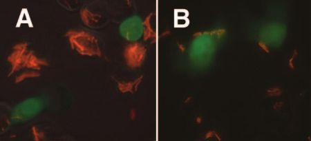
4806 MO ET AL. INFECT. IMMUN.
TABLE 1. Plasmids and strainsa
Reference or
Plasmid or strain Abbreviation Relevant characteristics
source
Plasmids
pMV206 Shuttle vector, Kmr 40
pMV261 Shuttle vector, Kmr 40
pMV361 Shuttle vector, Kmr 40
pYUB1058 pMV206, Hyr 40
pYUB1060 pMV261h pMV261, Hyr 40
pYUB1063 pMV206, Apr This study
pYUB1082 pGFPkP pMV261 with Phsp60::egfp, Kmr Hsub
pYUB1083 pGFPhE pYUB1058 with PCMV::egfp, Hyr This study
pYUB1084 pGFPaE pYUB1063 with PCMV::egfp, Apr This study
pYUB1085 pGFPhE/RFP pYUB1058 with Pmsp12::dsRed2 PCMV::egfp, Hyr This study
pYUB1086 pMV261 with Pmsp12::dsRed2, Kmr Ramakrishnanc
pYUB1143 pGFPhP pYUB1060 with Phsp60::egfp, Hyr Kood
pYUB1145 pCMVoriM pYUB1058 with PCMV, Hyr This study
Downloaded from http://iai.asm.org/ on February 15, 2021 by guest
pYUB1146 pgp120hE pYUB1058 with PCMV::gp120, Hyr This study
pYUB1147 pAB pMV361 with Phsp60::repA and repB, Kmr This study
pYUB1148 pA pMV361 with Phsp60::repA, Kmr This study
pYUB1149 pB pMV361 with Phsp60::repB, Kmr This study
pHIGH100 pHI p16R1 with copy-number-up mutations in oriM, Hyr Bourne
pHIGFPhE pYUB1143 with pHIGH100 oriM, Hyr This study
pHIgp120hE pYUB1146 with pHIGH100 oriM, Hyr This study
M. smegmatis strains
mc2155 155 Efficient plasmid transformation (ept-1) 35
mc21278 ask1::aph, diaminopalmitate auxotroph, Kmr 27
mc24519 ⌬esat6-cfp10 Lawrencef
mc24554 155(pMV261h) mc2155 with pYUB1060, Hyr 40
mc24556 155(pGFPkP) mc2155 with pYUB1082, Kmr Hsub
mc24557 155(pGFPhE) mc2155 with pYUB1083, Hyr This study
mc24559 155(pGFPhE/RFP) mc2155 with pYUB1085, Hyr This study
mKD211 211 Transposon mutation in Ms_orf, Kmr 11
mc24565 211Esx1 mKD211 complemented with M. tuberculosis esx-1, Kmr Hyr 11
mc24566 Transposon mutation in Ms4898, Kmr Derbyshireg
mc24571 211(pGFPhE) mKD211 with pYUB1083, Kmr Hyr This study
mc24572 211Esx1(pGFPaE) mc24565 with pYUB1084, Kmr Hyr Apr This study
mc24574 mc24566 with pYUB1083, Kmr Hyr Derbyshireg
mc24575 mc21278 with pYUB1083, diaminopalmitate auxotroph, Kmr Hyr This study
mc24576 mc24519 with pYUB1083, Hyr This study
mc25100 155AB attB::pYUB1147, Kmr This study
mc25101 155A attB::pYUB1148, Kmr This study
mc25102 155B attB::pYUB1149, Kmr This study
mc25103 155N attB::pMV361, Kmr This study
mc25104 155AB(pGFPhP) mc25100 with pYUB1143, Kmr Hyr This study
mc25105 155A(pGFPhP) mc25101 with pYUB1143, Kmr Hyr This study
mc25106 155B(pGFPhP) mc25102 with pYUB1143, Kmr Hyr This study
mc25107 155N(pGFPhP) mc25103 with pYUB1143, Kmr Hyr This study
mc25109 155AB(pGFPhE) mc25100 with pYUB1083, Kmr Hyr This study
mc25110 155A(pGFPhE) mc25101 with pYUB1083, Kmr Hyr This study
mc25111 155B(pGFPhE) mc25102 with pYUB1083, Kmr Hyr This study
mc25112 155N(pGFPhE) mc25103 with pYUB1083, Kmr Hyr This study
mc25114 155AB(gp120hE) mc25100 with pYUB1146, Kmr Hyr This study
mc25115 155A(gp120hE) mc25101 with pYUB1146, Kmr Hyr This study
mc25117 155N(pCMVoriM) mc2155 with pYUB1145, Hyr This study
mc25118 155N(gp120hE) mc25103 with pYUB1146, Kmr Hyr This study
mc25119 155(pHI) mc2155 with pHI, Hyr Bourne
mc25120 155(pHIGFPhE) mc25119 with pYUB1083, Hyr This study
mc25121 155(pHIgp120hE) mc25119 with pYUB1146, Hyr This study
M. bovis BCG strains
Pasteur BCG Vaccine strain
mc21604 ⌬lysA, lysine auxotroph 28
mc24580 BCG(pMV261) Pasteur BCG with pMV261, Kmr This study
mc24582 BCG(pGFPhE) Pasteur BCG with pYUB1083, Hyr This study
mc24585 mc21604 with pYUB1083, lysine auxotroph, Hyr This study
mc26000 ⌬panCD, pantothenate auxotroph, Hyr Sambandamurthyh
mc25122 BCG(pHI) BCG with pHI, Hyr This study
mc25123 BCG(pHIGFPhE) BCG with pHIGFPhE, Hyr This study
mc25124 BCG(pHIgp120hE) BCG with pHIgp120hE, Hyr This study
a
Abbreviations: Km and subscript k, kanamycin; Hy and subscript h, hygromycin; Ap and subscript a, apramycin; superscript P, prokaryotic promoter; superscript
E, eukaryotic promoter; subscript N, contains integrative pMV361 plasmid without rep insert.
b
Hsu, T. Hsu, laboratory of W. R. Jacobs, Jr. (unpublished).
c
Ramakrishnan, laboratory of L. Ramakrishnan (unpublished).
d
Koo, M. Koo, laboratory of W. R. Jacobs, Jr. (unpublished).
e
Bourn, W. Bourn, laboratory of P. van Helden (submitted) (GenBank accession no. EF216316).
f
Lawrence, K. Lawrence, laboratory of W. R. Jacobs, Jr. (unpublished).
g
Derbyshire, laboratory of K. Derbyshire (unpublished).
h
Sambandamurthy, V. Sambandamurthy, laboratory of W. R. Jacobs, Jr. (unpublished).VOL. 75, 2007 GENETIC ALTERATION OF M. SMEGMATIS 4807
Measurement of plasmid copy number. The relative plasmid copy numbers of (PBMCs) and splenocytes, blood was obtained from the retroorbital plexus and
RepHigh, pHIGH100, and RepWt derivatives were determined by comparing the spleens were harvested 7 days after inoculation. H-2Dd tetrameric complexes
amounts of plasmid DNA extracted from the derivatives. The results were corrob- folded with the P18 peptide (RGPGRAFVTI) (5), a sequence found in the V3
orated by analyzing the distribution and intensity of GFP expression in populations loop of HIV-1 HXBc2 envelope protein, were prepared as described previously
of various Rep derivatives of M. smegmatis expressing GFP. M. smegmatis was grown (5). Fresh blood samples (200 l from each mouse) or splenocyte suspensions
in 6 ml of Middlebrook 7H9 medium to log phase (optical density at 600 nm, 0.8 ⫾ (recovered after passage through a 70-m nylon cell strainer) were diluted in 3
0.02) before plasmid extraction using a modified Qiaprep kit (QIAGEN) protocol. ml RPMI medium with 40 U/ml heparin and layered over Ficoll-Hypaque (lym-
Briefly, pelleted M. smegmatis was resuspended with 250 l of P1 buffer containing pholyte-M) before centrifugation at 400 ⫻ g for 20 min at 20°C. The lymphocyte
10 mg/ml lysozyme and incubated at 37°C for 4 h in the presence of RNase for 10 layer was carefully transferred to a fresh tube, diluted with 10 ml of PBS, and
min, and then it was lysed at room temperature for 5 min with 300 l of P2 lysis then pelleted and washed in 1 ml PBS with 2% FCS before resuspension in 100
buffer, which was then neutralized with 350 l of prechilled N3 buffer. Aliquots of l (final volume) of the solution. The cells were stained with P18-tetramer-
serial twofold dilutions of extracted plasmid DNA were run on a 0.8% agarose gel phycoerythrin, vortexed briefly, and incubated at 20°C for 20 min, and this was
and stained with ethidium bromide. Plasmid quantities were estimated using ImageJ followed by staining with APC-CD8 for 20 min at 20°C. To control for nonspe-
software, version 1.34n (Wayne Rasband; http://rsb.info.nih.gov/ij/) after calibration cific fluorescence, samples were incubated with no monoclonal antibody, with
with the DNA High Mass Ladder (Invitrogen). For estimation by fluorescence- only APC-CD8, and with only phycoerythrin-CD4. The cells were washed with 5
activated cell sorting (FACS) analysis, a 500-l suspension of each clone was washed ml PBS at room temperature, resuspended in 2% formaldehyde in PBS, vor-
texed, and analyzed with a FACSCalibur cytometer. A minimum of 104 cells were
Downloaded from http://iai.asm.org/ on February 15, 2021 by guest
twice with an equal volume of PBS and then resuspended in 1 to 2 ml PBS. The
distribution and intensity of GFP expression among 50,000 bacilli were determined analyzed for each sample.
by FACS analysis using a BD Biosciences FACScan flow cytometer and were ana- Statistical analysis. Statistical tests were performed using the Student t test or
lyzed by using CellQuest software (Becton Dickinson, Mountain View, CA). one-way analysis of variance with Dunnett’s posttest by Prism 4.01 for Windows
Infection of mammalian cell cultures with mycobacteria. RAW 264.7 murine (GraphPad Software, Inc., San Diego, CA). P values of ⬍0.05 were considered
macrophage and HeLa (human cervical adenocarcinoma) cell lines were grown significant.
in Dulbecco’s modified Eagle’s Medium (DMEM) supplemented with 10% fetal
bovine serum (FBS), 2% HEPES buffer, and 5% NCTC-109 medium (Gibco).
J774 murine macrophage cells were grown in DMEM supplemented with 10% RESULTS
FBS. Cells were transferred at a concentration of 1 ⫻ 105 to 2 ⫻ 105 cells/well
into a 48-well plate and incubated at 37°C in the presence of 10% CO2 for 2 to M. smegmatis and M. bovis BCG transfer reporter plasmids
24 h before infection to generate semiconfluent lawns of cells. To determine the to eukaryotic cell lines. Mycobacteria are promising vectors for
optimal inoculum for each cell line, freshly grown mycobacteria were added to DNA vaccine delivery. Our goal is to develop an attenuated
the cells at a multiplicity of infection (MOI) of 1, 3, 10, 50, or 100 to obtain a total Mycobacterium tuberculosis, mutant BCG, or M. smegmatis vec-
volume of no more than 350 l/well in a 48-well plate. To determine whether
viable bacteria are required for gene transfer to mammalian cells, mycobacteria
tor for DNA immunization. To develop a system to measure
were killed by heating them to 80°C for 5 min prior to infection of RAW 264.7 the efficiency with which various mycobacterial strains can de-
cells. After certain infections, isoniazid (25 g/ml) was added immediately to liver DNA plasmids to eukaryotic cells, an extrachromosomal
induce premature lysis. Extracellular bacteria were killed by addition of 50 g/ml mycobacterial reporter plasmid, pGFPhE (the superscript E
of gentamicin (Gibco). To compare the efficiency of lipid-mediated DNA plas-
indicates that the plasmid contains the eukaryotic promoter
mid transfection to the efficiency of bactofection, lipofectamine (Invitrogen) was
diluted 1:50 in Opti-Mem medium (Gibco) and then incubated at room temper- PCMV expressing egfp; the subscript h indicates that the plas-
ature for 5 min, added to 10 g of DNA in an equal volume of medium, and mid encodes resistance to hygromycin) was constructed and
incubated for 20 min at room temperature before it was added to RAW 264.7 or transformed into M. smegmatis (Fig. 1A). No fluorescence of
HeLa cells. After incubation for 3 to 5 h, cells were washed three times with M. smegmatis strain 155(pGFPhE) was detected by FACS, con-
DMEM and resuspended in 0.5 to 2 ml of medium. Cells were examined by
fluorescence microscopy and FACS analysis (after trypsinization and resuspen-
firming the absence of cryptic mycobacterial promoters up-
sion in 4% FBS-PBS) at various intervals for up to 5 days after infection. stream of egfp (Fig. 1C). By contrast, GFP expression was
Microscopic imaging and FACS analysis. Live RAW 264.7 and HeLa cell readily detected in strain 155(pGFPkP), which contained the
samples were examined after infection using an Olympus IX 81 microscope mycobacterial Phsp60::egfp expression cassette (the superscript
(Melville, NY) equipped with a Cooke Sensicam QE air-cooled charge-coupled
P indicates that the plasmid contains the prokaryotic promoter
device camera and a mercury lamp for fluorescence illumination. Images were
collected using IPLab Spectrum software (Scanlytics, Rockville, MD) at a mag- Phsp60 expressing egfp; the subscript k indicates that the plas-
nification of ⫻10 or ⫻40. Adobe Photoshop (Adobe Systems, San Diego, CA) mid encodes resistance to kanamycin) (Fig. 1B and D).
was used to restore color and merge images captured using different fluorescence Macrophages and dendritic cells are the primary targets
filters or normal light (phase-contrast) illumination. The fluorescence in a min- during mycobacterial infection. To determine whether myco-
imum of 105 cells per sample was measured by FACS using a FACScan or
bacterial infection, DNA transfer into the nucleus, and subse-
FACSCalibur cytometer and CellQuest software (Becton Dickinson). Data were
further analyzed with the FloJo software (Tree Star, Inc., Ashland, OR). quent eukaryotic GFP expression (and fluorescence) could be
Animals and immunization. Six-to-eight-week-old female BALB/c mice observed in a macrophage cell line, RAW 264.7 cells were
(Charles River Laboratories) were inoculated with 108 CFU of RepHigh M. infected with strains 155(pGFPkP) and 155(pGFPhE), which
smegmatis strains (which overexpress Rep proteins and elevate the plasmid copy harbor the Phsp60::egfp and PCMV::egfp expression cassettes,
number) harboring pgp120hE or of BCG or M. smegmatis strains harboring
pHIgp120E via the intraperitoneal route (Table 1). The relative plasmid copy
respectively. Twenty-four hours after infection with 155(pG
number per bacterium of pHIgp120E or RepHigh and RepWt strains was con- FPkP) at an MOI of 10, one or more internalized bacteria were
firmed prior to and after immunization using the agarose gel density method. To observed by fluorescence microscopy in 96.5% ⫾ 1.6% of the
compare the immunogenicity of M. smegmatis having the wild-type plasmid copy RAW 264.7 cells (data not shown). However, a striking differ-
number, groups of control mice were inoculated with the corresponding RepWt
ence in the patterns of GFP expression in RAW 264.7 and
M. smegmatis strain. To compare the effects of the usual route of DNA vacci-
nation on tetrameric responses, intramuscular purified pgp120hE was adminis- HeLa cells was observed after infection by the two strains. In
tered at a dose of 50 g per mouse (25 g/gastrocnemius muscle) via intramus- RAW 264.7 and HeLa cells infected with 155(pGFPkP), indi-
cular injection. To control for the nonspecific effects of mycobacteria or DNA vidual mycobacteria expressing GFP were visible (Fig. 2A and
vaccination on tetrameric responses, groups of mice were inoculated with 108 2D, respectively). By contrast, a small percentage of RAW
CFU of 155N(pCMVoriM) or BCG(pHI) or with purifed pCMVoriM DNA by the
same method.
264.7 and HeLa cells infected with 155(pGFPhE) fluoresced
Tetramer staining and flow cytometric analysis. To determine the frequency throughout the cytoplasm, indicating that host cells expressed
of gp120-specific tetrameric responses in peripheral blood mononuclear cells GFP (Fig. 2B and 2E, respectively). No eukaryotic GFP ex-4808 MO ET AL. INFECT. IMMUN.
Downloaded from http://iai.asm.org/ on February 15, 2021 by guest
FIG. 1. M. smegmatis expresses GFP from a mycobacterial promoter but not from a eukaryotic promoter. Plasmids that replicate in mycobacteria encoding
GFP under the control of a eukaryotic immediate-early CMV promoter/enhancer (A) or the mycobacterial hsp60 promoter (B) were constructed. Abbreviations:
oriE, origin of replication in E. coli; oriM, origin of replication in mycobacteria; Kanr, kanamycin resistance gene; Hygr, hygromycin resistance gene; EGFP,
enhanced GFP; SV40 poly A, simian virus 40 late polyadenylation signal. M. smegmatis strains transformed with pGFPhE [strain 155(pGFPhE)] or pGFPkP [strain
155(pGFPkP)] were cultured and analyzed by flow cytometry for GFP expression (C and D). FSC, forward scatter.
pression was detected within RAW 264.7 cells infected with GFP expression than mycobacterial bactofection in RAW
heat-killed 155(pGFPhE), suggesting that viable bacteria are 264.7 cells yielded (0.0385% ⫾ 0.01%) after 24 h.
required for gene transfer to mammalian cells (data not To more precisely identify cells with internalized bacteria
shown). that expressed the GFP transgene, RAW 264.7 and HeLa cells
BCG or other mycobacteria harboring DNA plasmid vac- were infected with M. smegmatis harboring plasmid pGFPhE/
cines could be given orally. DNA has been observed to be RFP (which contains the PCMV::egfp and Pmsp12::dsRed2 ex-
transiently expressed by tissue-specific epithelial cells, and pression cassettes). By using microscopy, it was observed that
DNA vaccines can induce immune responses after intranasal a single internalized red mycobacterium was sufficient to per-
or oral administration; dendritic cells may capture antigens mit GFP expression by RAW 264.7 or HeLa cells (Fig. 3).
from mucosal epithelial or epidermal tissues and migrate to M. bovis BCG was demonstrated to mediate bactofection by
draining lymph nodes for antigen presentation to T cells in vivo infecting HeLa or J774 cells with BCG harboring pGFPhE (Fig.
(8). To determine whether HeLa cells permit plasmid transfer 4). To control for nonspecific fluorescence, HeLa or J774 cells
from mycobacteria, HeLa cells were infected with 155(pG were infected with BCG Pasteur harboring the vector
FPkP) or 155(pGFPhE). To compensate for the lower bacterial pMV261. After 24 h of infection, FACS analysis revealed that
uptake by this nonphagocytic cell line, a 5- to 10-fold-higher statistically significant (P ⬍ 0.05) percentages of HeLa cells
MOI was used in HeLa cell infections than in the RAW 264.7 (0.093% ⫾ 0.032%) and J774 cells (0.023% ⫾ 0.0058%) ex-
cell infection experiments. After infection with 155(pGFPhE), pressed GFP compared to the background fluorescence cells
a significantly higher proportion of HeLa cells (Fig. 2F, right infected with BCG harboring pMV261 (Fig. 4).
panel) than of RAW 264.7 cells (Fig. 2C, right panel) ex- Lysis-susceptible mycobacteria do not improve DNA trans-
pressed GFP, despite the fact that the efficiency of uptake of fer. Mutants of Shigella that are auxotrophic for diaminopalmi-
mycobacteria by HeLa cells (Fig. 2F, middle panel) was signif- tate undergo rapid lysis after they invade mammalian cells and
icantly less than the efficiency of uptake by RAW 264.7 cells are very efficient vectors for bactofection (33). To test whether
(Fig. 2C, middle panel). Transfection with purified pGFPhE lysis-susceptible mycobacteria mediate bactofection more effi-
DNA and lipofectamine yielded only slightly higher rates of ciently than wild-type parent strains, RAW 264.7 and HeLaVOL. 75, 2007 GENETIC ALTERATION OF M. SMEGMATIS 4809
Downloaded from http://iai.asm.org/ on February 15, 2021 by guest
FIG. 2. Eukaryotic GFP expression is observed in RAW 264.7 and HeLa cells infected with 155(pGFPhE). RAW 264.7 cells were infected with
155(pGFPkP) (A) or 155(pGFPhE) (B) at an MOI of 10. HeLa cells were infected with 155(pGFPkP) (D) or 155(pGFPhE) (E) at an MOI of 100.
Both groups were observed by fluorescence microscopy 24 h postinfection, and the original images were taken at a magnification of ⫻40. Cells
infected with 155(pMV261h), 155(pGFPkP), or 155(pGFPhE) at an MOI of 10 (RAW 264.7 cells) (C) or 50 (HeLa cells) (F) were also collected
5 days postinfection for flow cytometric analysis. Data representative of a minimum of three experiments are shown, and the quadrant axes are
aligned to reduce the background in high-percentage (axis 1) and low-percentage (axis 2) GFP-expressing samples. FSC, forward scatter.4810 MO ET AL. INFECT. IMMUN.
covered that transposon insertion mutations within and near the
Esx-1 locus of the M. smegmatis chromosome lead to a hypercon-
jugation phenotype (11). The Esx-1 locus is thought to encode a
specialized secretory apparatus responsible for secreting at least
two proteins (EsxA and EsxB), which are encoded within the Esx
locus (4, 7). The insertions are predicted to disrupt Esx-1 func-
tions that normally suppress conjugation, perhaps by interfering
with EsxA and EsxB secretion. Complementation of the mutants
with the wild-type Esx-1 region of M. tuberculosis reduces or
eliminates the hyperconjugative phenotype (11).
FIG. 3. Eukaryotic GFP expression in RAW 264.7 and HeLa cells
requires the presence of intracellular M. smegmatis 155(pGFPhE/RFP). HeLa cells were infected with a hyperconjugating M. smeg-
RAW 264.7 cells were infected with M. smegmatis 155(pGFPhE/RFP) matis mutant (MKD211) harboring pGFPhE. We observed a
at an MOI of 10 (A), and HeLa cells were infected at an MOI of 100 significantly higher frequency of GFP expression in HeLa cells
(B). Fluorescing mycobacteria (red) in RAW 264.7 cells expressing after infection with MKD211 than after infection with the
Downloaded from http://iai.asm.org/ on February 15, 2021 by guest
GFP (green) were observed 24 h after infection. Red and green images
were combined, and enhanced emission was recorded using phase- wild-type parent strain (P ⫽ 0.04) (Fig. 5). Complementation
contrast and red and green filter fluorescence microscopy. The original of the mutation with the M. tuberculosis Esx-1 locus suppressed
images were taken at a magnification of ⫻40. the increased transfer of pGFPhE to infected cells relative to
the mutant and wild-type strains (Fig. 5). In addition, an M.
smegmatis mutant with disruption of the lpqM gene that con-
cells were infected with pGFPhE-containing M. bovis BCG jugates 1,000-fold less than the wild type was still able to
strain mc21604 or mc26000 or M. smegmatis strain mc21278 mediate bactofection, albeit inefficiently (data not shown).
(which are auxotrophic for lysine, pantothenate, and diamino- A primary role of Esx-1 is the secretion of a heterodimer of
palmitate, respectively, and undergo lysis soon after infection EsxA and EsxB (4, 7). To determine whether EsxA and EsxB
of mammalian cells). Isoniazid also causes bacterial lysis after suppress plasmid transfer into nuclei, an M. smegmatis mutant
only a few generations due to inhibition of cell wall synthesis; with a precise deletion of the esxA and esxB genes (strain
therefore, RAW 264.7 and HeLa cells were also treated with mc24519) was transformed with pGFPhE to generate mc24576.
isoniazid after infection with pGFPhE-containing M. smegmatis Surprisingly, the same frequency of GFP expression was ob-
and M. bovis BCG. Neither the premature lysis mutants nor served among HeLa cells infected with strain mc24576 and
isoniazid treatment increased gene transfer frequency com- among HeLa cells infected with mc24557 [155(pGFPhE)] (data
pared to that observed with the wild type (data not shown). not shown). Together, these results suggest that the secreted
Therefore, early lysis of mycobacteria does not appear to im- forms of EsxA and EsxB do not inhibit bactofection but that it
prove the delivery of DNA into the host cell. is the disruption of the Esx-1 translocation apparatus that
Hyperconjugation mutants of M. smegmatis transfer DNA alleviates bactofection inhibition.
plasmids to mammalian cells more efficiently than the wild Overexpression of Rep proteins increases the copy number
type. We predicted that an M. smegmatis conjugation system that of oriM-based plasmids in M. smegmatis and enhances the gene
mediates DNA transfer to other mycobacteria (26) may contrib- transfer frequency to mammalian cells. A potential limitation
ute to plasmid transfer to mammalian cells. It was recently dis- of the use of mycobacteria as vectors for DNA vaccines is that
FIG. 4. M. bovis BCG mediates plasmid transfer to infected HeLa (A) and J774 (B) cells. (A) HeLa cells were infected with BCG(pGFPhE)
(BCG Pasteur harboring pGFPhE that contains the eukaryotic PCMV::egfp expression cassette) at an MOI of 50 and analyzed by flow cytometry
24 h after infection. The mean (and standard error of the mean) proportion of HeLa cells expressing GFP is shown based on the results of three
experiments. The proportion of HeLa cells expressing GFP by 24 h after infection with BCG(pGFPhE) was significantly higher than the background
frequencies after infection with control strain BCG(pGFPhE) (one asterisk, P ⬍ 0.05). (B) J774 cells were infected with BCG harboring plasmid
pHI [BCG(pHI)], pGFPhE [BCG(pGFPhE)], or pHIGFPhE [BCG(pHIGFPhE)] at an MOI of 50 and analyzed by flow cytometry 24 h after
infection. The mean (and standard error of the mean) proportion of J774 cells expressing GFP is shown based on the results of three experiments.
The proportion of J774 cells expressing GFP by 24 h after infection with BCG(pHIGFPhE) was significantly higher than the proportion after
infection with control strain BCG(pGFPhE) (two asterisks, P ⬍ 0.01).VOL. 75, 2007 GENETIC ALTERATION OF M. SMEGMATIS 4811
increased plasmid copy number in M. smegmatis correlates
with enhanced plasmid transfer to mammalian cells.
We were unable to recover BCG clones that express the RepA
and/or RepB protein in trans from the chromosome in BCG. As
an alternative to Rep overexpression, BCG was transformed with
high-copy-number plasmid pHIGFPhE, derived from the copy-
number-up mutant plasmid pHIGH100. Plasmid pHIGH100 is
maintained at a level of 32 to 64 copies per bacterium and is stably
maintained in BCG (Bourn et al., submitted). The ability of the
resulting strain, designated BCG(pHIGFPhE), to transfer pHIG
FPhE into phagocytic J774 cells compared to that of BCG har-
boring the regular-copy-number plasmid pGFPhE [strain BCG
(pGFPhE)] was examined. The frequency of GFP-expressing J774
cells infected with BCG(pHIGFPhE) was up to 3.5-fold higher by
Downloaded from http://iai.asm.org/ on February 15, 2021 by guest
24 h postinfection than the frequency of GFP-expressing J774
FIG. 5. Hyperconjugative M. smegmatis strain 211(pGFPhE) mediates cells infected with BCG(pGFPhE) (Fig. 4B). The BCG(pHIG
transfer of pGFPhE to HeLa cells more efficiently than wild-type M. FPhE) strain was not tested in HeLa cells. The frequency of
smegmatis. HeLa cells were infected with 211(pGFPhE), 155(pGFPhE) GFP-expressing J774 cells infected with strain 155 containing
(wild-type M. smegmatis harboring pGFPhE), or 211Esx1(pGFPaE) pHIGFPhE was up to 1.56-fold higher by 12 h postinfection than
[211(pGFPhE) complemented with the Esx-1 region of M. tuberculosis] at
an MOI of 100 and collected 4 to 5 days postinfection for flow cytometric the frequency of GFP-expressing J774 cells infected with RepHigh
analysis. The P value for a comparison of the percentage of HeLa cells strain 155AB(pGFPhE) (0.20% versus 0.13%), although the dif-
expressing GFP after infection with 155(pGFPhE) and the percentage of ference was not statistically significant (P ⬎ 0.05) (data not
HeLa cells expressing GFP after infection with 211(pGFPhE) was ⬍0.05 shown).
(indicated by an asterisk). The data are representative of a minimum of
Immunization with recombinant M. smegmatis strains har-
three experiments. The mean (and standard error of the mean) peak
intensity of GFP expression is shown for each strain. boring an HIV gp120 eukaryotic expression plasmid generates
gp120-specific CD8 T cells in mice. Several candidate immuno-
gens, including recombinant live attenuated viruses (23) and bac-
teria (22), are being studied for the generation of T-cell responses
mycobacterial plasmids exist at a low copy number, restricting against HIV. To determine the effect of a higher plasmid copy
the amount of transferable plasmid. pAL5000, the best-char- number on the frequency of gp120-specific T-cell responses, we
acterized mycobacterial plasmid, is present at a level of only compared the frequencies of gp120-specific tetrameric responses
five copies per bacterium (40). pAL5000 encodes two proteins, after immunization with recombinant M. smegmatis and BCG
RepA and RepB, which are thought to form an essential rep- strains harboring pHIgp120hE. The frequency of P18 tetramer
lication initiation complex that recognizes and initiates repli- staining among CD8⫹ T cells from mice immunized with strain
cation from the origin of replication, oriM (2, 3, 37). A negative 155AB(pgp120hE) was approximately twofold higher (P ⬍ 0.05)
regulatory circuit appears to control mRNA synthesis of RepA for splenocytes and threefold higher (P ⬍ 0.001) for PBMCs
and RepB (38) and thus reduce their expression, which directly (0.12% ⫾ 0.065%) than the frequency in mice immunized with
impacts the initiation of replication and copy number. RepWt strain 155N(pgp120hE) (Fig. 8). The magnitude of the P18
We investigated whether overexpression of Rep proteins tetrameric responses among PBMCs after 155AB(pgp120hE) im-
resulted in increased replication of pAL5000. Three M. smeg- munization was approximately one-half (48.2%) the magnitude
matis RepHigh strain derivatives were constructed expressing detected after intramuscular pgp120hE immunization (Fig. 8).
either RepA, RepB, or RepAB from the chromosome (desig- Agarose gel analysis of plasmids recovered from M. smegmatis
nated 155A,155B, and 155AB). pGFPhP was introduced into prior to inoculation confirmed that strain 155AB(pgp120hE) had a
each strain, and the relative quantities of pGFPhP extracted fivefold-higher copy number of pgp120hE per bacterium on aver-
from the strains were compared (Fig. 6). In each strain, ele- age than strain 155N(pgp120hE) (data not shown). Surprisingly,
vated expression of Rep proteins resulted in increased yields of no gp120-specific responses above the background level were
plasmid DNA that were up to 10-fold greater than the wild- detected among splenocytes 1 week after immunization with
type yields (Fig. 6A and 6B). BCG(pHIgp120hE) in control BCG-immunized mice (data not
FACS analyses with the RepHigh derivatives demonstrated shown). Also, the frequency of tetrameric responses among
that an increase in copy number correlated with an increase in splenocytes 1 week after immunization were 2.19-fold higher after
the level of mycobacterial GFP expression from pGFPhP. The intraperitoneal inoculation of RepWt strain 155N(pgp120hE) than
maximal intensity of GFP expression among populations of after inoculation of 155(pHIgp120hE) (data not shown).
RepHigh M. smegmatis was four- to sixfold higher than that
among the wild-type control population (Fig. 6C and 6D).
DISCUSSION
The ability of RepHigh strains to transfer pGFPhE into HeLa
or J774 cells was examined. The frequency of transfer of pG Intracellular bacterial vectors harboring eukaryotic ex-
FPhE into HeLa cell nuclei infected with a RepAHigh strain was pression plasmids may be more effective immunogens than
up to 3.5-fold higher by day 3 postinfection and up to 3-fold bacterial vectors harboring prokaryotic expression plasmids.
higher by 12 h postinfection in J774 cells than the frequency of Although attenuated strains of Shigella flexneri (34), L. mono-
transfer with the controls (Fig. 7). These results suggest that an cytogenes (21), Salmonella (36), M. smegmatis (5, 20), and M.4812 MO ET AL. INFECT. IMMUN.
Downloaded from http://iai.asm.org/ on February 15, 2021 by guest
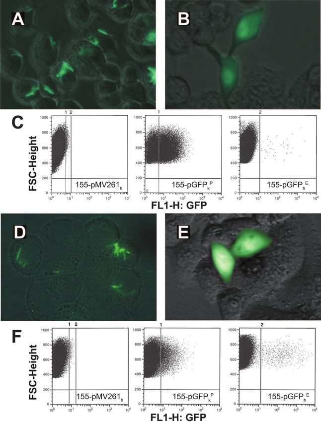
FIG. 6. Measurement of relative copy number of plasmid pGFPhP per bacterium in various recombinant RepHigh and RepWt M. smegmatis
strains. Following extraction of pGFPhP from 108 CFU of each strain, DNA was eluted with 40 l of distilled H2O, and aliquots of each sample
were loaded on a 0.7% agarose gel. (A) Agarose gel comparing relative amounts of pGFP isolated from each strain. The sample volumes loaded
are indicated above the lanes. The RepWt strain 155N contains pMV361 without the repAB gene insert. (B) Estimates of the amount of plasmid
DNA recovered from 108 CFU of each strain based upon quantification of the intensity of plasmid bands on the agarose gel by the software ImageJ,
version 1.34n. Two asterisks indicate that the P value is ⬍0.01 for comparisons of RepHigh strains 155AB(pGFPhP), 155A(pGFPhP), and 155B(pG
FPhP) with strain 155N(pGFPhP). The mean (and standard error of the mean) amounts of plasmid DNA shown are based on the results of three
experiments. (C) Representative flow cytometry analysis of various RepHigh M. smegmatis strains harboring pGFPhP (which contains the Phsp60::egfp
cassette) indicated by different colors. A population consisting of 50,000 bacteria of each strain was sorted by flow cytometry. Strain mc2155 without
a plasmid (indicated by purple shading) served as a negative control. (D) Peak intensity of GFP expression. The peak intensity of GFP expression
by populations of each strain was measured by FACS. The mean (and standard error of the mean) peak intensities of GFP expression shown are
based on the results of three experiments. The P values for comparisons of the peak GFP intensities with that of RepWt strain 155N(pGFPhP) were
⬍0.01 for RepHigh strains 155AB(pGFPhP) and 155A(pGFPhP) (two asterisks) and ⬍0.05 for 155B(pGFPhP) (one asterisk).
bovis bacillus Calmette-Guérin (48) which express heterolo- can mediate plasmid delivery to and subsequent transgene
gous antigens have shown promise as vaccine vectors, their expression by infected mice (46). We demonstrate here for the
efficacy is limited by reduced expression and incomplete pro- first time that BCG can also transfer genes to mammalian cells
cessing of full-length recombinant polypeptides within the bac- and define several critical parameters affecting bactofection
teria (9) and the failure to engender strong immune responses determined through genetic manipulation of M. smegmatis.
to nonsecreted recombinant antigens (19, 30). By contrast, an Macrophages are the natural target and host for mycobac-
attenuated bacterial vector that can deliver a DNA vaccine into teria in vivo. Despite the high rate of infection (⬎95%) of
mammalian cells has the distinct advantage of ensuring correct RAW 264.7 murine macrophage cells by recombinant M. smeg-
endogenous expression and processing of recombinant poly- matis observed in the present study, only a very low proportion
peptides for major histocompatibility complex class I and II (0.036% ⫾ 0.1%) of the infected cells expressed GFP. Never-
presentation to T cells, as well as proper glycosylation and theless, the efficiency of mycobacterial bactofection in RAW
folding of conformational B-cell epitopes, facilitating a robust 264.7 cells was comparable to that observed after lipo-
antigen-specific immune response. M. smegmatis is a promising fectamine transfection (0.0385% ⫾ 0.01%). A higher propor-
recombinant candidate vaccine vector. We observed previously tion of HeLa cells than of RAW 264.7 cells infected with M.
that recombinant M. smegmatis generates heterologous anti- smegmatis expressed GFP, despite a much lower frequency of
gen-specific T-cell responses despite its rapid clearance in mice bacterial uptake in HeLa cells than in RAW 264.7 cells. Al-
(5, 47). Other workers have demonstrated that M. smegmatis though the overall efficiency of mycobacterial pGFPhE bacto-VOL. 75, 2007 GENETIC ALTERATION OF M. SMEGMATIS 4813
Downloaded from http://iai.asm.org/ on February 15, 2021 by guest
FIG. 7. RepHigh M. smegmatis strains mediate bactofection in HeLa (A) or J774 (B) cells more effectively than RepWt M. smegmatis. HeLa cells
(A) or J774 cells (B) were infected with RepHigh M. smegmatis strain 155AB(pGFPhE), 155A(pGFPhE), or 155B(pGFPhE) or with RepWt strain
155N(pGFPhE). The proportion of HeLa cells expressing GFP was measured at different time points by FACS after infection. The mean (and
standard error of the mean) proportions of HeLa or J774 cells expressing GFP shown are based on the results of three experiments. The proportion
of HeLa cells expressing GFP by 72 h after infection with RepHigh strains 155AB(pGFPhE), 155A(pGFPhE), and 155B(pGFPhE) was significantly
higher than the proportion after infection with RepWt strain 155N(pGFPhE) (two asterisks, P ⬍ 0.01); the proportion of J774 cells expressing GFP
by 12 h after infection with RepHigh strain 155AB(pGFPhE) was significantly higher than the proportion after infection with RepWt strain
155N(pGFPhE) (two asterisks, P ⬍ 0.01).
fection per cell was fivefold lower than the efficiency of lipo- single mycobacterium was sufficient for GFP transgene expres-
fectamine transfection, we estimated that the efficiency of the sion in the mammalian cells.
former was 100,000-fold higher per microgram of plasmid Limitations to the use of mycobacteria as vectors for DNA
DNA. Notably, microscopic analysis of RAW 264.7 and HeLa plasmid transfer include their exclusive residence in the vacu-
cells infected with M. smegmatis harboring the dual-color flu- ole of infected antigen-presenting cells, which restricts their
orescence plasmid pGFPhE/RFP revealed that infection with a ability to release plasmids directly to the host cell cytoplasm,
FIG. 8. RepHigh M. smegmatis strain 155AB(pgp120hE) elicits a higher frequency than RepWt M. smegmatis strain 155N(pgp120hE): tetramer
staining and flow cytometric analysis of PBMCs (A) and splenocytes (B) after immunization of BALB/c mice with purified pgp120hE, RepHigh, or
RepWt strains harboring pgp120hE. PBMCs or splenocytes were recovered from groups of three to five mice 7 days after immunization with 108
bacilli. The gated population represented both CD8 and P18 tetramer-staining-positive T cells. The mean (and standard error of the mean) percent
gp120 P18 tetramer-positive CD8 T cells among PBMCs or splenocytes is shown for each group of mice. After subtraction of background responses
in control mice immunized with 155N(pCMVoriM) or 155N(pCMVoriM) (not shown), the frequencies of CD8 and P18 tetramer staining responses
were significantly higher after RepHigh strain immunization than after RepWt strain immunization (for PBMCs, P ⬍ 0.01 [two asterisks]; for
splenocytes, P ⬍ 0.05 [one asterisk]). The frequencies of HIV-1 P18-specific CD8⫹ T-cell responses among PBMCs were significantly higher after
intramuscular pgp120hE immunization than after strain 155AB(pgp120hE) immunization (one asterisk, P ⬍ 0.05).4814 MO ET AL. INFECT. IMMUN.
and the low copy number of mycobacterial plasmids (about five both the exponential and stationary phases of bacterial growth
copies per cell). To overcome these restrictions, we deter- (data not shown). The most likely explanation for the differ-
mined whether M. smegmatis with an enhanced-conjugation or ence in these and previous results is the location of the repAB
premature-lysis phenotype or M. smegmatis or BCG with an genes supplied in trans. In the RepHigh strains 155AB, 155A,
increased-plasmid-replication phenotype would facilitate DNA and 155B, integration of the repAB genes into the chromosome
plasmid transfer to the mammalian cell nucleus. ensured their constitutive and stable expression. By contrast,
We did not observe an increase in the frequency of plasmid introducing a second plasmid also encoding RepAB may result
transfer in HeLa cells infected with mycobacterial auxotrophs in plasmid incompatibility, a lower copy number of both plas-
that lysed prematurely compared to the cells infected with mid types, and inevitable plasmid loss.
wild-type strains. This is in contrast to observations of other pAL5000 encodes a negative regulatory circuit, which ap-
workers, who found that auxotrophic mutants of Shigella that pears to control mRNA synthesis of RepA and RepB (38)
lyse prematurely are highly effective vectors for plasmid gene from the native promoter. One explanation for our inability to
transfer to mammalian cells (33). Unlike Shigella and Listeria, recover BCG clones, which overexpress the Rep proteins from
wild-type M. smegmatis and BCG do not escape the endosome.
Downloaded from http://iai.asm.org/ on February 15, 2021 by guest
the constitutive Hsp60 promoter, is that these proteins are
This suggests that DNA release, mediated by premature lysis, toxic or interfere with replication of slowly growing mycobac-
is not a limiting factor in gene transfer to mammalian cell teria, such as BCG. Indeed, we consistently observed that
nuclei. This observation, combined with the effect of copy RepHigh M. smegmatis strains exhibited a significantly lower in
number and the inhibitory influence of Esx-1, also indicates vitro growth rate than the RepWt strain (data not shown),
that the mechanism of bactofection by M. smegmatis is funda- suggesting that even in rapid growers RepAB overexpression
mentally different than the mechanisms studied previously. exacts a toll on mycobacterial growth. We were surprised at the
Consistent with our prediction that hyperconjugation in my- lack of an immune response induced by BCG(pHIgp120E).
cobacteria would correlate with an enhanced ability to transfer This may have been due to the relative inefficiency with which
genes to mammalian cells, a significantly (albeit only 1.7-fold) BCG induces apoptosis in infected phagyocytic cells compared
higher frequency of GFP expression was observed in HeLa to M. smegmatis. Of note, we observed that a markedly higher
cells infected with a hyperconjugating mutant of M. smegmatis
proportion of THP1 cell lines infected with M. smegmatis than
(11) than in cells infected with the wild-type parent. This effect
of cells infected with BCG undergo apoptosis as detected by
was suppressed by genetic complementation of the mutant
terminal deoxynucleotidyltransferase-mediated dUTP-biotin
strain with the Esx-1 region from M. tuberculosis, suggesting
nick end labeling staining after 72 h (36.7 and 4.6%, respec-
that, similar to conjugation between mycobacteria, the Esx-1
tively) (data not show). After M. smegmatis induces apoptosis
apparatus secretes proteins that inhibit gene transfer to mam-
in infected phagocytic cells in vivo, vesicles carrying plasmid
malian cells. We currently have no definitive explanation for
DNA may be taken up by bystander antigen-presenting cells.
why the presence of the M. tuberculosis Esx-1 apparatus would
The basis for the inferior immunogenicity of M. smegmatis
suppress bactofection to almost background levels. One possi-
harboring pHIgp120hE compared to that of M. smegmatis
bility is that the M. tuberculosis locus encodes proteins not
RepWt strain 155N(pgp120hE) is not known. One possibility is
present in M. smegmatis which have a greater inhibitory effect
on bactofection. that the episomal plasmids are maintained more stably in the
To distinguish between EsxA and EsxB inhibition and other former strain in vivo, resulting in a lower efficiency of plasmid
effects of Esx-1 on bactofection, the two genes encoding EsxA transfer to macrophages; unlike pMV261, p16R1 and its de-
and EsxB were deleted; wild-type levels of bactofection were rivative pHIGH100 has the full-length rap (14). This may lead
observed (Fig. 5). This suggests that another protein(s) se- to increased stability of the plasmid in mycobacteria.
creted by M. smegmatis Esx-1 normally suppresses bactofection No naturally occurring mycobacterial plasmid has been ob-
or that the apparatus itself interferes with plasmid transfer. served to be maintained at a level of more than five copies per
Disruption of the entire locus would prevent either structural bacterium on average. To our knowledge, this work demon-
interference or secretion of other proteins and thus allow el- strates, for the first time, that plasmid replication to obtain
evated levels of bactofection. An M. smegmatis strain that con- ⱖ30 copies per bacterium can be achieved in mycobacteria by
tains a mutation that reduces its conjugation efficiency 1,000- overexpression of copies of repA and repB in trans from the
fold was still able to transfect cells (although at reduced levels), chromosome. Furthermore, the higher copy number is stably
suggesting that the conjugation system supports—but is not maintained in vitro and is associated with enhanced eukaryotic
necessary for—the release of DNA into the host cell by myco- gene transfer into mammalian cells both in vitro and in vivo.
bacteria. The use of RepHigh strains should reduce, by up to 10-fold, the
The plasmid-encoded RepA and RepB proteins were over- amount of M. smegmatis cells required for plasmid extraction
expressed from the hsp60 promoter to increase the pAL5000 compared to the amount of wild-type cells. This expands the
copy number. Attempts by other workers to overexpress the utility of M. smegmatis as a surrogate system for the manipu-
pAL5000 Rep proteins in trans from a second episomal plas- lation and study of genes from M. tuberculosis and other slowly
mid (39) in M. smegmatis resulted in a less-than-twofold in- growing mycobacteria. The effect of Rep overexpression in M.
crease in pAL5000 copy number. By contrast, we observed that smegmatis hyperconjugation mutants or M. smegmatis strains
overexpression of copies of repA and repB in trans from the that harbor pHIGH100 on plasmid copy number and transfec-
chromosome consistently resulted in a 5- to 10-fold increase in tion efficiency will be studied in future experiments; we antic-
the copy number of pAL5000-based plasmids in M. smegmatis. ipate that it should increase the efficiency of gene transfer
In addition, the episomal plasmid was maintained stably in further. Also, the effects of immunization with mutant strainsYou can also read