Novel Cell Culture Paradigm Prolongs Mouse Corneal Epithelial Cell Proliferative Activity in vitro and in vivo - Frontiers
←
→
Page content transcription
If your browser does not render page correctly, please read the page content below
ORIGINAL RESEARCH
                                                                                                                                               published: 30 June 2021
                                                                                                                                        doi: 10.3389/fcell.2021.675998

                                             Novel Cell Culture Paradigm
                                             Prolongs Mouse Corneal Epithelial
                                             Cell Proliferative Activity in vitro and
                                             in vivo
                                             Xiaoya An 1 , Guoliang Wang 1,2 , Mengyi Jin 1,2 , Xiaoping Zhou 1 , Shubin Gao 1 ,
                                             Jingyao Chen 3 , Peter S. Reinach 4 , Zuguo Liu 1,2 , Yuhua Xue 1* and Cheng Li 1,2*
                                             1
                                              Eye Institute & Affiliated Xiamen Eye Center, School of Pharmaceutical Sciences, School of Medicine, Xiamen University,
                                             Xiamen, China, 2 Fujian Provincial Key Laboratory of Ophthalmology and Visual Science, Fujian Engineering and Research
                                             Center of Eye Regenerative Medicine, Xiamen, China, 3 Yan’An Hospital Affiliated to Kunming Medical University, Kunming,
                                             China, 4 School of Ophthalmology and Optometry, Eye Hospital, Wenzhou Medical University, Wenzhou, China

                                             It has been a long-standing challenge to obtain from cell cultures adequate amounts of
                             Edited by:
                                             mouse corneal epithelial cells (mCEC) to perform transplantation surgery. This limitation
                           Shamik Sen,
         Indian Institute of Technology      is attributable to the passage dependent declines in their proliferative activity. We
                          Bombay, India      describe here development of a novel 6C medium that contains six different modulators
                        Reviewed by:         of different signaling pathways, which control proliferative mCEC activity. Its usage
                  Stéphanie Thebault,
      Universidad Nacional Autónoma          shortens the time and effort required to obtain epithelial sheets for hastening healing
                   de México, Mexico         of an epithelial wound in an experimental animal model. This serum-free 6C medium
                      Daniele Vergara,
                                             contains:Y27632, forskolin, SB431542, DAPT, IWP-2, LDN-193189 and also DermaLife
            University of Salento, Italy
                        Prakriti Tayalia,    K keratinocyte calcium. Their inclusion inhibits rises in four specific markers of epithelial
        Indian Institute of Technology       mesenchymal transdifferentiation:ZEB1/2, Snail, β-catenin and α-SMA. This medium
                         Bombay, India
                                             is applied in a feeder-free air-lifted system to obtain sufficient populations of epithelial
                  *Correspondence:
                           Cheng Li          progenitor cells whose procurement is facilitated due to suppression of progenitor
                cheng-li@xmu.edu.cn          epithelial cell transdifferentiation into epithelial-mesenchymal cells. Diminution of this
                          Yuhua Xue
                                             decline in transdifferentiation was confirmed based on the invariance of P63, K14, Pax6,
               xueyuhua@xmu.edu.cn
                                             and K12 gene expression levels. This cell culture technique is expected to facilitate
                    Specialty section:       ex vivo characterization of mechanisms underlying cell fate determination. Furthermore,
         This article was submitted to
         Cell Adhesion and Migration,
                                             its implementation will improve yields of progenitor mouse corneal epithelial cells, which
                a section of the journal     increases the likelihood of using these cells as a source to generate epithelial sheets for
  Frontiers in Cell and Developmental        performing transplantation surgery to treat limbal stem cell deficiency in a clinical setting.
                                Biology
                                             In addition, the novel insight obtainable from such studies is expected to improve the
           Received: 04 March 2021
            Accepted: 07 June 2021           outcomes of corneal regenerative medicine.
           Published: 30 June 2021
                                             Keywords: cell culture, mouse corneal epithelial cells, EMT, small molecules, tissue engineering
                                Citation:
       An X, Wang G, Jin M, Zhou X,
   Gao S, Chen J, Reinach PS, Liu Z,
     Xue Y and Li C (2021) Novel Cell
                                             INTRODUCTION
   Culture Paradigm Prolongs Mouse
    Corneal Epithelial Cell Proliferative
                                             The cornea is a convex and highly transparent tissue providing approximately 75% of the ocular
            Activity in vitro and in vivo.   refractive power needed for normal visual acuity (DelMonte and Kim, 2011). Its outward epithelial
      Front. Cell Dev. Biol. 9:675998.       layer facing the tear film is unique because it is made up of non-keratinized squamous cells
      doi: 10.3389/fcell.2021.675998         organized in 5–6 layers (Kinoshita et al., 2001). These properties are essential for maintaining

Frontiers in Cell and Developmental Biology | www.frontiersin.org                  1                                             June 2021 | Volume 9 | Article 675998
An et al.                                                                                                      Corneal Epithelial Cells Culture Paradigm

corneal transparency. They provide a physical barrier against               in serum and feeder cell quality, as well as inconsistent
pathogenic infiltration and injury to layers beneath the tissue’s           manipulation techniques. Resolution of these types of problems
outer surface (Ouyang et al., 2014). Preservation of these                  are expected to hasten obtainment of larger yields of functional
tight junctional and barrier functions is dependent on the                  proliferating CEC that readily attach to the insert. Such an
ability of the epithelial cells to undergo continuous renewal to            outcome is expected to increase insert yields needed for corneal
replace terminally differentiated cells in the uppermost layers.            reconstruction surgery, providing patients with a therapeutic
Otherwise, failure to rapidly restore tight junctional integrity            option to counter losses in vision caused by severe limbal
increases the likelihood that environmental pathogens will enter            stem cell deficiency (Pellegrini et al., 1997; Schwab et al.,
into the cornea interior. If the outer layer barrier function               2000). Improved management of this condition will make
is compromised, this can result in opacification, scarring,                 it possible to better control this condition and make it less
inflammation and neovascularization. Under normal conditions,               difficult for the patient to await until a suitable cornea becomes
the epithelial cells of the full thickness epithelium originate from        available for performing a keratoplasty (Ang et al., 2006).
a small stem cell population located in microenvironments at                Alternatively, improved outcome of the corneal epithelial
the junction between the corneal periphery and the adjoining                transplantation procedure may lessen the need to perform a
conjunctiva. This unique domain is requisite for perpetuating               corneal transplantation procedure in the future (Koizumi et al.,
these slow cycling cells which give rise to a continuous supply             2001; Rama et al., 2010).
of proliferating progenitor cells. They are indispensable because               Compared with other common experimental species, primary
they ultimately differentiate into unique cell types in the                 mCEC are more difficult to culture in vitro because of their poor
different epithelial cell layers. Therefore, studies focused on             adherence, weak proliferative capacity and propensity to undergo
clarifying the underlying mechanisms controlling this chain of              epithelial-mesenchymal cells transition (EMT) (Kawakita et al.,
events are relevant for delineating the factors optimizing this             2008). Small molecules with well-defined structures and target
renewal process.                                                            genes have been confirmed as effective tools for manipulating
    Limbal stem cell deficiency is a clinical condition characterized       the fate, and the functional states of various stem cell and
by losses in functional corneal limbal stem cells (Ghareeb et al.,          progenitor cell progeny. There has been significant progress in
2020). Such malfunction can impair the ability of this layer                using small molecules to either sustain pluripotency or induce
to mediate net fluid transport from the stroma and establish                the differentiation of different cell types. Sun et al. succeeded
effective barriers against the aforementioned stresses. If such             in culturing human CEC in the absence of feeders by using
functions are compromised, corneal swelling develops, which                 Y-27632, a ROCK inhibitor (Sun et al., 2015). While this has
can lead to visual impairment and even blindness (Schwartz                  been a successful strategy for different types of cell expansion, its
and Holland, 1998). Restoration of normal corneal epithelial                supplementation failed to inhibit spontaneous EMT and thereby
function in a clinical setting may warrant corneal transplantation          improve functional epithelial cell maintenance. Recently, Deng
surgery. To optimize the outcome of this procedure, cell culture            et al. reported that a reagent cocktail designated as 5C composed
techniques are continuously being modified to improve the yields            of forskolin, SB431542, DAPT, IWP-2 and LDN-193189 reduced
of viable epithelial cells used for this purpose. Limbal epithelial         the expression of EMT marker genes, effectively inhibited fibrosis
transplantation is predicted to mitigate the limited availability           of liver cells during in vitro culture and supported long-
of eye bank corneas for transplantation surgery (Pellegrini et al.,         term Hepatitis B virus (HBV) infection in vitro (Xiang et al.,
1997). The success of this procedure is dependent on increasing             2019). Each compound contained in this 5C cocktail has been
the yields of proliferating precursor epithelial cells. Even though         proven to improve corneal epithelium homeostasis maintenance
substantive progress has been made in this regard, it remains               (Nakamura et al., 1998; Li et al., 2013; Tsai et al., 2014; Choudhary
a stumbling block warranting further study to generate more                 et al., 2017; Lee et al., 2017; Kamarudin et al., 2018). In the current
readily available adequate populations of proliferating epithelial          study, we compared on the mCEC culture the individual and
cells for corneal reconstruction surgery.                                   combined effects of Y-27632 and 5C namely 6C.
    In vivo stabilization of corneal epithelial cell functionality              Here, we show that this novel air lifting modified cell
and identity are highly dependent on precise spatiotemporal                 culture paradigm using the 6C cocktail to supplement the
gene expression regulation by micro-environmental signals.                  DermalLife medium increased the epithelial cell proliferation
It is known that yields of isolated primary corneal epithelial              and sustained the expression patterns of progenitor cell function
cells (CEC) are limited because they have trouble maintaining               gene expression levels and suppressed EMT. This innovation
their core gene expression characteristics and specific functions.          lays a foundation for undertaking CEC plasticity research and it
Substantial progress has been made to reverse these declines                also provides a promising novel approach for improving corneal
through developing innovative defined conditions for increasing             regenerative medicine.
CEC proliferation and adherence in culture. One alternative is
the air lifted feeder-layer system which significantly increases the
clonal formation efficiency of limbal stem and progenitor cells             MATERIALS AND METHODS
(Tseng et al., 1996). Feeder-conditioned media supplemented
with serum inhibits spontaneous differentiation and allows                  Materials and Reagents
indefinite self-renewal. On the other hand, downstream                      DermaLife K Keratinocyte Calcium-Free Medium Kit was
experimental results can be erratic owing to intrinsic variability          purchased from Life Line (Oceanside, CA, United States).

Frontiers in Cell and Developmental Biology | www.frontiersin.org       2                                        June 2021 | Volume 9 | Article 675998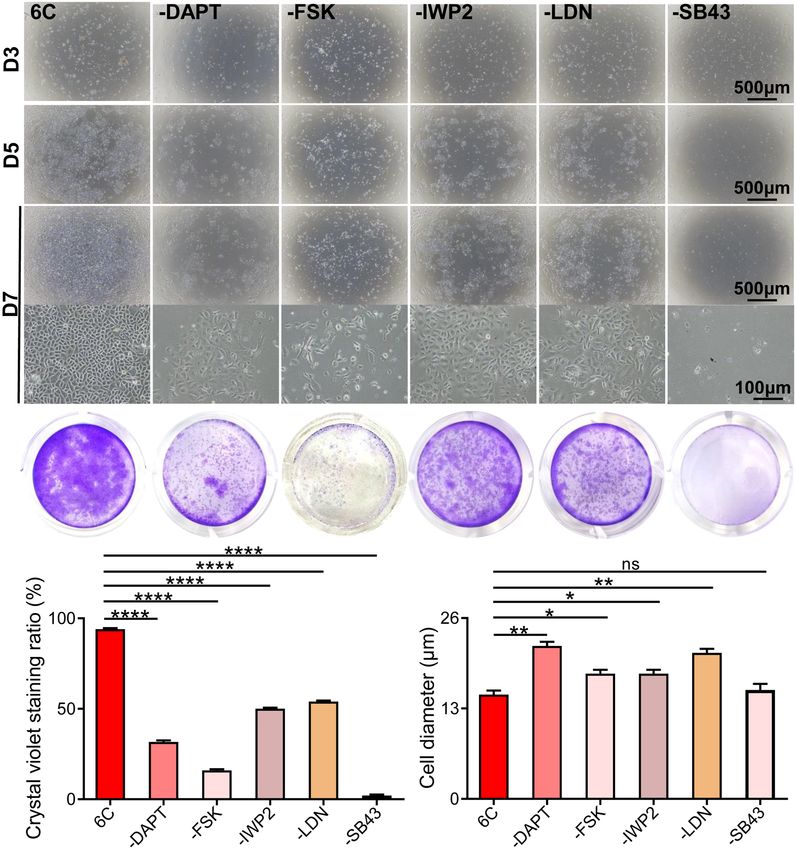
An et al.                                                                                                  Corneal Epithelial Cells Culture Paradigm

Dulbecco’s Modified Eagle Medium, Ham’s F-12 medium, fetal               inserts in 12-well plates containing DermaLife K keratinocyte
bovine serum (FBS), mouse epidermal growth factor (EGF),                 calcium and serum-free medium with or without 6C for 1 week.
HEPES buffer, gentamicin, amphotericin B, trypsin-EDTA,                  To expose an epithelial explant to air, 0.5 ml of culture
TRIzol and the antibody of anti-ZO1 (339100) were purchased
            R
                                                                         medium was added to each well. This amount was sufficient
from Invitrogen (Carlsbad, CA, United States). Dispase II was            to only immerse the underlying stromal cells in medium. The
purchased from Roche (Basel, Switzerland). Y-27632, Triton X-            corneal epithelium and epithelial single cells were harvested as
100, bovine serum albumin (BSA), insulin-transferrin-sodium              described above. The planting area of the epithelial sheet was
selenite media supplement, hydrocortisone, cholera toxin and             about one-tenth of the area of the insert, and the planting
dimethyl sulfoxide (DMSO) were purchased from Sigma (St.                 density of epithelial single cells was about 5,000/cm2 . They
Louis, MO, United States). Alexa Fluor 488− and Alexa Fluor              were submerged in the medium until the cell density covered
594-conjugated IgG were purchased from Life Technologies                 at least 90% of the inserts, and then the cultures were air
(Carlsbad, CA, United States). Forskolin, SB431542, DAPT, IWP-           lifted for 1 week.
2, and LDN-193189 were purchased from Apexbio (Boston, MA,
United States). The antibodies of rabbit anti-Ki67 (ab16667),            Cell Proliferation Assay
rabbit anti-Pax6 (ab195045), rabbit anti-P63 (ab124762), rabbit          To evaluate the cell proliferative activity, the cells were
anti-cytokeratin 14 (K14) (ab181595), and mouse anti-alpha               trypsinized and harvested daily from three randomly selected
smooth muscle actin (α-SMA) (ab18147) were purchased                     wells and manually counted using a hemocytometer (Stewart
from Abcam (Cambridge, MA, United States). The antibodies                et al., 2000). The protocol involved counting the number of cells
specific for goat anti-cytokeratin 12 (K12) (sc-17101) and               in the upper left, lower left, upper right, and lower right grids of
rabbit anti-β-catenin (sc7199) were purchased from Santa Cruz            the hemocytometer. The cells in each well were counted 3 times,
Biotechnology (Santa Cruz, CA, United States). The antibodies            and the average cell density was used to generate a growth curve.
of rabbit anti-ZEB1 (a5600) and rabbit anti-Snail (a5243) were              Crystal violet staining was also used to detect cell proliferation
purchased from Abclonal (Boston, MA, United States). ExScript            rates. After culturing for 1 week, cells were fixed with 4%
RT Reagent kit and SYBR Premix Ex Taq Kit were obtained                  paraformaldehyde at room temperature for 15 min, then washed
from Takara Bio (Shiga, Japan). 12-well culture inserts used in          three times with PBS for 5 min each and stained with 0.05%
this study were purchased from Millipore Corporation (Billerica,         crystal violet at room temperature for 30 min, then washed three
MA, United States).                                                      times with PBS for 5 min each and digital images were captured
                                                                         with a camera attached to a microscope.
Cell Culture
C57BL/6 mice (6∼8 weeks) were obtained from the Experimental             RNA Isolation, Reverse Transcription,
Animal Center of Xiamen University. All experimental
procedures used for animals in ophthalmology and vision
                                                                         and Quantitative Real-Time PCR
research are in compliance with the regulations of the Association       (qRT-PCR)
for Research in Vision and Ophthalmology (ARVO) and have                 The total cellular RNA was extracted with TRIzol and then
been approved by the Experimental Animal Ethics Committee of             reverse transcribed into cDNA using ExScript RT kit. qRT-PCR
Xiamen University.                                                       was performed with the SYBR Premix Ex Taq kit and the
    After mice were sacrificed by cervical dislocation, their            StepOne Real-Time PCR detection system (Applied Biosystems,
eyeballs were harvested and incubated with 10 mg/ml dispase II           Darmstadt, Germany) according to the manufacturer’s
at 4◦ C for 18 h. The corneal epithelium was isolated and digested       instruction. The amplification procedure included an initial
with 0.05% Trypsin-EDTA at 37◦ C for 15 min. Cells were seeded           denaturation step at 95◦ C for 10 min, followed by denaturation
at a density of 5,000 cells/cm2 in a 24-well dish. They were             at 95◦ C for 10 s, and annealing and extension at 60◦ C for 30 s,
incubated at 37◦ C, 5% CO2 and 95% air, and the DermaLife K              for 40 cycles. Using β-actin as an internal control, the results of
keratinocyte calcium and serum-free medium was replaced every            relative qRT-PCR were analyzed by the comparative CT method
2 days. The cell morphology was observed and digital images were         (2-11Ct ) (Suzuki et al., 2000). Table 1 shows the primers used to
captured using a microscope (Olympus CKX53, Japan).                      amplify specific gene products obtained from mCEC cDNA.
    The experimental groups are as follows: Control group;
Solvent group, containing DMSO (0.1%); Y-27632 group,                    Colony-Forming Efficiency Assay
containing Y-27632 (10 µmol/l); 5C group, containing Forskolin           Clonal culture was performed according to Rheinwald and
(20 µmol/l), SB431542 (10 µmol/l), DAPT (5 µmol/l), IWP-2                Green (R&G)’s method (Rheinwatd and Green, 1975). 3T3
(0.5 µmol/l), LDN-193189 (0.1 µmol/l); 6C group, containing              fibroblasts were treated with mitomycin C (5 µg/ml) at
Forskolin (20 µmol/l), SB431542 (10 µmol/l), DAPT (5 µmol/l),            37◦ C for 3 h, and then digested with 0.25% Trypsin-
IWP-2 (0.5 µmol/l), LDN-193189 (0.1 µmol/l), Y-27632                     EDTA at 37◦ C for 3 min, and terminated with SHEM
(10 µmol/l).                                                             medium (comprising an equal volume of HEPES-buffered
                                                                         DMEM containing bicarbonate and Ham’s F-12 medium,
Air-Lifted Exposure Culture                                              2 ng/ml mouse EGF, 5 mg/ml insulin, supplemented with 5%
Mouse corneas were isolated with ophthalmic scissors, and then           FBS, 0.5% dimethyl sulfoxide, 5 mg/ml transferrin, 5 ng/ml
cut into petals with a blade. Next, explants were placed on              selenium, 0.5 mg/ml hydrocortisone, 1 nM cholera toxin,

Frontiers in Cell and Developmental Biology | www.frontiersin.org    3                                       June 2021 | Volume 9 | Article 675998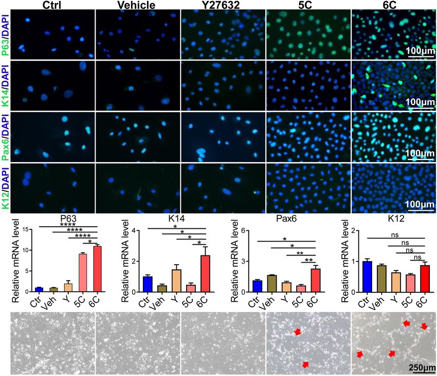
An et al. Corneal Epithelial Cells Culture Paradigm
TABLE 1 | Primer sequences for qRT-PCR of indicated gene transcripts. Statistical Analysis
Gene Sense Antisense ImageJ software was used to analyze the time dependent declines
name in fluorescein sodium staining area in a debrided region of
the epithelium and crystal violet staining area of the cultured
Ki67 CACTCCAAAGAAACCCACAA CTCATCTGCTGCTGCTTCTC
cells. Statistical analysis was performed with one-way analysis
P63 ATGTCACCGAGGTTGTGAAA GAATTCAGTGCCAACCTGTG
of variance analysis with GraphPad Prism 8.0.1 software. Unless
K14 CCCACCTTTCATCTTCCCAATT AAGCCTGAGCAGCATGTAGCAG
otherwise stated in all figures, data are shown as mean ± SEM
K12 AAACCGCAGACACCATCAGT ATGAGACCACTTCGCCATTC
(n = 3). ∗ P < 0.05, ∗∗ P < 0.01, ∗∗∗ P < 0.001, ∗∗∗∗ P < 0.0001.
Pax6 AGTGTCTACCAGCCAATCCC CATGGAACCTGATGTGAAGG
ZEB1 CCACTGTGGAGGACCAGAAT GTGAGGCCTCTTACCTGTGT
ZEB2 AAGTACCGCCACGAGAAGAA TTTGGTGCTGATCTGTCCCT
α-SMA CTCCCTGGAGAAGAGCTACG CGCTGACTCCATCCCAATGA
RESULTS
β-catenin ACAAGAAGCGGCTTTCAGTC CTGCAGTCTCATTCCAAGCC
β-actin GAGACCTTCAACACCCCAGC ATGTCACGCACGATTTCCC
6C Treatment Increases Adherence and
Proliferation of Primary mCEC
It is well known that the mCEC were poorly adherent when
50 mg/ml gentamicin, and 1.25 mg/ml amphotericin B). 3T3 cultured in vitro (Kawakita et al., 2004), but by the third
fibroblasts and mCEC (isolated as described above) were day, most of the cells in the 6C treatment group adhered
co-cultured at a density of 4.5 × 10⁴ /cm2 and 2,000/cm2 and the proliferation rate far exceeded that of the other four
in a 12-well dish. treatment groups (Figure 1A). Cell growth curves were generated
to determine if culturing mCEC exposed to the 6C cocktail
promoted their adherence and increased cell proliferation
Histological Characteristics and (Figure 1B). By day 7, the cells in both the control group and
Immunostaining the vehicle group had irregular morphology and fell off. In both
Corneal epithelial cell sheets were imbedded in opti-mum the 5C and the Y-27632-treated groups, the cells had a larger
cutting temperature (OCT) compound, and cryostat sections volume. It is apparent that the cells treated with 6C had a very
(5 µm) were obtained. Cultured mCEC and frozen sections were regular oval morphological structure, and the cells were small
rehydrated in PBS after being fixed in 4% paraformaldehyde for and densely arranged (Figure 1C). The crystal violet cell staining
20 min at room temperature, followed by incubation in 0.2% area in the 6C group was greater than the other four treatment
Triton X-100 for 10 min. After rinsing with PBS three times for groups, indicating that 6C treatment enhanced cell proliferation
5 min each and preincubating with 2% BSA to block non-specific (Figures 1D,E). Furthermore, Ki67 immunofluorescence staining
staining, the sections were incubated with primary antibodies and qRT-PCR analysis of proliferation related gene expression
overnight at 4◦ C with different dilutions [P63, β-catenin and levels confirmed that 6C treatment promoted cell proliferation
α-SMA all at 1:100, Ki67, Pax6, K12, ZEB1, and Snail all at (Figures 1F,G). Fibrillar actin (F-actin) and tight junctional ZO-
1:200, K14 (1:1000), ZO-1 (5–10 µg/ml)]. After washing with 1 proteins play important roles in sustaining both cell adhesion
PBS three times for 10 min each, cells or sections were incubated and cell-cell tight junctional connectivity (Higbee and Hazlett,
with Alexa Fluor-conjugated secondary antibodies for 1.5 h. After 1983; Ryeom et al., 2000). When the cells were treated with
rinsing each section three times with PBS for 10 min, they were 6C in vitro, F-actin immunostaining appeared tight and regular,
counterstained with DAPI and then mounted. A fluorescence but its pattern in the other four treatment groups was less
microscope was used to view and analyze each section (Leica, well defined and more irregular (Figure 1H). In addition, the
Germany and Olympus FV1000MPE-B, Japan). expression of ZO-1 in the 6C-treated group was significant
For morphological analysis, hematoxylin and eosin (H&E) whereas in the other groups it was erratic (Figure 1I). These
staining was performed according to standard procedures. Digital results show that 6C treatment promoted cell adhesion and
images of representative areas were captured with the light intercellular adhesion.
microscope (Eclipse 50i, Nikon, Tokyo, Japan).
Effects of Each Component in 6C on the
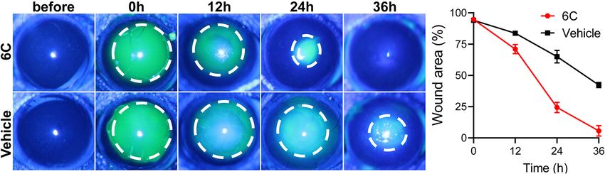
Corneal Epithelial Wound Healing Growth of mCEC
The mice were anesthetized by intraperitoneal injection of To determine if each component in the 6C cocktail is essential
pentobarbital (40 mg/kg). A corneal trephine with a diameter for the proliferation of mCEC, we compared the effects of a
of 1.5 mm was used to make a circular mark on the cornea single component at a time on cell growth (Figure 2A). The
of the mouse, and the circular area was scraped off with a results showed that omission of any one compound caused the
corneal spatula. Then the eyes were spotted with 6C solution cell proliferation to decline (Figures 2B,C) and compromised
and the solvent control group was set. The corneal epithelial the cell morphology relative to that in the cultures treated with
defects were visualized at 0, 12, 24, and 36 h after injury by 6C cocktail (Figure 2D). In addition, the results showed that
instillation of 0.5% fluorescein sodium and were examined and the cells in the treatment group without SB431542 almost lost
photographed with a camera mounted on a slit-lamp microscope their proliferative activity, which means that SB431542 plays an
(Haag-StreitAG, Switzerland). essential role in regulating the proliferation of mouse corneal
Frontiers in Cell and Developmental Biology | www.frontiersin.org 4 June 2021 | Volume 9 | Article 675998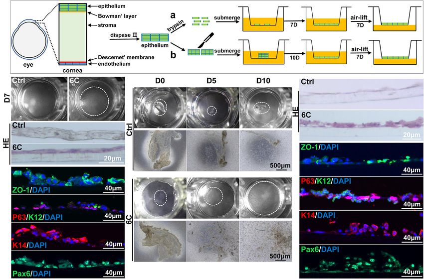
An et al. Corneal Epithelial Cells Culture Paradigm
FIGURE 1 | 6C can promote the proliferation of primary mCEC. (A) Morphological comparison of mCEC cultured with different compositions. Images were taken at
day 3 and day 7 post seeding. (B) Cell growth curve. (C) A statistical graph of the average diameter of the cells after 1 week of culture. (D) Crystal violet staining
results after 1 week of cell culture. 6C group had the most stained area. (E) Statistical analysis of crystal violet staining results after 1 week of cell culture. (n = 5
biological replicates). (F) qRT-PCR analysis of Ki67 gene expression (normalized to ctrl). (G) Ki67 (green) immunofluorescence staining. Nuclei were stained with
DAPI (blue). (H,I) Immunofluorescence staining of F-actin (red) and ZO-1 (green). Nuclei were stained with DAPI (blue). The expression of F-actin and ZO-1 in 6C
group was distributed on the cell membrane, and the connection between cells was tight, but the distribution of other treatment groups was scattered or missing.
Data expressed as the means ± SEM from three separate experiments (**P < 0.01, ***P < 0.001, ****P < 0.0001).
epithelial cells. The growth of cells in the treatment group without in vivo, the in vitro cell types do not correspond to those
FSK was more scattered and the cell status was poor. proliferating in vivo even though there are no definitive
biomarkers of stem cells. In vivo in this niche there are few cells
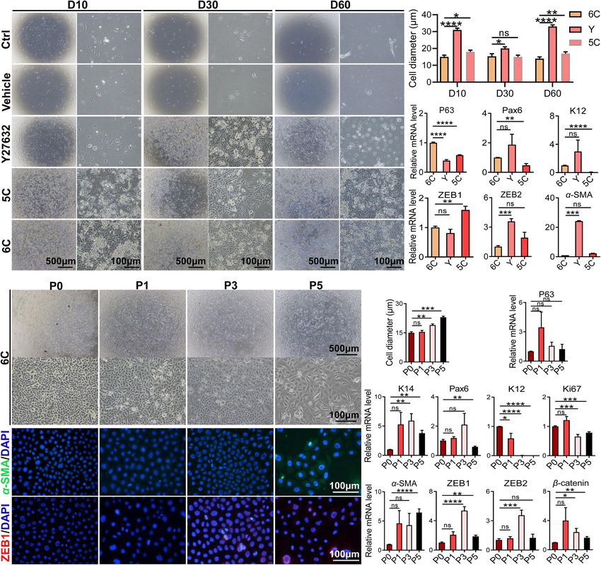
6C Treatment Preserves Function of undergoing an EMT whereas in vitro they are easily transformed
into mesenchymal cells. Nevertheless, in vitro 6C treatment
Primary mCEC
significantly down-regulated this transition because ZEB1 and
Immunofluorescent staining showed that after 6C treatment the
Snail immunofluorescence were both significantly lower than
P63 and K14 expression levels increased, which is indicative
their levels in the other four groups (Figure 4A). Corresponding
of more abundant proliferating epithelial cell types (Guo et al.,
measurements of the declines in the mRNA expression levels
2018; Figure 3A). Such increases were consistent with rises in
of ZEB1/2 and Snail, which are EMT transcription factors
the Pax6 expression levels, which is another corneal epithelial
(Tiwari et al., 2017), also show that 6C treatment caused their
specific marker (Kitazawa et al., 2019), whereas expression of
levels to decline relative to those in the four other treatment
the differentiation marker, K12, was only marginally expressed
groups (Figure 4B). These effects are consistent with the declines
in each group. The results of qRT-PCR analysis showed that
in β-catenin and α-SMA expression levels, which are other
the increases in the mRNA level of each interrogated gene
characteristic markers of this cell type transition. These results
was consistent with their corresponding protein expression
effectively illustrate that 6C treatment significantly inhibited the
levels (Figure 3B). In addition, we also conducted a 3T3-clone
EMT and thereby plays an important role in maintaining the
formation assay and found that 6C treatment significantly
proliferating phenotype of mCEC.
promoted the formation of cloned cells, which also shows that
We determined if 6C treatment suppressed FBS-induced
the cells treated with 6C possess a phenotype more characteristic
EMT in cultured mCEC. Following exposure to the 6C
of proliferating progenitor cells (Figure 3C).
cocktail for 3 days, the cell confluence had reached about
40% (Figures 4Ca,b). At this time, the cell morphology was
6C Treatment Inhibits Spontaneous and regular, and their volume was small (Figure 4E). In one group,
FBS-Induced EMT 6C treatment was terminated and the culture was continued
As the cell culture condition cannot mimic the unique until four more days had passed (Figure 4Cb). In this group
microenvironment in the niche supporting stem cell activity the nuclear and cell volumes enlarged (Figure 4E) whereas
Frontiers in Cell and Developmental Biology | www.frontiersin.org 5 June 2021 | Volume 9 | Article 675998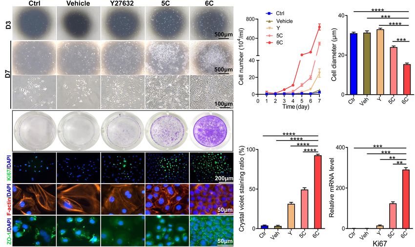
An et al.                                                                                                                     Corneal Epithelial Cells Culture Paradigm

  FIGURE 2 | Effects of subtracting a compound on the proliferation of mCEC. (A) Morphological comparison of mCEC cultured with subtracting one compound from
  6C or 6C, respectively (The group without Y-27632 has been described above). Images were taken at day 3, day 5 and day 7 post seeding. The cell proliferation rate
  of the 6C treatment group was much better than that of the treatment group minus any compound. (B) Crystal violet staining results after 1 week of cell culture. 6C
  group had the most stained area. (C) Statistical analysis of crystal violet staining results. Data expressed as the means ± SEM from three separate experiments
  (*P < 0.05, **P < 0.01, ****P < 0.0001, ns: no significance). (D) Statistical analysis of the diameters of the cells.

in another group in which 6C treatment was continued, they                            group, α-SMA expression was lower, which is an EMT biomarker.
remained very small (Figure 4Ca). The effects were evaluated                          Similarly, in the 6C supplemented with 10% FBS group, α-SMA
of serum following 3 days of 6C treatment on cell growth and                          expression was lower than that in the 10% FBS treated group.
differentiation (Figures 4Da’,b’). The cell volumes had increased                     These results substantiate the notion that 6C treatment promoted
in both the 6C supplemented with 10% FBS and the other group                          increases in cell proliferation and inhibited EMT.
supplemented only with 10% FBS (Figure 4E). However, the
degree of differentiation of cells treated with only 10% FBS was
pronounced because, the cell boundaries were blurred, and it
                                                                                      Effects of 6C Treatment on Long-Term
was difficult to recognize the morphology of individual cells.                        Maintenance of mCEC Morphology and
The qRT-PCR results showed that the 6C-treated group had                              Function
higher expression levels of progenitor cell markers P63 and K14,                      To determine whether 6C protects cells from undergoing declines
differentiation markers Pax6 and K12, as well as higher Ki67                          in proliferative activity and changes in phenotype during an
expression (Figures 4F,G). On the other hand, in the 6C-treated                       extended period in culture, they were cultured for 60 days.

Frontiers in Cell and Developmental Biology | www.frontiersin.org                 6                                              June 2021 | Volume 9 | Article 675998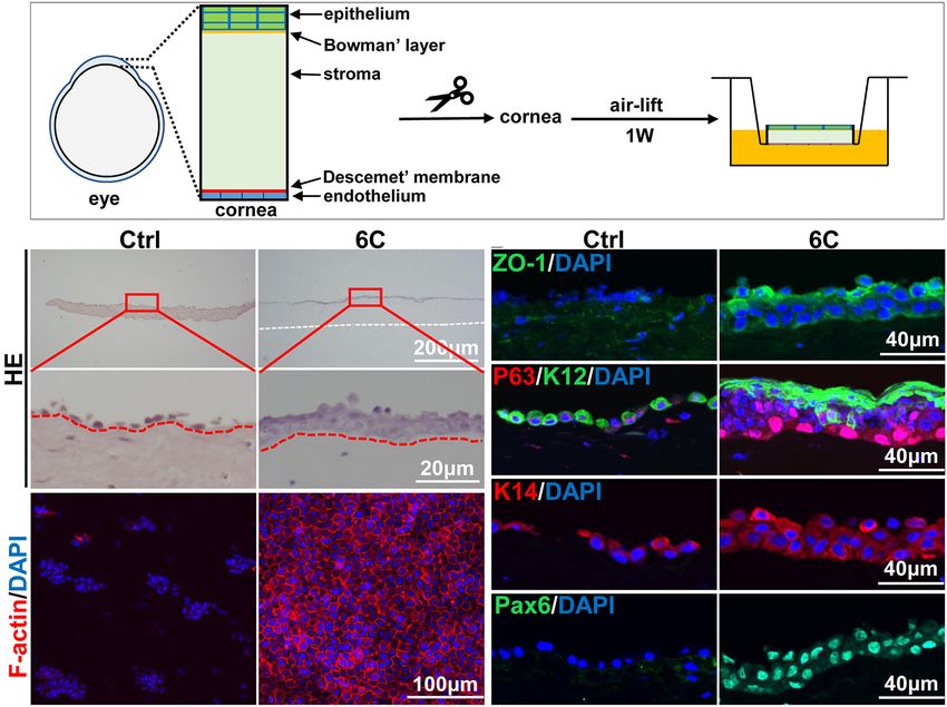
An et al.                                                                                                                         Corneal Epithelial Cells Culture Paradigm

  FIGURE 3 | 6C can effectively maintain the expression of primary mCEC related genes. (A) Immunofluorescence staining of mCEC function markers (green). Nuclei
  were stained with DAPI (blue). P63 and K14 are markers representing the characteristics of corneal epithelial stem cells. Pax6 is a key transcription factor regulating
  corneal epithelial cell fate. K12 is a marker of mCEC differentiation. (B) qRT-PCR analysis of P63, K14, Pax6 and K12 gene expression (normalized to ctrl). Data
  expressed as the means ± SEM from three separate experiments (*P < 0.05, **P < 0.01, ****P < 0.0001, ns: no significance). (C) 6C can promote the formation of
  clones of mCEC (red arrow).

Initially, the microscopic images reveal that the control group and                      in their integrity resulting from transitioning into a cell type
the vehicle group were poorly adherent and weakly proliferative                          expressing EMT markers.
(Figure 5A). After additional time, the cells gradually detached                             Next, we further explored the effects of 6C treatment on
and died. The development of the proliferating status was                                primary mCEC subcultures. At an initial stage, the mCEC were
delayed in the Y-27632-treated group in the first 10 days,                               oval, and as the number of passages increased some cells acquired
but later it was transiently enhanced. By the 60th day, their                            a long spindle shape (Figure 5D), and their volume increased
growth activity had markedly declined and structural integrity                           (Figure 5E). At the mRNA expression level, the expression
became less distinct. The 5C-treated group had the second                                of the progenitor cell marker P63 did not change, but the
fastest proliferation rate, which was only exceeded by the                               expression of K14 was up-regulated (Figure 5F). The expression
6C-treated group. After culturing the 6C-treated group for                               of the transcription factor Pax6 was down-regulated by the
60 days, the interconnections between cells were tight and                               fifth passage. The differentiation marker K12 was significantly
regular and their apparent cell volumes remained invariant                               down-regulated. The cell proliferation marker Ki67 was down-
from those at earlier times (Figure 5B). The results of qRT-                             regulated from the third generation. At the same time, as the
PCR showed that the expression levels of P63, Pax6, and K12                              number of passages increased, the expression levels of EMT-
in group 6C were significantly higher than that in the Y-                                related genes α-SMA, ZEB1/2, β-catenin were up-regulated. By
27632 and 5C-treated groups, and the expression of ZEB1/2 and                            the fifth generation, α-SMA immunofluorescent staining became
α-SMA in group 6C was significantly lower on the 60th day                                evident and ZEB1 expression already appeared in the third
of culture (Figure 5C). These results substantiate that 6C can                           generation (Figure 5G). The structural integrity and functional
effectively maintain mCEC proliferative activity and reduce losses                       activity were only initially preserved of these proliferating cell

Frontiers in Cell and Developmental Biology | www.frontiersin.org                    7                                              June 2021 | Volume 9 | Article 675998
An et al.                                                                                                                       Corneal Epithelial Cells Culture Paradigm

  FIGURE 4 | 6C can suppress the occurrence of EMT. (A) Immunofluorescence staining of EMT-related genes after 1 week of cell culture. ZEB1 and Snail are
  transcription factors of EMT, β-catenin and α-SMA are markers of EMT (green). Nuclei were stained with DAPI (blue). The expression of EMT-related genes in group
  6C was significantly reduced. (B) qRT-PCR analysis of ZEB1/2, Snail, β-catenin and α-SMA gene expression (normalized to ctrl) after 1 week of cell culture.
  (C) Comparison of cell morphology. a, Treat with 6C for 1 week. b, Treat with 6C for 3 days, then remove 6C and continue to cultivate for 4 days. (D) Comparison of
  cell morphology. a’, It was first treated with 6C for 3 days, and then cultured with 6C plus 10% FBS for another 4 days. b’, Treat with 6C for 3 days, then remove 6C
  and continue to incubate with 10% FBS for 4 days. (E) Statistical analysis of cell diameter after C and D treatment. (F) After C treatment, qRT-PCR analysis of P63,
  K14, K12, Pax6, Ki67, and α-SMA gene expression (normalized to 6C). (G) After D treatment, qRT-PCR analysis of P63, K14, K12, Pax6, Ki67, and α-SMA gene
  expression (normalized to 6C plus 10% FBS). Data expressed as the means ± SEM from three separate experiments (*P < 0.05, **P < 0.01, ***P < 0.001,
  ****P < 0.0001, ns: no significance).

Frontiers in Cell and Developmental Biology | www.frontiersin.org                  8                                              June 2021 | Volume 9 | Article 675998
An et al.                                                                                                                        Corneal Epithelial Cells Culture Paradigm

  FIGURE 5 | 6C can maintain the morphology and function of mCEC in long-term culture and subculture. (A) Morphological comparison of mCEC cultured with
  different compositions in long-term culture. (B) Statistical analysis of the diameters of long-term cultured cells. (C) qRT-PCR analysis of P63, Pax6, K12, ZEB1/2, and
  α-SMA gene expression (normalized to 6C). (D) Morphology of mCEC subculture. (E) Statistical analysis of the diameters of the cells. (F) qRT-PCR analysis of
  EMT-related genes, mouse corneal epithelial function-related genes and cell proliferation marker Ki67 (normalized to P0). (G) Immunofluorescence staining of
  EMT-related genes α-SMA (green) and ZEB1 (red). Nuclei were stained with DAPI (blue). Data expressed as the means ± SEM from three separate experiments
  (*P < 0.05, **P < 0.01, ***P < 0.001, ****P < 0.0001, ns: no significance).

types following treatment of the mCEC subcultures with 6C. At                           maintains itself (Figure 6A). H&E staining results showed that
later times, 6C treatment failed to provide long term protection                        the control groups underwent severe epithelial shedding leaving
since the mCEC subcultures appeared to gradually become more                            remaining less than one layer of epithelial cells (Figure 6B).
similar to other cultures not treated with 6C.                                          In contrast, the 6C treatment group had developed a relatively
                                                                                        complete full thickness stratified epithelium. Furthermore, the
                                                                                        distribution of F-actin in the 6C treatment group was regular in
6C Treatment Maintains Mouse Corneal                                                    these cultures, while the control group had scant levels F-actin
Tissue Cultures                                                                         expression (Figure 6C). In the 6C treatment group, tight junction
Freshly isolated mouse corneal tissues were air lifted and cultured                     protein ZO-1 was expressed in the entire epithelium, but was
to simulate the in vivo conditions in which the normal cornea                           almost absent in the control group (Figure 6D). In the 6C

Frontiers in Cell and Developmental Biology | www.frontiersin.org                   9                                               June 2021 | Volume 9 | Article 675998
An et al. Corneal Epithelial Cells Culture Paradigm
FIGURE 6 | The effect of 6C on mouse cornea ex vivo culture. (A) Schematic diagram of air-exposed culture of mouse cornea. (B) HE staining results of mouse
cornea after air exposure culture. The epithelium in the 6C treatment group was relatively intact, while the control group had severe shedding (the white dotted line
represents the edge of the corneal stroma and the red dotted line represents the boundary between the epithelial layer and the stromal layer). (C) Wholemount
immunostaining of F-actin (red). Nuclei were stained with DAPI (blue). (D) Immunofluorescence staining of ZO-1 (green), P63 (red), K12 (green), K14 (red) and Pax6
(green). Nuclei were stained with DAPI (blue).
treatment group, mouse corneal epithelial progenitor cell marker membranes in the 6C treatment group. However, the cells were
P63 was strongly expressed in the basal and middle part of the poorly adherent in the control groups not treated with the 6C
epithelium. The mouse corneal epithelial differentiation marker cocktail (Figure 7B). Then both the control and the 6C-treated
K12 was expressed at the epithelial surface, while the control groups were air-lifted for 1 week to characterize the mCEC
group had weaker expression of P63 and K12. K14 was used as a differentiation process. The results of H&E staining showed that
limbal progenitor cell marker to further characterize the effect of the cells in the 6C treatment group were stratified to about two
6C treatment cocktail on the behavior of corneal epithelial cells layers (Figure 7C). ZO-1 was nearly expressed throughout an
in culture (González et al., 2019). Both K14 and the epithelial entire constructed mouse corneal epithelial sheet (Figure 7D).
differentiation regulator Pax6 were almost present throughout P63 was expressed in the basal layer of a cell sheet. K14 and Pax6
the entire epithelium in the 6C treatment group, while only K14 were expressed in almost all layers. K12 was expressed in the
was expressed in the control group. These results show that 6C surface layer. The effects of 6C treatment indicate that it is also
improves maintenance of the morphology and function of the beneficial to construct tissue-engineered mouse corneal epithelial
mouse cornea cultured ex vivo. sheets with a small amount of mouse corneal epithelium
(Figures 7E–G). The epithelial sheets constructed by the former
6C Treatment Facilitates the method have tighter connections between cells because of the
Construction of Tissue Engineered higher expression levels of ZO-1. The expression levels of other
genes are almost the same between the two methods.
Mouse Corneal Epithelial Sheets
As 6C treatment improved the growth and decreased terminal
differentiation of corneal explants, we determined if these two Effect of 6C Treatment on Mouse Corneal
effects alter the outcome of tissue engineered mouse corneal Epithelial Wound Healing Duration
epithelial sheets generated for corneal reconstruction surgery. Treatment with eye drops containing the 6C cocktail significantly
Trypsin was used to detach the mouse corneal epithelium, shortened the wound healing time in the corneal epithelium
which was then layered on a Transwell membrane (Figure 7A).
R
scraping model (Figure 8A). Twenty-four hours after
One week later, the cells had nearly completely covered the epithelial scraping, the remaining wounded area of the
Frontiers in Cell and Developmental Biology | www.frontiersin.org 10 June 2021 | Volume 9 | Article 675998An et al. Corneal Epithelial Cells Culture Paradigm FIGURE 7 | 6C helps to construct tissue engineered mouse corneal epithelium. (A) Schematic diagram of tissue engineering mouse corneal epithelial sheet culture. (a) The isolated mouse corneal epithelium was digested into single cells with trypsin, and then planted into insert. After the cells were confluent, they were cultured in air exposure. (b) Divide the isolated corneal epithelium into two and plant it on 12-well cell culture inserts. After the cells are covered with the insert, do air exposure culture. (B) When epithelial single cells were cultured to the seventh day, the cells in the 6C treatment group were almost confluent. (C) HE staining results of mCEC after air exposure culture. The cells in the 6C treatment group were stratified to two layers. (D) Immunofluorescence staining of Z0-1 (green), P63 (red), K12 (green), K14 (red) and Pax6 (green). Nuclei were stained with DAPI (blue). (E) Morphological comparison of corneal epithelial sheets cultured with corneal epithelium. In the 6C treatment group, during the culture of the epithelial sheet, the epithelial cells crawled out from the edge, and the surface cells gradually fell off. By the tenth day of culture, the cells were almost confluent, but the epithelium of the control group hardly grew. (F) HE staining results of corneal epithelium after air exposure. (G) Immunofluorescence staining of Z0-1 (green), P63 (red), K12 (green), K14 (red) and Pax6 (green). Nuclei were stained with DAPI (blue). FIGURE 8 | The effect of 6C on wound healing of corneal epithelium in mice. (A) Fluorescein sodium nodded and photographed with a slit lamp. 6C can significantly promote wound healing. (B) Statistical analysis of wound healing. corneal epithelium in the 6C treatment group was 25% of a large area defect of 48%. The improved healing of corneal that in the control group (Figure 8B). By the 36th hour, the epithelial wounds in the 6C is likely attributable to larger wound in the 6C treatment group had healed completely, increases in cell proliferation and cell migration in mice while the corneal epithelium in the solvent group still had (Kao, 2020). Frontiers in Cell and Developmental Biology | www.frontiersin.org 11 June 2021 | Volume 9 | Article 675998
An et al.                                                                                                       Corneal Epithelial Cells Culture Paradigm

DISCUSSION                                                                    cells (Ban et al., 2003), resulting from improved tight junction
                                                                              structural integrity (Anderson, 1977; Sugrue and Zieske, 1997; Ko
Corneal epithelial renewal and maintenance of function                        et al., 2009). Such suppression of EMT was described in other
is supported by preservation of different niches whose                        cell types in vitro, as follows. Forskolin’s modes of action stems
microenvironment is needed to support different cell phenotypes               from: reducing EMT possibly due to its activation of cAMP linked
(Schermer et al., 1986; Ang et al., 2004). This continuous renewal            signaling pathways, which are known to promote rat corneal
process maintains a functional epithelial layer that supports                 epithelium secretion of mucin-like glycoprotein (Nakamura et al.,
normal vision through acting as a barrier against pathogenic                  1998). It is suggested that this response thereby protects cells
infiltration. Specifically, the niche in which the slow cycling               from external stimulation by cytokines and lubricates their cell
stem cells reside in the limbus is different from those that                  surface (Devine and McKenzie, 1992; Cone, 2009; Baudouin et al.,
promote their stepwise transition to proliferating progenitor                 2019). SB431542 is a selective and potent inhibitor of the TGF-
cells and differentiating suprabasal cells. The coordination and              β/Activin/NODAL pathway that promotes the differentiation of
control of the program that directs this renewal process is very              human induced pluripotent stem cells into corneal epithelial-
complex and is known to involve a host of different cytokines                 like cells by restoring the level of endogenous BMP signaling
controlling different steps in this renewal process as well as other          (Kamarudin et al., 2018). DAPT prevents EMT in cultured limbal
factors involved in controlling the expression of these unique                epithelial stem cells and corneal endothelial cells by inhibiting the
microenvironments (Schofield, 1978; Keung et al., 2010).                      Notch signaling pathway (Li et al., 2013; Tsai et al., 2014). IWP-
    The insight gained from studies delineating the complexity of             2 is one of the Wnt/β-catenin signaling inhibitors, which can
this regenerative has improved the outcome of corneal epithelial              affect the differentiation of human limbal epithelial progenitors
reconstruction surgery. Such progress stems from larger yields                (Lee et al., 2017). LDN- 193189 inhibits the BMP signaling
of proliferating corneal epithelial cells needed for generating cell          pathway, mediating the differentiation of pluripotent stem cells
sheets used to resurface the corneal epithelium. The current study            into retinal pigment epithelial cells (Choudhary et al., 2017). Y-
is instructive because it identifies a novel procedure for enhancing          27632 is a Rho/Rock signaling pathway inhibitor that promotes
in a corneal epithelial cell culture the yields of proliferating              corneal limbal cell epithelial proliferation and wound healing
corneal epithelial cells. This was achieved by treating them with             (Chen et al., 2008; Yin and Yu, 2008; Miyashita et al., 2013; Sun
cocktails of different cell signaling modulators which extend                 et al., 2015). It is conceivable that these agents have overlapping
their proliferative lifetimes through altering signaling events               inhibitory modes of action since omission of one of them in
inducing terminal differentiation and cell death (Xiang et al.,               the previously described 5C cocktail, namely Y-27632, markedly
2019). Specifically, the cocktails contained six common inhibitors            reduced the inhibitory efficacy of the novel 6C cocktail on EMT
which reduced the constraints of cell expansion by perpetuating               and generation of proliferating epithelial cells. The 6C cocktail
their proliferative status as progenitor surface adherent cells and           effectiveness in inhibiting EMT was presumably greater than that
presumably blocking signals that trigger them instead to undergo              of the 5C cocktail through larger upregulation of characteristic
declines in cell vitality and undergo terminal differentiation as the         progenitor genes (P63 and K14) and Pax6. Therefore, the 6C
cell density increases on the inserts.                                        cocktail under serum-free and trophoblast-free conditions is an
    It is difficult to maintain for extended periods growth and               effective short term preserver of the mouse corneal epithelium
differentiation of corneal epithelial cells in vitro. One of the              linage (Ouyang et al., 2014).
constraints stems from the loss of the regulatory effect of the                   Even though the long-term cultivation of bovine corneal
in vivo microenvironment signals. They are essential to preserve              endothelial cells in vitro increases their cell surface area and cell
the proliferative status of progenitor cells and prevent the cells            size (D’Hondt et al., 2009), in our case 6C culture treatment
from entering into a differentiating phase resulting in EMT and               was needed to extend the short-term proliferative potential of
termination of cell expansion (Thiery and Sleeman, 2006; Tiwari               mCEC and the maintenance of tissue-specific cell phenotype. In
et al., 2017). EMT is induced by increases in the expression                  addition, we used 6C culture conditions to subculture mCEC. As
of a group of transcription factors including members of the                  the number of culture passages increased, the progenitor P63 and
Snail, ZEB families and key markers such as β-catenin, α-SMA                  K14 cell markers continued to be expressed. Their perpetuation
(Kato et al., 2007; Aomatsu et al., 2011; Lamouille et al., 2014;             has an important regulatory role in maintaining the self-renewal
Puisieux et al., 2014; Tiwari et al., 2017). During this process,             of corneal epithelial cells in vivo and can expedite repair of
losses occur in cell-to-cell coupling in polarized epithelial cells as        damaged epithelium. This outcome is supported by the fact that
they undergo transformation into very stable mesenchymal-like                 their uniform morphology was better maintained than in a basic
cells (Shibata et al., 2019).                                                 serum-free medium. Nevertheless, the expression of EMT-related
    Interestingly, our novel 6C cocktail inhibited the expression of          genes rose as the number of passages increased.
spontaneous EMT-related genes and curtailed both this transition                  It was reported that a permanent rabbit corneal epithelial cell
and FBS-induced stimulation of this process. Moreover, 6C                     line was established by long-term continuous passage culture
treatment rendered changes in the cells morphology resulting                  on a 3T3 feeder layer (Castromunozledo, 1994). Kawakita
in their appearance becoming more regular due to increases                    also established the mouse corneal epithelial cell line TKE2
in the expression levels of the cytoskeletal proteins and tight               using the extended culture method (Kawakita et al., 2008).
junctional proteins at the cell membrane surface. These changes               There are many reports showing that karyotype changes occur
resulted in the formation of a continuous belt encircling the                 with this methodology used to establish an immortalized cell

Frontiers in Cell and Developmental Biology | www.frontiersin.org        12                                       June 2021 | Volume 9 | Article 675998
An et al. Corneal Epithelial Cells Culture Paradigm
line during culture (Kahn et al., 1993; Skopinski et al., 1998; cell availability for corneal epithelial reconstruction surgery
Jozwiak et al., 2001). Although the 6C culture conditions can transplantation in a clinical setting.
maintain the phenotype of mCEC in short-term subculture, their
phenotypes may also be unstable due to karyotype changes after
multiple passages. DATA AVAILABILITY STATEMENT
As it was possible to culture mCEC with 6C supplementation,
we explored the effect of 6C on native corneal tissue stability The original contributions presented in the study are included
ex vivo. Furthermore, single cells or small-area epithelial sheets in the article/supplementary material, further inquiries can be
were used to construct functional tissue-engineered corneal directed to the corresponding authors.
epithelial sheets. There are many reports about the in vitro
culture of human and rabbit limbal tissues (Qi et al., 2008;
Miyashita et al., 2013; Suárez-Barrio et al., 2019), but it is still ETHICS STATEMENT
very difficult to culture the mouse corneal epithelium. Human
corneal epithelial cells normally express K12 when cultured The animal study was reviewed and approved by the
in vitro, but K12 expression is barely detectable in mouse Experimental Animal Ethics Committee of Xiamen University.
corneal epithelial cells. Even though K12 is rarely expressed
in submerged cultured cells, it continues to be expressed
in air-lifted corneal tissues and engineered epithelial sheets.
AUTHOR CONTRIBUTIONS
This difference indicates that the 6C culture system maintains CL and YX conceived and designed the experiments. XA
the progenitor potential of epithelial cells and subsequent air performed the research, and collected and analyzed the data.
lifting of the cultures effectively simulates the differentiation GW, MJ, XZ, SG, JC, and ZL analyzed the data. CL, XA, and
conducive environment in vivo and accordingly promotes cell PR wrote the manuscript. All authors read and approved the
differentiation. These findings are likely to be insightful in final manuscript.
identifying other novel procedures for controlling the balance
between mouse corneal epithelial proliferating progenitor and
differentiating cells. In addition, in vivo experiments have shown FUNDING
that 6C can promote wound healing by stimulating the rapid
proliferation of epithelial cells, which may be achieved by This study was supported in part by grants from The
maintaining the limbal proliferating stem and progenitor cell National Key R&D Program of China (2020YFA0908103 and
phenotype (Li et al., 2007). 2018YFA0107301), the National Natural Science Foundation of
Limbal stem cell deficiency can lead to the development China (NSFC No. 82070931, 81770891, 81672955, 81900822, and
of various ocular surface dysfunctions and even blindness. 82000869), and the Huaxia Translational Medicine Fund for
To treat this disease, limbal stem cell transplantation is an Young Scholars (No. 2017-A-001).
effective method to treat ocular surface diseases and reconstruct
corneal epithelial structure (Jackson et al., 2020). Our 6C
culture system provides novel insight on the identity of targets ACKNOWLEDGMENTS
whose modulation provides both a feasible approach for the
in vitro expansion of limbal cells and even limbal tissues. Its The authors thank Jing-Ru Huang from the Biomedical Science
implementation may ultimately improve viable corneal epithelial Core Facility of Xiamen University for technical assistance.
REFERENCES corneal epithelium. Exp. Eye Res. 76, 663–669. doi: 10.1016/s0014-4835(03)
00054-x
Anderson, R. A. (1977). Actin filaments in normal and migrating corneal epithelial Baudouin, C., Rolando, M., Benitez Del, Castillo, J. M., Messmer, E. M., Figueiredo,
cells. Invest Ophthalmol. Vis. Sci. 16, 161–166. F. C., et al. (2019). Reconsidering the central role of mucins in dry eye and
Ang, L. P., Nakamura, T., Inatomi, T., Sotozono, C., Koizumi, N., Yokoi, N., ocular surface diseases. Prog. Retin. Eye Res. 71, 68–87. doi: 10.1016/j.preteyeres.
et al. (2006). Autologous serum-derived cultivated oral epithelial transplants for 2018.11.007
severe ocular surface disease. Arch. Ophthalmol. 124, 1543–1551. doi: 10.1001/ Castromunozledo, F. (1994). Development of a Spontaneous Permanent Cell-
archopht.124.11.1543 Line of Rabbit Corneal Epithelial-Cells That Undergoes Sequential Stages of
Ang, L. P., Tan, D. T., Beuerman, R. W., and Lavker, R. M. (2004). Development of a Differentiation in Cell-Culture. J. Cell Sci.. 107, 2343–2351.
conjunctival epithelial equivalent with improved proliferative properties using a Chen, J., Guerriero, E., Lathrop, K., and SundarRaj, N. (2008). Rho/ROCK
multistep serum-free culture system. Invest Ophthalmol. Vis. Sci. 45, 1789–1795. signaling in regulation of corneal epithelial cell cycle progression. Invest
doi: 10.1167/iovs.03-1361 Ophthalmol. Vis. Sci. 49, 175–183. doi: 10.1167/iovs.07-0488
Aomatsu, K., Arao, T., Sugioka, K., Matsumoto, K., Tamura, D., Kudo, K., et al. Cone, R. A. J. A. D. D. R. (2009). Barrier properties of mucus. Adv. Drug Deliv. Rev.
(2011). TGF-β induces sustained upregulation of SNAI1 and SNAI2 through 61, 75–85.
Smad and non-Smad pathways in a human corneal epithelial cell line. Invest D’Hondt, C., Ponsaerts, R., Srinivas, S. P., Vereecke, J., and Himpens, B. (2009).
Ophthalmol. Vis. Sci. 52, 2437–2443. doi: 10.1167/iovs.10-5635 Reduced intercellular communication and altered morphology of bovine
Ban, Y., Dota, A., Cooper, L. J., Fullwood, N. J., Nakamura, T., Tsuzuki, M., et al. corneal endothelial cells with prolonged time in cell culture. Curr. Eye Res. 34,
(2003). Tight junction-related protein expression and distribution in human 454–465. doi: 10.1080/02713680902913022
Frontiers in Cell and Developmental Biology | www.frontiersin.org 13 June 2021 | Volume 9 | Article 675998An et al. Corneal Epithelial Cells Culture Paradigm DelMonte, D. W., and Kim, T. (2011). Anatomy and physiology of the cornea. progenitors according to culture condition. Sci. Rep. 7:15241. doi: 10.1038/ J. Cataract. Refract. Surg. 37, 588–598. doi: 10.1016/j.jcrs.2010.12.037 s41598-017-15454-x Devine, P. L., and McKenzie, I. F. (1992). Mucins: structure, function, and Li, C., Dong, F., Jia, Y., Du, H., Dong, N., Xu, Y., et al. (2013). Notch signal regulates associations with malignancy. Bioessays 14, 619–625. doi: 10.1002/bies. corneal endothelial-to-mesenchymal transition. Am. J. Pathol. 183, 786–795. 950140909 doi: 10.1016/j.ajpath.2013.05.025 Ghareeb, A. E., Lako, M., and Figueiredo, F. C. (2020). Recent Advances in Stem Li, W., Hayashida, Y., Chen, Y. T., and Tseng, S. C. (2007). Niche regulation of Cell Therapy for Limbal Stem Cell Deficiency: A Narrative Review. Ophthalmol. corneal epithelial stem cells at the limbus. Cell Res. 17, 26–36. doi: 10.1038/sj.cr. Ther. 9, 809–831. doi: 10.1007/s40123-020-00305-2 7310137 González, S., Oh, D., Baclagon, E. R., Zheng, J. J., and Deng, S. X. (2019). Wnt Miyashita, H., Yokoo, S., Yoshida, S., Kawakita, T., Yamagami, S., Tsubota, K., et al. Signaling Is Required for the Maintenance of Human Limbal Stem/Progenitor (2013). Long-Term Maintenance of Limbal Epithelial Progenitor Cells Using Cells In Vitro. Investigat. Opthalmol. Visual Sci. 60:25740. doi: 10.1167/iovs.18- Rho Kinase Inhibitor and Keratinocyte Growth Factor. Stem Cells Translational. 25740 Med. 2, 758–765. doi: 10.5966/sctm.2012-0156 Guo, Z. H., Zhang, W., Jia, Y. Y. S., Liu, Q. X., Li, Z. F., and Lin, J. S. (2018). An Nakamura, M., Endo, K. I., and Nakata, K. (1998). Mucin-like glycoprotein Insight into the Difficulties in the Discovery of Specific Biomarkers of Limbal secretion is mediated by cyclic-AMP and protein kinase C signal transduction Stem Cells. Int. J. Mol. Sci. 19:19071982. doi: 10.3390/ijms19071982 pathways in rat corneal epithelium. Exp. Eye Res. 66, 513–519. doi: 10.1006/ Higbee, R. G., and Hazlett, L. D. (1983). Actin filament localization and distribution exer.1997.0457 in the young adult mouse cornea: a correlative immunofluorescent and Ouyang, H., Xue, Y., Lin, Y., Zhang, X., Xi, L., Patel, S., et al. (2014). WNT7A cytochemical study. Exp. Eye Res. 36, 171–180. doi: 10.1016/0014-4835(83) and PAX6 define corneal epithelium homeostasis and pathogenesis. Nature 511, 90003-9 358–361. Jackson, C. J., Myklebust Erno, I. T., Ringstad, H., Tonseth, K. A., Dartt, D. A., and Choudhary, P., Booth, H., Gutteridge, A., Surmacz, B., Louca, I., Steer, J., et al. Utheim, T. P. (2020). Simple limbal epithelial transplantation: Current status (2017). Directing Differentiation of Pluripotent Stem Cells Toward Retinal and future perspectives. Stem Cells Transl. Med. 9, 316–327. doi: 10.1002/sctm. Pigment Epithelium Lineage. Stem Cells Transl. Med. 6, 490–501. 19-0203 Pellegrini, G., Traverso, C. E., Franzi, A. T., Zingirian, M., Cancedda, R., and Jozwiak, J., Skopinski, P., Komar, A., Wojcik, A., and Malejczyk, J. (2001). De Luca, M. (1997). Long-term restoration of damaged corneal surfaces with Characterisation of epithelial cell line from rat cornea. Eye 15, 82–88. doi: autologous cultivated corneal epithelium. Lancet 349, 990–993. doi: 10.1016/ 10.1038/eye.2001.19 s0140-6736(96)11188-0 Kahn, C. R., Young, E., Lee, I. H., and Rhim, J. S. (1993). Human corneal epithelial Puisieux, A., Brabletz, T., and Caramel, J. (2014). Oncogenic roles of EMT-inducing primary cultures and cell lines with extended life span: in vitro model for ocular transcription factors. Nat. Cell Biol. 16, 488–494. doi: 10.1038/ncb2976 studies. Invest. Ophthalmol. Vis. Sci. 34, 3429–3441. Qi, H., Shine, H. D., Li, D. Q., de Paiva, C. S., Farley, W. J., Jones, D. B., et al. (2008). Kamarudin, T. A., Bojic, S., Collin, J., Yu, M., Alharthi, S., Buck, H., et al. (2018). Glial cell-derived neurotrophic factor gene delivery enhances survival of human Differences in the Activity of Endogenous BMP Signalling Impact on the Ability corneal epithelium in culture and the overexpression of GDNF in bioengineered of Induced Pluripotent Stem Cells to Differentiate to Corneal Epithelial Like constructs. Exp. Eye Res. 87, 580–586. doi: 10.1016/j.exer.2008.09.012 Cells. Stem Cells 36, 337–348. Rama, P., Matuska, S., Paganoni, G., Spinelli, A., De Luca, M., and Pellegrini, G. Kao, W. W. (2020). Keratin expression by corneal and limbal stem cells during (2010). Limbal stem-cell therapy and long-term corneal regeneration. N. Engl. development. Exp Eye. Res. 200:108206. doi: 10.1016/j.exer.2020.108206 J. Med. 363, 147–155. doi: 10.1056/NEJMoa0905955 Kato, N., Shimmura, S., Kawakita, T., Miyashita, H., Ogawa, Y., Yoshida, S., et al. Rheinwatd, J. G., and Green, H. (1975). Seria cultivation of strains of human (2007). Beta-catenin activation and epithelial-mesenchymal transition in the epidemal keratinocytes: the formation keratinizin colonies from single cell is. pathogenesis of pterygium. Invest Ophthalmol. Vis. Sci. 48, 1511–1517. doi: Cell 6, 331–343. 10.1167/iovs.06-1060 Ryeom, S. W., Paul, D., and Goodenough, D. A. (2000). Truncation mutants of the Kawakita, T., Espana, E. M., He, H., Yeh, L. K., Liu, C. Y., and Tseng, S. C. tight junction protein ZO-1 disrupt corneal epithelial cell morphology. Mol. (2004). Calcium-induced abnormal epidermal-like differentiation in cultures of Biol. Cell. 11, 1687–1696. doi: 10.1091/mbc.11.5.1687 mouse corneal-limbal epithelial cells. Invest Ophthalmol Vis. Sci. 45, 3507–3512. Schermer, A., Galvin, S., and Sun, T. T. (1986). Differentiation-related expression doi: 10.1167/iovs.04-0266 of a major 64K corneal keratin in vivo and in culture suggests limbal location of Kawakita, T., Shimmura, S., Hornia, A., Higa, K., and Tseng, S. C. (2008). corneal epithelial stem cells. J. Cell Biol. 103, 49–62. doi: 10.1083/jcb.103.1.49 Stratified epithelial sheets engineered from a single adult murine corneal/limbal Schofield, R. (1978). The relationship between the spleen colony-forming cell and progenitor cell. J. Cell Mol. Med. 12, 1303–1316. doi: 10.1111/j.1582-4934.2008. the haemopoietic stem cell. Blood Cells 4, 7–25. 00297.x Schwab, I. R., Reyes, M., and Isseroff, R. R. (2000). Successful transplantation Keung, A. J., Kumar, S., Schaffer, D. V. J. A. R. O. C., and Biology, D. of bioengineered tissue replacements in patients with ocular surface disease. (2010). Presentation Counts: Microenvironmental Regulation of Stem Cells by Cornea 19, 421–426. doi: 10.1097/00003226-200007000-00003 Biophysical and Material Cues. Annu Rev. Cell Dev. Biol. 26, 533–556. Schwartz, G. S., and Holland, E. J. (1998). Iatrogenic limbal stem cell deficiency. Kinoshita, S., Adachi, W., Sotozono, C., Nishida, K., Yokoi, N., Quantock, A. J., Cornea 17, 31–37. doi: 10.1097/00003226-199801000-00006 et al. (2001). Characteristics of the human ocular surface epithelium. Prog. Shibata, S., Hayashi, R., Okubo, T., Kudo, Y., Baba, K., Honma, Y., et al. (2019). Retin. Eye Res. 20, 639–673. doi: 10.1016/s1350-9462(01)00007-6 The secretome of adipose-derived mesenchymal stem cells attenuates epithelial- Kitazawa, K., Hikichi, T., Nakamura, T., Nakamura, M., Sotozono, C., Masui, mesenchymal transition in human corneal epithelium. Regen Ther. 11, 114–122. S., et al. (2019). Direct Reprogramming Into Corneal Epithelial Cells Using doi: 10.1016/j.reth.2019.06.005 a Transcriptional Network Comprising PAX6, OVOL2, and KLF4. Cornea Skopinski, P., Jozwiak, J., Lamprecht, J., Drobecka-Brydak, E., and Malejczyk, J. 38(Suppl. 1), S34–S41. doi: 10.1097/ico.0000000000002074 (1998). [Morphological characteristics of epithelial cell line from rat cornea]. Ko, J. A., Yanai, R., and Nishida, T. (2009). Up-regulation of ZO-1 expression and Klin Oczna 100, 355–358. barrier function in cultured human corneal epithelial cells by substance P. FEBS Stewart, N. T., Byrne, K. M., Hosick, H. L., Vierck, J. L., and Dodson, M. V. (2000). Lett. 583, 2148–2153. doi: 10.1016/j.febslet.2009.05.010 Traditional and emerging methods for analyzing cell activity in cell culture. Koizumi, N., Inatomi, T., Suzuki, T., Sotozono, C., and Kinoshita, S. (2001). Methods Cell Sci. 22, 67–78. doi: 10.1023/a:1009839501174 Cultivated corneal epithelial transplantation for ocular surface reconstruction Suárez-Barrio, C., Etxebarria, J., Hernáez-Moya, R., Del Val-Alonso, M., in acute phase of Stevens-Johnson syndrome. Arch. Ophthalmol. 119, 298–300. Rodriguez-Astigarraga, M., Urkaregi, A., et al. (2019). Hyaluronic Acid Lamouille, S., Xu, J., and Derynck, R. (2014). Molecular mechanisms of epithelial- Combined with Serum Rich in Growth Factors in Corneal Epithelial Defects. mesenchymal transition. Nat. Rev. Mol. Cell Biol. 15, 178–196. doi: 10.1038/ Int. J. Mol. Sci. 20:20071655. doi: 10.3390/ijms20071655 nrm3758 Sugrue, S. P., and Zieske, J. D. (1997). ZO1 in corneal epithelium: association Lee, H. J., Wolosin, J. M., and Chung, S. H. (2017). Divergent effects of Wnt/beta- to the zonula occludens and adherens junctions. Exp Eye Res. 64, 11–20. doi: catenin signaling modifiers on the preservation of human limbal epithelial 10.1006/exer.1996.0175 Frontiers in Cell and Developmental Biology | www.frontiersin.org 14 June 2021 | Volume 9 | Article 675998
You can also read